University of South Carolina
Scholar Commons
Theses and Dissertations
2018
Dually Modified Transmembrane Proteoglycan
TβRIII/Betaglycan In Cell Signaling And Cancer
Laura M. JenkinsUniversity of South Carolina
Follow this and additional works at:https://scholarcommons.sc.edu/etd Part of theChemistry Commons
This Open Access Dissertation is brought to you by Scholar Commons. It has been accepted for inclusion in Theses and Dissertations by an authorized administrator of Scholar Commons. For more information, please contactdillarda@mailbox.sc.edu.
Recommended Citation
D
UALLYM
ODIFIEDT
RANSMEMBRANEP
ROTEOGLYCANT
β
RIII/B
ETAGLYCAN INC
ELLS
IGNALING ANDC
ANCERby
Laura M. Jenkins
Bachelor of Science Clemson University, 2008
Master of Science Claflin University, 2013
Submitted in Partial Fulfillment of the Requirements
For the Degree of Doctor of Philosophy in
Chemistry
College of Arts and Sciences
University of South Carolina
2018
Accepted by:
Mythreye Karthikeyan, Major Professor
Caryn Outten, Committee Member
John Lavigne, Committee Member
Fabienne Poulain, Committee Member
DEDICATION
This dissertation is dedicated to my parents, sister and fiancé. Thank you for your
ACKNOWLEDGEMENTS
I am grateful to my advisor Dr. Mythreye Karthikeyan for providing the support,
guidance, patience and enthusiasm needed to guide my scientific journey. I thank her for
providing me with the freedom to explore and learn from my successes and failures while
continuously helping me understand my research and myself more and more each day.
The training and strong scientific background that I have developed while in her lab will
serve me well as I pursue my scientific passions in the future. I am also grateful to Anne
Serrao, Ben Horst, Pratik Patel and my undergraduate team (Haley F., Shreya S., Carly
L., Mariah H. and Juliet J.) for their continued support of my research and passions. I
would also like to thank my extraordinary thesis committee as they have guided me along
this process by giving me constructive criticism and guidance.
This work would not have been possible without the financial support of the
following organizations and I would like to personally thank them for their support over
the years: National Institutes of Health (NIH) (Grants P20-GM109091 and
GM122379-01), Marsha Rivkin Foundation, USC SPARC Graduate Research Program, USC Center
for Colon Cancer Research and the American Association of University Women.
To my family, friends and fiancé: Thank you. Thank you for your dedication to
my career in science and support of my dreams. I could not have done any of this without
you all. You all have been continuous sources of love, motivation and encouragement for
toes.” It’s an honor to be your older sister. Finally, thank you Jeremy for being my rock.
You know how to be a friend, critic, problem-solver and listener and you have truly
ABSTRACT
During tumor progression, cancer cells undergo a number of alterations to evade
tumor inhibitory mechanisms, proliferate, invade surrounding tissues and metastasize to
distant sites. These properties reflect changes in their cell signaling pathways that, in
normal cells, control aberrant cell proliferation, motility, and survival. Over the past few
decades, hyperactive Wnt/β-catenin signaling has been linked to the formation of
multiple cancers, including malignant ovarian cancer, making identification of molecules
regulating Wnt/β-catenin signaling crucial to the development of early detection methods
and cancer treatment strategies. Proteoglycans, which regulate transmission of cellular
signals, are implicated in the pathophysiology of diseases, including cancer, where
signals and tissue interactions malfunction. These cell surface molecules consist of a core
protein decorated extracellularly with covalently linked heparan sulfate (HS), chondroitin
sulfate (CS), dermatan sulfate and/or keratan sulfate glycosaminoglycan (GAG) chain
modifications. A small group of proteoglycans that contain the two most common GAG
chain attachments, HS and CS chains, span the cell membrane to regulate signal
transduction and are hereby referred to as dually modified transmembrane proteoglycans
(DMTPs). Two examples of DMTPs are betaglycan (TβRIII) and syndecan-1 (SDC1).
These DMTPs play important roles in modulating key cell signaling pathways to affect
epithelial cell biology and cancer progression. The overall goal of my dissertation
in Wnt signaling. Specifically, depending on its GAG chain modification, betaglycan can
either enhance or suppress canonical Wnt/β-catenin signaling. My findings implicate
interactions between Wnt and betaglycan’s HS chains in inhibiting Wnt signaling, likely
via Wnt sequestration, while the CS GAG chains on betaglycan promote Wnt signaling.
My studies reveal a novel, dual role for TβRIII/betaglycan and define a key requirement
for the balance between CS and HS chains in dictating ligand responses. Ultimately, my
findings will help unravel the complex roles that DMTPs and their GAG chains play in
TABLE OF CONTENTS
Dedication ... iii
Acknowledgements ... iv
Abstract ... vi
List of Figures ... x
List of Abbreviations ... xii
Chapter 1: Introduction to Dually Modified Transmembrane Proteoglycans (DMTPs) ... 1
1.1: DMTP Structure and Features ... 3
1.2: DMTPs in Development ... 8
1.3: DMTPs in Disease ... 9
Chapter 2: Wnt signaling regulation by Betaglycan ... 14
2.1: Introduction ... 15
2.2: Experimental Procedures ... 16
2.3: Betaglycan suppresses Wnt/β-catenin activity at the level of signal reception ... 21
2.4: TGF-β signaling does not limit betaglycan’s ability to suppress Wnt/β-catenin signaling ... 24
2.5: GAG chains of betaglycan regulate Wnt signaling ... 26
2.6: Betaglycan interacts with Wnt and the balance between HS and CS chains determine betaglycan’s ability to regulate Wnt/β-catenin signaling ... 27
2.7: Discussion and Conclusions ... 30
3.1: Introduction ... 44
3.2: Experimental Procedures ... 46
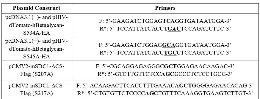
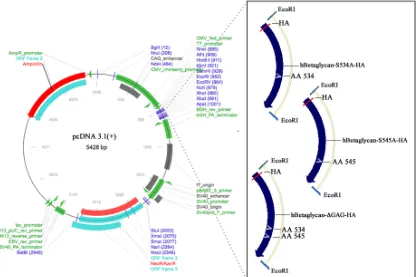
3.3: Sequence and generation of DMTP constructs ... 49
3.4 Characterization of human betaglycan-S534A-HA and -S545A-HA ... 50
3.5: Characterization of pCMV2-mSDC1-ΔCS-Flag ... 52
3.6: Conclusions ... 53
Chapter 4. Unique roles for individual betaglycan GAG chains in cell signaling and cancer cell biology ... 62
4.1: Introduction ... 63
4.2: Experimental Procedures ... 66
4.3: Effect of betaglycan’s individual GAG chains on TGFβ superfamily and FGF signaling ... 67
4.4. Impact of betaglycan’s individual GAG chains in regulating cancer cell behavior ... 68
4.5. Conclusions ... 69
Chapter 5. Summary and Future Directions ... 74
References ... 78
Appendix A: Betaglycan in Growth Factor Signaling ... 95
LIST OF FIGURES
Figure 1.1. Structure and features of DMTPs betaglycan and SDC1 ... 13
Figure 2.1. TβRIII suppresses Wnt/β-catenin activity at the level of signal reception ... 34
Figure 2.2. TβRIII suppresses Wnt induced β-catenin cytoplasmic accumulation and transcriptional activity ... 35
Figure 2.3. TGF-β signaling does not limit TβRIII’s ability to suppress Wnt/β-catenin signaling ... 37
Figure 2.4. GAG chains of TβRIII suppress Wnt signaling ... 38
Figure 2.5. TβRIII interacts with Wnt through its GAG chains ... 40
Figure 2.6. The balance between sulfated heparan and chondroitin chains on TβRIII determines TβRIII’s ability to regulate Wnt/β-catenin signaling ... 41
Figure 3.1. Human betaglycan-FL-HA amino acid sequence with indicated serines for HS (Serine 534 in red) or CS (Serine 545 in blue) GAG chain attachment in the pcDNA3.1(+) plasmid backbone ... 55
Figure 3.2. Mouse SDC1 amino acid sequence with indicated serines for HS (Serines 37, 45, 47 in red) or CS (Serines 207 and 217 in blue) GAG chain attachment in the pCMV2 plasmid backbone ... 56
Figure 3.3. pcDNA3.1(+)-hBetaglycan-S534A-HA, -S545A-HA, ΔGAG-HA plasmid maps ... 57
Figure 3.4. pCMV2-mSDC1-ΔCS-Flag plasmid map ... 58
Figure 3.5. Characterization of human betaglycan-S534A-HA and -S545A-HA ... .59
Figure 3.6. Characterization of pCMV2-mSDC1-ΔCS-Flag ... 61
Figure 4.1. DMTPs in growth factor signaling pathways ... .71
Figure A.1: Additional roles for betaglycan in Wnt signaling ... 99
Figure A.2: Betaglycan alters matrix metalloproteinases (MMPs) and TGFβ
LIST OF ABBREVIATIONS
ACTR ... Activin Type Receptor
ALK ... Activin Receptor-like Kinase
BMP ... Bone Morphogenetic Protein
BMPR ... Bone Morphogenetic Protein Receptor
CS ... Chondroitin Sulfate
DMTP ... Dually Modified Transmembrane Proteoglycan
ECD ... Extracellular Domain
ECM ... Extracellular Matrix
EMT ... Epithelial-Mesenchymal Transformation
FGF ... Fibroblast Growth Factor
FGFR ... Fibroblast Growth Factor Receptor
FZD ... Frizzled
GAG ... Glycosaminoglycan
HSPG ... Heparan Sulfate Proteoglycan
HS ... Heparan Sulfate
LRP ... Low-density Lipoprotein Receptor (LDLR)-related Proteins
MAPK/ERK ... Mitogen-Activated Protein Kinase/Extracellular Signal-Regulated Kinase
MMP ... Matrix Metalloproteinases
PDZ ... Postsynaptic Density-95/Disc Large Protein/Zonula occludens-1
R-SMAD ... Receptor-regulated Smad
SDC1 ... Syndecan-1
sDMTP ... Soluble Dually Modified Transmembrane Proteoglycan
sSDC1 ... Soluble/Shed SDC1
TβR ... TGF-β Receptor
TGF-β ... Transforming Growth Factor β
TP ... Transmembrane Proteoglycans
WNT ... Wingless/Integrated
CHAPTER
1
INTRODUCTION TO DUALLY MODIFIED TRANSMEMBRANE
PROTEOGLYCANS (DMTPS)
1
1Jenkins, L. M., Horst, B., Lancaster, C. L., and Mythreye, K. (2017). Dually modified
transmembrane proteoglycans in development and disease. Cytokine & growth factor
reviews. In press.
Transmembrane proteoglycans (TP) are members of the heparan sulfate
proteoglycan (HSPG) family whose core domains span the cell membrane to modulate a
multitude of intracellular and extracellular activities(1-10). TP extracellular domains
(ECD) contain covalently attached glycosaminoglycan (GAG) chain modifications that
include heparan sulfate (HS), chondroitin sulfate (CS), dermatan sulfate and/or keratan
sulfate, with HS and CS chains being the most common(2). As members of the GAG
family, HS and CS chains are characterized by repeating disaccharide units containing
glucuronic acid and either N-acetylgalactosamine for HS or N-acetylglucosamine for
CS(1). Of the known human transmembrane proteoglycans, only a few can present both
HS and CS chains and are referred to here as dually modified transmembrane
proteoglycans (DMTP). Human DMTP members include the Type III Transforming
Growth Factor β Receptor/Betaglycan, Syndecans-1 and -3, CD44 and Neuropilin-1.
Syndecan-3 (SDC3), similar to betaglycan and syndecan-1 (SDC1), contains both CS and
HS chains but is found almost exclusively in neuronal and musculoskeletal tissue(11)
with defined roles in skeletogenesis(12) and neurite growth(13). Unlike SDC3, CD44 is
expressed in many different cell types(14) and in various isoforms, which can exhibit HS
and/or CS chains depending on the splice variant(2,15-18). In epithelial cells, the primary
form of CD44, known as CD44E or CD44v8-10, lacks the variable region (exon v3)
required for HS chain attachment and therefore can only express CS chains(14,18-21).
Neuropilin-1 (NRP1), which is prominent in neuronal and endothelial cells, has clearly
outlined roles in axonal growth/guidance as well as physiological and pathological
angiogenesis(22-24). Since NRP1 only contains one serine residue (Ser612) for GAG
hybrid of the two(24-27). DMTPs are present in both membrane-bound and/or
shed/soluble forms with these forms able to impact cell behavior by either mimicking or
opposing each other’s cellular functions. Contributions from the core domain’s
scaffolding properties can also influence GAG chain-ligand binding(7), highlighting the
importance of the HSPGs’ core domain in delineating roles for GAG chains.
Consequently, shed/soluble DMTPs and their attached GAG chains can affect ligand
availability, downstream signaling and promote interactions between GAG chains,
ligands and the extracellular environment(28).
The background summarizes regulatory roles for both betaglycan and SDC1 in
growth factor signaling with the dissertation work focused primarily on betaglycan in
Wnt signaling. I emphasize the underappreciated mutual dependence of HSPGs’ GAG
chains and core protein and their impact in disease, with a focus on cancer progression.
Finally, I provide comprehensive information on these DMTPs for the future
development of therapeutic techniques to modulate growth factor/ligand availability and
thus control downstream cell signaling pathways involved in human disease.
1.1 DMTP Structure and Features
1.1.i. Core Structure
Both SDC1 and betaglycan are proteoglycans containing a large ECD, a
transmembrane domain and a short cytoplasmic domain (~200 amino acids or less) that
lacks intrinsic kinase activity. Betaglycan’s ECD contains a zona pellucida domain (ZP1
and ZP2, Figure 1.1), which is important for protein polymerization and protein-protein
interactions(29). Specifically, betaglycan’s ZP domain contains two distinct
(ZP1)(Figure 1.1 and (30)) and one in the membrane-proximal ZP region (ZP2)(Figure
1.1 and (31)). TGF-β ligands TGF-β1/2/3, Bone Morphogenetic Proteins (BMPs) 2/4/7
and Growth Differentiation Factor 5 (GDF-5) can bind both regions while the TGF-β
member Inhibin only binds the ZP region (ZP2)(32,33). Unlike betaglycan, little is
known about SDC1’s ectodomain core independent of its GAG chain functions. SDC1’s
235 amino acid(34) extracellular core, however, does support fibroblast cell
adhesion(35), interacts with integrins αvβ3 and αvβ5 during angiogenesis and αvβ1
integrin during re-epithelialization of lung tissue(36-40).
The short cytoplasmic domains of DMTPs are serine/threonine rich, frequently
phosphorylated and contain a PDZ (Postsynaptic Density-95/Disc large protein/Zonula
occludens-1) binding motif at the extreme carboxy terminus. These cytoplasmic regions
commonly interact with scaffolding proteins to control DMTP internalization and cell
signaling (Figure 1.1 and (2,34,41,42)). Betaglycan’s short cytoplasmic domain, through
its Class I PDZ binding motif, binds the scaffolding protein GAIP-interacting protein C
terminus (GIPC) to modulate TGF-β signaling (43).
Additionally, the Type II TGF-β Receptor (TβRII) can phosphorylate
betaglycan’s cytoplasmic domain at Thr841 to promote β-arrestin2-betaglycan interactions
and receptor internalization (44). SDC1’s cytoplasmic domain is divided into two
conserved regions, C1 and C2 (Figure 1.1), which are common regions between all
syndecans, and a central variable region (V, Figure 1.1), which confers specific properties
on each syndecan (Figure 1.1 and (45)). SDC1’s V domain regulates cell spreading and
actin cytoskeleton assembly(46) while its C1 domain, adjacent to the membrane,
C2 carboxyl-terminal tetrapeptide sequence can bind PDZ-domain-containing
proteins(41,42), which may function as membrane scaffold proteins that recruit signaling
and cytoskeletal proteins to the plasma membrane. Contributions from the core domain’s
scaffolding properties are likely to influence GAG chain-ligand binding by either
immobilizing the chains to specific sites within the plasma membrane(7) or by altering
the internalization of ligands bound to GAG chains (48). Additionally, the position of the
GAG chains on the core protein (plasma membrane distal or proximal) could impact
GAG chain association with receptors and subsequent DMTP function.
1.1.ii. GAG Chain Structure
On the extracellular core of DMTPs are sites for linear polysaccharide GAG chain
attachments (Figure 1.1 and (1,2,30,34,49)). Synthesis of DMTP GAG chains begins with
covalent attachment of a linkage tetrasaccharide, glucuronic
acid-galactose-galactose-xylose (GlcAβ1-3Galβ1-3Galβ1-4Xylβ1-O-), to a serine hydroxyl group on the core
protein followed by addition of repeating disaccharide regions composed of hexosamine
and hexuronic acids (Figure 1.1 and (49)). The repeating disaccharide units distinguish
HS chains from CS chains. During initial synthesis of HS chains, a N-acetylglucosamine
(GlcNac) is attached to the linkage tetrasaccharide by α
1,4-N-acetylglucosaminyltransferase-I (α4GlcNAcT-I). On the other hand, synthesis of the CS
chains occurs when β1,4-N-acetylgalactosaminyltransferase-I (β4GalNAcT-I) attaches a
N-acetylgalactosamine disaccharide to the linkage tetrasaccharide(49). The first
hexosamine residue thus determines which chain is made (HS or CS). After attachment of
this fifth saccharide, the chondroitin synthase family (for CS chains) or exostosins (for
sulfated at the N, 3-O and 6-O positions of glucosamine and the 2-O position of uronic
acid residues by sulfotransferases(50) while sulfate groups on CS chains are added at C-4
and/or C-6 of N-acetyl-galactosamine and/or C-2 of glucuronic acid(51). In vertebrates,
SDC1 is one of four syndecan family members and is the primary syndecan expressed in
epithelial cells(52). As a “full-time” proteoglycan (no GAG-less form in vivo), SDC1’s
ECD contains three attachments sites for HS chains (Ser37, Ser45 and Ser47) and two sites
for CS chain attachment (Ser210 and Ser220)(Figure 1.1 and (34,53,54)). While SDC1 is an
obligate or “full-time” DMTP, betaglycan is commonly referred to as a “part-time”
proteoglycan since it can be expressed with or without GAG chains(1,2,49,55) in vivo.
CS chains attach to betaglycan’s 100 kDa protein core primarily at Ser545. CS chains can
also attach at Ser534, but HS chains usually predominate at this site (Figure 1.1 and
(30,49,56)). Both alterations in core protein expression and GAG chain density and/or
composition (HS vs. CS) can impact DMTP functions.
1.1.iii. Shedding and Intracellular Domain (ICD) Cleavage
Several DMTP functions can be attributed to ectodomain shedding which leads to
DMTP expression in both membrane-bound and shed/soluble forms. Both forms can
impact whether GAG chains are anchored to the membrane or present as soluble GAGs
in the extracellular matrix (ECM). Cleavage occurs near the transmembrane domain to
release a soluble ectodomain containing GAG chains (Figure 1.1). This ectodomain
shedding is highly regulated by the enzymatic activity of several proteases(2,39,57-61)
including matrix metalloproteinases (MMP) and metalloprotease disintegrins. Enzymatic
activity by pervanadate, a general tyrosine phosphatase inhibitor, stimulates cleavage of
matrilysin(65), MMP-9(66) and MMP-1(60) cleave SDC1’s ECD. Intriguingly, shedding
for SDC1 has also been shown to be regulated by intracellular interaction of the small
GTPase Rab5 with SDC1’s intracellular domain as Rab5’s dissociation from SDC1’s
cytoplasmic domain triggers SDC1 ectodomain shedding at the cell surface(63). It is
postulated that activated GTP-Rab5 dissociates from SDC1’s cytoplasmic domain,
associates with β1 integrin and stimulates the internalization of β1 integrin and
dissociation of β1 integrin from SDC1. This cascade of events exposes SDC1’s ECD
cleavage site to sheddases(63).Whether such intracellular control of betaglycan shedding
exists has not been tested. However, the ECD of betaglycan is cleaved by MMP1 and
-3(59). Both SDC1 and betaglycan’s transmembrane/cytoplasmic fragments can be
cleaved by γ-secretase to release both DMTPs’ cytoplasmic domains intracellularly
(Figure 1.1 and (67,68)). Functions for these cytoplasmic domains are reviewed in
(67,68).
Expression of different GAG chains on membrane bound DMTPs can impact
soluble DMTP (sDMTP) pools, as observed with membrane bound SDC1’s HS chains,
where higher membrane bound HS modified SDC1 leads to lower SDC1 shedding(69).
Altering the balance of membrane bound versus soluble DMTPs could, in effect, impact
cell signaling by altering ligand interactions with sDMTPs’ ECD/GAG chains (See
Chapters 2-4). In the cell media, for example, increased levels of sDMTPs containing HS
chains exclusively could suppress signaling as they may act as ligand traps capable of
reducing ligand availability to the membrane-bound cognate receptors. Cleaved DMTP
ectodomains containing CS chains could prevent ligand aggregation in the cell media,
the current impact of DMTPs in regulating cell signaling, as outlined in Chapters 2-4, an
in-depth analysis of the GAG chain composition on membrane-bound and sDMTPs may
clarify roles for DMTPs and their GAG chains in modulating growth factor signaling and
cell biology.
1.2. DMTPs in Development
SDC1 is expressed during development to modulate tissue morphogenesis as well
as during wound healing, with several of its developmental functions mediated by its
GAG chains’ interactions with a number of extracellular adhesion proteins and growth
factors(46,52,70). SDC1 interaction with laminin, for example, is completely dependent
on both its HS and CS chains and leads to increased cell adhesion to the basement
membrane(71). However, SDC1’s CS chains promote Slit signaling to drive axon and
myotube guidance when bound to the core protein(28,72), suggesting differential roles
for the independent GAG chains. Betaglycan’s significance in development is
underscored by the observed embryo lethality in betaglycan knockout mice after 13.5
days(73,74). This lethality is partly due to significant impairment of coronary
vasculogenesis in betaglycan-null mice, demonstrating a requirement for betaglycan in
coronary vessel development and embryonic viability(74). Additionally,
betaglycan-specific antisera inhibits mesenchyme formation and endothelial cell migration in the
atrioventricular (AV) cushion, where cardiac endothelial cells lining the lumen of the AV
cushion undergo epithelial-mesenchymal transformation (EMT) to participate in valve
and membranous septa formation(75-77). Moreover, betaglycan overexpression in
non-transforming ventricular endothelial cells confers transformation in response to TGF-β2,
part of the adaptive immune response, T-cell receptors (TCRs), which are expressed on T
lymphocytes, recognize antigens associated with molecules of the major
histocompatibility complex(78). In both lymphoid and stromal cells of the thymus,
betaglycan has been shown to promote T-cell development by protecting thymocytes
from apoptosis(79). Individual functions for betaglycan’s HS and CS chains during
development are still under investigation.
1.3. DMTPs in Disease
Over the past few decades, an increasing body of evidence suggests that HS and
CS chains heavily influence cell biology and cancer progression(80). GAG chains, which
are present at the cell surface of almost all tumor cells(80), can regulate growth factor
signaling, tumor cell survival, angiogenesis, growth, invasion and metastasis(80). HSPGs
can exhibit both pro-tumorigenic(3-6) and anti-tumorigenic effects depending on their
GAG modifications(7-10). In addition to cancer, changes in HSPG expression/function
are also observed in other human diseases, including arthritis, inflammation(3),
Alzheimer’s/dementia(81), osteoporosis(82), fibrosis(83) and ischemic heart disease(84).
Furthermore, HSPGs have been shown to alter wound healing and matrix remodeling,
outcomes that can impact a broad range of pathologies(3,73,84,85), making our
understanding of HSPG/GAG chain roles in the context of the cellular environment
critical. Since much of the research emphasis has been placed on HSPG/GAG chain roles
in cancer (>8% of all PubMed articles), my dissertation work will focus on summarizing
1.3.i. DMTPs in Cancer
Both SDC1 and betaglycan have established roles in regulating tumor progression
with specific roles in apoptosis, adhesion, angiogenesis, proliferation, migration, invasion
and metastasis in vitro and in vivo(2,9,39,52,86-96). The membrane-bound form of SDC1
is capable of suppressing apoptosis as SDC1 knockdown in myeloma cells induce growth
arrest and apoptosis(4). Other reports, however, indicate that recombinant human SDC1
ectodomains induce apoptosis in breast cancer cells(8). Whether differential GAG
modifications in the recombinant SDC1 ectodomains versus SDC1 in myeloma account
for the differences observed is unclear. Moreover, while increased SDC1 levels correlate
with a more favorable prognosis in mesothelioma, head and neck, pancreatic, lung and
invasive breast cancers(2,3,39,97,98), both tumor promoting and tumor suppressing
effects have been noted in colorectal and prostate cancers(39). These seemingly
contradictory functions for SDC1 emphasize the variability of DMTP roles in cancer.
Similarly, in the case of betaglycan, loss or reduced expression has been demonstrated in
tumors(86-88,91,99). In particular, betaglycan expression has been shown to be a strong
predictor of overall and recurrence-free patient-survival in certain cancers, such as breast
cancer and neuroblastoma(86,90,100). Although betaglycan expression is commonly lost
in most tumor types, acting primarily as a tumor suppressor(2,86-88,90,100), betaglycan
expression has been shown to be elevated in colon cancer, a subset of triple negative
breast cancers and high-grade lymphomas, indicating context dependent functions for
betaglycan(5,6,101). Whether or not betaglycan’s differential role in cancer can be
attributed to its HS and CS chain functions remains to be determined.
deliver growth factors in a paracrine manner to neighboring cells in the environment.
sSDC1 from multiple myeloma cells, for example, transports hepatocyte growth factor
(HGF) to bone marrow stromal cells to regulate gene expression(39,102). These
HGF/sSDC1 interactions in the ECM likely cooperate with already established roles for
membrane SDC1 in promoting myeloma cell growth, survival and spread through HS
chains interactions with HGF(3). Betaglycan levels in the tumor stroma may vary
depending on the tumor type. For instance, betaglycan expression is significantly higher
in the stroma of early-stage, stroma-rich neuroblastomas(100), yet is down regulated in
the tumor stroma of breast tumors(103). In neuroblastoma, increased stromal expression
confers a significant survival advantage as patients with increased stromal betaglycan
exhibited significantly higher event-free survival due, in part, to increased Fibroblast
Growth Factor (FGF) signaling and differentiation(100). Contrastingly, in breast tumor
stroma, decreased betaglycan mRNA and protein expression is linked to poorer patient
outcome(103), supporting betaglycan’s contextual roles in cancer. Whether stromal cells
can take up sBetaglycan from tumor cells to impact gene expression, like in the case of
sSDC1, has not been determined. Taken together, these data suggest that DMTPs can
have important effects on cancer progression as potent regulators of tumor stromal
interactions mediated partly through their GAG chains.
In the tumor environment, cancer cells can subvert the host immune system with
mechanisms similar to those they use to evade immunotherapeutic strategies(104-106).
Additionally, effector cell dysfunction and an increased number of regulatory/suppressor
T-cells in cancer patients may further limit the efficacy of immunotherapeutic
significantly and preferentially expressed on naïve and central memory T-cells and is
upregulated upon TCR stimulation. Treating cells with sBetaglycan decreased
regulatory/suppressor FoxP3+ T-cell generation, in a TGF-β-dependent manner,
suggesting a promoter role for betaglycan in T-cell tolerance and immune responses(78).
In both murine breast cancer and melanoma, progressive loss of betaglycan expression
generates an immunotolerant tumor microenvironment whereby TGF-β signaling is
enhanced and upregulates both immunoregulatory enzyme indoleamine 2,3-dioxygenase
in plasmacytoid dendritic cells (DCs) and the CCL22 chemokine in myeloid DCs(104).
This mechanism, coupled with the elevated TGF-β levels, creates an immunotolerant
environment and may facilitate tumor progression and metastasis(104). Contrary to
betaglycan’s ability to increase tumor sensitivity to immune defenses, SDC1 appears to
aid in cancer’s subversion of the host immune system. For example, myeloma cells
coated with anti-SDC1 antibodies displayed enhanced cross-presentation of
tumor-derived cellular antigens and generated tumor specific–killer T cells by DCs(108),
suggesting an immunosuppressive role for SDC1. Additionally, anti-SDC1 immunotoxin
(IT, B-B4-SO6), in combination with the chemotherapeutic drug doxorubicin, led to
increased cell death in doxorubicin-sensitive multiple myeloma cells(109), further
supporting SDC1’s tumor promoter role. An in-depth analysis of DMTPs and their GAG
chains in modulating interactions between the immune system and malignant cells is
warranted as it could strongly augment the design of clinically more efficient
Figure 1.1. Structure and features of DMTPs betaglycan and SDC1. (A) Structures of DMTP CS (top) and HS (lower) glycosaminoglycan (GAG) chains. Both GAG chains share a common linkage tetrasaccharide covalently attached to a serine hydroxyl group on the DMTP core protein. These serine residues have an adjacent glycine residue on the carboxy-terminal side and usually lie within an area rich in acidic residues. (B) Betaglycan’s core protein contains two ZP domains in its ECD for ligand binding and
both DMTPs contain a small cytoplasmic domain, which can be cleaved by γ-secretase to
release the cytoplasmic fragment. A PDZ motif in betaglycan’s cytoplasmic domain is frequently phosphorylated and regulates betaglycan cell surface expression and intracellular protein interactions. SDC1’s cytoplasmic domain is divided into two conserved regions and a variable region, which are responsible for intracellular protein binding. SDC1 can also interact with proteins containing a PDZ motif. Betaglycan and SDC1 both undergo ectodomain shedding by MMPs to release (C) soluble forms of each receptor, which can both bind growth factors and modulate their activity. Abbreviations: C1: Conserved Domain 1, C2: Conserved Domain 2, MMP: Matrix Metalloproteinases, PDZ: Postsynaptic Density-95/Disc large protein/Zonula occludens-1, sBetaglycan: Soluble/shed betaglycan, sSDC1: Soluble/shed SDC1, V: Variable Domain, ZP: Zona
CHAPTER 2
WNT SIGNALING REGULATION BY BETAGLYCAN 2
2Jenkins, L. M., Singh, P., Varadaraj, A., Lee, N. Y., Shah, S., Flores, H. V., O'Connell,
K., and Mythreye, K. (2016) Altering the Proteoglycan State of Transforming Growth Factor beta Type III Receptor (TbetaRIII)/Betaglycan Modulates Canonical
Wnt/beta-Catenin Signaling. The Journal of biological chemistry291, 25716-25728.
2.1. Introduction
Wnt glycoproteins regulate three distinct Wnt signaling pathways to mediate cell
fate, proliferation and apoptosis as well as cancer initiation and progression in multiple
cancers, including ovarian (110-118). Activation of the canonical Wnt/β-catenin pathway
begins with binding of Wnt to its cell surface receptors, Frizzled and low-density
lipoprotein receptor (LDLR)-related proteins LRP5/6, followed by phosphorylation of
LRP5/6, recruitment of Dishevelled (Dsh) to the plasma membrane to interact with
Frizzled and stabilization of cytosolic β-catenin (119). Axin interaction with
phosphorylated LRP5/6 and Dsh leads to inactivation of the β-catenin destruction
complex, accumulation of β-catenin and translocation to the nucleus to regulate Wnt
target genes by binding to TCF/LEF transcription factors (119,120). The Wnt signaling
cascade is controlled, in part, by transmembrane proteoglycans, which interact with Wnt
signaling components and can either stimulate or inhibit signaling activity. For instance,
the HSPGs Glypican-3 and Syndecan-1 stimulate canonical Wnt signaling (121,122)
while others, including Glypican-1 and Glypican-6 suppress Wnt signaling (122,123).
The Type III TGF-β receptor (TβRIII)/Betaglycan is a transmembrane
proteoglycan with loss resulting in embryonic lethality in mice (73). Beyond its roles in
regulating TGF-β signaling, betaglycan also controls several other pathways to inhibit
cell migration, invasion, cell growth and angiogenesis in both in vitro and in vivo cancer
models (2,86-91) and regulating differentiation through FGF2 signaling (9).
Mechanistically, betaglycan regulates these pathways by either altering the actin
cytoskeleton, via betaglycan/β-arrestin2 cytoplasmic interactions (124), or through GAG
chain interactions with FGF2 (9). Overall, betaglycan also acts as a tumor suppressor in
prostate (89), lung (125), pancreatic (88) and breast cancer (86,90,93,95) but has been
Although betaglycan’s core can bind TGF-β superfamily members with high
affinity (30,44,91) the extracellular domain also contains two sites of heparan and
chondroitin sulfate GAG chain modifications resulting in betaglycan existing in multiple
forms in vivo (30,126,127). Given that Wnt glycoproteins have a high affinity for both
heparan and chondroitin GAG chains on proteoglycans (50,122), we initiated studies to
determine the possible role of betaglycan on canonical Wnt3a signaling.
We find, using both cancer and normal epithelial cells and a combination of loss
and gain-of-function approaches, that betaglycan suppresses Wnt3a signaling both at the
signal reception level and through inhibition of β-catenin transcriptional activity by
binding Wnt3a via its sulfated GAG chains. In contrast, betaglycan’s chondroitin sulfate
chains can promote Wnt3a signaling, suggesting that the composition of GAG chains
may significantly alter cellular response to betaglycan and thereby Wnt signaling.
Consistent with a lack of a role for betaglycan’s GAG chains in betaglycan’s functions as
a TGF-β co-receptor (30), betaglycan’s suppression of canonical Wnt3a signaling is
independent of TGF-β signaling and independent of betaglycan’s cytoplasmic domain
interactions described previously (43,124,128). These results demonstrate an intricate
mode of Wnt3a signaling regulation by betaglycan, mediated largely by its heparan and
chondroitin chains, and lay the foundation to advance current understanding of the
various roles proteoglycans, with different GAG chains, can have in maintaining cellular
homeostasis, specifically through control of Wnt availability and signaling.
2.2. Experimental Procedures
Cell Lines and Reagents–Ovarian epithelial carcinoma cell lines SKOV3, and OVCA429 were obtained from Duke Gynecology/Oncology Bank (Durham, NC).
Authentication of cell lines was carried out at the University of Colorado (Denver, CO)
mammary tumor cell line 4T1 (ATCC® CRL2539™), normal Chinese hamster ovarian
epithelial cell lines pgsA-745 (ATCC® 2242™) and pgsD-677 (ATCC®
CRL-2244™) were obtained from ATCC (Manassas, VA). Epithelial carcinoma cell lines
SKOV3, 4T1 and OVCA429 were cultured in RPMI-1640 (ATCC® 30-2001™)
containing L-glutamine, 10% FBS, and 100 U of penicillin-streptomycin. COS-7 cells
were maintained in DMEM (ATCC® 30-2002™) containing 10% FBS, and 100 U of
penicillin-streptomycin. CHO cell lines pgsA-745 and pgsD-677 were cultured in
Kaighn's Modification of Ham's F-12 Medium (ATCC® 30-2004™) containing
L-glutamine, 10% FBS, and 100 U of penicillin-streptomycin. All cells lines were
maintained at 37°C in a humidified incubator at 5% CO2. Antibodies: phospho-LRP6
(Ser1490) (#2568), LRP6 (#2560), β-catenin (D10A8) XP® Rabbit mAb (#8480),
GAPDH Rabbit mAb (#14C10), HA Rabbit mAb (#3724) and Wnt3a (C64F2) Rabbit
mAb (#2721) were from Cell Signaling Technology (Danvers, CA). Mouse E-cadherin
mAb was purchased from BD Biosciences (#610181) (San Jose, CA). Human betaglycan
Antibody (#AF-242-PB) was purchased from R&D Biosystems (Minneapolis, MN) and
Actin (#A2228) from Sigma-Aldrich (St. Louis, MO). Mouse HA antibody (#32-6700)
from Invitrogen (Carlsbad, CA). Inhibitor SB431542 hydrate (#S4317) was purchased
from Sigma-Aldrich. Sodium chlorate (NaClO3) was obtained from Thomas Scientific
(Swedesboro, NJ) and sodium sulfate anhydrous (Na2SO4) (#S421-500) from
ThermoFisher Scientific (Waltham, MA). Heparinase III (#H8891) and Chondroitinase
ABC (#C3667) were obtained from Sigma-Aldrich. Recombinant TGF-β1, TGF-β2 and
Wnt3a were purchased from R&D Systems.
study have been described previously (9,86,89,129,130). Full length betaglycan consists
of betaglycan/TβRIII-HA in pcDNA 3.1(+) as described previously (44,129). pcDNA
3.1(+) betaglycan/TβRIII-ΔGAG construct consists of human betaglycan-HA, with
serine-to-alanine point mutations at amino acids 534 and 545 to prevent GAG attachment
(44,94,131,132). rTβRIII consists of the rat betaglycan sequence with HA tag in the
pcDNA 3.1 vector (89). Adenoviral constructs were used at MOIs between 5-100
particles/cell and infections performed as described previously (9,90,124). shRNA
sequences for betaglycan were obtained from Sigma-Aldrich with the following
sequences: shRNA33430 (shTβRIII-1):
CCGGCCAAGCATGAAGGAACCAAATCTCGAGATTTGGTTCCTTCATGCTTGGT
TTTTG and shRNA33432 (shTβRIII-2):
CCGGCGTGCTTTATCTCTCCATATTCTCGAGAATATGGAGAGATAAAGCACGT
TTTTG in a pLKO.1-puro backbone (betaglycan shRNA construct and non-targeted
control). Lentiviral particles were generated at the Center for Targeted Therapeutics
Core Facility and the University of South Carolina (Columbia, SC). For betaglycan
knockdown, SKOV3 cells were infected with 1X betaglycan shRNA lentivirus. Cells
were then selected in the presence of 1 µg ml-1 puromycin. Stable cell lines were
maintained in 0.5 µg ml-1 puromycin.
Wnt3a-HA (#18030) and TβRII-ΔCyto (#14051) plasmids were purchased from
Addgene (Cambridge, MA) (133). The soluble human betaglycan construct was a kind
gift from Blobe, G. (Duke University, Durham, NC). Conditioned media containing
soluble TβRIII/betaglycan was generated by transfecting cells with the indicated
Transient DNA transfections were performed using Lipofectamine 2000 (#11668019)
from Life Technologies (Carlsbad, CA) or FuGENE® 6 (#E2691) from Promega
(Madison, WI) according to manufacturer’s instructions. Cell fractionation kit to analyze
β-catenin localization came from Cell Signaling (#9038). Luciferase assay kit (#E1500)
came from Promega and M50 Super 8x TOPFlash (134) used to measure luciferase
activity was a gift from Randall Moon (Addgene plasmid #12456).
Quantitative Polymerase Chain Reaction (qRT-PCR)–For qRT-PCR, total RNA
was isolated from approximately 200K cells using Trizol reagent (Invitrogen). RNA was
retro-transcribed using iScript™ Reverse Transcription Supermix (#1708841) and
SsoAdvanced Universal SYBR Green Supermix (#1725271) from Bio-Rad (Hercules,
CA). qRT-PCR primer sequences used were: RPL13A-F: AGATGGCGGAGGTGCAG,
RPL13A-R: GGCCCAGCAGTACCTGTTTA, Betaglycan-F:
CGTCAGGAGGCACACACTTA, Betaglycan-R: CACATTTGACAGACAGGGCAAT.
Immunoprecipitation and Western Blotting–Immunoprecipitation and western blotting were performed using standard techniques as described previously (90,93,135).
For co-immunoprecipitation in COS-7 cells, betaglycan-expressing cells were transfected
with Wnt3a-HA construct indicated and culture media was collected 48 h after
transfection in serum-free conditions. betaglycan was then immunoprecipitated by
incubating the cell lysates overnight with anti-human betaglycan antibody. The next day,
protein G–Sepharose beads were added to the lysates for 2 h at 4°C. The beads were then
washed three times with cold phosphate buffer solution (PBS) and resuspended in sample
buffer. The amount of betaglycan or Wnt3a bound to the beads was detected by western
Wnt3a-Betaglycan Pull-down Assay–This was performed as described previously
(122,136). Briefly, OVCA429 cells were lysed in non-denaturing COIP lysis buffer (50
mM TrisHCl, pH 7.5, 150 mM of NaCl, 1% Nonidet P-40, 10% glycerol, 1 mM DTT, 25
mM NaF, 1 mM Na3VO4 and 1X protease inhibitor cocktail [#P8340, Sigma-Aldrich]).
betaglycan-HA was then immunoprecipitated by incubating the cell lysates overnight
with an anti-human betaglycan antibody. The next day, protein G–Sepharose beads were
added to the lysates for 2 h at 4°C. Beads were then washed three times with PBS, and
incubated with 20 nM Wnt3a-conditioned media for 2 h at 4°C. After two more washes
with PBS, the beads were resuspended in sample buffer and the amount of Wnt3a bound
to betaglycan was detected by western blot using anti-Wnt3a and anti-betaglycan
antibodies.
Luciferase Assay–Indicated cells were seeded in 24-well plates and were
co-transfected with a luciferase reporter vector containing a β-catenin-responsive promoter
[to drive luciferase expression (TOPFlash, #12456, Addgene)] and SV40 (Renilla internal
control vector). One day after transfection and infection, cells were incubated overnight
with 50 ng ml-1 Wnt3a and then lysed. Luciferase activity (Luciferase Assay System,
Promega) was measured by calculating the ratio between luciferase and Renilla activities
(to normalize for transfection efficiency) and then normalizing values to the untreated
sample.
Immunofluorescence and Intensity Analysis–Indicated cells were seeded onto
coverslips in 12-well plates at a density of 5x10⁴ cells per well. After infections and
treatment with 50 ng ml-1 Wnt3a, cells were washed with ice-cold PBS and fixed with
0.1% Triton X-100 in PBS and then blocked with 3% BSA or 0.2% gelatin in PBS for 30
min at room temperature, followed by an overnight incubation at 4°C with a rabbit anti-β
-catenin antibody. After extensively washing with PBS, cells were incubated with an
Alexa-conjugated secondary antibody (Molecular Probes, Eugene, OR). Cells were
mounted in mounting medium and analyzed under an Olympus IX81 motorized inverted
microscope (Shinjuku, Tokyo, Japan). Fluorescence intensity for the β-catenin was
analyzed using ImageJ 1.50d software (National Institutes of Health, USA) by drawing a
fixed line of interest over the membrane and cytoplasm followed by averaging maximum
intensities obtained from the plot profile plugin. To estimate the change in β-catenin
localization after Wnt treatment, in the presence and absence of betaglycan, the ratio
between membrane and cytoplasmic fraction of β-catenin fluorescence was calculated.
Statistical significance of the data was analyzed in SigmaPlot version 11software.
P-values <0.05 were considered to be statistically significant.
Subcellular Fractionation–Indicated cells were seeded in 12-well plates and
infected to express betaglycan. 48 h post infection, cells were treated with 50 ng ml-1
Wnt3a for 1 h and then lysed. Subcellular fractionation of β-catenin, cytoplasmic marker
GAPDH and plasma membrane marker E-cadherin was carried out using the cell
fractionation kit (Cell Signaling) according to manufacturer’s instructions.
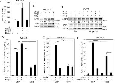
2.3. Betaglycan suppresses Wnt/β-catenin activity at the level of signal reception.
To investigate the role of betaglycan on signaling by Wnt glycoproteins, which
have high affinities for both HSPGs and CSPGs (50,137,138), we expressed betaglycan
in the ovarian cancer cell line OVCA429 that we, and others, have established express
expression of betaglycan by shRNA-mediated knockdown in the ovarian cancer cell line
SKOV3, which express higher levels of betaglycan (Figure 2.1A.iii) and (139)). We
examined whether betaglycan can affect canonical Wnt signaling as determined by
phosphorylation of coreceptor LRP6, one of the first steps initiated by Wnts binding to
their signaling coreceptors (140). We find that while Wnt3a robustly phosphorylated
LRP6 at serine 1490 (141) in OVCA429 cells (low betaglycan levels), transiently
increasing betaglycan expression in OVCA429 cells suppressed Wnt induced LRP6
phosphorylation in a betaglycan dose dependent manner (Figure 2.1B). Total LRP6 levels
remained stable in betaglycan-expressing OVCA429 cells when compared to OVCA429
cells with low betaglycan levels (Figure 2.1B). In SKOV3 cells, which express high
levels of betaglycan (Figure 2.1A.i), reducing betaglycan expression using shRNA
resulted in increased LRP6 phosphorylation when compared to Wnt3a stimulated SKOV3
control cells expressing high endogenous betaglycan (Figure 2.1C-D). To confirm that
the effect of shBetaglycan was specific to betaglycan, we utilized shRNA resistant rat
betaglycan (9,90) to rescue betaglycan expression and examined Wnt induced LRP6
phosphorylation. We find that rescue of betaglycan expression in shBetaglycan cells
(Figure 2.1A.iii) suppressed Wnt induced LRP6 phosphorylation compared to cells
containing endogenous betaglycan (Figure 2.1C). Total LRP6 levels were not
significantly altered by shRNA to betaglycan or transient expression of rat betaglycan in
SKOV3 cells when compared to control cells (Figure 2.1C). Consistently, a second
shRNA to betaglycan (shTβRIII-2) also resulted in increased LRP6 phosphorylation
that betaglycan may regulate Wnt signaling at the signal reception level by suppressing
canonical Wnt signaling.
Activation of the canonical Wnt pathway leads to stabilization and accumulation
of cytosolic β-catenin, which then enters the nucleus and regulates Wnt target genes
(119). Consistent with reduced LRP6 phosphorylation, Wnt induced β-catenin cytosolic
accumulation was significantly reduced in the presence of betaglycan (Figure 2.2A-B).
Upon β-catenin accumulation and stabilization, activation of TCF/LEF-sensitive
transcription by β-catenin provides a robust read-out of the Wnt-stimulated canonical
pathway (142). To test if betaglycan-mediated changes on LRP6 phosphorylation and β
-catenin accumulation translated to downstream effects on TCF/LEF activity, we analyzed
the activity of a TCF/LEF-sensitive reporter, which contains multiple β-catenin binding
sites (134). We find that Wnt3a significantly increases TCF/LEF reporter activity in
OVCA429 cells (Figure 2.2C). Increasing betaglycan expression in these cell lines
resulted in a significant suppression of Wnt3a induced activation of the
TCF/LEF-reporter compared to control Wnt treated cells (Figure 2.2C). Similar to trends seen in
OVCA429 cells, overexpressing betaglycan in SKOV3 cells (betaglycan–high) resulted
in suppression of Wnt3a induced TCF/LEF activity compared to control Wnt treated cells
(Figure 2.2C). Side-by-side analysis of Wnt3a stimulated TCF/LEF activity in SKOV3
(high betaglycan) and OVCA429 (low betaglycan) cells in the same experiment revealed
lower Wnt3a induced TCF/LEF activity in SKOV3 cells, when compared to Wnt3a
treated ovarian cancer OVCA429 cells (Figure 2.2D), which we hypothesize is in part
due to higher endogenous betaglycan expression in SKOV3 cells (Figure 2.1A, left
(Figure 2.1A, right graph) that resulted in enhanced Wnt induced TCF/LEF-reporter
activity compared to control cells (Figure 2.2E). This increased Wnt signaling in
shBetaglycan cells was suppressed upon restoring betaglycan expression using shRNA
resistant rat betaglycan (Figure 2.2E), consistent with increased LRP6 activation
observed in SKOV3 cells upon knockdown of betaglycan (Figure 2.1C). Regulation of
TCF/LEF reporter activity by betaglycan was not restricted to ovarian cancer cells as
betaglycan expression also repressed Wnt induced TCF/LEF reporter activity in 4T1
(breast cancer) cells (Figure 2.4D), indicating broad impacts of betaglycan on Wnt
signaling regulation.
2.4. TGF-β signaling does not limit betaglycan’s ability to suppress Wnt/β-catenin
signaling
To begin elucidating the mechanisms by which betaglycan regulates Wnt
signaling, we examined whether the presence of TGF-β a high affinity ligand for
betaglycan’s core domain (143-145), impacted the ability of betaglycan to suppress Wnt
signaling. We find that both TGF-β1 and TGF-β2 enhance Wnt induced LRP6
phosphorylation and TCF/LEF activity (Figure 2.3A-B), in OVCA429 cells, but to a
lesser extent in SKOV3 cells (high betaglycan) (Figure 2.3C, Lanes 1-4), indicating a
cooperative role for TGF-β ligands in Wnt signaling that may be repressed by betaglycan.
Treating betaglycan-knockdown SKOV3 cells (shTβRIII) with TGF-β resulted in an
enhancement of the Wnt3a-TGF-β cooperativity, compared to control
betaglycan-expressing SKOV3 cells treated with Wnt3a and TGF-β (Figure 2.3C, Lanes 5-8). Since
TGF-β2 binds the core domain of betaglycan with higher affinity than TGF-β1 (146) and
this ligand was chosen to determine TGF-β signaling mediated changes on suppression of
Wnt3a induced TCF/LEF activity by betaglycan. We find that Wnt induced TCF/LEF
activity, both in the absence and presence of TGF-β2, was dampened by betaglycan
expression in OVCA429 cells (Figure 2.3D).
To confirm that betaglycan does not require TGF-β signaling receptors to
suppress Wnt signaling, we first utilized SB431542 (inhibitor of TβRI kinase activity)
and analyzed Wnt induced TCF/LEF activity in OVCA429 cells. We find that inhibition
of TβRI suppressed Wnt signaling independent of betaglycan expression in control cells
(Figure 2.3E). However, inhibition of TβRI did not affect betaglycan’s ability to suppress
Wnt induced TCF/LEF activity in OVCA429 cells when compared to control cells
(Figure 2.3E) indicating that Wnt signaling repression by betaglycan is independent of
TβRI kinase activity. Several TGF-β independent roles for betaglycan have been reported
through its interactions with the Type II TGF-β receptor TβRII (143). However, transient
expression of TβRII lacking its cytoplasmic domain (and therefore unable to interact with
betaglycan) (TβRII–ΔCyto) (133,143), did not affect betaglycan’s ability to suppress Wnt
induced TCF/LEF activity when compared to control cells (Figure 2.3F). Similar to
Figure 2.3E, removal of TβRII’s cytoplasmic domain (TβRII–ΔCyto) in GFP-expressing
cells led to a suppression of Wnt induced TCF/LEF activity when compared to control
cells (Figure 2.3F). These betaglycan independent observations of TβRII–ΔCyto and
SB431542 on TCF/LEF activity may point to autocrine TGF-β-Wnt signaling
mechanisms unrelated to betaglycan’s ability to suppress Wnt dependent Wnt signaling.
TGF-β2, the absence of TGF-β signaling and betaglycan-TβRII interaction, betaglycan is still
able to suppress Wnt signaling.
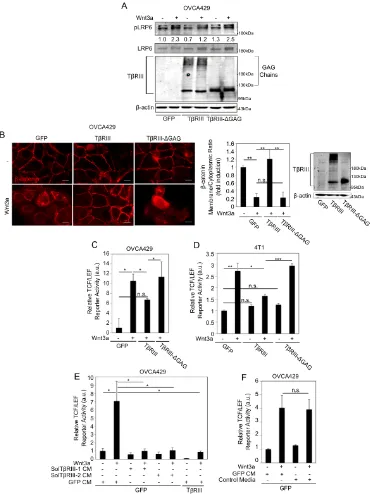
2.5. GAG chains of betaglycan regulate Wnt signaling
Wnt glycoproteins have been shown to have a high affinity for GAG chains on
transmembrane proteoglycans (50) and the extracellular betaglycan domain contains two
sites of HS and CS GAG chains (9,131). To determine if the chains on betaglycan are
involved in the suppressive effects on Wnt signaling, we expressed either full-length
betaglycan (TβRIII), betaglycan lacking GAG chain modifications (TβRIII-ΔGAG) (30)
or control vectors in OVCA429 cells and assessed the levels of phosphorylation of LRP6,
cytosolic β-catenin accumulation and TCF/LEF activity induced by exogenous Wnt3a.
We find that, unlike full-length betaglycan, TβRIII-ΔGAG failed to suppress LRP6
phosphorylation in OVCA429 cells (Figure 2.4A). Consistently, TβRIII-ΔGAG did not
suppress Wnt3a-dependent β-catenin cytoplasmic accumulation compared to full-length
betaglycan; instead, β-catenin cytoplasmic accumulation in the presence of Tβ
RIII-ΔGAG resembled cytoplasmic β-catenin levels observed in Wnt3a treated control cells
(Figure 2.4B). TβRIII-ΔGAG cells also failed to suppress TCF/LEF activity when
compared to full-length betaglycan (Figure 2.4C). The effect of betaglycan’s GAG
chains on Wnt signaling was not restricted to ovarian cells as TβRIII-ΔGAG also failed
to suppress Wnt signaling when compared to full length betaglycan in the murine
mammary 4T1 cells (Figure 2.4D).
To test whether the extracellular domain (ECD) of betaglycan was sufficient to
suppress Wnt induced signaling, we used two parallel approaches. We treated OVCA429
cells expressing only betaglycan’s ECD (Sol-TβRIII-1) (86,88,144) or CM from cells
expressing full-length betaglycan containing soluble betaglycan in the media due to
shedding (Sol-TβRIII-2) (30,144) (Figure 2.4E). CM from control vector (GFP)
expressing cells was used as control (GFP-CM, Figure 2.4E). These conditions were
compared to OVCA429 cells expressing full-length betaglycan in the same experiment
(Figure 2.4E). We find that both shed and soluble forms of betaglycan were able to
significantly suppress Wnt induced TCF/LEF activity to the same extent as expressing
full-length betaglycan (Figure 2.4E). To control for possible artifacts associated with
infection of vectors, we also tested media from uninfected cells (Figure 2.4F) and find
that infection with GFP did not impact TCF/LEF activity (Figure 2.4F). Taken together,
these data confirm that betaglycan’s ECD and GAG chains are sufficient at suppressing
Wnt induced signaling.
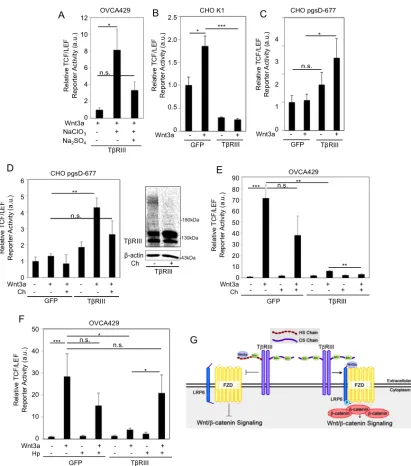
2.6. Betaglycan interacts with Wnt and the balance between HS and CS chains
determine betaglycan’s ability to regulate Wnt/β-catenin signaling
To determine whether betaglycan binds Wnt3a, we used co-immunoprecipitation
of recombinant Wnt3a and betaglycan, a methodology commonly used to study Wnt
interactions with its receptors (122,136). We find a Wnt dose dependent interaction
between betaglycan and Wnt3a in OVCA429 cells (Figure 2.5A). Consistent with the
extracellular domain of betaglycan being sufficient to suppress Wnt signaling (Fig. 4E),
we find that soluble betaglycan was also able to interact with Wnt3a, as determined by
using CM from COS-7 cells expressing full length betaglycan and HA tagged Wnt3a
(Figure 2.5B). To determine if the betaglycan–Wnt3a interaction is mediated through
incubated OVCA429 cell lysates with recombinant Wnt3a and performed
co-immunoprecipitation in cells expressing full-length betaglycan (TβRIII), TβRIII-ΔGAG
or control (Experimental Procedures). We observed immunoprecipitation of Wnt3a and
betaglycan that is reduced to background levels in cells expressing TβRIII-ΔGAG (Figure
2.5C). These data indicate that the interaction/binding capacity of TβRIII-ΔGAG is
significantly less than full-length betaglycan. These findings are consistent with Tβ
RIII-ΔGAG being unable to inhibit Wnt3a signaling (Figure 2.4B-C).
Since betaglycan represses Wnt signaling and appears to interact with Wnt3a
through its GAG chains, we aimed to test whether Wnt signaling regulation by
betaglycan’s GAG chains are dependent on the sulfation state of betaglycan’s GAG
chains. We treated betaglycan-expressing OVCA429 cells with sodium chlorate, a
competitive inhibitor of ATP-sulfurylase, which results in proteoglycans arriving at the
cell surface bearing nonsulfated heparan sulfate or chondroitin sulfate chains (147). We
find that non-sulfated GAG chains on betaglycan significantly stimulated Wnt induced
TCF/LEF activity (Figure 2.6A). Treatment with sodium sulfate, which overcomes the
effects of sodium chlorate and restores sulfation of proteoglycans (147), decreased Wnt
induced TCF/LEF activity compared to betaglycan-expressing OVCA429 cells only
treated with sodium chlorate (Figure 2.6A). These results demonstrate that the sulfation
of betaglycan’s GAG chains is required for betaglycan-mediated suppression of Wnt
signaling and loss of sulfation results in increased Wnt induced signaling.
Since the GAG chains on betaglycan comprise both HS and CS chains (127), we
aimed to isolate the individual effects of the different GAG chains of betaglycan on Wnt
signaling was conserved in parental Chinese Hamster Ovarian (CHO) K1 cells, where
betaglycan expresses both HS and CS chains (148). Although CHO cells have a modest
response to Wnt stimulation as observed previously (50,149) and by us (Figure 2.6B-C),
we observed a significant decrease in Wnt signaling upon betaglycan-expression in CHO
K1 cells compared to control cells (Figure 2.6B), consistent with our observations in
ovarian and breast cancer cells (Figs. 2.2 and 2.4). To determine the role of betaglycan’s
CS chains in Wnt signaling, we utilized the CHO cell line derivative pgsD-677, which
lack both N-acetylglucosaminyltransferase and glucuronyltransferase activities and are
unable to synthesize heparan sulfate, but can produce high amounts of chondroitin sulfate
(148). We increased betaglycan expression in pgsD-677 (ΔHS) cells (as described in
Experimental Procedures) and examined Wnt induced TCF/LEF activity. Strikingly, we
observed a significant increase in Wnt signaling, in betaglycan-expressing pgsD-677 cells
compared to control cells (Figure 2.6C). Furthermore, removal of betaglycan’s CS chains
with chondroitinase (Ch) (Figure 2.6D, right panel) reduced Wnt induced TCF/LEF
activity in betaglycan-expressing pgsD-677 cells (Figure 2.6D). Since pgsD-677 cells
express only CS GAG chains (148), we tested whether CS chains promote Wnt signaling
in cells that made both HS and CS GAG chains. Similar to our results in pgsD-677 cells
(Figure 2.6D), we find that betaglycan was able to further repress Wnt signaling in
OVCA429 cells treated with chondroitinase as compared to control cells (2x repressed,
Figure 2.6E). In contrast, heparanase (Hp) treatment of betaglycan-expressing OVCA429
cells resulted in increased TCF/LEF activity compared to heparanase untreated cells (5x
increased, Figure 2.6F). These data suggest that HS and CS chains on betaglycan
we propose that the HS chains of betaglycan are responsible for Wnt3a sequestration and
subsequent betaglycan–mediated suppression of Wnt3a signaling. In contrast
betaglycan’s CS chains increase Wnt availability and signaling (Figure 2.6G).
2.7. Discussion and Conclusions
We provide novel evidence for TβRIII/Betaglycan-mediated regulation of
canonical Wnt signaling through distinct functions of its HS and CS GAG chains. Our
studies demonstrate that the HS chains of betaglycan are responsible for suppression of
Wnt3a signaling, most likely via sequestering Wnt, in contrast with the CS chains of
betaglycan, which promote Wnt signaling. Based on our findings, we propose that Wnt
interactions with the HS chains on betaglycan result in sequestration of Wnt away from
LRP6 and Frizzled, which decreases the levels of signaling-productive complexes
between the ligand and its receptors. This hypothesis is confirmed upon examining the
inability of betaglycan to suppress Wnt signaling upon removal of its GAG chains
(Figure 2.4). Mechanistically, our pull-down assays in betaglycan-expressing cells
(Figure 2.5) indicate interaction between betaglycan and Wnt glycoproteins, which have
a high affinity for polyanionic compounds such as heparin (150), and reveal that the
GAG chains significantly increase Wnt-betaglycan interaction to suppress Wnt signaling.
Strikingly, betaglycan’s CS chains promote Wnt3a signaling in the absence of its HS
chains (Figure 2.6B,D). To support this conclusion, chondroitinase treatment in pgsD-677
and OVCA429 cells resulted in a loss of Wnt signaling, thus indicating an exciting new
role for the chondroitin chains of betaglycan in stimulating Wnt signaling.
The role of GAG chains in Wnt signal transduction may also depend on the core
betaglycan’s HS and CS chains in Wnt signaling. To support our hypothesis, it has been
shown that exogenous chondroitin sulfate, heparin, and GAGs are unable to stimulate
Wnt3a signaling while endogenous CSPGs promote Wnt signaling in mouse L cell
fibroblasts, suggesting that the core proteins of CSPGs may be involved in regulating
Wnt3a activity (137). We speculate that localization, sulfation and/or chain length of
GAG chains attached to core proteins could contribute to differences in ligand
availability and signaling.
Studies have also shown that cell context can determine the role proteoglycans
and GAG chains play in cancer progression. For instance, enzymatic elimination of
chondroitin sulfate molecules in primary breast tumors, for example, increases lung
metastases in mice (151) while digestion of cell surface CS on lung cancer cells injected
into tail veins leads to a reduction in the number of tumor cells able to populate and
metastasize (152). These results suggest that CS molecules may have opposing roles
during cancer progression: an anti-metastatic function in primary tumor tissue and a
pro-metastatic role during extravasation (circulating cancer cell interaction with endothelial
cells) (153). Other proteoglycans have also been shown to function as either tumor
promoters or suppressors depending on the protein core, GAG chains attached, associated
molecules, proteoglycan localization and tumor type (154). Perlecan, for example, can
both promote tumor invasiveness (155) and inhibit angiogenesis (156) while glypicans
and syndecans may promote local cancer cell growth and metastatic potential in some
cancer tissues (138,157), but inhibit tissue growth, invasion and metastasis in others
(158,159). Together, these data show a requirement for the proteoglycan’s core domain
In addition to the contributions made by the proteoglycan’s core domain and
environment, the sulfation state of the proteoglycan also plays a major role in its ability to
regulate signaling pathways. Upon treatment of our betaglycan-expressing OVCA429
cells with sodium chlorate, an ATP-sulfurylase competitive inhibitor that causes
proteoglycans to arrive at the cell surface bearing nonsulfated HS or CS chains (147), we
find betaglycan unable to repress Wnt signaling, indicating that the sulfation of
betaglycan’s GAG chains is required for proper Wnt signal regulation by betaglycan (Fig.
6A), consistent with previous reports for Glypican-1 (50). Studies in Drosophila have
also shown that, upon treatment of Drosophila cells with sodium chlorate or in the
absence of an HS N-deacetylase/N-sulfotransferase, cells are completely deficient in HS
chain sulfation and Wingless [Wg] signaling is disrupted (160-163). HS chain sulfation
plays a vital role in regulating FGF signaling as well. Consistently, the HS chains of
betaglycan can also regulate FGF signaling and play a critical role in tumor progression
(9).
Previous reports indicate that FGF signal transduction is dependent on sulfation of
the 2-O and 6-O positions on HS chains, which control FGF1 binding to heparin and
FGF1-dependent dimerization and activation of the FGFR1 receptor, respectively
(164-166). In articular cartilage, studies reveal a Wnt signaling promoter role for CS chains
that is dependent on the sulfation of the CS chain (167). Taken together, these studies,
combined with our data, suggest that sulfation plays a significant role in growth factor
signaling regulation by GAG chains on proteoglycans.
It is possible that different expression levels of β