77:13 (2015) 99–109 | www.jurnalteknologi.utm.my | eISSN 2180–3722 |
Jurnal
Teknologi
Full Paper
M
ICROBIAL
I
NACTIVATION
U
SING
P
ULSED
L
IGHT
-
A
R
EVIEW
Moses Elisha Kundwal
a,c, Mohd Nizam Lani
b, Abd Rahman Tamuri
a*a
Department of Physics, Faculty of Science, Universiti Teknologi
Malaysia, 81310 UTM Johor Bahru, Johor, Malaysia
b
School of Food Science and Technology, Universiti Malaysia
Terengganu, 21030 Kuala Terengganu, Terengganu, Malaysia
c
Department of Physics, School of Science, Federal College of
Education,Yola, Adamawa State, Nigeria
Article history
Received
15 April 2015
Received in revised form
29 September 2015
Accepted
12 November 2015
*Corresponding author
[email protected]
Graphical abstract
Abstract
The use of pulse light to inactivate microbes has recently attracted interest of many researchers. Consequently, there are various reports on the efficacy and advantages of pulsed light in microbial inactivation; and also the potentiality of pulsed light systems being adopted for industrial use in this regards. Here, we review some of the works done in relation to microbial inactivation with pulse light, with emphasis on the role played by pulse light parameters such as fluence, spectral range, pulse power, pulse width, pulse frequency, etc. We focussed in particular on factors that make pulsed light systems more effective than their continuous wave counterpart and also proffer suggestions on possible areas for improvement in future study. The use of pulsed lasers in inactivating microbes was briefly appraised. Also, prospects and challenges in the use of pulsed light for inactivation were highlighted.
Keywords: Pulsed light, inactivation, bacteria
© 2015 Penerbit UTM Press. All rights reserved
1.0 INTRODUCTION
The use of light to inactivate microbes has continued to attract the attention of researchers for some obvious reasons. Traditional methods of pasteurization or sterilization using thermal or chemical treatments have generally been effective but, nevertheless, they have certain limitations. Pasteurization of food materials and other methods of thermal sterilization results into rise in temperature in the bulk of the material, which may be undesirable. Pasteurization also leads to the emergence of pasteurization-resistant bacteria. There is also the issue of wastage of energy and time in some instances; for example, thermally treated sterile air has to be cooled before being used to aerate pasteurised food for the purpose of packaging. For the above and other similar reasons,
there is currently considerable interest in developing alternative methods for the control of microorganisms; methods which will be effective in inactivating the microbes and yet have leave less or minimal damaging effects on the material being processed.
Pulsed light is one such emerging technology that has the potential to inactivate microorganisms very rapidly on exposed surfaces. It is a relatively novel technology which could be an effective alternative to traditional thermal treatment in order to assure the microbial quality and safety of food products [1]. Previous studies conducted [2-6] indicate the potentiality of pulsed light (especially of the UV domain) for inactivation of bacteria. These studies suggest pulsed light as a more efficient non-thermal technology for decontamination and sterilization. The pulsed light disinfection systems can therefore be used
Dimerization of the
DNA thymine
Mutations of genes
Impairment of gene
replication and transcription
Death of
to destroy dangerous microorganisms in water treatment systems, in air and on contaminated surfaces.
Pulsed lasers also play some role in microbial inactivation. One area in which lasers are put into applications which are more closely related to human systems is in the interaction of lasers with bio-samples. Studies in this area are often fashioned out in such a way that most of the resulting applications are tailored towards medicine, surgery or dentistry. Practically all of these applications are for curative purpose. Study of the interaction of lasers with microbial organisms (particularly the pathogenic types) also leads to applications such as food preservation, food safety,
decontamination of immediate environment,
sterilization of equipment, etc, which are preventive rather than curative.
In this paper , In this paper , a review of the some works done in relation to microbial inactivation with pulse light was attempted, with emphasis on the role played by pulse light parameters such as fluence, spectral range, pulse power, pulse width, pulse frequency, etc. We focussed in particular on factors that make pulsed light systems more effective than their continuous wave counterpart and also proffer suggestions on possible areas for improvement in future study. The use of pulsed lasers in inactivating microbes was briefly appraised. Finally, prospects and challenges in the use of pulsed light for microbial inactivation were highlighted.
2.0 PULSED LIGHT SYSTEMS
A pulsed light system (PLS) can be described as one involving the conversion of short-duration, high-power electric pulses into short-duration, high-power pulses of radiation. The radiation produced could be in form of broad spectrum light (with spectral distribution from UV to IR), any of the constituents of the broad spectrum light (e.g. pulsed UV) or a particular wavelength of the spectrum (e.g. laser), depending on relative magnitudes of electrical input parameters and method of conversion used.
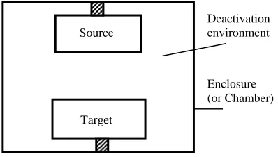
Generally, a pulsed light system for microbial inactivation is composed of the source (where the pulsed light is produced and controlled), the target (which contains the microbial organism) and the enclosure or chamber (which houses the environment for inactivation). This is illustrated in Figure 1.
Figure 1 Block diagram of a pulsed light system for inactivation of microbial organisms
2.1 Source
The source produces the light of required power and spectral distribution for the purpose of inactivation of microorganisms. It is composed basically of an electrical unit and a lamp unit. The electrical unit produces and controls electrical pulses, which are sent to the lamp unit. The lamp unit converts the electrical pulses to light pulses of similar characteristics to those of the electrical pulses.
2.2 Target
The target is the material containing the microbial organism, earmarked for inactivation. It could be in form of solid or fluid. Fluid materials are usually enclosed in a containing vessel made of transparent materials like glass, plastic, etc.
2.3 Chamber
Both source and target are enclosed in a chamber where inactivation takes place. The chamber is fitted with some control and monitoring instruments such as thermometer and barometer. Some chambers are also fitted with mechanisms to provide for relative motion between source and target. In order to maximize interaction of the pulsed light with the target, the inner surface of the chamber is coated with highly reflective material so that light rays that were initially not directed at the target can undergo multiple reflections until they get to the target.
2.4 Principle of Pulsed Light
The operational principles of pulse light have been elaborately treated in [9]. In summary, electrical power (in form of a.c.) is first converted to high power d.c. by means of storage capacitors before being released in form of d.c. pulses unto a discharge lamp. The lamp then converts the d.c. pulses to light pulses of same duration and frequency as that of the received d.c. pulses. An example of a pulsed UV system arrangement is shown in Figure 2.
Source
Target
Figure 2 Block diagram of a pulsed UV system (adapted from [2])
3.0 PULSED
LIGHT
PARAMETERS
AND
MICROBIAL INACTIVATION
The most important physical parameters encountered when dealing with PLS can be classified into two groups viz: source parameters and target parameters as shown in Table 1. Source parameters, being the main focus of this paper, are given preferential treatment in this review. In some studies conducted so far, efficiency of inactivation has been found to vary with lamp output power-related parameters such as pulse width and fluence [8]. Thus, we shall consider some of the pulse light parameters in relation to microbial inactivation.
Table 1 Pulsed light parameters used for microbial inactivation
Source Target
Wavelength distribution Transparency Energy density or Fluence (F) Size
Pulse duration (τ) Surface condition
Number of the pulses applied (n) Temperature
Pulse frequency (f)
3.1 Spectral Distribution
Spectral distribution and fluence (see section 4.2) are said to be the most important source parameters in pulsed light system and microbial inactivation [9]. Spectral distribution refers to the range in wavelength of the pulsed light. It has been suggested [9] that, since spectral distribution of the light from flash lamps depends strongly on current density of the pulses that excite the gas in the lamp, desired wavelength can be obtained by proper adjustment of the electrical pulse current density by means of the control switches and photo-detectors. Alternatively, appropriate light filters can be used to block unwanted frequencies in the pulsed light before reaching the target.
A number of investigations have been conducted to ascertain the effects of spectral range on inactivation of microbial organisms. In a study [10], a monochromator was used to study the spectral
response to the inactivation of Escherichia coli using
broad spectrum flash lamp with spectral range extending from UV to IR. The most efficient inactivation was found to occur at around 270 nm (within UV),
where a peak value for E. coli population reduction of
0.43 log per mJ/cm2 was measured, and no
inactivation was observed above 300 nm. A more elaborate discussion on the significance of spectral distribution in inactivating microbes is done in section 4.1.
3.2 Fluence (or Energy Density) F
This is defined as the energy dose received per unit
surface area of a material. It is measured in J/m2 (or in
KJ/m2). For a pulse light system, it can be defined as
pulse energy over irradiated area [11]. Fluence is said to be the most important parameter when characterising pulse light treatment [12] as the effects of radiation upon a material body can be better evaluated using the fluence [9]. The combination of pulse radiation intensity and the pulse duration on a material surface translates into fluence. A number of published works that investigated the effects of fluence on bacterial inactivation [8] indicate that generally, effectiveness of inactivation increases with the value of total fluence delivered. In some cases, a saturation fluence value is reached beyond which further increase in fluence value does not result into any change in inactivation efficiency.
In spite of the importance attached to fluence, it is stated [12] that proper determination of this parameter is often overlooked in some reported works dealing with both continuous wave and pulsed light treatment of materials. This was attributed to inadequate background in Physics [12], since determination of fluence requires a good knowledge of the properties of light. In pulsed light treatment, it is the fluence that is incident on the sample that is of importance. Since energy emitted by the lamp is not same as energy incident on sample, experimental results will be error-bound if the latter is mistaken for the former. Factors such as source – target separation and medium of propagation (air, water, etc) will affect the amount of energy that is ultimately incident on the sample.
According to [9], the rate of both the heat transfer and the material temperature increase as a result of application of pulsed light depends on the intensity and duration of the incident radiation and on the thermal properties of the material. Hence, for a given material, the photo-thermal effects of pulsed light on the material will also depend, to some extent, on the fluence delivered to the material. For higher values of fluence, both photo-thermal effects and lamp heating result in increase in temperature of the process material [8]. Cooling systems are therefore required to remove heat generated from the above effects. [8]
Dual switch mode power supply
Discharge voltage, DC 400-1000v
Trigger voltage, Pulse 22kv Mains
External trigger
Flash lamp box Lamp Discharge control
was able to achieve a reduction in temperature range
of about 10oC by using an external water–ethylene
glycol cooling system cooling system for the process sample, in addition to the factory fitted blower kit which blows out the hot air generated by lamp-heating within the inactivation chamber. Another technique is to feed in the process material into the inactivation chamber at a lower temperature [8]. The lower temperature at the inlet will compensate for some of the heat that will be absorbed in the chamber so that the processed material is received from the outlet at a better preferred temperature.
3.2.1 Calculation of Fluence
Fluence plays a vital role in pulsed light experiments and is also a key parameter in pulsed laser processing [11, 15]. Apart from determining the rate at which some photodynamic processes occur, knowledge of pulse fluence is also important in order to
predetermine threshold fluence of optical
crystals/filters so as to protect them from optical damage [15-17]. Hence, it is important to determine fluence value as accurately as possible.
The total energy delivered by a pulsed system is determined by the cumulative energy of the individual pulses delivered. Thus the initial step towards calculating total fluence in a pulsed light system is to calculate the pulse fluence. Table 2 summarises the laser parameters that are used to characterise pulse fluence and other output parameters of a pulsed light.
3.2.2 Bunsen-Roscoe Law
The Bunsen-Roscoe law is of great significance in photochemical processes. In simple terms, the law states that the intensity of light multiplied by the time of
exposure equals a constant. By implication of the law, it is required that high fluence value can be attained by using either high fluence rate with low time of exposure or low fluence rate with high time of exposure. However, a caution was sounded against setting a definitive conclusion on the use of this law when dealing with microbial inactivation (especially when considering peak power), and various cases were cited where there was a departure from the above law [12]. These cases require some different
explanation. It should be noted that the Bunsen
-Roscoe law was developed based on classical theory of light. Perhaps taking a look at the law from the quantum point of view may shade more light on the observed departures.
3.3 Pulse Duration (or Pulse Width) t
Pulse duration and pulse frequency are the two important time parameters relating to pulse light systems. Pulse duration refers to the length of time taken to deliver a single pulse. For a fixed input current density, pulse power (and consequently, fluence) is determined by the pulse duration. High pulse power can be achieved with a low current density if the pulse duration can be shortened so that the generated light energy can be dissipated within a short time.
3.4 Pulse Frequency f
The number of pulses delivered per second is termed the pulse frequency. The maximum achievable frequency of a pulse light system is usually limited by the design features of the lamp. However, higher pulse frequency can be achieved by using two or more lamps, arranged and flashed in sequence.
Table 2 Relations for calculation of some output parameters of pulsed light (modified from [16])
Parameter Symbol Unit Key Relations
Pulse duration s
𝜏 =𝐷 𝑅
Pulse frequency or Repetition rate R Hz 𝑅 = 𝐷 𝜏
Peak Power Ppeak W
𝑃𝑃𝑒𝑎𝑘=
𝐸 𝜏
Average Power Pav W 𝑃𝑎𝑣= 𝑃𝑃𝑒𝑎𝑘× 𝐷 = 𝐸 × 𝑅
Pulse energy E J 𝐸 = 𝑃
𝑝𝑒𝑎𝑘× =
𝑃𝑎𝑣
𝑅
Pulse spot area A cm2 𝐴 = 𝜋 × 𝑟2
Peak Intensity Ipeak W/cm2
𝐼𝑝𝑒𝑎𝑘 =
𝐹
Average Intensity Iav W/cm2 𝐼𝑎𝑣 = 𝐼𝑝𝑒𝑎𝑘 × 𝐷 = 𝐹 × 𝑅
Pulse fluence Fpulse J/cm2
𝐹𝑝𝑢𝑙𝑠𝑒=
𝐸
𝐴= 𝐼𝑝𝑒𝑎𝑘× = 𝐼𝑎𝑣
3.5 Number of Pulses Applied n
The total fluence delivered on a given surface depends on the pulse power as well as the number of pulses delivered. Limiting pulse frequency also implies limitation in number of pulses that can be delivered on a given surface per second. Hence number of pulses delivered is usually not tied to unit time.
In some experiments, especially where the pulse fluence is low, there exists a threshold for number of pulses, below which no inactivation is achieved. This often results into a sigmoid-shaped inactivation curve [18], similar to that which is obtained in treatment with continuous wave UV light [19]. The initial plateau was due to injury inflicted on the cells of the microbial organism [12]. Beyond the injury stage, lethal action sets in and survivor number decreases drastically with minimal exposure to the pulsed light.
3.6 Target Parameters
Target parameters are usually target-specific. Effectiveness of inactivation is affected by size of material, its absorption coefficient and transmission coefficient [9], which in turn are determined by opaqueness (or transparency), colour and viscosity of material [8]. For example, transparent liquids allow high percentage of light to penetrate into the bulk of the material, resulting into higher absorption by microbial organisms contained in the liquid. However, some materials (such as glass), though transparent to visible light, are not transparent to the UV spectrum that plays a significant role in microbial inactivation. Also, solids with rough surfaces and minute grooves can shadow some microorganisms from light and reduce the effectiveness of inactivation. Therefore samples meant for microbial inactivation with pulsed light should have smooth surface and be placed in containers that are transparent to at least the UV (if not the entire) range of the spectrum.
4.0 MECHANISMS
OF
BACTERIAL
INACTIVATION USING PULSED LIGHT
Although details of the mechanism by which pulsed light inactivates microbial organisms is still being developed, it is generally accepted that it is the UV region of the broad spectrum of pulsed light that contributes more to the inactivation process [1]. The main effects of pulsed light on microorganisms is said to be due to the photochemical action of (mainly the UV-C region of) the pulsed light, with about 54% of the emitted energy coming from the ultraviolet range [7]. Basically, the germicidal effects of pulsed light on microbial organisms are attributed mainly to DNA mutations induced from the absorption of UV portion of the pulsed light by the DNA molecules of the organism [1, 7, 10, 20]. This is referred to as
photochemical effects. The UV light causes dimerization of the DNA thymine, resulting in mutations, damage to the genetic information, impairment of replication and gene transcription and then in the death of the microorganism [9, 21] (Figure 3).
Figure 3 Stages in photochemical effects during microbial inactivation with UV light
Additional lethal effects of UV light on microbial organisms is the photo-thermal effects, caused by localised heating of microbial cells and the release of energy of several intense flashes per second which increases the instantaneous energy intensity that contributes to microbial inactivation [7]. While photochemical effects in microbial inactivation with pulsed light is attributed mainly to the UV-C region of the spectrum, the photo-thermal effects of same process is said to be due to the entire broad band spectrum (from IR to UV). A possible coexistence of both photochemical and photo-thermal mechanisms had been suggested [12] such that the relative importance of any of the two would depend on the fluence and target.
Other structural damages induced in cell walls, membranes and some internal structures could also be some minor effects involved in pulsed light inactivation efficiency [3, 10]. The exact origin of these minor effects is not yet ascertained but experimental report [22] suggests that they are observed after treatment of yeast with pulsed UV light but not observed after similar treatment with continuous wave UV light. The concern here is of the possibility of these minor effects interfering with the original structural composition of host substrates of microbial organisms. Knowing the actual mechanism behind these effects may help in mitigating whatever negative attachment they may have.
Hence, lethal effects of pulsed light is said to depend on the energy dose (or fluence) incident on the sample [3] in addition to the composition of the emitted light spectrum. Other factors include the distance of the sample from the light source, the thickness, colour, opacity, viscosity and product flow conditions of the liquid samples, as well as the presence of particulate material [8.
Dimerization of the DNA thymine
Mutations of genes
Impairment of gene replication and transcription Death of
4.1 The Ultra-Violet (UV) Spectrum and Microbial Inactivation
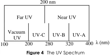
The UV spectrum is the portion of the electromagnetic spectrum between visible light and X-rays, with wavelength ranging from about 400 nm to about 100 nm. Depending on its spectral properties and applications, the UV spectrum is subdivided into four regions as shown in Figure 4. Another classification mode of the UV spectrum refers to the longer wavelengths up to 200 nm as near-UV while those above 200 nm are called far-UV. Table 3 shows the Characteristic effects of each off the four regions of the UV spectrum on living cells.
Figure 4 The UV Spectrum
Table 3 Ultra-violet light characteristics [23]
Type Wavelength
Region Range (nm) Characteristics UV-A Long 320-400 Tanning
UV-B Medium 280-320 Skin burning (cancer) UV-C Short 200-280 Germicidal range
Vacuum UV range UV-V Very short 100-200
Although the broad spectrum from infrared to UV can be used for microbial inactivation, most of the germicidal effects have been attributed to the UV region, particularly the UV-C. Absorption of radiation within the ultraviolet range originates mainly from protein, which is the building block of most vital components of biological tissues or organelles. Values of bond energies and corresponding wavelengths of some of the common bonds that characterise biological systems are listed in Table 4
[24]. Also, the photon energy (hν = hc/λ),
corresponding to each region of the UV and visible portions of the electromagnetic spectrum are shown in Table 5.
Comparison of last column of Table 4 with second column of Table 5 indicates that the bond energies in microbial molecules are generally coincident with photon energies in the UV portion of the spectrum. This assertion has been supported by several documented studies [4, 6, 19, 26-28], which indicate that UV light generally can be effective for various
decontamination and sterilization processes.
Specifically, pulsed UV light has been reported to be more efficient in inactivation of bacteria and other microbial organisms than continuous flow UV light [1, 4-6]. The transmission electron micrographs of intense
pulsed light (IPL-) and UVC-induced cell damage [1, 13] indicate that bacterial cell structures were destroyed by IPL treatment but not by UVC treatment. However, within the UV domain, the UVC component was observed to be most efficient in inducing structural damage of cells. Figure 5 shows scanning electron micrographs revealing how some bacteria cell damage are induced by a UV aided photo-catalytic process, using titanium dioxide [21]. Clearly, the pictures indicates that UV-C induced more structural damage than under UV-B or UV-A for the three bacteria studied.
Table 4 Typical bond energies of important biological moieties and their corresponding wavelengths [24]
Bond Typical bond energy (kJ/mole)
Corresponding wavelength (nm)
O-H 460 260
C-H 410 290
N–H 390 310
C-O 370 320
C═C 830 140
C═C 620 190
C≡N 850 140
C═O 740 160
C═N 600 200
Table 5 Characteristic wavelengths and corresponding photon energies of radiation within UV and visible portions of the electromagnetic spectrum [25]
Portion of
spectrum Radiation type Characteristic wavelength (nm)
Characteristic photon energy (eV)
UV Vacuum 50-200 24.0-6.0
C 200-270 6.0-4.4
B 270-330 4.4-3.6
A 330-400 3.6-3.0
Visible Violet 420 2.8
Blue 470 2.5
Green 530 2.3
Yellow 580 2.1
Orange 620 1.9
Red 700 1.7
Figure 5 Scanning electron photomicrograph of Escherichia coli (A), Listeria monocytogenes (B) and Salmonella typhimurium (C) treated in a UV-assisted TiO2
-PCO reaction [21])
Treatment time (min)
Escherchia coli
UV-A UV-B UV-C
Listeria monocytogenes
UV-A UV-B UV-C
Salmonella Typhimurium
UV-A UV-B UV-C
0
3
30
60
200 nm
Far UV Near UV
Vacuum
UV UV-C UV-B UV-A
280 λ (nm)
200
The complete UV spectrum is present in natural sunlight. However, the UV-A wavelengths are the predominant UV wavelengths reaching the earth’s surface. UV-B wavelengths are mostly absorbed by stratospheric ozone of the atmosphere, although increased levels are now reaching the earth’s surface due to the hole in the ozone layer. The UV-C range does not reach the earth because it is completely absorbed by ozone and oxygen in the earth’s atmosphere. Thus the natural light present in the earth surface becomes of little consequence in environmental situations with regards to effective microbial inactivation. This has led to the development of a variety of artificial UV light sources for use in disinfection technologies. These include low- and medium-pressure mercury arcs, excimer lamps and flashlamps. A summary of such UV sources and their basic characteristics are shown in Table 6.
Practical UV disinfection systems have traditionally used low-pressure mercury lamps as the source of germicidal radiation. Low-pressure mercury lamps, at pressures of 100-200 Pa, provide several emission lines in the visible, and two lines in the UV region at wavelengths of 185 nm and 254 nm. Most of the output power occurs at 254 nm (40 to 50%), and since other lines, including the 185 nm UV line, are outside the region for microbial inactivation, low-pressure mercury lamps are considered to provide monochromatic radiation as far as disinfection is concerned. Medium-pressure mercury UV lamps are now often used as an alternative to low-pressure lamps. These medium-pressure lamps use mercury pressures of hundreds of kPa, provide increased power, and emit several broad lines over the germicidal wavelength range between 200 nm and 300 nm, together with a continuum due to
recombination radiation. Although the medium-pressure lamp is less electrically efficient than the low-pressure lamp, it emits a substantially higher light intensity and thus provides a given UV germicidal dose in a much shorter irradiation time. The UV radiation from both types of mercury lamp is emitted continuously (CW).
An alternative to mercury lamps as an intense source of UV radiation is the pulsed xenon arc, or xenon flashlamp. The xenon pressure is usually in the range 50-100 kPa; and, under pulsed conditions, a xenon flashlamp emits several strong UV lines suitable for inactivation of microorganisms. When operated continuously, the xenon arc is an inefficient light source with a significant part of its light emission being in the infrared as a result of excitation of atomic xenon, giving XeI lines [29, 30]. On the other hand, when a high-energy pulse is applied to produce a high-current arc for a few ms, excitation of xenon ions takes place and many of the resultant XeII lines are in the UV region [29, 31].
Generally, it is easier to develop and work with UV sources in the continuous mode than in the pulsed mode. The pulsed light system requires a further step of converting the continuous to a pulsed mode. As can be seen from the Table 4, most of the developed UV sources work in the continuous mode rather than the pulsed mode. It will be economically less viable and therefore needless to insist on using the pulsed mode without some derivable benefits. We shall compare the efficacies of the continuous and pulsed modes in a subsequent section. Before then, we briefly consider the historical development of pulsed light as used for microbial disinfection.
Table 6 Summary of UV sources and their basic characteristics (Modified from [25])
Radiation Source UV Emission
Spectrum UV-C Wavelength (nm)
Electrical Efficiency (%)
Lamp Surface Temperature (°C)
Deployment Method
Low-Pressure Hg Arc Monochromatic 254 30-50 40 Continuous Medium-Pressure Hg Arc Monochromatic 200-300 15-30 400-1000 Continuous
Excimer Monochromatic Tunable 10-35 Ambient Continuous
Flashlamp Monochromatic 200-300 15-20 - Pulsed
5.0 TRENDS IN MICROBIAL INACTIVATION
USING PULSED LIGHT
According to literature [12], the first published works on microbial inactivation using pulsed light were reported by Bank in the early 90s [32, 33]. However, early works on microbial disinfection using flash lamps were said to have taken place in Japan in the late 70s, with a reported patent [34]. Before then, microbial inactivation using continuous wave UV-C has been in existence as far back as the 1960s [12]. There are quite a good number of microbes (both pathogenic and non-pathogenic) that may be of
interest to researchers in the area of microbial disinfection with pulse light. By now, a considerable number of works must have been done in this regards. However, for the purpose of comparison and critical review, we focus on some past work done on inactivation of two common bacteria
species- Escherichia coli and Listeria monocytogenes
– with flash lamps. Tables 7a and 7b show a summary of some selected works that were done in relation to
inactivation of Escherichia coli and Listeria
Table 7(a) Some reported inactivation of Escherichia coli using Xenon Flash Lamp
Lamp Spectral range (nm)
Treatment time (s)
Pulse power (W/m2)
Pulse Duration (μs)
Total Fluence (J/cm²)
Pulse Per Sec (Hz)
Host Medium Log
Reduction Sample- lamp distance (cm)
Ref.
- 60 455 10 - 5 0.1M K2HPO4 buffer 7 12 [1]
- 150 376 10 - 5 0.1M K2HPO4 buffer 7 12 [1]
- 17* 20 # - - 1 Sterile ringer solution 8 8 [2]
200-1100 3.3 ** - 360 4 3 Apple Juice 3.1 1.9 [7]
200-1100 4.2 ** - 360 5.1 3 Apple Juice 4.9 1.9 [7]
100-1100 - - 360 4 3 Apple Juice 4 5.8 [8]
100-1100 - - 360 4 3 Orange Juice 2.9 5.8 [8]
- 60 455 10 - 5 0.1M K2HPO4 buffer 7 12 [1]
- 150 376 10 - 5 0.1M K2HPO4 buffer 7 12 [1]
- 60 455 10 - 5 0.1M K2HPO4 buffer 7 12 [1]
- 150 376 10 - 5 0.1M K2HPO4 buffer 7 12 [1]
* No of pulses ** Av No of pulses
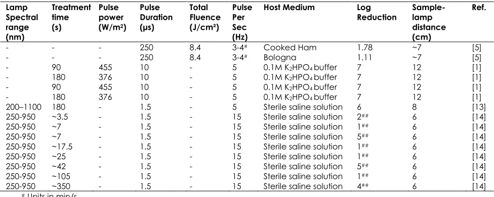
Table 7(b) Some reported inactivation of Listeria monocytogenes using Xenon Flash Lamp
Lamp Spectral range (nm)
Treatment time (s)
Pulse power (W/m2)
Pulse Duration (μs)
Total Fluence (J/cm²)
Pulse Per Sec (Hz)
Host Medium Log
Reduction Sample- lamp distance (cm)
Ref.
- - - 250 8.4 3-4# Cooked Ham 1.78 ~7 [5]
- - - 250 8.4 3-4# Bologna 1.11 ~7 [5]
- 90 455 10 - 5 0.1M K2HPO4 buffer 7 12 [1]
- 180 376 10 - 5 0.1M K2HPO4 buffer 7 12 [1]
- 90 455 10 - 5 0.1M K2HPO4 buffer 7 12 [1]
- 180 376 10 - 5 0.1M K2HPO4 buffer 7 12 [1]
200–1100 180 - 1.5 - 5 Sterile saline solution 6 8 [13]
250-950 ~3.5 - 1.5 - 15 Sterile saline solution 2## 6 [14]
250-950 ~7 - 1.5 - 15 Sterile saline solution 1## 6 [14]
250-950 ~7 - 1.5 - 15 Sterile saline solution 5## 6 [14]
250-950 ~17.5 - 1.5 - 15 Sterile saline solution 1## 6 [14]
250-950 ~25 - 1.5 - 15 Sterile saline solution 1## 6 [14]
250-950 ~42 - 1.5 - 15 Sterile saline solution 5## 6 [14]
250-950 ~105 - 1.5 - 15 Sterile saline solution 1## 6 [14]
250-950 ~350 - 1.5 - 15 Sterile saline solution 4## 6 [14]
# Units in min/s
## Values are of survival fraction
One thing that is apparent from the data in Tables 7 (a and b) is absence of standardised values for the different parameters which could make for easy comparison. Values for parameters such as number of pulses/treatment time, pulse energy/power, sample-flash lamp separation, pulse frequency, etc, are chosen and used loosely at the discretion of the researcher. For example, stating only the fluence or the log reduction does not give adequate information for comparison with data from other similar experiments. A higher log reduction could have resulted from too much fluence either due to high exposure time or high energy pulses. Hence, there is a need to have a specific parametric value (e.g. log reduction per unit fluence at standard values of other parameters) that can make for easy comparison. Also, in spite of the importance attached to spectral range in inactivation process, some authors still could not comprehensively state the parameter in their reports
A fluence-related parameter which can be used to compare the effectiveness of pulsed light in inactivating different microbial organisms is called “decimal reduction fluence, DF1” [9]. It is defined as
the fluence required for a single pulse to inactivate a
microorganism by 1log (or reduce population by 90%). This is illustrated in Figure 6.
For a linear graph, DF1 is given by the slope of the
graph. The significance of DF1 lies in the fact that the
lower the value of DF1, the more effective is the
pulsed light in inactivating the organism. On the
other hand, higher value of DF1 implies that the
Figure 6 Example of relationship between inactivation effects by a single-pulsed light and fluence (adapted from [9])
5.1 Continuous Flow UV Light versus Pulsed UV Light
The great inactivating effects of pulsed light has been attributed to the near total absence of photo repair enzymes in the DNA damaged molecules of cells irradiated with pulsed light [9]. In cells irradiated with continuous UV radiation, the effects of inactivation are reversible due to the presence of photo repair enzymes. The power and close repetitive pattern of pulsed light does not give time for the DNA cells to undergo or complete any repair or adaptation process.
5.2 Pulsed Laser
Laser irradiation has been adopted as a technique for curing or controlling various bacteria-related diseases in medicine and dentistry [35-38]. The use of lasers in these and other health-related fields is dependent on the absorption ability of interacting targets or media. Like other pulsed lights, the mechanisms of pulsed laser action on bacteria are still being developed. So far, explanations for interactions of both pulsed light and pulsed laser are of closed similarities. A conducted study [37] suggests that irradiation of bacteria with laser causes some changes in the bacterial cells which inhibits the process of cell metabolism. The essence of that study was to use total adenosine triphosphate (ATP) estimation in bacteria as a measure of bacterial survival. The result shows that irradiation for shorter durations (about 3-5min) resulted in reduced ATP concentrations but not increased bacterial death. Increased bacterial mortality resulted only when reduced ATP concentrations were sustained. Moreover, regression analysis between the total viable count (TVC) and ATP concentration in the
irradiated samples indicate no significant
relationship. The lack of correlation is taken as a sort of supportive evidence to imply the occurrence of a laser-mediated inhibition of metabolic processes in living bacterial cells.
Just like pulsed light, the impact of pulsed laser irradiation on bacteria depends on wavelength, power density and duration of exposure and the resultant laser-bacteria interaction can be from photochemical, photo-thermal photo-ablative or photomechanical reactions [37]. Low power laser irradiation for a sufficient time period results into photochemical reactions which cause damage within reparable limit. When a high power laser is used, photo-thermal reaction also sets in, causing vaporization and denaturing of cell components thereby resulting into permanent damage. With very high power laser beam, there are photo-ablative (breaking of chemical bonds) and photomechanical (creation of a localized plasma) reactions, the combined effects of which causes instant fatality. Peak power of laser radiation were observed to influence bactericidal effects as the use of longer pulse durations were seen to eliminate higher percentages of bacteria, at greater depth [36].
Obviously the level of impact made by pulsed laser irradiation should depend on total fluence received since there cannot be interaction without the application of energy. Wavelength, power and exposure time can therefore serve as fluence-determining parameters. A range of lasers of various wavelengths, power densities and/or other related parameters can therefore be used to experimentally determine their effectiveness in inactivating bacteria.
6.0 PROSPECTS AND CHALLENGES
The use of pulsed light for bacterial and other microbial inactivation tend to gain higher acceptance in the medical pathology and food preservation industries over the use of chemicals or thermal technology for some reasons. First is that generally, the use of light has less damaging effects on treated samples and reduces after –treatment
effects such as corrosion, odour, protein
denaturation, altered chemical composition, etc, which are often associated with chemical or thermal treatment [20]. In water treatment, the use of chlorine as a major disinfectant has generated some concerns because of the formation of additional potentially toxic disinfection by-products [2]. Moreover, the presence of some microorganisms,
such as the oocysts of cryptosporidium and Giardia,
which are highly resistant to chemical treatments, calls for the need for other alternative disinfectants of which UV light has been identified as a viable option [2, 4].
In comparison to continuous wave light, pulsed light can be used to deliver high energy dose within shorter time, which is of practical importance for situations requiring rapid disinfection. Also, the pulsed light obtained from xenon flash lamps is mercury-free and do not require a warm-up period (i.e. they are instant-on) [4]. This makes them more environment friendly than continuous wave UV light.
1000000
100000
10000
1000
100
10
1 2
0 4 6 8 10 12 14 16
DF1
N
u
m
b
er
o
f
su
rv
iv
o
rs
The major challenge in the use of pulsed light is in the area of sample heating resulting from photo-thermal effects or lamp heating. To mitigate the effects of sample heating, experiments and patents should be designed with improved cooling systems to remove the heat generated. In addition, the use of bowers in inactivation chambers serve the dual purpose of removing both the generated heat as well as the excess ozone generated by the UV component of the pulsed light [8].
Another possible limitation in the use of pulsed light is in the depth of penetration of light in the materials being processed. Depth of penetration is usually limited by the power of the pulsed light in addition to the level of transparency of the sample material being processed. It has been stated [12] that there are no independent experimental reports to confirm some claims of pulse light having more penetrating power than continuous wave light. The use of lasers has resulted in improved penetrating power in dentistry [36]. However, the diffraction-limited nature of laser beams also limits the amount of material surface that can be treated within a specified time when compared to other pulsed light systems.
7.0
CONCLUSION
Generally, light has less damaging effects to the surrounding environment when used as agents for materials processing. This unique advantage is being harnessed in the interaction of pulsed light with microbes. The fluence (or any other parameter) of a pulsed light system could be manipulated so that bacteria are destroyed with minimal damaging effects on their host substrate. Whereas broad band UV light may be more convenient only in superficial treatments, well directed pulsed light, such as laser, can be delivered to more remote and hidden positions in substrates, where bacteria are often well accommodated.
References
[1] Cheigh, C., M. Park, M. Chung, J. Shin and Y. Park. 2012. Comparison of Intense Pulsed Light- and Ultraviolet (UVC)-Induced Cell Damage in Listeria Monocytogenes and Escherichia coli O157:H7.Food Control. 25(2): 654-659. [2] Sharifi-Yazdi, M. and H. Darghahi. 2006. Inactivation of
Pathogenic Bacteria Using Pulsed UV-Light and its Application in Water Disinfection and Quality Control.
Acta Medica Iranica. 44(5): 305-308.
[3] Artíguez, M., A. Lasagabaster, and I. Marañón. 2011. Factors Affecting Microbial Inactivation by Pulsed Light In A Continuous Flow-Through Unit For Liquid Products Treatment.Procedia Food Science. 1(0): 786-791. [4] Bohrerova, Z., H. Shemer, R. Lantis, C. Impellitteri and K.
Linden. 2008. Comparative Disinfection Efficiency of Pulsed and Continuous-Wave UV Irradiation Technologies.
Water Research. 42(12): 2975-2982.
[5] Hierro, E., E. Barroso, L. la Hoz, J. Ordóñez, S. Manzano and M. Fernández. 2011. Efficacy of Pulsed Light for Shelf-Life
Extension and Inactivation of Listeria Monocytogenes on Ready-to-Eat Cooked Meat Products. Innovative Food
Science & Emerging Technologies. 12(3): 275-281.
[6] Farrell, H., J. Hayes, J. Laffey, and N. Rowan. 2011. Studies on the Relationship Between Pulsed UV Light Irradiation and the Simultaneous Occurrence of Molecular and Cellular Damage in Clinically-Relevant Candida Albicans.
Journal of Microbiological Methods. 84(2): 317-326.
[7] Muñoz, A., I. Caminiti, I. Palgan, G. Pataro, F. Noci, D. Morgan, D. Cronin, P. Whyte, G. Ferrari, and J. Lyng. 2012. Effects on Escherichia Coli Inactivation and Quality Attributes in Apple Juice Treated by Combinations of Pulsed Light and Thermosonication. Food Research
International. 45(1): 299-305.
[8] Pataro, G., A. Muñoz, I. Palgan, F. Noci, G. Ferrari, and J. Lyng. 2011. Bacterial Inactivation in Fruit Juices Using a Continuous Flow Pulsed Light (PL) System.Food Research
International. 44(6): 1642-1648.
[9] Palmieri, L. and D. Cacace. 2005. High Intensity Pulsed Light Technology. In: Da-Wen, S. ed. Emerging
Technologies for Food Processing. London: Academic
Press. 279-306;
[10] Wang, T., S. MacGregor, J. Anderson, and G. Woolsey. 2005. Pulsed Ultra-Violet Inactivation Spectrum of Escherichia coli.Water Research. 39(13): 2921-2925. [11] Farkas, B. and Z. Geretovszky. 2006. On Determining the
Spot Size for Laser Fluence Measurements. Applied
Surface Science. 252(13): 4728-4732.
[12] Gómez-López, V., P. Ragaert, J. Debevere, and F. Devlieghere. 2007. Pulsed Light for Food Decontamination: A Review. Trends in Food Science &
Technology. 18(9): 464-473.
[13] Cheigh, C., H. Hwang, and M. Chung. 2013. Intense Pulsed Light (IPL) and UV-C Treatments for Inactivating Listeria Monocytogenes on Solid Medium and Seafoods. Food
Research International. 54(1): 745-752.
[14] Choi, M., C. Cheigh, E. Jeong, J. Shin, and M. Chung. 2010. Nonthermal Sterilization of Listeria Monocytogenes in Infant Foods by Intense Pulsed-Light Treatment.Journal of
Food Engineering. 97(4): 504-509.
[15] Brygo, F., C. Dutouquet, F. Le Guern, R. Oltra, A. Semerok, and J. Weulersse. 2006. Laser Fluence, Repetition Rate and Pulse Duration Effects on Paint Ablation. Applied
Surface Science. 252(6): 2131-2138.
[16] Simrock. Laser Damage Threshold. 2013. Available: http://www.laser2000.co.uk/technotes/TN_LaserDamageT hreshold.pdf.
[17] Wang, B., G. Dai, H. Zhang, X. Ni, Z. Shen, and J. Lu. 2011. Damage Performance of Tio2/Sio2 Thin Film Components Induced By A Long-Pulsed Laser.Applied Surface Science. 257(23): 9977-9981.
[18] Geeraerd, A., V. Valdramidis, and J. Van Impe. 2005. GInaFiT, A Freeware Tool to Assess Non-Log-Linear Microbial Survivor Curves. International Journal of Food
Microbiology. 102(1): 95-105.
[19] Marquenie, D., A. Geeraerd, J. Lammertyn, C. Soontjens, J. Van Impe, C. Michiels and B. Nicolaı̈. 2003. Combinations of Pulsed White Light and UV-C or Mild Heat Treatment to Inactivate Conidia of Botrytis Cinerea and Monilia Fructigena. International Journal of Food
Microbiology. 85(1-2): 185-196.
[20] Turtoi, M. and A. Nicolau. 2007. Intense Light Pulse Treatment as Alternative Method for Mould Spores Destruction on Paper–Polyethylene Packaging Material.
Journal of Food Engineering. 83(1): 47-53.
[21] Kim, S., K. Ghafoor, J. Lee, M. Feng, J. Hong, D. Lee, and J. Park. 2013. Bacterial Inactivation in Water, DNA Strand Breaking, and Membrane Damage Induced by Ultraviolet-Assisted Titanium Dioxide Photocatalysis.Water
Research. 47(13): 4403-4411.
[22] Takeshita, K., J. Shibato, T. Sameshima, S. Fukunaga, S. Isobe, K. and M. Itoh. 2003. Damage of Yeast Cells Induced by Pulsed Light Irradiation.International Journal
[23] Guerrero-Beltr·n, J. and G. Barbosa-C·novas. 2004. Advantages and Limitations on Processing Foods by UV Light.Food Science and Technology International. 10(3): 137-147.
[24] Smith, M. 2013. March's Advanced Organic Chemistry:
Reactions, Mechanisms, and Structure. 7th Edition: Wiley.
[25] Blatchley III, E. and M. Peel. 2001. Disinfection by ultraviolet irradiation. In: Block, S. S. ed. Disinfection, sterilization, and
preservation. Philadealphia, USA: Lippincott Williams and
Wilkins. 823- 829.
[26] Ramos-Villarroel, A. Y., Aron-Maftei, N., Martín-Belloso, O. and Soliva-Fortuny, R. 2012. Influence of Spectral Distribution on Bacterial Inactivation and Quality Changes of Fresh-Cut Watermelon Treated With Intense Light Pulses.
Postharvest Biology and Technology. 69(0): 32-39.
[27] Matafonova, G. and V. Batoev. 2012. Recent Progress on Application of UV Excilamps for Degradation of Organic Pollutants and Microbial Inactivation. Chemosphere. 89(6): 637-647.
[28] Azar Daryany, M., S. Hosseini, M. Raie, J. Fakharie, and A. Zareh. 2009. Fakharie, J. and Zareh, A. Study on Continuous (254 Nm) and Pulsed UV (266 and 355 Nm) Lights on BVD Virus Inactivation and Its Effects on Biological Properties of Fetal Bovine Serum. Journal of
Photochemistry and Photobiology B: Biology. 94(2):
120-124.
[29] Saloman, E. 2004. Energy Levels and Observed Spectral Lines of Xenon, Xe I through Xe LIV.Journal of Physical and
Chemical Reference Data. 33(3): 765-921.
[30] Meggers, W., T. De Bruin, and C. Humphreys. 1929. The First Spectrum of Xenon. Science (New York, N.Y.). 69(1789): 406.
[31] Humphreys, C. 1939. Second Spectrum of Xenon.J. Res.
Natl. Bur. Stand.(US). 22: 19-53.
[32] Bank, H., J. John, M. Schmehl, and R. Dratch. 1990. Bactericidal Effectiveness of Modulated UV Light.Applied
and Environmental Microbiology. 56(12): 3888-3889.
[33] Tessman, I. and H. Bank. 1992. Documentation of the Claim that Modulation of UV Light Pulses Increases the Bactericidal Effectiveness of the Light [2]. Applied and
Environmental Microbiology. 58(2): 778-779.
[34] Hiramoto, T. 1984. Method of Sterilization. 4,464,336. [35] Andrade, A., I. Feist, C. Pannuti, S. Cai, D. Zezell, and G.
Micheli. 2008. Nd:YAG Laser Clinical Assisted in Class II Furcation Treatment.Lasers in Medical Science. 23(4): 341-347.
[36] Franzen, R., N. Gutknecht, S. Falken, N. Heussen, and J. Meister. 2011. Bactericidal Effect of a Nd:YAG Laser on Enterococcus Faecalis at Pulse Durations of 15 And 25 Ms in Dentine Depths of 500 and 1,000 μm.Lasers in Medical
Science. 26(1): 95-101.
[37] Nandakumar, K., H. Obika, T. Shinozaki, T. Ooie, A. Utsumi, and T. Yano. 2003. Laser Impact on Bacterial ATP: Insights into the Mechanism of Laser-Bacteria Interactions.
Biofouling. 19(2): 109-114.
![Figure 2External trigger Block diagram of a pulsed UV system (adapted from [2])](https://thumb-us.123doks.com/thumbv2/123dok_us/1309868.1163603/3.612.63.285.436.538/figure-external-trigger-block-diagram-pulsed-uv-adapted.webp)
![Table 2 Relations for calculation of some output parameters of pulsed light (modified from [16])](https://thumb-us.123doks.com/thumbv2/123dok_us/1309868.1163603/4.612.109.511.495.684/table-relations-calculation-output-parameters-pulsed-light-modified.webp)

![Table 6 Summary of UV sources and their basic characteristics (Modified from [25])](https://thumb-us.123doks.com/thumbv2/123dok_us/1309868.1163603/7.612.89.524.487.557/table-summary-uv-sources-basic-characteristics-modified.webp)
![Figure 6 Example of relationship between inactivation effects by a single-pulsed light and fluence (adapted from [9])](https://thumb-us.123doks.com/thumbv2/123dok_us/1309868.1163603/9.612.56.286.60.213/figure-example-relationship-inactivation-effects-single-fluence-adapted.webp)