extreme environments
A thesis submitted to the University of London for the
Degree of Doctor of Philosophy by
Rory A. Cameron
Department of Biochemistry and Molecular Biology
University College London
Gower Street
London W CIE 6BT
All rights reserved
INFORMATION TO ALL USERS
The quality of this reproduction is dependent upon the quality of the copy submitted.
In the unlikely event that the author did not send a complete manuscript and there are missing pages, these will be noted. Also, if material had to be removed,
a note will indicate the deletion.
uest.
ProQuest 10015790
Published by ProQuest LLC(2016). Copyright of the Dissertation is held by the Author.
All rights reserved.
This work is protected against unauthorized copying under Title 17, United States Code. Microform Edition © ProQuest LLC.
ProQuest LLC
789 East Eisenhower Parkway P.O. Box 1346
Abstract
In a study prior to this work, screening experiments for thermophilic nitrile degrading
organisms led to the identification of a moderately thermophilic Bacillus sp. (strain
RAPc8). The organism was shown to constitutively express a nitrile hydratase
(NHase), which was purified and extensively characterised.
This work reports the cloning of a gene cluster containing the NHase genes of
Bacillus sp. RAPc8 into Escherichia coli. Sequencing of a 5.9kb section of cloned DNA revealed the presence of eight complete open reading frames, encoding (in order),
amidase (belonging to the nitrilase related aliphatic amidase family), nitrile hydratase (3
and a subunits (of the cobalt containing class), a putative 122 amino acid protein of unknown function, designated P14K, a homologue of the 2Fe-2S class of ferredoxins
and three putative proteins with distinct homology to the cobalt uptake proteins cbiM,
cbiN and cbiQ of the S. typhimurium LT2 cobalamin biosynthesis pathway. The NHase
operon shares an exceptionally high sequence identity with that of Bacillus sp. BR449. The deduced NHase a subunit N-terminal sequence bore no similarity the empirically
determined sequence of the previous study. However, in this study, N-terminal
sequencing of the recombinant protein confirmed the sequence predicted by gene
translation.
The amidase and nitrile hydratase genes of Bacillus sp. RAPc8 were subcloned and
actively expressed in E. coli. The recombinant NHase was partially purified and found
to exhibit catalytic behaviour very similar to the native protein. Substrate specificity
trials showed that the recombinant enzyme, as with the native enzyme, has broad
Structure was built using the crystal structure of the NHase of Rhodococcus sp. R312 as a template. Aromatic residues in the active site, identified from the model were
replaced with non-polar or charged residues using site directed mutagenesis. Of the six
mutants investigated, none demonstrated any catalytic activity on the aromatic nitriles
tested. However, two mutants, PY67E and pW76G, did display an apparent decrease
benzonitrile inhibition (rise in Ki of ~6.5mM and 29.25mM respectively). A further
two mutants (p36L and PY67A) also showed a marked increase in specific activity in
crude cell extracts compared with that of the wild type enzyme.
A PCR based method for the identification of NHase genes in environmental soil and
sediment samples was developed and employed to verify the presence of such genes in
samples taken from New Zealand hot pools. Six unique sequences corresponding to the
partial sequence of the a subunit gene were identified. At the DNA level, these
sequences showed significant homology with the Bacillus NHase sequences (between
83% and 93% sequence identity with the Bacillus sp. RAPcS gene). DNA isolated from
NHase positive sediment samples was used to create a substantial multi-genomic library
for tthe purpose of screening for full-length NHase genes. However, attempts to identify
functional nitrile hydratase genes through complementation screening on succinate-
Acknowledgements
I would like to acknowledge the Biotechnology and Biological Sciences Research Council for provision o f the funding for my research at UGL.
I thank my supervisor, Professor D on Cowan for his support, his faith in my abihties and for providing me with the opportunity to undertake this research.
I gratefully acknowledge the invaluable help and advice of Professor Peter Piper, Dr. John Ward, Dr. Haitham Hussain, Dr. Stefan MiUson and Dr. Mehdi MoUapour in various aspects o f microbiology and molecular biology: without their help, this work would have taken a good deal more blood, sweat and tears than it already has!
Many thanks to all past and present members o f the Ground Floor Labs for the requisite distractions and for helping me to see the light side of the dark days.
Paul, Emma, Rekha, Latha, George, Sam, Steve and Hannah I remain deeply indebted to you for your friendship, for your belief in me, for lending me your strength when mine was waning and generally for helping to keep my life rolling as smoothly as possible down a path that, over the past eight years has had it's fair share of daunting twists. I’U not forget you.
Finally, to my family I owe a similar debt for their love and continued support since year dot - I won't forget them either!
Table of contents
A b s t r a c t ...2
A c k n o w l e d g e m e n t s ...4
T a b l e o f c o n t e n t s ...5
L i s t o f f i g u r e s ...? L i s t o f t a b l e s ...10
A b b r e v i a t i o n s ... u C h a p t e r 1 : I n t r o d u c t i o n ...13
1.1 Nitriles...13
1.2 Nitrilase...16
1.3 Nitrile Hydratase...20
1.4 Amidase...37
1.5 Applications o f NHases...40
1.6 Thermophiles...42
1.7 Thennostable enzymes...43
1.8 Bacillus sp. RAPcS...46
1.9 Bacillus sv. RAPcS NH ase...46
1.10 Aim s...48
C h a p t e r 2 : M a t e r i a l s a n d M e t h o d s ...49
2.1 Chemicals and reasents...49
2.2 Enzymes...49
2.3 Media...49
2.4 Buffers and solutions...52
2.5 Bacterial strains...54
2.6 Plasmids...55
2.7 Analytical Procedures...56
2.8 Growth and maintenance o f cultures...59
2.9 Preparation o f competent E. coli cells...59
2.10 Transformation o f competent E. coli cells...60
2.11 DNA Purification...61
2.12 Polymerase chain reaction (PCR)...63
2.13 Site-directed mutasenesis...63
2.14 Restriction endonuclease digestion o f DNA...64
2.15 DNA cloning...64
2.16 DNA hybridisation...67
2.17 Protein expression...70
2.18 Cell free extract preparation...71
2.19 Protein purification...71
2.20 Growth and expression curves... 72
2.22 Sequencins protocols...74
2.23 Computational techniques...74
C h a p t e r
3
: C l o n i n g t h e n i t r i l e
HYDRATASE OPERON FROM
BACILLUS
SP.
R A P cS ... 76
3.1 Introduction...76
3.2 Verification o f the identity o f Bacillus sp. RAPcS... 77
3.3 Clonins Stratesies...78
3.4 Nitrile hydratase operon structure...99
3.5 Discussion...110
CHAPTER
4
: EXPRESSION AND
CHARACTERISATION... ii8
4.1 Introduction...118
4.2 Expression strates y...119
4.3 Subclonins o f NHase and amidase senes...120
4.4 Expression studies...125
4.5 Purification...130
4.6 Characterisation o f recombinant Nitrile hydratase...136
4.7 Discussion...146
C h a p t e r
5
: E n z y m e e n g i n e e r i n g ... 153
5.1 Introduction...153
5.2 Homolosy m odellins...153
5.3 Site directed mutasenesis...163
5.4 Expression o f mutants...170
5.5 Thermostability...172
5.6 Benzonitrile Inhibition...173
5.7 Discussion...177
C h a p t e r
6
: T h e s e a r c h f o r n o v e l n i t r i l e
-METABOLISING ENZYMES...
184
6.1 Introduction...184
6.2 DNA Extraction...185
6.3 PCR analysis ofWHA30 environmental DNA sample....187
6.4 Library construction...191
6.5 Library screening...194
6.6 Discussion...195
C
h a p t e r
?: G
e n e r a l
d i s c u s s i o n
... 201
List o f figures
Fig. 1.1: Metabolism of nitriles via hydrolysis. 15
Fig. 1.2: Nitrilase mode of action. 17
Fig. 1.3: Stereoview of the aP heterodimers of Rhodococcus sp. R312 NHase. 22
Fig. 1.4: Superimposition of Co-type NHase and Fe-type NHase. 25
Fig. 1.5: Three putative reaction mechanisms of NHase. 27
Fig. 1.6: Alignment of NHase p subunit N-terminal sequences. 32
Fig. 1.7: Alignment of NHase a subunit N-terminal sequences. 32
Fig. 1.8: Arrangement of NHase gene clusters 35
Fig. 1.9: Mechanism of the amidase acyl transfer 39
Fig. 1.10: Hydration of adiponitrile. 41
Fig. 1.11: RAPcS NHase p subunit N-terminal sequence BLAST search. 47
Fig. 3.1: Organisation of the NHase operon of R. erythropolis. 80
Fig. 3.2: PCR amplification of R. erythropolis JCM6823 NHase a subunit gene. 80
Fig. 3.3: Hybridisation with Rhodococcus NHase a subunit gene. 81
Fig. 3.4: Alignment of the a subunit sequences of 10 recently cloned NHases. 82
Fig. 3.5: Degenerate oligonucleotides design. 83
Fig. 3.6: Hybridisation with DIG end-labelled NHFl probe. 85
Fig. 3.7: Hybridisation with DIG end-labelled NHR2 probe. 85
Fig. 3.8: Hybridisation with DIG end-labelled NHF2 probe. 86 Fig. 3.9: Schematics outlining the arrangement of RAPcS NHase PCR primers. 88 Fig. 3.10: PCR amplification using primers NHFl 4- NHRI and NHF2 + NHR2. 91
Fig. 3.11: Annealing temperature optimisation of PCR reaction. 91
Fig. 3.12: Translated BLAST searches with the inserts of pNHFl and pNHF2. 93
Fig. 3.13: Sequences of the inserts from pNHFl and pNHF2. 94
Fig. 3.14: Hybridisation with DIG labelled PCR products NHFl and NHF2. 95
Fig. 3.15: Organisation of NHase genes within the insert of pNHB4. 97
Fig. 3.16: Organisation and sequences of primers AMD3, AMD2 and C8B2. 98
Fig. 3.17: Amplification of the amidase and partial NHase P subunit genes. 99
Fig. 3.19: Alignment of NHase p subunit peptide sequences. 102
Fig. 3.20: Alignment of the translation product of 0R F4 with
homologous proteins. 103
Fig. 3.21: Hairpin loop structure found just downstream of the P14K gene. 104
Fig. 3.22: Layout of the Bacillus sp. RAPcS gene cluster containing
the NHase genes. 106
Fig. 3.23: Nucleotide sequence of the RAPcS NHase gene cluster. 107
Fig. 3.24: The order of the cobl opérons of B. megaterium and S. typhimurium. 114
Fig. 3.25: Comparison of hydrophobicity plots of S. typhimurium cbiQ and cbiN
with RAPcS homologues. 115
Fig. 4.1: Primers used for cloning the RAPcS NHase genes
for expression studies. 120
Fig. 4.2: PCR amplification of genes of the Bacillus sp. RAPcS NHase operon. 121
Fig. 4.3: Cloning and subcloning of the RAPcS amidase gene. 122
Fig. 4.4: Cloning and subcloning of the RAPcS NHase genes. 123
Fig. 4.5: Expression profile of recombinant amidase 125
Fig. 4.6: SDS-PAGE analysis amidase expressed in E. coli (BL21) 1DE3 pLysS. 125
Fig. 4.7: Expression profile of recombinant NHase. 127
Fig. 4.8: SDS-PAGE analysis NHase expressed in E. coli (BL21) 1DE3 pLysS. 12S
Fig. 4.9: Heat purification of RAPcS amidase. 130
Fig. 4.10: SDS-PAGE showing amidase purification. 131
Fig. 4.11: Heat purification of RAPcS NHase. 133
Fig. 4.12: SDS-PAGE of fractions from NHase ion exchange chromatography. 134
Fig. 4.13: Comparison of results of Edman sequencing of native and recombinant
NHase with the deduced NHase a and p subunit N-terminal sequences. 135
Fig. 4.14: Thermostability profile for the purified NHase. 137
Fig. 4.15: Thermostability of recombinant NHase. 137
Fig. 4.16: Kinetics of acetonitrile hydrolysis. 142
Fig. 4.17: Kinetics of acrylonitrile hydrolysis. 143
Fig. 4.18: Benzonitrile inhibition of acetonitrile hydrolysis. 144
Fig. 5.4: Space filled model of the active site region of RAPcS NHase a subunit. 159
Fig. 5.5: Space filled model of the RAPcS NHase holoenzyme. 160
Fig. 5.6: Space filled model of the RAPcS NHase holoenzyme. 160
Fig. 5.7: Space filled model of an aP dimer of RAPcS NHase. 161
Fig. 5.8: Schematic of the Quik-change mutagenesis mutagenesis procedure. 163
Fig. 5.9: Restriction map of the RAPcS NHase genes. 165
Fig. 5.10: Confirmation of the mutation of aW 124 to G by treatment with Xmal. 166
Fig. 5.11: Confirmation of the mutation of PP36 to L by treatment with HaeJR. 166
Fig. 5.12: Confirmation of the mutation of pF52 to G by treatment with HaeUl. 167
Fig. 5.13: Confirmation of the mutation of PY67 to A by treatment with Fokl. 167
Fig. 5.14: Confirmation of the mutation of PY67 to E by treatment with Fokl. 16S
Fig. 5.15: Confirmation of the mutation of pW76 to G by treatment with Ncol. 16S
Fig. 5.16: SDS PAGE gel of cell free extracts of the expressed mutant proteins. 170
Fig. 5.17: Thermostability profile of wild type and mutated NHases. 171
Fig. 5.18: Benzonitrile inhibition of mutant pF36L. 173
Fig. 5.19: Benzonitrile inhibition of mutant PF52G. 173
Fig. 5.20: Benzonitrile inhibition of mutant pY67A. 174
Fig. 5.21: Benzonitrile inhibition of mutant pY67E. 174
Fig. 5.22: Benzonitrile inhibition of mutant pYW76G. 175
Fig. 5.23: Benzonitrile inhibition of mutant aW124G. 175
Fig. 6.1: Typical result of environmental DNA extraction from sample WHA30. 1S5
Fig. 6.2: Amplification of NHase fragments using primers NHFl and NHRI. 1S6
Fig. 6.3: Alignments of the DNA and deduced amino acid sequences of the six
environmental PCR amplicons. 1S9
Fig. 6.4: Overview of environmental library construction using pCR-XL-TOPO. 191
Fig. 6.5: EcoRI-digested plasmids recovered en masse from the library. 192
Fig. 6.6: Primers used in amplification of NHase genes from soil in relation to the
List of tables
Table 1.1: Molecular and functional characteristics of nitrilases. 19
Table 1.2: Thermal stability of NHases. 30
Table 1.3: Structural and functional properties of NHases. 36
Table 2.1: Buffers and solutions. 52
Table 2.2: Bacterial strains. 54
Table 2.3: Plasmids. 55
Table 2.4: SDS PAGE resolving gel constituents. 57
Table 2.5: SDS PAGE stacking gel constituents. 57
Table 2.6: Typical PCR reaction constituents. 63
Table 3.1: Sequence identity and similarity of RAPcS NHase with other NHases. 100
Table 3.2: Summary of the properties of the genes in the RAPcS NHase locus. 105
Table 3.4: Comparison of the MW of RAPcS amidase and NHase with
the native proteins. 110
Table 4.1: Effect of cobalt addition to cell cultures on NHase activity. 127
Table 4.2: Heat purification of RAPcS Amidase 131
Table 4.3: Purification of RAPcS NHase. 134
Table 4.4: Comparison of NHase thermostability. 13S
Table 4.5: Comparison of NHase substrate specificity 140
Table 4.6: Comparison of the kinetic data of the native and recombinant
RAPcS NHase on acetonitrile and acrylonitrile substrates. 141
Table 5.1: Details of the mutagenic primers used. 164
Table 5.2: Specific activities of each mutant of the recombinant RAPcS NHase. 170
Table 5.3: Estimated benzonitrile Ki for each of the RAPcS NHase mutants. 172
Table 6.1: Identity and similarity of the DNA and translation products of the six amplicons with the DNA and amino acid sequences of
Abbreviations
À angstrom
abs absorbance (-log Transmitance)
Amp ampicillin
ATP adenosine triphosphate
bp base pairs
BSA bovine serum albumin
Da Dalton
DIG digoxigenin
g gram
GC gas chromatography
h hour
k kilo (10^)
kb kilobase
kDa kilodalton
Km Michaelis Menten constant
L liter
m milli (10^)
M molar
min minute
Mw relative molecular weight
NB nutrient broth
°C degrees Celsius
ORF open reading frame
PAGE polyacrylamide gel electrophoresis
PCR polymerase chain reaction
SDS sodium dodcyl sulphate
Topt temperature optimum
U enzyme activity: |xmoles product formed/minute
v/v volume per volume
Vmax rate of enzyme-catalysed reaction at infinit substrate concentration
w/v weight per volume
X-gal 5-bromo-4chloro-3-indolyl-p-D-galactoside
P-gal p-galactosidase
p, micro (10'^)
dATP deoxyadenosine triphosphate
dCTP deoxycytosine triphosphate
dGTP deoxyguanosine triphosphate
dTTP deoxythymidine triphosphate
dUTP deoxyuridine triphosphate
The Amino Acid Codes
A Ala Alanine
R Arg Arginine
N Asn Asparagine
D Asp Aspartic acid
C Cys Cysteine
Q Gin Glutamine
E Glu Glutamic acid
G Gly Glycine
H His Histidine
I He Isoleucine
L Leu Leucine
K Lys Lysine
M Met Methionine
F Phe Phenylalanine
P Pro Proline
S Ser Serine
T Thr Threonine
W Trp Tryptophan
Y Tyr Tyrosine
V Val Valine
The Nucleotide Codes
A Adenine
C Cytosine
G Guanine
Chapter 1: Introduction
1.1 Nitriles
1.1.1 Origins of nitriles
The nitriles, also known as organic cyanides are defined by the general formula R-C=N.
The presence throughout the natural environment of compounds containing this cyano
group has been well documented, with reports of their synthesis or metabolism by
members of the plant, animal and bacterial Kingdoms (Legras et al, 1990).
Many organisms rely on the toxicity of organocyanide compounds as a defence
mechanism against herbivores, predators and parasites (Jones, 1979). For example,
some insects have the ability to spray cyanide and mandelonitrile (Duffey, 1981), whilst
certain strains of Streptomyces species produce toyamycin, an antimicrobial nitrile (Kikuchi, 1955). However, the greatest group of nitrilated compounds, the cyanogenic
glycosides, are mainly produced by plants, often in seeds, roots and the epidermal layers
of leaves (Legras et a l, 1990). As the plant is masticated, the cyanogenic glycosides
decompose and give rise to cyanide. Nitrile based compounds, such as 3-
indoleacetonitrile also serve as growth hormones during germination (Mascharak,
2002).
Nitriles are also produced (on the megaton scale) by man, to be used as base chemicals
for a wide range of syntheses, these include acrylonitrile and the polymers derived from
it in the manufacture of plastics and acrylic fibres, and adiponitrile, used as a precursor
in nylon-66 production (Wyatt and Linton, 1988). Nitriles are also used in the production of herbicides, pharmaceuticals and amino acids (Jallégeas et a l, 1980, Nagasawa and Yamada, 1989). The industrial effluents of these syntheses, as well as
the herbicides containing the -C=N moiety, such as 3,5-dibromo-4-hydroxybenzonitrile
(Bromoxynil) and 3,5-di-iodo-4-hydroxybenzonitrile (loxynil) are the chief sources of
anthropogenic nitriles in the environment (Jallegeas et al, 1980, Mascharak, 2002). Acrylonitrile and the nitrile containing herbicides are released to such an extent that
of nitrile-containing industrial effluents precludes disposal by incineration; thus they are
usually disposed of either at sea or below the water table (Mascharak, 2002, Wyatt and
Knowles, 1996, Wyatt and Linton, 1988).
Despite the commercial interest in these chemicals, the presence of sensitive
functionalities on some of the nitrile compounds prevents their chemical modification -
due to the harsh conditions required (e.g. heating with acid or alkali). For this reason,
the industry has turned to microbial biotransformations for the conversion of nitriles
into commodity chemicals. There is also widespread interest in their use in
bioremediation of industrial effluents (Nagasawa and Yamada, 1989).
1.1.2 Enzymatic degradation of nitriles
Five possible mechanisms have been described for the detoxification of cyanide and
nitriles (Legras, 1990):
1. Hydrogenation (of a limited number of nitriles) by nitrogenase containing
nitrogen-fixing bacteria.
2. Oxidation by Pseudomonas fluorescens involving dioxygenase or
monooxygenase followed by a cyanase.
3. Decomposition to cyanide followed by conversion to (3-cyanoalanine by
Enterobacter sp. and Chromobacterium violaceum.
4. Decomposition to cyanide followed by conversion to thiocyanate by a
rhodanese.
5. Hydration/hydrolysis of the -C=N function by nitrilase, or hydration by nitrile
hydratase followed by an amidase catalysed hydrolysis.
The fifth mechanism is the most widely characterised, and perhaps the most useful
reaction to microorganisms since the nitrile is metabolised to provide a source of carbon
and/or nitrogen for growth (Nagasawa and Yamada, 1989). The nitrilase (Nase;
EC 3.5.5.1) pathway catalyses the direct conversion of a nitrile to its corresponding
carboxylic acid (Fig. 1.1 A). The nitrile hydratase (NHase; EC 4.2.1.84) pathway
Fig. 1.1: Metabolism of nitriles via hydrolysis: A - nitrilase pathway, B - nitrile hydratase pathway.
Nitrilase
R-CN+2H20--- ► R-COOH+NH
3
B
Nitrile Amidase
hydratase ^
R-CN + H2O ► R-CONH2 --- ^ R-COOH + NH3
The two pathways exist in both the Eucaryotic and Prokaryotic domains, but, to the
authors knowledge there are currently no reports of nitrile-degrading activity in the
Archaea. Although the majority of the enzymes described are bacterial, there are
reports of nitrilase activity in three of the 21 plant families (Graminae, Cruciferae and
Musaceae) (Thimann and Mahadevan, 1964), as well as several fungal species
{Fusarium solani, Aspergillus niger and Pénicillium chrysogenum) (Harper, 1977). However, it is impossible to assess the true diversity of these enzymes without more
1.2 Nitrilase
Nitrilases catalyse the direct conversion of nitriles to their corresponding acids. They
form a branch of the superfamily (consisting of 13 branches) of carbon-nitrogen
hydrolysing thiol enzymes, to which they give their name: the Nitrilase superfamily
(Pace and Brenner, 2001). Members of the nitrilase branch of the superfamily are
broadly distributed throughout the living world and can be found in plants, animals
(C. elegans), fungi and many types of bacteria.
1.2.1 Composition
All Nases characterised to date are composed of a single subunit. Subunit sizes vary
considerably, from 23kDa to 76kDa, with the average size being 40-45kDa (Table 1.1).
Whilst several organisms are reported to produce functional Nase monomers
{Comamonas testosteroni (Levy-schil et ah, 1995) and Arthrobacter sp. J-1
(Bandyopadhyay et a l, 1986)), the majority are homomultimers. Aggregation of non
specific numbers of subunits has also been reported (Goldlust and Bohak, 1989). This
phenomenon appears to be due to a substrate activation process, whereby the presence
of substrate induces a conformational change, which leads to aggregation and a resultant
increase in activity, known as cooperative activation (Stevenson et a l, 1992, Yamamoto
et a l, 1996).
Generally, the sequence similarities between Nases are low (Cowan et a l, 1998,
Kobayashi et a l, 1992a). However, three residues: glutamate, lysine and cysteine are strictly conserved throughout the nitrilase superfamily, the cysteine is involved in a
CWE motif that is conserved throughout the nitrilase branch (Pace and Brenner, 2001).
Mutagenesis studies on the Nases of Rhodococcus rhodochrous J l; /?. rhodochrous K22
and Alcaligenes faecalis JM3 have demonstrated that C165 {R. rhodochrous J1 numbering) is essential for catalytic activity (Kobayashi et a l, 1993, Kobayashi et al,
1.2.2 Reaction mechanism
The centrally conserved cysteine residue of the nitrilase family is believed to act as the
active site nucleophile (Novo et al, 1995). This hypothesis is further supported by several studies that demonstrate that mutation of this residue yields completely inactive
enzymes (Kobayashi et al, 1993, Kobayashi et al, 1992a, Kobayashi et a l, 1992c) and by a recent report of the generation of nitrilase activity in the cysteine protease papain
by a Glnl9Glu mutation (Dufour et al, 1995).
The reaction mechanism proposed for the Nase enzymes involves a nucleophilic attack
on the nitrile carbon atom by the sulphydryl group of this cysteine with concomitant
protonation of the nitrogen to form a thioimidate. This complex is then hydrolysed to
an acylenzyme complex - generating ammonia, followed by a second hydrolysis -
liberating the acid and regenerated enzyme (Fig. 1.2) (Mahadevan and Thimann, 1964,
Pace and Brenner, 2001, Stevenson et al, 1992)
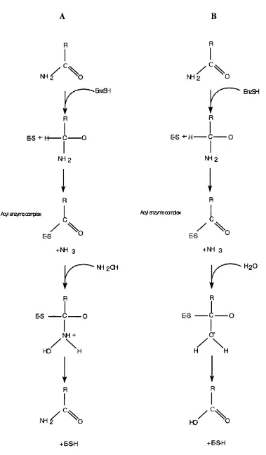
Fig. 1.2: Nitrilase mode of action.
EnzSH H2O
R-CN:
thioim idate
NH4+
\ .
S-E
Acyl e n z y m e com plex
H2O
E-S-H
O
\
OHHydrolysis of benzonitrile by F. solani Nase and phenylacetonitrile by Rhodococcus
ATCC 39484 Nase results in small percentages of the corresponding amide in the
product (Stevenson et a l, 1992). It has been suggested that since the rate of breakdown of the covalent intermediate is the rate limiting step, that a tetrahedral intermediate is
formed, which for certain substrates can break down to amide rather than the usual acid
1.2.3 Specificity
Early reports generally described the Nases as aromatic specific (Harper, 1977,
McBride et ah, 1986). However, over the past 12 years, a number of enzymes with a wide range of activities have been reported (Table 1.1), including the hydrolysis of
aliphatic mono- and dinitriles (Cramp et al, 1997b, Kobayashi et a l, 1990). Many of the Nases exhibit enantioselectivity and their ability to produce optically pure products
is of particular commercial interest. The Nase from Acinetobacter sp. AK226 has been
shown to convert racemic 2-(4’-isobutylphenyl) propionitrile to (S)-(+)-2-(4’-
isobutylphenyl) propionic acid (S-(+)-ibuprofen) with a 180 fold higher activity on the
S-(+)- substrate than the R-(-)-substrate (Yamamoto et al, 1990). Similar
enantioselectivity has been demonstrated by R. rhodochrous NCIMB 11216 Nase for
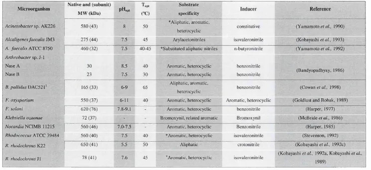
M icroorganism Native and (subunit)
MW (kDa) p H o p i
T o p t
CC)
Substrate
specificity Inducer Reference
Acinetobactersp. AK226 580 (43) 8 50 * Aliphatic, aromatic,
heterocyclic constitutive (Yamamoto era/., 1990)
Alcaligenes faecalisJM3 275 (44) 7.5 45 Arylacetonitriles isovaleronitrile (Kobayashi et al., 1993)
A. faecalisATCC 8750 460 (32) 7.5 40-45 ^Substituted aliphatic nitriles n-butyronitrile (Yamamoto e / a/., 1992)
A rth robactersp, J-1 Nase A
Nase B
30 23
8.5 7.5
40 30
Aromatic, heterocyclic Aromatic, heterocyclic
benzonitrile
benzonitrile (Bandyopadhyay, 1986)
B. pallidusT>AC52l^ 165 (33) 6-9 65 Aliphatic, aromatic,
heterocyclic benzonitrile (Cowan et al., 1998)
F. oxysporium 550 (37) 6-11 40 Aromatic, heterocyclic Aromatic, heterocyclic (Goldlust and Bohak, 1989)
F. solani 620 (76) 7.8-9.1 - Aromatic, heterocyclic benzonitrile (Harper, 1977)
Klebsiella ozaenae 72 (37) - - Bromoxynil, related aromatic Bromoxynil (McBride era/., 1986)
NocardiaFlClMR 11215 560 (46) 7.0-7.5 - Aromatic, heterocyclic Benzonitrile (Harper, 1985)
RhodococcusATCC 39484 560 (40) 7.5 40 * Aromatic, heterocyclic isovaleronitrile (Stevenson, 1992)
R. rhodochrous K22 650 (41) 5.5 50 Aliphatic crotonitrile (Kobayashi era/., 1992c)
R. rhodochrous ] I 78 (41) 7.6 45 Aromatic, heterocyclic isovaleronitrile
(Kobayashi et a i, 1992a, Kobayashi et ai,
1989)
Table 1.1: Molecular and functional characteristics of nitrilases. 'T h e rm o p h ilc o r g a n ism * E n a n tio se le c tiv e ‘^ R e g io se le c tiv e
1.3 Nitrile Hydratase
Since the initial isolation of an NHase from Nocardia rhodochrous LL-100-21 (Asano
et a l, 1980), a wide variety of microorganisms producing NHases have been described
and their enzymes characterised (Table 1.3). The Rhodococcus genera dominate the
known NHase producers, although recently a number of NHases from thermophilic
Bacillus sp. have been described (Cramp and Cowan, 1999, Padmakumar and Oriel, 1999, Pereira gr aZ., 1998, Takashima gr a/., 1998).
1.3.1 Size
Typically, NHases are heteromultimers composed of two distinct subunits, a and p. The
subunits have similar molecular weights ranging from 22 to 28kDa; by convention the p
subunit is the larger. It is commonly held that whilst the minimal functional unit is an
a p dimer, NHases are usually heterotetrameric in structure - (aP)%. R. rhodochrous J1 produces two NHases: L-NHase, which is tetrameric and H-NHase, which forms
multimers of up to 11 aP dimers (Nagasawa et a l, 1991). The molecular weight of
Rhodococcus sp. N-771 in solution appears to be in equilibrium between dimer and
tetramer, in the concentration range from 10^ - 1 mg/ml, where the tetramer
predominates (Nakasako et a l, 1999), similarly in the three crystal structures reported so far, the asymmetric unit consists of two dimers (Huang et a l, 1997, Miyanaga et al,
2001, Nagashima et a l, 1998). As NHase concentration in the cell is often high, these results suggest that the natural state in vivo is the heterotetramer.
1.3.2 Cofactors
Early studies of NHases indicated that pyrroloquinoline quinone (PQQ) was a cofactor
for Rhodococcus sp. R312 (Nagasawa and Yamada, 1987). However, this hypothesis was definitively disproved with the resolution of the enzyme’s crystal structure (Huang
et al, 1997). The crystal structures described to date (Huang et al, 1997, Miyanaga et a l, 2001, Nagashima et al, 1998), together with numerous spectroscopic studies, have shown that NHases contain either a low-spin non-heme Fe (III) ion per aP dimer, or a
Microorganism
Native MW (kDa)
N Subunit
MW (kDa) Cofactor To.,CC) pHopt
Substrate
preference Production
Coding of structural
genes
Reference
A., tumefaciens 69 a21 (327 - 40 7.0 Aromatic, cyclic
enantioselective Inducible
-(Bauer ef a/., 1994)
Bacillussp. BR449* - 025 P27 - 55 7.5 Constitutive p-a (Kim and Oriel, 2000)
Bacillussp. RAPcS* ' '
B. p allidusDA C 521'^
110 028 P29 - 60 7.0 Aliphatic, cyclic
aliphatic Constitutive
-(Pereira, 1998)
110 027 p29 - 50 7.0-7.5 Aliphatic Constitutive - (Cramp, 1997)
B. smithii* j 130 026 P29 Co"" 40 7.2 Aliphatic Constitutive p-a (Takashima ef a/., 1998)
P. chlororaphis 100 o22 p25 Fe'" 20 6.0-7.5 Aliphatic Inducible *a-P (Nagasawa e? a/., 1987)
P. putida 54, 95 o23 P24 Co'" 30 7.2 Aliphatic, dinitriles
enantioselective - a-P
(Payne e/a/., 1997)
Ps. thermophila * - 029 P32 Co'" 55 - - - P-a
(Yamaki et a i, 1997)
R. rhodochrousJ1
H-NHase 530 023 P26 Co'" 35-40 6.5-6.S Aliphatic, aromatic Inducible P-a
(Komeda et al, 1996a)
R. rhodochrousJ1
L-NHase 96 023 p25 Co'" 40 8.8 Aliphatic, aromatic Inducible P-a
(Komeda et al, 1996b)
R. erythropolis - 023 p24 Fe'"
-Aliphatic, aromatic Constitutive a-P (Duran era/., 1993)
Rhodococcus s p . R 312 « 95 o23 P24 Fe'" 35 7-8.5 Aliphatic, cyclic Constitutive a-P
(Nagasawa et a i, 1986)
Rhodococcussp. N-774 90 o23 P24 Fe"' 35 7.0-8.5 Aliphatic Constitutive a-P (Hashimoto e / a/., 1994)
Table 1.3: Structural and functional properties of NHases. 'Denotes the thermophilic organisms - Denotes undetermined.
1.3.3 Photoactivaton
The NHases from Rhodococcus sp. R312 (Duran et a l, 1992), Rhodococcus sp. N-771 (Nagamune et al, 1990), Rhodococcus sp. N-774 (Nakajima et a l, 1987) (which share
identical amino acid sequences), R. erythropolis (Cummings et a l, 1995), and
C. testosteroni N il (Bonnet et al, 1997) have all been shown to display a unique photoreactivity; both in vivo and in vitro. The enzymes are inactive in the dark but activated upon light irradiation. Incubation in the dark inactivates the active form.
Spectroscopic studies of the enzyme of Rhodococcus sp. N-771 indicated that the
photoreactive site was an iron complex located on the alpha subunit (Tsujimura et al,
1996). The inactivation was found to be due to a nitric oxide (NO) bound directly to the
non-heme iron (Tsujimura et al, 1997), which dissociates upon light irradiation causing a local conformational change of the iron centre a subunit residues and subsequent
activation of the enzyme (Nagashima et a l, 1998). This was the first example of NO regulation of enzyme activity in bacteria, though at present its physiological
significance is not known.
1.3.4 Structure
The structural characterisation of NHase has, to date, been chiefly focused on the iron-
NHases, particularly that of Rhodococcus sp. R312 NHase (Brennan et a l, 1996b, Jin et
a l, 1993, Nelson et a l, 1991, Scarrow et al, 1996). Initial studies involving a variety of spectroscopic techniques indicated that the active site contained a six-coordinate, low
spin Fe^'^ ion with an oxygen, two sulphur and three nitrogen ligands. The nitrogen
ligands were concluded to be the imidazole nitrogens of histidine residues and the
oxygen was thought to be an hydroxide ion (Jin et a l, 1993, Nelson et al, 1991).
These data have been re-evaluated following the determination of the crystal structures
of photoactivated Rhodococcus sp. R312 NHase (Huang et a l, 1997) and nitrosylated
NHase from Rhodococcus sp. N-771 (Nagashima et a l, 1998) to 2.3Â and 1.7Â
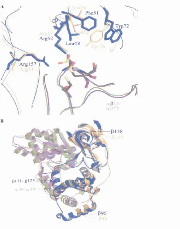
respectively. The two structures are very similar, indicating that the conformation is
subunit, a helical domain, and a C-terminal domain that is folded into a p roll. The two subunits form a tight dimer (Fig. 1.3), stabilised by the N-terminal loops of each subunit that w r ^ around the opposite subunit; these interactions suggest that dimer formation is a dynamic rather than simple docking process (Nakasako et al, 1999). The electrostatic properties of the interfacial surfaces of each subunit are quite different; Nakasako et al.
(1999) proposed that up to 50 hydration water molecules which are densely distributed over the negatively charged p subunit surface may aid in dimer stabilisation by moderating this charge and hence decreasing the free energy in heterodimer association.
V * X
Fig. 1.3; Stereoview of the ap heterodimers of Rhodococcus sp. R312 NHase. The location of the iron centre is indicated by a red sphere, the a subunit is in blue, the p subunit in yellow. (Taken from Huang et al, 1997)
The iron centre is located in a central cavity formed by the subunit interface. All protein ligands to the iron are provided by the a subunit. In accordance to the earlier spectroscopic data (Brennan era/., 1996b, Jin e/a/., 1993, Nelson er a/., 1991, Scarrow
et ai, 1996), the ligands were found on five vertices of an octahedron. However they did not comply with the 2S, 3N and O prediction (Jin et al, 1993, Nelson et al, 1991) and no histidine residues were found within the cavity. In fact, the ligands came from three cysteine thiolates (aCllO, 113 and 115) and two main-chain nitrogen atoms (aS114 and aCl 15). Huang et al (1997) reported that the sixth position was unoccupied, whilst Nagashima et al (1998) reported a nitric oxide hgand at this
position, illustrating the difference between the active and inactive states. The residues
C y sll3 and C y sll5 of Rhodococcus sp. N-771 are post-translationally oxidised to
cysteine-sulphinic (-SO2H) and sulphenic acids (-SOH) respectively in the NO
inactivated enzyme whilst just the sulphinic group is found in the light-activated
enzyme (Kobayashi and Shimizu, 1998). Despite having an identical amino acid
sequence and photoreactive behaviour, the crystal structure of Rhodococcus sp. R312 NHase does not display such modifications. This difference was attributed to the lower
resolution achieved in this study rather than structural differences with species N-771
(Nagashima et a l, 1998). The NO molecule is thought to be stabilised by the oxygen atoms of the three cysteines, which protrude into the iron centre termed by Nagashima
et a l (1998) as a “claw setting”. Light irradiation is thought to break the Fe-N (NO) bond and induce a structural change in the claw setting. a C y s ll3 and a C y s ll5 form
hydrogen bonds with PArg56 and PArgl41, which are conserved through all known
NHases (Huang gr a/., 1997, Nagashima gr aZ., 1998). Mutation of these residues results
in loss of activity, suggesting that they are involved in the stability of the claw setting
and that this setting is important for NHase activity (Piersma et al, 2000). A channel from the bulk solvent to the active centre is formed by the subunit interface, involving
five residues from the a subunit and four from the p subunit. A cluster of 10 aromatic
residues is found around this region and is believed to stabilise the channel (Nagashima
et a l, 1998). The entrance of the inactive N-771 enzyme, 10Â from the centre and only 4Â wide, is too narrow for substrates to enter. It is suggested therefore that dynamic
structural changes, as are observed during light-activation, are required to open and
close the channel during the catalytic cycle.
Until recently, little structural information was available concerning cobalt NHases.
EFR and EXAFS spectroscopy on the H-NHase of R. rhodochrous J 1 indicated that that
the ligand environment was very similar to the iron NHases (Brennan et a l, 1996a). The enzyme was found to contain a non-corrin Co^^ ion with a mixed S and N
octahedral ligand field. However, these studies ruled out the possibility of NO
Pseudonocardia thermophila NHase - a thermostable Co-type NHase was published very recently (Miyanaga et a l, 2001). Resolved to 1.8Â, the structure exhibits a very high similarity with the Fe-type NHase. The structure around the active centre is almost
identical (Fig. 1.4), and the cofactor ligands a C l l l and aC113 are post translationally
modified to form a similar claw setting to the Fe-type NHase.
The major deviation from the Fe-type structures involves the residues between p95 and
pl38. The central portion of this region corresponds in alignments (Figs. 3.19 and 5.1)
to an insertion in the Co-type NHases {cf. the Fe-type) of between 12 and 21 residues
(20 residues in the Ps. thermophila enzyme). The Ps. thermophila NHase structure
demonstrates that this region comprises an a helix (residues p ill-p l2 5 ), which
interacts with a second a helix on the external surface of the a subunit. It has been proposed that this interaction contributes to the thermostability of the Ps. thermophila
enzyme (Miyanaga et al, 2001). However, it should be noted that similar sequences are
also found in the Co-type NHases of Rhodococcus species. (Mayaux et al, 1991) and both R. rhodochrousJ1 NHases (Kobayashi et a l, 1991), none of which are particularly thermostable enzymes (Section 1.3.7). It is also noticable that an active site residue
pW72 o f the Ps. thermophila enzyme (conserved in the Co-type NHases) is orientated
in a different direction to pY76 of Rhodococcus sp. N-771 (conserved in Fe-type
NHases) (Nagashima et a l, 1998). This difference in orientation leads to a larger space in the substrate-binding site, which may account for the general differences in substrate
specificities (Section 1.3.6) of the two groups (Miyanaga et al, 2001).
Recent studies with the NHase of Rhodococcus sp. N-771 revealed that the enzyme
showed activity when the iron cofactor was substituted with cobalt ions, though
oxidation of both the cobalt to Co^^ and a C y sll3 to the cysteine-sulphinic acid was
essential for this activity (Nojiri et a l, 2000). This study supports the hypothesis that Co and Fe-type NHases share very similar structures and, presumably, reaction
/ Phc51
L eu48
A r g l5 7
»
31 1 1- p l 2 5
Fig. 1.4: Superimposition of the Co-type NHase and the Fe-type NHase from
Rhodococcus sp. N-771. The a (green) and p (blue) subunits of Co-type NHase, and a (purple) and p (orange) subunits of Fe-type are shown in different colors. The magenta
sphere is the cobalt atom in the active center, and the cyan one is the iron atom. A:
Active sites of the Co-type and the Fe-type NHase. The side chains of residues of the
Co-type NHase are represented by thick sticks and those of the Fe-type NHase by thin
ones. B: Ribbon diagram of the Co-type and Fe-type NHases. Helices interacting
between the a and p subunits (a36-a49, and p i l l - pi 25) are enriched by red dotted
1.3.5 Reaction Mechanism
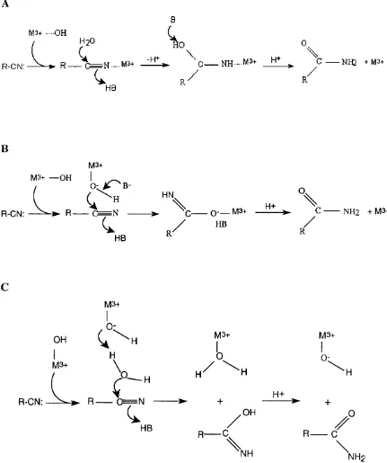
A number of mechanisms have been proposed for the hydrolysis of nitriles by NHases
(Huang et a l, 1997, Kobayashi, 1997, Nagasawa et a l, 1986). Based on the effects of addition of substrate or competitive inhibitors on the EPR and Resonance Raman
spectra, it is thought that the metal ion is involved as a Lewis-acid in catalysis (Huang et a l, 1997).
Three reaction models involving the metal ion as a Lewis acid have been proposed
(Huang era/., 1997):
1. The nitrile substrate binds directly to the metal ion, thereby activating it for
hydration. The nitrile carbon atom is then subjected to nucleophilic attack by a
water molecule held in place by a proximal residue (Tyr 72 has been indicated)
(Fig. 1.5a).
2. The nitrile substrate approaches a metal bound hydroxide ion, which acts as a
nucleophile on the nitrile carbon atom (Fig. 1.5b).
3. The final mechanism does not involve an enzyme bound intermediate, thus
avoiding ligand exchange. A metal-bound hydroxide activates a water molecule,
which then acts as the nucleophile, attacking the nitrile carbon atom (Fig. 1.5c).
Early hypotheses suggested that one of the conserved cysteine thiolates acted as a
nucleophile, followed by hydrolysis of the resulting thioimidate (Kopf et al, 1996). These have largely been discarded following the observation that those residues are
coordinated to the metal ion (Huang et a l, 1997, Miyanaga et a l, 2001, Nagashima et a l, 1998). Due to the slow rates of ligand exchange for trivalent metals, in particular for C o"\ it has been argued that mechanism 3 is the most likely since this does not
involve ligand exchange (Huang et al, 1997). Furthermore, the observation that the rates of hydration of certain nitriles are very similar between some Co-type and Fe-type
NHases, whilst the exchange kinetics of low spin Fe°^ and Co°* are very different has
also been interpreted as an indication that mechanism 3 is used (although there is
currently no empirical evidence for this) (Mascharak, 2002). However, as reported in
substrate analogue iodoacetonitrile is directly associated with the metal ion - replacing
the solvent derived hydroxide ligand (Doan et al, 1999). Furthermore, Co-chelate
complexes have shown to bind the nitrile reagent, leading to the suggestion that direct
association model is the correct mechanism (Nakasako et a l, 1999).
Fig. 1.5: Three putative reaction mechanisms of NHase.
M 3+ OH
L
(
C °
\
R-CN: ^ , R — C ^ H M3, C— NH M3. H - , C — N H 2 . M 3 .
HB R
R
/
B
M3+ M3+ — OH
I
R-CN:
HB
HN. O
\
V H +
R 0 = = = N ---0 --- O"— M3+ --- ^ N H 2 + M3+
R'' R
OH
I
M3+
R-CN:
M3+
O:
H
V
R
. e
NM3+
O
4-OH
M3+
I
O:
H
H+
1.3.6 Specificity
The general trend of substrate specificity amongst the NHases characterised to date
seems to be almost exclusively for aliphatic nitriles. However, some enzymes with
activity on aromatic nitriles have been described (Table 1.3). It has been claimed that
the iron-containing NHases are specific for aliphatic nitriles whereas the cobalt-
containing NHases are specific for aromatic nitriles (Kobayashi and Shimizu, 1998,
Miyanaga et a l, 2001), although this is not strictly true. The Fe-type NHase of R. erythropolis showed broad substrate specificity, and was capable of hydrating aliphatic, aromatic and heterocyclic nitrile compounds (Duran et a l, 1993) whilst the Co-type
enzymes of B. smithii, B. pallidus DAC521, Bacillus sp. RAPcS and P. putida
demonstrate no activity on aromatic nitriles (Cramp and Cowan, 1999, Payne et al,
1997, Pereira et a l, 1998, Takashima et a l, 1998).
It has been suggested that the lack of aromatic specificity is due to the narrow entrance
channel from the bulk solvent, which enables only small, aliphatic molecules to enter
the catalytic site (Nakasako et a l, 1999). A second hypothesis involving the spatial arrangement of the catalytic cavity suggests that a larger cavity caused by the
displacement by a leucine residue and a phenylalanine residue of pW72 of
Ps. thermophila NHase (in comparison to the corresponding PY76 of
Rhodococcus sp. N-771) may be responsible for the differences in substrate preferences between Co-type and Fe-type NHases.
Several NHases have been described with very broad substrate ranges. Bacillus sp. RAPc8 demonstrated activity only on aliphatic nitriles but catalysed the hydration of branched, linear, cyclic-aliphatic and dinitriles. The highest affinity recorded was for
acetonitrile, with a Km value of 8.8mM (Pereira, 1998). Both the L-NHase and H-
NHase of R. rhodochrous J1 have broad substrate ranges, with similar Km values for the
aliphatic nitriles (e.g., for acrylonitrile. Km values of 2.67mM and 1.89mM and specific
activities of 828U/mg and 1760U/mg for L-NHase and H-NHase respectively).
However, the affinity and activity of L-NHase for aromatic nitriles was significantly
higher than H-NHase (Wieser et al, 1998).
There are also reports of NHases with enantioselectivity. Agrobacterium tumefaciens
(S)-amides enantioselectively (>90% enantiomeric excess) from a number of racemic
phenylpropionitriles (Bauer et a l, 1994). P. putida NHase also produces (S)-amides, for example racemic 2-(4-chlorophenyl)-3-methylbutyronitrile was converted to the (S)-
amide at >90% enantiomeric excess (Fallon et al, 1997).
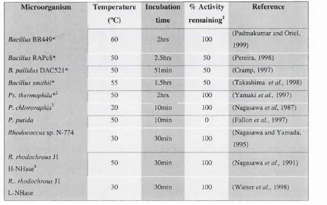
1.3.7 Stability
Most NHases are particularly thermolabile although it has been demonstrated that
n-butyric acid acts as a potent stabilising agent (Nagasawa et al, 1987). There appears to be a relation between stability and cofactor, with Co-type NHases being generally more
stable than Fe-type (Tables 1.2 and 1.3). In the past five years, at least five NHases
from moderate thermophiles have been characterised, four of which are thermophilic
Bacillus isolates, the fifth a Ps. thermophila strain. With the exception of B. pallidus
DAC521 (for which the cofactor is not known), all are Co-type enzymes.
Due to the different approaches of the various researchers in describing thermal
stability, comparison between the enzymes is not straightforward. Nevertheless, Table
Table 1.2: Thermal stability of NHases as defined by remaining activity following incubation for a predetermined time period at a specific temperature.
Microorganism Temperature Incubation % Activity Reference ("C) time ^
« remaining^
B R 4 4 9 * ^ 60 100 (Padmakumar and Oriel, 1999)
BacillusRAPcS* 50 2.5hrs 50 (Pereira, 1998)
B. pallidus ' [ 50 51min 50 (Cramp, 1997)
Bacillus smithii^ 55 »1.5hrs 50 (Takashima e ta l, 1998)
Ps. thermophila*^ 50 2hrs 100 (Yamaki e ta l, 1997)
P. chlororaphis^ 20 lOmin 100 (Nagasawa et al, 1987)
P. putida 50 lOmin 0 (Fallon e/a/., 1997)
Rhodococcussp. N-774
30 30min 100 (Nagasawa and Yamada, 1995)
R. rhodochrousJ 1
H-NHase" 50 30min 100 (Nagasawa gf a/., 1991)
R.. rhodochrous SI
L-NHase 30 30min 100 (Wieser e/a/., 1998)
* D e n o te s the th e r m o p h ilic o r g a n ism s
'w h e r e 100% is g iv e n as r em a in in g a c tiv ity , auth ors rep orted that a c tiv ity le v e ls d ro p p ed d ra m a tica lly a b o v e th o s e tem p eratu res.
^S tabilised in 3 4 m M u -b u ty ric acid ^ S tabilised in 2 2 m M « -b u ty ric acid " S tab ilised in 4 4 m M « -b u ty ric acid
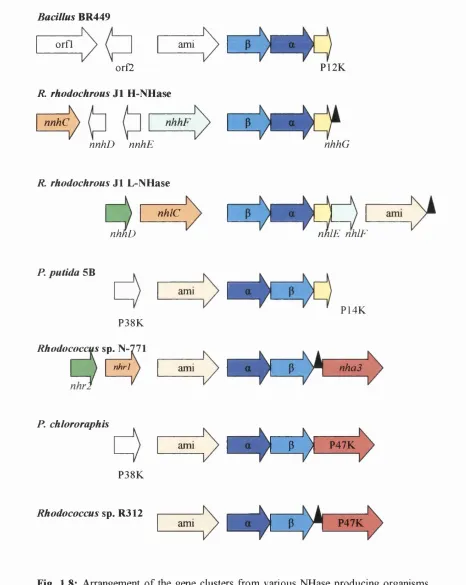
1.3.8 Gene structure
Over the past 13 years, the genes for a large number of the nitrile-modifying enzymes described so far have been cloned using a variety of techniques. The classic approach has been to determine N-terminal and internal amino acid sequences of the purified proteins, from which oligonucleotides were designed either for use as labelled probes (e.g. cloning of Rhodococcus sp. N-774 NHase (Ikehata et a l, 1989)) or to produce PCR products for use as probes (e.g. cloning of Ps. thermophila NHase genes (Yamaki
et al, 1997)). Other cloning strategies have involved the use of the N-774 NHase genes as probes in Southern hybridisations in order to isolate the NHase genes of
R. rhodochrous J1 (Kobayashi et al, 1991) and R. erythropolis (Duran et a l, 1993),
whilst PCR primers designed against the DNA sequence of the B. smithii NHase genes
(Takashima et a l, 1995) have been used in PCR for the isolation of the corresponding
genes of Bacillus BR449 (Kim and Oriel, 2000).
The a and P subunit genes are of similar size and range from 609bp to 660bp for the a
subunit and from 636bp to 706bp for the P subunit. The genes are found in both a-P
and P-a orders, usually separated by a short sequence of between 16bp and 29bp (Duran
et a l, 1993, Kim and Oriel, 2000). With the exception of the R. rhodochrous J1 genes, all NHases characterised to date are found just downstream of an amidase gene. No
amidase sequences were found close to the H-NHase genes of R. rhodochrous J1
although an amidase gene was found 1.9kb downstream of the a subunit gene of the L-
NHase gene (Kobayashi et a l, 1992b). The close location of the NHase and amidase genes in nitrile metabolising organisms supports the hypothesis that these enzymes are
involved in a two-step reaction in the nitrile degradation pathway. No transcription
terminators have been found between the amidase, a or p genes although stem-loop
structures that may act as transcription terminators have been identified immediately
downstream of the P subunit genes of P. chlororaphis (Nishiyama et a l, 1991) and
Rhodococcus sp. R312 (Bigey et al, 1999).
The translated peptide sequences of the NHases reveal a remarkably high degree of
sequence conservation despite the fact that many originate from distantly related
organisms. Generally the a subunit is more conserved than the p, containing 43 strictly
conserved amino acids, whilst the P subunit contains 28 (Payne et a l, 1997). A number
of conserved sections in the amino acid sequences reflect regions of structural and/or
functional significance. O f particular note is the sequence homology o f the p subunit N-
terminal sequences (Fig. 1.6), which may be explained by the observation from
crystallographic data that this region is involved in the stabilisation of the subunit
interface (Huang et a l, 1997, Miyanaga et a l, 2001, Nagashima et al, 1998). The region of the a subunit containing the cysteine ligands is also highly conserved,
the Co-type and Fe-type NHases respectively (Fig. 1.7) (Payne et ai, 1997). It has been suggested that these two differences are the determinants of the identity of the cofactor bound by a particular NHase (Kobayashi and Shimizu, 1998).
Fig. 1.6: Alignment of NHase P subunit N-terminal sequences, shaded at 60% conservation.
B a c i l l u s B R 4 4 9 B . s m i t h i i P . p u t i d a K l e b s i e l l a
R h o d o c o c c u s J 1 H R h o d o c o c c u s J 1 L R h o d o c o c c u s s p . P s . t h e r m o p h i l a S . m e l o t i
M . l o t i
R h o d o c o c c u s N 7 7 4 R h o d o c o c c u s R 3 1 2 R . e r y t h r o p o l i s P . c h l o r o r a p h i s
CiKDVGGMDGafiG- - K ^
tI H D Y S G M D G F - G — ]
iG IH D T d ^ H G fiG --:
Ig i h d l g g m hGl g— P f L M D G I H D T G G M T G # G — P | P M D G I H D L G G R A ^ L G — P # K r P D L ' G G T D G l G — p f o I G V Y D V G G T D G L G — P | N
IPiDL^QHGMG— p | a
; P Q D L G G Q M G # G — P » A m d g v h d l a g v q g f’g k v p h t W D G V H D i i A G V Q G F G K V P H T M D G V H D L A G V Q G F G K V P H S W D G F H D L G G F Q G F G K V P H T
Fig. 1.7: Alignment of the conserved NHase a subunit sequences containing the characteristic cofactor binding motif VC(T/S)LCSC. The alignment is shaded at 60% conservation.
B a c i l l u s B R 4 4 9 B . s m i t h i i P . p u t i d a K l e b s i e l l a
R h o d o c o c c u s J l H R h o d o c o c c u s J l L R h o d o c o c c u s s p . S . m e l o t i
P . t h e r m o p h i l a M . l o t i
R h o d o c o c c u s N 7 7 4 R h o d o c o c c u s R 3 1 2 R . e r y t h r o p o l i s P . c h l o r o r a p h i s
J T L ^ S C Y P H P L L G L P P S W Y : T L C S i ï P W P L L G L P P S 1 3 t f F V C T L C S C Y P B P T L G L P P A î r s e _ n V g î T L C S C Ÿ P W P V L G L P P V B Y H V W C T L C S C Y P H P V L G L P P A W ÿ i M W C T L C S C t P W P V L G L P P N W Ï H A I V C T L C S g t P W P V L G L P P N l i i r i i L I V C T L C S C f P H A V L G L P P V 1 B | H W V t T L C S C t P W P V L G L P P N l t “ j W C T L C S f i t P H S V L G L P P V B l r i v e S L C siT A W P I L G L P PTWie
r i V C S L C S | T A H P I L G L P P T H | r i v e S L C s I t A B P I L G L P P T W l ? r i V C S L e s i T N B P V L G L P P E B Y
1.3.9 Gene regulation and expression
Requirements for the production of active NHase in the native organisms vary. Some NHases are produced constitutively, irrespective of the presence or absence of nitriles, amides or metal cofactor ions, whilst others have been shown to be inducible (Table 1.3). Whilst no information appears to be available on the effect of supplementing the
growth media of Fe-type NHase producers with Fe^"^ ions, experiments have shown that
the specific activities of Bacillus sp. BR449, B. smithii and R. rhodochrous J l NHases
are significantly enhanced by addition of 0.1-0.5mM C0CI2 to the growth media
(Kobayashi et al, 1991, Padmakumar and Oriel, 1999, Takashima et a l, 1998).
Investigations with recombinant R. rhodochrous J l H-NHase have shown that cobalt is
required for activity rather than protein expression (Kobayashi et a l, 1991, Komeda et al, 1996b), similarly, recombinant Bacillus sp. BR449 NHase is expressed in a largely inactive form in the absence of cobalt ions but can be activated upon incubation in 5pM
C0CI2 (Kim and Oriel, 2000, Kim et al, 2001). These findings suggest that cobalt is involved in enhancing protein folding rather than induction of NHase expression
(Cowan et a l, 1998, Kobayashi et al, 1992b, Kobayashi et a l, 1991).
The inducible NHases are generally induced by their amide reaction products rather
than by nitriles (Kobayashi and Shimizu, 1998). This is also found to be the case for the
NHase-associated amidases. This observation, together with the close location of the
structural genes and the absence of transcriptional terminators between them, indicates
that amidases and NHases are under the control of the same regulatory elements. It has
been proposed that the open reading frames encoding the NHase a and (3 subunits and
the amidase monomer are cotranscribed as a single, polycistronic mRNA (Duran et a l,
1993, Nishiyama et a l, 1991). Indeed, this has been shown to be the case for the
NHase operon of Rhodococcus sp. ACV2 (Bigey et a l, 1999).
Early attempts to express recombinant NHase in E. coli often resulted in the formation of inactive inclusion bodies or no expression at all (Kobayashi et al, 1992b, Nishiyama
et a l, 1991). However, further studies revealed that in many cases, the inclusion of upstream and/or downstream open reading frames in expression constructs significantly
enhanced expression of activity. Overproduction of P. putida NHase was found to
require co-expression of P14K, the gene for which is found immediately downstream of
the NHase p subunit (Wu et al, 1997). Similar proteins, also with P-subunit homology
have been found downstream of the NHase genes of Bacillus sp. BR449 and
genes (Bigey et a l, 1999, Nishiyama et a l, 1991, Nojiri et al, 1999). Although the precise function of these proteins is not known, it has been suggested that they are
involved in incorporation of the cofactor ion into the active site (Bunch, 1998). The
observation that their presence is required for the expression of constitutively produced
enzymes (Bigey et a l, 1999, Nojiri et al, 1999), indicates that they are not involved in regulation of expression. It is noteworthy that the p-homologue proteins are associated
with the Co-type NHases whereas the P47K proteins are Fe-type associated.
1.3.9.1 Regulation of the NHase genes of R. rhodochrous J l
The regulation of the R. rhodochrous J l genes has been particularly well characterised
(Kobayashi and Shimizu, 1998). Both H-NHase and L-NHase are induced by
crotonamide. However urea or cyclohexanecarboxamide induce either H-NHase or L-
NHase genes respectively (Yamada and Kobayashi, 1996).
The H-NHase operon contains two regulatory genes, nhhC and nhhD, both located
upstream of the NHase genes. NhhC, which is homologous to the negative regulator
AmiC of the P. aeruginosa aliphatic amidase, exhibits a positive regulation in the
presence of amide, of NhhD production. Presence of NhhD then induces NHase
expression (Kobayashi and Shimizu, 1998).
The 5’ upstream region of the L-NHase operon is also required for NHase expression;
two genes, nhlD and nhlC serve positive and negative regulatory roles respectively. In the presence of amide, NhlC (also an AmiC homologue) inhibits NhlD repression of
NHase production (Komeda et al, 1996b).
An additional gene, nhlF/nhhF is found within both NHase opérons and has been shown
to produce a cobalt uptake protein that confers cobalt uptake on Rhodococcus strains (Komeda et a l, 1997) and is believed to play a role in providing cobalt to the NHase (Kobayashi and Shimizu, 1998). Both opérons also contain insertion sequences, which
are believed to be responsible for the unique arrangement and regulation of the
Bacillus BR449
orfl ami
orfZ
R. rhodochrous J l H-NHase
nnhC nhhF
nnhD nnhE
P12K
nhhG
R rhodochrous J l L-NHase
nhlC
nhhD nhlIE nhlF
P. putida 5B
P38K
Rhodococcus sp. N-771
nhrl
ami
ami
P14K
nhaS
P. chlororaphis
P38K
ami P \ ) ---P47K
V
---Rhodococcus sp. R312
ami P47K