Drug Design, Development and Therapy
Dove
press
O r i g i n a l r e s e a r c h
open access to scientific and medical research
Open access Full Text article
inhibition of lymphangiogenesis in vitro and
in vivo by the multikinase inhibitor nintedanib
Tong lin1,2
lan gong1,2
1Department of Ophthalmology, eye
and enT hospital, Fudan University,
2Key laboratory of Myopia, Ministry
of health, shanghai, People’s republic of china
Purpose: To investigate the feasibility of nintedanib, a novel triple angiokinase inhibitor, for inhibiting lymphatic endothelial cell (LEC)-induced lymphangiogenesis in vitro and inflammatory corneal lymphangiogenesis in vivo.
Materials and methods: Methylthiazolyldiphenyl-tetrazolium bromide (MTT) test, transwell system, and tube-formation assay were used to evaluate the effects of nintedanib on the proliferation, migration, and tube formation of LECs stimulated by vascular endothelial growth factor-C (VEGF-C), basic fibroblast growth factor (bFGF), or platelet-derived growth factor-BB (PDGF-BB). The murine model of suture-induced corneal neovascularization was used to assess the anti-hemangiogenic and anti-lymphangiogenic effects of nintedanib via systemic and topical applications. Corneal flatmounts were stained with lymphatic vessel endothelial hyaluronan receptor-1 (LYVE-1) and CD31, and the areas of involved blood and lymph vessels were analyzed morphometrically. Corneal cryosections were stained with F4/80 to evaluate inflammatory cell recruitment.
Results: We observed a significant enhanced effect of LEC proliferation, migration, and tube formation with the administration of VEGF-C, PDGF-BB, and bFGF, respectively, which was diminished by nintedanib. Both topical and systemic applications of nintedanib inhibited suture-induced hemangiogenesis and lymphangiogenesis in the murine cornea. A reduction in F4/80+ cell infiltration was observed at day 14 after corneal suture for both systemic and topical
applications of nintedanib. In comparison with controls, 61% of F4/80+ cell recruitment was
inhibited via the systemic application of nintedanib, while 49% of F4/80+ cell recruitment was
inhibited with the topical application of nintedanib.
Conclusion: Nintedanib was shown to inhibit in vitro lymphangiogenesis stimulated by VEGF-C, bFGF, and PDGF-BB. Applied topically or systemically, it effectively inhibited corneal hemangiogenesis and lymphangiogenesis, accompanied by reduced inflammatory cell recruitment, which represents a new promising treatment for graft rejection after penetrating keratoplasty.
Keywords: kinase inhibitor, lymphangiogenesis, in vitro, in vivo
Introduction
Lymphangiogenesis is defined as the formation of new lymphatic vessels from preexisting ones, and it involves proliferation, migration, and tube formation of the lymphatic endothelial cells (LECs).1 Growth factors, adhesion molecules, chemokines,
and the extracellular matrix play important roles in lymphangiogenesis.2–4 Previous
studies have shown that an extensive variety of proangiogenic factors, including vascular endothelial growth factor (VEGF), fibroblast growth factor (FGF), and platelet-derived growth factor (PDGF), could be also involved in the development of lymphangiogenesis.5–8 It is well established that vascular endothelial growth factor
correspondence: lan gong Department of Ophthalmology, eye and enT hospital, Fudan University, 83 Fenyang road, shanghai 200031, People’s republic of china Tel/fax +86 21 6431 0068 email 13501798683@139.com
Journal name: Drug Design, Development and Therapy Article Designation: Original Research
Year: 2017 Volume: 11
Running head verso: Lin and Gong
Running head recto: Nintedanib inhibits lymphangiogenesis DOI: http://dx.doi.org/10.2147/DDDT.S130297
Drug Design, Development and Therapy downloaded from https://www.dovepress.com/ by 118.70.13.36 on 21-Aug-2020
For personal use only.
Number of times this article has been viewed
This article was published in the following Dove Press journal: Drug Design, Development and Therapy
Dovepress
lin and gong
(VEGF) receptor-3 (VEGFR-3) is a specific receptor of VEGF-C and VEGF-D, and upregulation of the expression
of VEGF-C and VEGF-D promotes lymphangiogenesis.9,10
Meanwhile, basic fibroblast growth factor (bFGF) has been demonstrated to promote lymphangiogenesis directly and to act in an indirect role involving the upregulation of VEGF-C and VEGF-D expression in lymphangiogenesis.11 It has
previously been reported that PDGF-BB can also stimulate the proliferation and migration of LECs, and blockage of the VEGF-C/VEGF-D/VEGFR-3 pathway fails to inhibit PDGF-BB-induced lymphangiogenesis; this suggests that PDGF-BB also has a direct effect on LECs.12
In pathological conditions, such as transplant rejection and cancer metastasis, numerous angiogenic factors arise to stimulate angiogenesis and lymphangiogenesis; this could contribute to the exacerbation of these diseases.13–17
Inhibition of lymphangiogenesis is suggested to be a pow-erful selective therapy for reducing lymphatic metastasis of tumor cells and graft rejection. Recently, a novel, potent, triple angiokinase inhibitor called nintedanib was devel-oped. Nintedanib competitively binds to the adenosine-5′-triphosphate (ATP)-binding site of receptor tyrosine kinases (RTKs), including VEGFR, fibroblast growth factor receptor (FGFR), and platelet-derived growth factor receptor (PDGFR) and then inhibits their downstream intracellular signaling.18 Nintedanib has displayed potent antiangiogenic
effects in vitro by inhibiting endothelial, pericyte, and smooth muscle cell proliferation in culture and antitumor activity in several tumor xenograft models in vivo by reducing tumor microvascular density and the number of perivascular cells.18,19 Moreover, this multi-targeted angiokinase
inhibi-tor has also demonstrated to be effective in several human cancer trials.20–22 Furthermore, nintedanib was found to be an
effective and potent inhibitor of the pre-retinal pathological neovascularization in a murine model of vasoproliferative retinopathy.23 Although the antiangiogenic effect of
ninte-danib is well recognized, its effect on lymphangiogenesis remains unclear. This investigation was designed to deter-mine the feasibility of the multikinase inhibitor, nintedanib, for inhibiting LEC-induced lymphangiogenesis in vitro and inflammatory corneal lymphangiogenesis in vivo.
Materials and methods
Mice
Six-week-old male C57BL/6 mice purchased from SIPPR-Bk Laboratory Animal Co., Ltd. (Shanghai, China) were used throughout this study. The animals were maintained with a 12-h light–dark cycle and had free access to food and water.
Animal experiments were performed in accordance with the Association for Research in Vision and Ophthalmology (ARVO) Statement for the Use of Animals in Ophthalmic and Vision Research; the study was approved by Fudan University’s Animal Ethical Committee.
cell culture
Primary human lymphatic endothelial cells (HLECs) were purchased from PromoCell (Heidelberg, Germany) and cultured in endothelial basal medium (EBM; Lonza, Walkersville, MD, USA) supplemented with 10% fetal calf serum (FCS; Biowest, Riverside, MO, USA) and other supplements, as previously described.24 HLECs at passages
4–8 were used in this study.
nintedanib
Nintedanib was purchased from Selleckchem (Houston, TX, USA). For in vitro assays, nintedanib was solubilized in dimethyl sulfoxide (DMSO; Sigma-Aldrich, St Louis, MO, USA) and used at 0.01, 0.1, or 1 μM in culture medium. For in vivo assays, nintedanib was solubilized in 0.5% carboxy-methyl cellulose. Fifty mg/kg was administered daily by oral gavage, and 5 mg/mL was used as topical administration. Control mice received vehicle.
cell proliferation assay
The HLEC proliferation assay was performed using methylthiazolyldiphenyl-tetrazolium bromide (MTT; Sigma-Aldrich) assay. HLECs were seeded onto each
well of 96-well plates in a total volume of 150 μL EBM
containing 2% (vol/vol) FCS at a density of 5×10³ cells/
well. After incubation for 24 h, cells were treated with 50 ng/mL recombinant VEGF-C (PeproTech, Rocky Hill, NJ, USA), PDGF-BB (PeproTech), or bFGF (PeproTech) in the presence or absence of nintedanib at concentrations of 0.01, 0.1, and 1 μM. The control cells were treated with 0.1% DMSO. After incubation at 37°C for 48 h, 20 μL of MTT solution (5 mg/mL) was added to each well, and the cells continued to be incubated for 4 h. Following removal of
the medium containing MTT, 200 μL of DMSO was added
to dissolve the formazan crystals formed by live cells. The absorbance was measured at 490 nm with an absorbance microplate reader (BioTek, Winooski, VT, USA). The assay was repeated six times.
Migration assay
A transwell system (Corning Costar, Acton, MA, USA) was used to study HLECs’ ability to migrate through a micropore
Drug Design, Development and Therapy downloaded from https://www.dovepress.com/ by 118.70.13.36 on 21-Aug-2020
Dovepress nintedanib inhibits lymphangiogenesis
nitrocellulose filter (8-μm pore size). In brief, each stimulating factor (VEGF-C, PDGF-BB, or bFGF) was added to the lower chamber at a concentration of 50 ng/mL in 2% (vol/vol) FCS-basal EBM. Approximately 1×10⁵ HLECs in serum-free
EBM in the presence or absence of 0.1 μM nintedanib were seeded in each well of the upper chamber. The control cells were treated with 0.1% DMSO. The cells were incubated for 12 h. Non-migrating cells were removed from the top surface of the membrane using a cotton swab. Migrating cells adhering to the undersurface of the filters were fixed with methanol, followed by staining with a 4′ ,6-diamidino-2-phenylindole (DAPI, 1:1,000; Sigma-Aldrich) solution, and observed using a fluorescence microscope (DM 4000B; Leica Microsystems, Wetzlar, Germany), as described in a previous study.25 The cells that migrated into the lower chamber were
counted in five fields for each well. The experiments were performed in triplicate.
Tube-formation assay
Tube formation was assayed as previously described.26
In brief, 200 μL of Matrigel (BD Biosciences, Bedford, MA, USA) was added to each well in 24-well plates and allowed to polymerize for 30 min at 37°C. After trypsini-zation, HLECs were seeded on the gel at a density of 1×10⁵ cells/well and incubated in the medium containing
VEGF-C, FGF-2, or PDGF-BB in the presence or absence of 0.1 μM nintedanib for 24 h. The control cells were treated with 0.1% DMSO. Capillary-like structures organized by the cells were viewed and photographed at a magnifica-tion of 200×. The area and length of the tube network in five fields for each well were quantified by ImageJ soft-ware (National Institutes of Health). The experiment was performed in triplicate.
corneal neovascularization assay
immunostainings
The mouse model of suture-induced inflammatory corneal neovascularization was used as previously described.27
First, each animal was deeply anesthetized with an intra-muscular injection of ketamine (8 mg/kg) and xylazine (0.1 mL/kg). The central cornea was marked with a 2 mm diameter trephine, and three 11-0 nylon sutures (Lingqiao; Ningbo Medical Needle Co., Ltd., Ningbo, China) were then placed in the intrastromal position extending over 120° of the corneal circumference to obtain standardized angiogenic responses. Sutures were left in place for 2 weeks.
The systemic treatment group received nintedanib (50 mg/kg, daily) by oral gavage for 14 days after suture
surgery. Control mice received equal amounts of the substance vehicle. For enhancing the penetration of nintedanib into the corneal stroma, the central 2 mm of the corneal epithelium was scraped off before suturing, and the mice received nintedanib (5 mg/mL, four times daily) in eye drops for 12 days. Control mice received an equal volume of the substance vehicle. After 2 weeks, mice were sacrificed and corneas were prepared for immunofluores-cence assay.
corneal immunostainings
Corneal whole mounts were used to determine corneal lymphangiogenesis and hemangiogenesis. Mice were sacrificed at planned times, and the eyes were removed and dissected; following this, whole-mounted corneas
were fixed in 4% paraformaldehyde overnight at 4°C and
blocked in 5% donkey serum albumin (Solarbio, Beijing, China) for 1 h. For double lymphatic vessel endothelial hyaluronan receptor-1 (LYVE-1) and CD31 immunostain-ings, corneas were incubated overnight with polyclonal rabbit anti-mouse LYVE-1 (1/200; Abcam, Cambridge, UK) and rat anti-mouse CD31 (1/100; BD Biosciences Pharmingen, San Jose, CA, USA) antibodies, as described previously.28 Following this, incubations with Alexa Fluor
488-coupled donkey anti-rabbit antibody (1/200; Abcam) and Cy3-conjugated donkey anti-rat immunoglobulin G (IgG; Jackson ImmunoResearch, West Grove, PA, USA) were performed overnight. Corneas flat mounted on a microscope slide with antifade solution (AR1109; Boster, Wuhan, China) were examined using a confocal microscope (TCS SP8; Leica, Heidelberg, Germany). Pictures were taken at 100× magnification and automatically assembled to reconstitute the whole cornea picture. The area of corneal lymphangiogenesis and hemangiogenesis was quantified by ImageJ software.
For corneal inflammatory cell recruitment assay, corneal cryosections (8 μm) were fixed in 4% paraformaldehyde for 5 min.29 After blocking with 5% donkey serum albumin in
phosphate-buffered saline (PBS), sections were incubated overnight at 4°C with rabbit polyclonal F4/80 antibody (1:100; Abcam). After washing with PBS, the sections were incubated for 2 h at room temperature with Alexa Fluor 488-coupled donkey anti-rabbit antibody (1/200). Next, sections were washed with PBS, followed by staining with a DAPI (1:1,000) solution; pictures were taken at
400× magnification using a fluorescence microscope (DM
4000B). Five representative pictures per cornea were taken, and the F4/80+ cells were counted in each section. The
Drug Design, Development and Therapy downloaded from https://www.dovepress.com/ by 118.70.13.36 on 21-Aug-2020
Dovepress
lin and gong
mean cell number of the control section was set as 100%; the numbers of F4/80+ cells per section were then related to
this value (cell ratio).
statistical analysis
Data are expressed as the mean ± standard deviation (SD). The Student’s t-test and one-way analysis of variance with Scheffe’s post hoc multiple comparison analysis were per-formed to analyze the data statistically. A value of P,0.05 was considered as statistically significant.
Results
in vitro effect of nintedanib on
lymphangiogenesis
effect of nintedanib on growth factor-induced proliferation of hlecs
We observed a significant pro-proliferative effect of VEGF-C, PDGF-BB, and bFGF on HLECs compared with the negative control group (all P,0.01; Figure 1A–C). How-ever, the enhanced effect was significantly eliminated when nintedanib was administered at a concentration of $0.1 μM,
and the inhibiting effect exhibited concentration dependence (Figure 1A–C).
nintedanib suppresses growth factor-induced migration of hlecs
Next, we investigated the effect of nintedanib on growth factor-induced migration of HLECs at a concentration of
0.1 μM. As expected, VEGF-C, PDGF-BB, and bFGF
(50 ng/mL) significantly stimulated migration of HLECs from the upper chamber to the bottom of the filter (Figure 2). The migration of HLECs stimulated by each growth factor was suppressed with the administration of 0.1 μM nintedanib (all P,0.05; Figure 2E).
nintedanib inhibits growth factor-induced tube formation of hlecs
Meanwhile, we investigated the effect of nintedanib on growth factor-induced tube formation of HLECs at a concentration of 0.1 μM. After incubation for 24 h, the cells organized into capillary-like structures in Matrigel basement membrane matrix. Tube formation of HLECs was evaluated in terms
Figure 1 effect of BiBF 1120 (nintedanib) on growth factor-induced proliferation of hlecs.
Notes: (A) VEGF-C (50 ng/mL) enhanced proliferation of HLECs significantly. The enhanced effect was significantly reduced by 0.01 μM nintedanib (P,0.05) and almost eliminated by 0.1 μM nintedanib (P,0.01). (B) PDGF-BB (50 ng/mL) enhanced proliferation of HLECs significantly. The enhanced effect was significantly reduced by 0.01 μM nintedanib (P,0.05) and almost eliminated by 0.1 μM nintedanib (P,0.01). (C) A concentration of 50 ng/mL of bFGF enhanced proliferation of HLECs significantly. The enhanced effect was significantly reduced by 0.1 μM nintedanib (P,0.05) and almost eliminated by 1 μM nintedanib (P,0.01). *P-value ,0.05. **P-value ,0.01.
Abbreviations: hlecs, human lymphatic endothelial cells; VegF-c, vascular endothelial growth factor-c; PDgF-BB, platelet-derived growth factor-BB; bFgF, basic fibroblast growth factor; OD, optical density.
&RQWURO %,%)0
9(*)&QJP/
3UROLIHUDWLRQ 2'QP
&RQWURO %,%)0
3'*)%%QJP/
3UROLIHUDWLRQ 2'QP
$
%
&RQWURO %,%)0
E)*)QJP/
3UROLIHUDWLRQ 2'QP
&
Drug Design, Development and Therapy downloaded from https://www.dovepress.com/ by 118.70.13.36 on 21-Aug-2020
Dovepress nintedanib inhibits lymphangiogenesis
of the length and area of the tube structures. Both the length and area of the tube structures clearly increased with the administration of VEGF-C, PDGF-BB, or bFGF at a concen-tration of 50 ng/mL (Figure 3). The tube formation of HLECs stimulated by each growth factor was significantly suppressed by 0.1 μM nintedanib (all P,0.05; Figure 3E and F).
in vivo effect of nintedanib on
lymphangiogenesis
effect of systemic application of nintedanib on hemangiogenesis and lymphangiogenesis
We investigated the effect of systemic application of nintedanib on the outgrowth of blood and lymphatic vessels in a suture-induced corneal neovascularization assay. The treatment group (n=8) showed a significant decrease in angiogenesis (P,0.01) and lymphangiogenesis (P,0.01) compared with the controls (n=8; Figure 4).
effect of topical application of nintedanib on hemangiogenesis and lymphangiogenesis
We sought to determine whether the topical application of nintedanib also significantly affected corneal neovasculariza-tion. Therefore, we scraped off the central 2 mm of the corneal
epithelium before suture placement to promote penetration of the drug into the corneal stroma. By application of 5 mg/mL nin-tedanib as eye drops four times daily, the outgrowths of blood (P,0.01) and lymphatic (P,0.01, n=8) vessels were signifi-cantly inhibited compared with the controls (n=8; Figure 5).
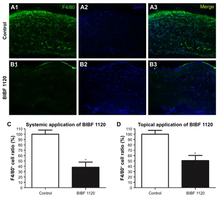
Nintedanib inhibits inflammatory cell recruitment
in the sutured cornea
Finally, we evaluated whether the application of nintedanib suppresses inflammatory cell (F4/80+ cell) recruitment in the
sutured cornea. Notably, a reduction in F4/80+ cell infiltration
was observed at day 14 in corneas from both systemic (n=6) and topical (n=6) applications of nintedanib. In comparison with controls, recruitment of F4/80+ cells was inhibited by
61% with the systemic application of nintedanib (P,0.01; Figure 6C) and by 49% with the topical application of nintedanib (P,0.01; Figure 6D).
Discussion
The results of our current study allow the following four conclusions to be drawn:
1. Nintedanib, a potent triple angiokinase inhibitor, inhibits both lymphangiogenesis and hemangiogenesis.
1XPEHURIPLJUDWHG
FHOOVPP
&RQWURO9(*)& 3'*)%%
9(*)&%,%)
3'*)%%%,%)
E)*)%,%) E)*)
3
3
3
&RQWURO
$
%
9(*)&&
3'*)%%'
E)*)9(*)& %,%)
%
3'*)%% %,%)
&
E)*) %,%)
'
(
Figure 2 BiBF 1120 (nintedanib) suppressed growth factor-induced migration of hlecs.
Notes: (A) cells treated with 0.1% DMsO were used as controls. (B1 and B2) VEGF-C (50 ng/mL) promoted migration of HLECs significantly, and the enhanced effect was significantly reduced by 0.1 μM nintedanib. (C1 and C2) PDGF-BB (50 ng/mL) promoted migration of HLECs significantly, and the enhanced effect was significantly reduced by
0.1 μM nintedanib. (D1 and D2) A concentration of 50 ng/mL of bFGF enhanced migration of HLECs significantly, and the enhanced effect was significantly reduced by 0.1 μM nintedanib. (E) The number of migrated cells stimulated by VegF-c, PDgF-BB, or bFgF at a concentration of 50 ng/ml in the presence or absence of 0.1 μM nintedanib. a–D at 200× magnification; scale bar =50 μm.
Abbreviations: hlecs, human lymphatic endothelial cells; DMsO, dimethyl sulfoxide; VegF-c, vascular endothelial growth factor-c; PDgF-BB, platelet-derived growth factor-BB; bFGF, basic fibroblast growth factor.
Drug Design, Development and Therapy downloaded from https://www.dovepress.com/ by 118.70.13.36 on 21-Aug-2020
Dovepress
lin and gong
2. Nintedanib prevents enhanced effects of cell proliferation, migration, and tube formation of HLECs stimulated by VEGF-C, PDGF-BB, and bFGF.
3. Both topical and systemic applications of nintedanib inhibit suture-induced hemangiogenesis and lymphangio-genesis in the murine cornea.
4. Nintedanib inhibits inflammatory cell recruitment in the sutured murine cornea.
The first commercially available antiangiogenic agent for cancer treatment was bevacizumab, a humanized monoclonal antibody that blocks VEGF-A.30 However, a proposed
mech-anism of resistance to agents that inhibit VEGF-mediated angiogenesis is tumor cells’ ability to escape from sustained VEGF inhibition by upregulating other proangiogenic fac-tors, such as PDGF and FGF.31–33 This finding highlights
the importance of developing multi-targeted tyrosine kinase
&RQWURO 9(*)& 3'*)%% E)*)
9(*)&%,%) 3'*)%%%,%) E)*)%,%)
%
&
'
$
%
&
'
/HQJWKRIWXEH
VWUXFWXUHVPPFP
3 3
3
&RQWURO 9(*)&9(*)& %,%)
3'*)%%3'*)%% %,%)
E)*)E)*) %,%)
$UHDRIWXEH
VWUXFWXUHVPP
FP
3
3
3
&RQWURO 9(*)&9(*)& %,%)
3'*)%%3'*)%% %,%)
E)*) E)*) %,%)
(
)
P P
P
P P P P
Figure 3 BiBF 1120 (nintedanib) inhibited growth factor-induced tube formation of hlecs.
Notes: (A) cells treated with 0.1% DMsO were used as controls. (B1 and B2) VEGF-C (50 ng/mL) enhanced tube formation of HLECs significantly, and the enhanced effect was significantly reduced by 0.1 μM nintedanib. (C1 and C2) PDGF-BB (50 ng/mL) enhanced tube formation of HLECs significantly, and the enhanced effect was significantly
reduced by 0.1 μM nintedanib. (D1 and D2) A concentration of 50 ng/mL of bFGF enhanced tube formation of HLECs significantly, and the enhanced effect was significantly
reduced by 0.1 μM nintedanib. (E) The area of the tube structure after being stimulated by VegF-c, PDgF-BB, and bFgF at a concentration of 50 ng/ml in the presence or
absence of 0.1 μM nintedanib. (F) The length of the tube structure after being stimulated by VegF-c, PDgF-BB, and bFgF at a concentration of 50 ng/ml in the presence
or absence of 0.1 μM nintedanib. a–D at 100× magnification.
Abbreviations: hlecs, human lymphatic endothelial cells; DMsO, dimethyl sulfoxide; VegF-c, vascular endothelial growth factor-c; PDgF-BB, platelet-derived growth factor-BB; bFGF, basic fibroblast growth factor.
Drug Design, Development and Therapy downloaded from https://www.dovepress.com/ by 118.70.13.36 on 21-Aug-2020
Dovepress nintedanib inhibits lymphangiogenesis
inhibitors (TKIs) that can block several key receptors and simultaneously maintain the selectivity profile of kinases in terms of safety and tolerability. Novel multi-targeted agents that have antiangiogenic properties, such as sorafenib, suni-tinib, vandetanib, and others, have been developed in recent years.34 These agents all inhibit additional pathways beyond
VEGF signaling. Nintedanib, a triple angiokinase inhibitor
of VEGFR, PDGFR, and FGFR, is one of these investiga-tional targeted agents. Nintedanib has been used for treating several malignancies, including non-small cell lung cancer, prostate cancer, and ovarian cancer, because of its potent antiangiogenic ability.35–37
Like angiogenesis, lymphangiogenesis has been consid-ered as one of the major components of tumor progression
'
(
&RQWURO %,%) &RQWURO %,%)
3 3
$UHDFRYHUHGE\ O\PSKDWLFYHVVHOV
PP
$UHDFRYHUHGE\
EORRGYHVVHOVPP
/<9( &' 0HUJHG
$
$
$
%
%
%
&
&
&
1RQVXWXUHG FRUQHD
6XWXUHGFRUQHD
FRQWURO
6XWXUHGFRUQHD %,%)
Figure 4 Systemic application of BIBF 1120 (nintedanib) significantly decreased angiogenesis and hemangiogenesis in the cornea in comparison with the controls. Notes: (A1–A3) non-sutured corneas were used as negative controls. sutured corneas with nintedanib treatment (C1–C3) were compared to those without nintedanib
treatment (B1–B3). Significant inhibition of (D) lymphangiogenesis (P,0.01, n=8) and (E) hemangiogenesis (P,0.01, n=8) after systemic treatment with nintedanib over 14 days was observed compared with the controls without nintedanib treatment. a–c at 100× magnification.
Abbreviation: lYVe-1, lymphatic vessel endothelial hyaluronan receptor-1.
Drug Design, Development and Therapy downloaded from https://www.dovepress.com/ by 118.70.13.36 on 21-Aug-2020
Dovepress
lin and gong
3
&RQWURO
$UHDFRYHUHGE\
O\PSKDWLFYHVVHOVPP
%,%)
3
&RQWURO
$UHDFRYHUHGE\
EORRGYHVVHOVPP
%,%)
$
%
Figure 5 Topical application of BIBF 1120 (nintedanib) significantly decreased lymphangiogenesis and hemangiogenesis in the cornea in comparison with the controls. Note: Significant inhibition of (A) lymphangiogenesis (P,0.01, n=8) and (B) hemangiogenesis (P,0.01, n=8) after topical treatment with nintedanib over 12 days was observed compared with the controls without nintedanib treatment.
&RQWURO
'$3, 0HUJH
$
)$
$
%,%)
%
&RQWURO
6\VWHPLFDSSOLFDWLRQRI%,%)
%,%)
)
FHOOUDWLR
&
%
%
&RQWURO
7RSLFDODSSOLFDWLRQRI%,%)
%,%)
)
FHOOUDWLR
'
Figure 6 Treatment with BIBF 1120 (nintedanib) significantly diminished the recruitment of inflammatory cells to the cornea.
Notes: (A1–A3) Sutured corneas without systemic nintedanib treatment were infiltrated by numerous F4/80+ cells. (B1–B3) sutured corneas with systemic nintedanib treatment were infiltrated by a few F4/80+ cells. Significant inhibition of inflammatory cell recruitment was observed both with systemic nintedanib treatment and topical
nintedanib treatment. in comparison with controls, 61% of F4/80+ cell recruitment was inhibited via the systemic application of nintedanib (C), while 49% was inhibited with
the topical application of nintedanib (D). *P-value ,0.01. a–B at 400× magnification. Abbreviation: DaPi, 4′,6-diamidino-2-phenylindole.
Drug Design, Development and Therapy downloaded from https://www.dovepress.com/ by 118.70.13.36 on 21-Aug-2020
Dovepress nintedanib inhibits lymphangiogenesis
and metastasis in recent years.38–40 Anti-lymphangiogenic
treatment has been considered as an effective way to reduce tumor metastasis. Yet, so far, little research has been con-ducted to investigate the effect of nintedanib on lymphangio-genesis. Thus, our current study provided the evidence of the anti-lymphangiogenic property of nintedanib. In terms of in vitro lymphangiogenesis assay, we evaluated the effect of nintedanib on the biological behavior of growth factor-stimulated HLECs, which included cell proliferation, migration, and tube formation.
Cao et al41 previously showed that VEGF-C and bFGF
independently stimulate LEC proliferation and migration; they also found that VEGF-C and bFGF collaboratively stimulated lymphangiogenesis, which occurred under pathological conditions, such as in the tumor environment and corneal neovascularization. Meanwhile, it has been reported that PDGF-BB could independently stimulate LEC proliferation and migration and stimulate both tumor and corneal lymphangiogenesis.8,12 In the current study,
we found that VEGF-C, PDGF-BB, and bFGF could all stimulate LEC proliferation and migration independently, which was similar to the results of previous studies.12,41
Furthermore, we observed that these three growth factors could enhance the tube formation of HLECs in vitro. Nin-tedanib’s targets include PDGFR, FGFR, and VEGFR, and its activity results in the blockade of signaling of VEGF/
VEGFR, FGF/FGFR, and PDGF/PDGFR.18 Our in vitro
experiment showed that this triple angiokinase inhibitor could eliminate the proliferation, migration, and tube forma-tion of HLECs enhanced by VEGF-C, PDGF-BB, and bFGF, thereby indicating its ability to inhibit growth factor-induced lymphangiogenesis.
Following the in vitro assay, we also investigated the in vivo effect of nintedanib on the outgrowth of blood and lymphatic vessels in a suture-induced corneal neovascularization assay. Considering that the cornea is one of the few tissues in the body that is normally devoid of both blood and lymphatic vessels, the murine cornea has been extensively exploited as a tool for vascular studies.42
More recently, the use of this unique tissue in the new area of lymphangiogenesis has started to generate promising data.43
It is noteworthy that both topical and systemic applications of nintedanib inhibited suture-induced hemangiogenesis and lymphangiogenesis in the murine cornea in the current study. Nintedanib’s dual blocking effect on hemangiogenesis and lymphangiogenesis provides more evidence to verify its clinical efficacy for several malignancies by inhibiting tumor progression and metastasis.
In addition to tumor metastasis, lymphangiogen-esis also has been suggested to play a key role in graft rejection.44,45 Immune-mediated graft rejections remain the
most common cause of graft failure after organ and tissue transplantation. A great medical need exists for pharma-cological strategies to promote graft survival. Moreover, there has been extensive research concerning the role of lymphangiogenesis in corneal graft rejection.46–48 The three
structural components of the immune system allowing for immune responses against foreign tissue after transplan-tation are afferent lymphatic vessels (the afferent arm of the immune reflex arc), regional lymph nodes (the central processing unit), and efferent blood vessels (the effer-ent arm of the immune reflex arc).46 Lymph angiogenesis
provides a route for antigens and antigen-presenting cells to reach the regional lymph nodes; thus, it constitutes one of the earliest events in the immune cascade lead-ing to rejection. Anti-lymphangiogenic treatment could be a selective strategy to prevent graft rejection. Thus, it has been suggested that nintedanib has the potential to improve graft survival after penetrating keratoplasty by reducing the ingrowths of both blood and lymphatic ves-sels, thereby interrupting the so-called immune reflex arc. Since the topical application of nintedanib was proved to be effective for inhibiting corneal hemangiogenesis and lymphangiogenesis in our current study, this suggested a promising topical administration of nintedanib in clinical conditions of ophthalmology. The topical administration is the most common drug delivery and allows easier usage of nintedanib in ophthalmology.
We also provided evidence that, in addition to reducing blood and lymphatic vessel growth, nintedanib was able to inhibit F4/80+ cell recruitment. F4/80+ cells include
macrophages and dendritic cells. Accordingly, other TKIs, such as PTK787/ZK222584, ZK261991, and sunitinib, have been reported to display similar inhibition of inflam-matory cell recruitment in a corneal neovascularization model.49,50 There were several possible explanations for
this finding. First, reduced F4/80+cell infiltration might
result from the inhibition of VEGF-A/VEGFR-1 path-way by nintedanib. Infiltrating macrophages represent a major source of prolymph/angiogenic factors, such as VEGF-A, VEGF-C, and VEGF-D, leading to an immune amplification.5 Nintedanib might also abrogate the effects
of these prolymph/angiogenic factors secreted by mac-rophages in addition to its impact on inflammatory cell recruitment. Besides, nintedanib also potently inhibits Lck, a downstream target of the T-cell receptor,18 which
Drug Design, Development and Therapy downloaded from https://www.dovepress.com/ by 118.70.13.36 on 21-Aug-2020
Dovepress
lin and gong
may reduce the chemokine for macrophage recruitment produced by effective T-cells.
It has been reported that macrophages can also take part in the formation of lymphatic vessels by transdifferentiating into an endothelial phenotype and incorporating into the lymphatic wall.51,52 This evidence suggests an indirect effect
of nintedanib on lymphangiogenesis by reducing F4/80+ cell
recruitment.
Numerous studies have suggested that T-cells play an important role in corneal graft rejection,53–55 and the
Lck inhibitors have proved to be able to prevent solid organ transplant rejection, including kidney and heart transplantation.56–58 This evidence indicated that nintedanib,
with the ability of inhibiting Lck, might have an effect to prevent corneal graft rejection by immune suppression. We also verified nintedanib’s anti-lymphangiogenic effect in the current study. Therefore, nintedanib, with both properties of anti-lymphangiogenesis and immune suppression, may be more effective in maintaining corneal allograft survival. However, further study should be conducted to confirm this hypothesis in the future.
The previous studies have also demonstrated that corneal fibroblasts play an important role in corneal neovasculariza-tion. It was proved that corneal fibroblasts exhibited enhanced synthesis and secretion of a wide variety of proangiogenic factors, including extracellular matrix-degrading enzymes (matrix metalloproteinases 2, 3, and 14) and VEGF in the mouse model of corneal alkali burn.59 It was also reported
that enhanced VEGF-A expression by corneal fibroblasts was dependent on the bFGF-induced pathway.60 Nintedanib
has been proved to inhibit fibroblast activation,61,62 and this
could be another possible reason for the anti-corneal neo-vascularization by nintedanib.
There were some limitations to this study. We did not investigate the safety of topical application of nintedanib in an in vivo experiment. Since the safety evaluation is quite necessary for the clinical application of a drug, we are planning a further study in order to determine the optimal dosage and safety of topical application of nintedanib for anti-corneal neovascularization. Furthermore, the lymp-hangiogenesis is generally accompanied by angiogenesis in numerous pathological conditions, such as infection, inflammation, chemical trauma, and tumor,63 and these two
processes share some common proangiogenic factors such as VEGF, FGF, and PDGF,43 which made it difficult to research
on lymphangiogenesis completely independent of angio-genesis. Thus, our current study failed to differentiate the specific mechanism of nintedanib’s anti-lymphangiogenic functions versus its anti-angiogenesis functions.
Conclusion
In summary, nintedanib, a novel potent triple angioki-nase inhibitor, was shown to inhibit lymphangiogenesis both in vitro and in vivo, in addition to its antiangiogenic property. Nintedanib inhibits lymphangiogenesis in vitro stimulated by growth factors, including VEGF-C, bFGF, and PDGF-BB. Applied topically or systemically, it effectively alleviates suture-induced corneal hemangio-genesis and lymphangiohemangio-genesis accompanied by reducing inflammatory cell recruitment, thereby representing a new promising treatment for graft rejection after penetrating keratoplasty.
Acknowledgment
We gratefully acknowledge Gang Li and Rong Zhang from the Department of Ophthalmology, Eye and ENT Hospital, Fudan University, and Key Laboratory of Myopia, Ministry of Health for their excellent technical assistance.
Disclosure
The authors report no competing interests or conflicts of interest in this work.
References
1. Detry B, Bruyere F, Erpicum C, et al. Digging deeper into lymphatic vessel formation in vitro and in vivo. BMC Cell Biol. 2011;12:29. 2. Jussila L, Alitalo K. Vascular growth factors and lymphangiogenesis.
Physiol Rev. 2002;82(3):673–700.
3. Avraamides CJ, Garmy-Susini B, Varner JA. Integrins in angiogenesis and lymphangiogenesis. Nat Rev Cancer. 2008;8(8):604–617. 4. Paupert J, Sounni NE, Noël A. Lymphangiogenesis in post-natal tissue
remodeling: lymphatic endothelial cell connection with its environment.
Mol Aspects Med. 2011;32(2):146–158.
5. Cursiefen C, Chen L, Borges LP, et al. VEGF-A stimulates lymphangio-genesis and hemangiolymphangio-genesis in inflammatory neovascularization via macrophage recruitment. J Clin Invest. 2004;113(7):1040–1050. 6. Hajrasouliha AR, Funaki T, Sadrai Z, Hattori T, Chauhan SK, Dana R.
Vascular endothelial growth factor-C promotes alloimmunity by ampli-fying antigen-presenting cell maturation and lymphangiogenesis. Invest
Ophthalmol Vis Sci. 2012;53(3):1244–1250.
7. Chang LK, Garcia-Cardeña G, Farnebo F, et al. Dose-dependent response of FGF-2 for lymphangiogenesis. Proc Natl Acad Sci U S A. 2004; 101(32):11658–11663.
8. Cao Y. Direct role of PDGF-BB in lymphangiogenesis and lymphatic metastasis. Cell Cycle. 2005;4(2):228–230.
9. Karkkainen MJ, Haiko P, Sainio K, et al. Vascular endothelial growth factor C is required for sprouting of the first lymphatic vessels from embryonic veins. Nat Immunol. 2004;5(1):74–80.
10. Al-Rawi MAA, Jiang WG. Lymphangiogenesis and cancer metastasis.
Front Biosci. 2011;16:723–739.
11. Tzeng HE, Chang AC, Tsai CH, Wang SW, Tang CH. Basic fibroblast growth factor promotes VEGF-C-dependent lymphangiogenesis via inhibition of miR-381 in human chondrosarcoma cells. Oncotarget. 2016;7(25):38566–38578.
12. Cao R, Björndahl MA, Religa P, et al. PDGF-BB induces intratumoral lymphangiogenesis and promotes lymphatic metastasis. Cancer Cell.
Drug Design, Development and Therapy downloaded from https://www.dovepress.com/ by 118.70.13.36 on 21-Aug-2020
Dovepress nintedanib inhibits lymphangiogenesis
13. Cao Y, Zhong W. Tumor-derived lymphangiogenic factors and lym-phatic metastasis. Biomed Pharmacother. 2007;61(9):534–539. 14. Van der Auwera I, Cao Y, Tille JC, et al. First international consensus
on the methodology of lymphangiogenesis quantification in solid human tumours. Br J Cancer. 2006;95(12):1611–1625.
15. Chen L. Ocular lymphatics: state-of-the-art review. Lymphology. 2009;42(2):66–76.
16. Alitalo K, Tammela T, Petrova TV. Lymphangiogenesis in development and human disease. Nature. 2005;438(7070):946–953.
17. Cueni LN, Detmar M. The lymphatic system in health and disease.
Lymphat Res Biol. 2008;6(3–4):109–122.
18. Hilberg F, Roth GJ, Krssak M, et al. BIBF 1120: triple angiokinase inhibitor with sustained receptor blockade and good antitumor efficacy.
Cancer Res. 2008;68(12):4774–4782.
19. Kutluk Cenik B, Ostapoff KT, Gerber DE, Brekken RA. BIBF 1120 (nintedanib), a triple angiokinase inhibitor, induces hypoxia but not EMT and blocks progression of preclinical models of lung and pan-creatic cancer. Mol Cancer Ther. 2013;12(6):992–1001.
20. Mross K, Stefanic M, Gmehling D, et al. Phase I study of the angiogenesis inhibitor BIBF 1120 in patients with advanced solid tumors. Clin Cancer Res. 2010;16(1):311–319.
21. Reck M, Kaiser R, Eschbach C, et al. A phase II double-blind study to investigate efficacy and safety of two doses of the triple angiokinase inhibitor BIBF 1120 in patients with relapsed advanced non-small-cell lung cancer. Ann Oncol. 2011;22(6):1374–1381.
22. Ledermann JA, Hackshaw A, Kaye S, et al. Randomized phase II placebo-controlled trial of maintenance therapy using the oral triple angiokinase inhibitor BIBF 1120 after chemotherapy for relapsed ovarian cancer. J Clin Oncol. 2011;29(28):3798–3804.
23. Rivera JC, Noueihed B, Omri S, Barrueco J, Hilberg F, Chemtob S. BIBF1120 (Vargatef) inhibits preretinal neovascularization and enhances normal vascularization in a model of vasoproliferative retinopathy.
Invest Ophthalmol Vis Sci. 2015;56(13):7897–7907.
24. Aguilar B, Choi I, Choi D, et al. Lymphatic reprogramming by Kaposi sar-coma herpes virus promotes the oncogenic activity of the virus-encoded G-protein-coupled receptor. Cancer Res. 2012;72(22):5833–5842. 25. Ding D, Wei S, Song Y, et al. Osthole exhibits anti-cancer property
in rat glioma cells through inhibiting PI3K/Akt and MAPK signaling pathways. Cell Physiol Biochem. 2013;32(6):1751–1760.
26. Min JK, Cho YL, Choi JH, et al. Receptor activator of nuclear factor (NF)-kappaB ligand (RANKL) increases vascular permeability: impaired permeability and angiogenesis in eNOS-deficient mice. Blood. 2007;109(4):1495–1502.
27. Truong T, Huang E, Yuen D, Chen L. Corneal lymphatic valve formation in relation to lymphangiogenesis. Invest Ophthalmol Vis Sci. 2014;55(3):1876–1883.
28. Anderson C, Zhou Q, Wang S. An alkali-burn injury model of corneal neovascularization in the mouse. J Vis Exp. 2014;(86):51159. 29. Kojima T, Chang JH, Azar DT. Proangiogenic role of ephrinB1/
EphB1 in basic fibroblast growth factor-induced corneal angiogenesis.
Am J Pathol. 2007;170(2):764–773.
30. Ferrara N, Hillan KJ, Gerber HP, Novotny W. Discovery and development of bevacizumab, an anti-VEGF antibody for treating cancer. Nat Rev Drug Discov. 2004;3(5):391–400.
31. Erber R, Thurnher A, Katsen AD, et al. Combined inhibition of VEGF and PDGF signaling enforces tumor vessel regression by interfering with pericyte-mediated endothelial cell survival mechanisms. FASEB J. 2004;18(2):338–340.
32. Bergers G, Song S, Meyer-Morse N, Bergsland E, Hanahan D. Benefits of targeting both pericytes and endothelial cells in the tumor vasculature with kinase inhibitors. J Clin Invest. 2003;111(9):1287–1295. 33. Casanovas O, Hicklin DJ, Bergers G, Hanahan D. Drug resistance by
evasion of antiangiogenic targeting of VEGF signaling in late-stage pancreatic islet tumors. Cancer Cell. 2005;8(4):299–309.
34. Santos ES, Gomez JE, Raez LE. Targeting angiogenesis from multiple pathways simultaneously: BIBF 1120, an investigational novel triple
35. Caglevic C, Grassi M, Raez L, et al. Nintedanib in non–small cell lung cancer: from preclinical to approval. Ther Adv Respir Dis. 2015;9(4): 164–172.
36. Roth GJ, Binder R, Colbatzky F, et al. Nintedanib: from discovery to the clinic. J Med Chem. 2015;58(3):1053–1063.
37. du Bois A, Kristensen G, Ray-Coquard I, et al; AGO Study Group led Gynecologic Cancer Intergroup/European Network of Gynaecologic Oncology Trials Groups Intergroup Consortium. Standard first-line chemotherapy with or without nintedanib for advanced ovarian cancer (AGO-OVAR 12): a randomised, double-blind, placebo-controlled phase 3 trial. Lancet Oncol. 2016;17(1):78–89.
38. Garmy-Susini B, Avraamides CJ, Schmid MC, et al. Integrin alpha4-beta1 signaling is required for lymphangiogenesis and tumor metastasis.
Cancer Res. 2010;70(8):3042–3051.
39. Cao Z, Shang B, Zhang G, et al. Tumor cell-mediated neovasculariza-tion and lymphangiogenesis contrive tumor progression and cancer metastasis. Biochim Biophys Acta. 2013;1836(2):273–286.
40. Hirakawa S, Brown LF, Kodama S, Paavonen K, Alitalo K, Detmar M. VEGF-C-induced lymphangiogenesis in sentinel lymph nodes promotes tumor metastasis to distant sites. Blood. 2007;109(3):1010–1017. 41. Cao R, Ji H, Feng N, et al. Collaborative interplay between FGF-2
and VEGF-C promotes lymphangiogenesis and metastasis. Proc Natl
Acad Sci U S A. 2012;109(39):15894–15899.
42. Kather JN, Kroll J. Transgenic mouse models of corneal neovasculariza-tion: new perspectives for angiogenesis research. Invest Ophthalmol
Vis Sci. 2014;55(11):7637–7651.
43. Ellenberg D, Azar DT, Hallak JA, et al. Novel aspects of corneal angiogenic and lymphangiogenic privilege. Prog Retin Eye Res. 2010; 29(3):208–248.
44. Kerjaschki D, Huttary N, Raab I, et al. Lymphatic endothelial pro-genitor cells contribute to de novo lymphangiogenesis in human renal transplants. Nat Med. 2006;12(2):230–234.
45. Yamamoto I, Yamaguchi Y, Yamamoto H, et al. A pathological analysis of lymphatic vessels in early renal allograft. Transplant Proc. 2006;38(10):3300–3303.
46. Dietrich T, Bock F, Yuen D, et al. Cutting edge: lymphatic vessels, not blood vessels, primarily mediate immune rejections after transplanta-tion. J Immunol. 2010;184(2):535–539.
47. Tang XL, Sun JF, Wang XY, Du LL, Liu P. Blocking neuropilin-2 enhances corneal allograft survival by selectively inhibiting lymp-hangiogenesis on vascularized beds. Mol Vis. 2010;16:2354–2361. 48. Zhang H, Grimaldo S, Yuen D, Chen L. Combined blockade of
VEGFR-3 and VLA-1 markedly promotes high-risk corneal transplant survival. Invest Ophthalmol Vis Sci. 2011;52(9):6529–6535. 49. Hos D, Bock F, Dietrich T, et al. Inflammatory corneal
(lymph)angio-genesis is blocked by VEGFR-tyrosine kinase inhibitor ZK 261991, resulting in improved graft survival after corneal transplantation.
Invest Ophthalmol Vis Sci. 2008;49(5):1836–1842.
50. Detry B, Blacher S, Erpicum C, et al. Sunitinib inhibits inflammatory corneal lymphangiogenesis. Invest Ophthalmol Vis Sci. 2013;54(5): 3082–3093.
51. Gordon EJ, Rao S, Pollard JW, Nutt SL, Lang RA, Harvey NL. Mac-rophages define dermal lymphatic vessel calibre during development by regulating lymphatic endothelial cell proliferation. Development. 2010;137(22):3899–3910.
52. Maruyama K, Ii M, Cursiefen C, et al. Inflammation-induced lymp-hangiogenesis in the cornea arises from CD11b-positive macrophages.
J Clin Invest. 2005;115(9):2363–2372.
53. Nicholls SM, Mitchard LK, Laycock GM, et al. A model of corneal graft rejection in semi-inbred NIH miniature swine: significant T-cell infiltration of clinically accepted allografts. Invest Ophthalmol Vis Sci. 2012;53(6):3183–3192.
54. Tan Y, Cruz-Guilloty F, Medina-Mendez CA, et al. Immunological dis-ruption of antiangiogenic signals by recruited allospecific T cells leads to corneal allograft rejection. J Immunol. 2012;188(12):5962–5969. 55. Tan Y, Abdulreda MH, Cruz-Guilloty F, et al. Role of T cell recruitment
and chemokine-regulated intra-graft T cell motility patterns in corneal
Drug Design, Development and Therapy downloaded from https://www.dovepress.com/ by 118.70.13.36 on 21-Aug-2020
Drug Design, Development and Therapy
Publish your work in this journal
Submit your manuscript here: http://www.dovepress.com/drug-design-development-and-therapy-journal
Drug Design, Development and Therapy is an international, peer-reviewed open-access journal that spans the spectrum of drug design and development through to clinical applications. Clinical outcomes, patient safety, and programs for the development and effective, safe, and sustained use of medicines are the features of the journal, which
has also been accepted for indexing on PubMed Central. The manu-script management system is completely online and includes a very quick and fair peer-review system, which is all easy to use. Visit http://www.dovepress.com/testimonials.php to read real quotes from published authors.
Dovepress
Dove
press
lin and gong
56. Borhani DW, Calderwood DJ, Friedman MM, et al. A-420983: a potent, orally active inhibitor of lck with efficacy in a model of transplant rejection. Bioorg Med Chem Lett. 2004;14(10):2613–2616.
57. Stachlewitz RF, Hart MA, Bettencourt B, et al. A-770041, a novel and selective small-molecule inhibitor of Lck, prevents heart allograft rejection. J Pharmacol Exp Ther. 2005;315(1):36–41.
58. Burchat A, Borhani DW, Calderwood DJ, Hirst GC, Li B, Stachlewitz RF. Discovery of A-770041, a src-family selective orally active lck inhibitor that prevents organ allograft rejection. Bioorg Med
Chem Lett. 2006;16(1):118–122.
59. Zhou Q, Yang L, Qu M, et al. Role of senescent fibroblasts on alkali-induced corneal neovascularization. J Cell Physiol. 2012;227(3): 1148–1156.
60. Han KY, Fahd DC, Tshionyi M, et al. MT1-MMP modulates bFGF-induced VEGF-A expression in corneal fibroblasts. Protein Pept Lett. 2012;19(12):1334–1339.
61. Hostettler KE, Zhong J, Papakonstantinou E, et al. Anti-fibrotic effects of nintedanib in lung fibroblasts derived from patients with idiopathic pulmonary fibrosis. Respir Res. 2014;15:157.
62. Huang J, Beyer C, Palumbo-Zerr K, et al. Nintedanib inhibits fibroblast activation and ameliorates fibrosis in preclinical models of systemic sclerosis. Ann Rheum Dis. 2016;75(5):883–890.
63. Karpanen T, Alitalo K. Molecular biology and pathology of lymp-hangiogenesis. Annu Rev Pathol. 2008;3:367–397.
Drug Design, Development and Therapy downloaded from https://www.dovepress.com/ by 118.70.13.36 on 21-Aug-2020