1556-6811/10/$12.00
doi:10.1128/CVI.00338-10
Copyright © 2010, American Society for Microbiology. All Rights Reserved.
Porcine Reproductive and Respiratory Syndrome Virus Infection at
the Time of Porcine Circovirus Type 2 Vaccination Has No
Impact on Vaccine Efficacy
䌤
A. Sinha,
1H. G. Shen,
1S. Schalk,
1N. M. Beach,
2Y. W. Huang,
2P. G. Halbur,
1X. J. Meng,
2and T. Opriessnig
1*
Department of Veterinary Diagnostic and Production Animal Medicine, College of Veterinary Medicine, Iowa State University,
Ames, Iowa,
1and Department of Biomedical Sciences and Pathobiology, Center for Molecular Medicine and
Infectious Diseases, College of Veterinary Medicine, Virginia Polytechnic Institute and
State University, Blacksburg, Virginia
2Received 17 August 2010/Returned for modification 22 September 2010/Accepted 28 September 2010
Several porcine circovirus type 2 (PCV2) vaccines are now commercially available and have been shown to
be effective at decreasing the occurrence of porcine circovirus-associated disease (PCVAD). Many herds are
coinfected with PCV2 and porcine reproductive and respiratory syndrome virus (PRRSV). Some producers and
veterinarians are concerned that if pigs are vaccinated for PCV2 at or near the time that they are typically
infected with PRRSV, the efficacy of the PCV2 vaccine will be compromised. The impact of PRRSV on PCV2
vaccination is unclear and has not been investigated under controlled conditions. The objective of the present
study was to determine whether the presence of PRRSV viremia has an effect on the efficacy of commercial
PCV2 vaccinations. Three-week-old PCV2-negative conventional pigs with passively derived PCV2
anti-bodies were either vaccinated with one of three commercial PCV2 vaccines or left nonvaccinated. A portion of
the pigs were infected with PRRSV 1 week prior to PCV2 vaccination. To determine vaccine efficacy, a PCV2
challenge was conducted at 8 weeks of age. PCV2 vaccination, regardless of PRRSV infection status at the time
of vaccination, was similarly effective in inducing an anti-PCV2 IgG response in the presence of maternally
derived immunity and in protecting the pigs from PCV2 challenge, as determined by a reduction in the level
of PCV2 viremia and a reduction in the prevalence and amount of PCV2 antigen in lymphoid tissues in
vaccinated pigs compared to nonvaccinated pigs. The results indicate that acute PRRSV infection at the time
of PCV2 vaccination has no adverse effect on PCV2 vaccine efficacy.
Porcine circovirus (PCV) type 2 (PCV2) is a single-stranded,
circular, nonenveloped DNA virus with an icosahedral
symme-try (6, 44). It has an ambisense genome of approximately 1.8 kb
and belongs to the family
Circoviridae
, members of which are
known to be very host specific and often associated with
sub-clinical infections (36, 45). PCV-associated disease (PCVAD),
first recognized in 1991 as postweaning multisystemic wasting
syndrome (PMWS) (15), has spread throughout the global
swine population. Besides the systemic presentation of PCV2
infection, other disease manifestations of PCVAD include
re-spiratory disease, enteritis in grow-finisher pigs, reproductive
failure, and porcine dermatitis and nephropathy syndrome
(PDNS) (27). Lymphoid depletion and granulomatous
inflam-mation in several organ systems are the hallmark lesions of
PCVAD (41).
Pigs concurrently infected with PCV2 and other pathogens
have a greater potential of developing clinical PCVAD (27). In
a retrospective study in South Korea, 76.2% (80/105) of pigs
suffering from porcine respiratory disease complex (PRDC)
were found to be coinfected with PCV2 and various other
pathogens, including porcine reproductive and respiratory
syn-drome virus (PRRSV), swine influenza virus (SIV), and
por-cine parvovirus (PPV) (18). In another investigation in the
United States, PRRSV was present in 51.9% of 484 cases of
systemic PCVAD and was the most frequent pathogen (34). In a
cross-sectional study performed with 583 conventionally reared
pigs of different ages, coinfections of PCV2 with PRRSV, SIV,
and
Mycoplasma hyopneumoniae
had the greatest effect on the
development of diseases in the early to late nursery phases (8).
In a case-control study of pigs with and without PCVAD, pigs
with concurrent PCV2 and PRRSV infections had a higher
odds ratio of developing PCVAD (35).
PRRSV is a single-stranded, positive-sense, enveloped RNA
virus that belongs to the family
Arteriviridae
, genus
Arterivirus
(4). PRRSV primarily infects the monocyte/macrophage cell
line (11) via CD163 scavenger receptor and heparin sulfate and
sialoadhesin receptors (10, 46), which are also involved in the
primary defense of the innate immune system (9, 21). It causes
reproductive failure in pregnant sows and respiratory disease
in pigs of all ages and is also associated with neonatal diarrhea
(1, 3, 23, 37). Under experimental conditions, with concurrent
PRRSV and PCV2 infection in 1- to 2-day-old
colostrum-deprived (CD) pigs (2), 5-week-old PCV2-seropositive pigs
(38), or 3-week-old cesarean-derived colostrum-deprived (CD/
CD) pigs (16), more severe clinical disease and lesions as well
as enhanced replication and distribution of PCV2 were
ob-* Corresponding author. Mailing address: Department of
nary Diagnostic and Production Animal Medicine, College of
Veteri-nary Medicine, Iowa State University, Ames, IA 50011. Phone: (515)
294-1137. Fax: (515) 294-3564. E-mail: tanjaopr@iastate.edu.
䌤
Published ahead of print on 6 October 2010.
1940
on August 17, 2020 by guest
http://cvi.asm.org/
served compared to the conditions for pigs inoculated with
PCV2 alone. These results suggest that immune modulation by
PRRSV may be a key factor in the development of PCVAD.
PRRSV can be transmitted transplacentally, resulting in
in-fected neonates, and PRRSV viremia may last for up to 154
days, with virus being detectable in tissues for up to 202 days
(20). PRRSV seroconversion and viremia typically occur
dur-ing the nursery phase of production. It is a common practice
for veterinarians and producers to adjust the timing of
admin-istration (delay or move earlier) of common vaccinations, such
as vaccination for
M. hyopneumoniae
(9), to avoid vaccine
failure associated with PRRSV-induced immunosuppression.
At present, no information exists on the efficacy of PCV2
vaccination in PRRSV viremic pigs. The objective of this study
was to determine the effect of PRRSV infection at the time of
PCV2 vaccination on PCV2 vaccine efficacy. Three currently
available commercial PCV2 vaccines were compared in the
conventional pig model using pigs with passively acquired
anti-PCV2 antibodies with or without experimental PRRSV
infec-tion.
MATERIALS AND METHODS
Animals and housing. Ninety-nine specific-pathogen-free (SPF) pigs were weaned at 2 weeks of age from high-health sows free of PRRSV and SIV. PCV2 vaccination was not used in the breeding herd. The dams of the piglets used in this study were not PCV2 viremic (negative for PCV2 DNA on serum evaluation) but were positive for PCV2 antibody, as determined by PCV2 enzyme-linked immunosorbent assay (ELISA) approximately 1 week before farrowing. Upon arrival at the research facility at Iowa State University, the pigs were housed in four separate rooms: the noninfected negative-control group and the group infected with PRRSV alone were housed in separate rooms, and all other pigs regardless of treatment group were randomly assigned to one of two rooms, which each contained four pens. The pens were 2 m by 2.3 m in size, and each one was equipped with one nipple drinker and a self feeder that contained a complete feed ration free of animal proteins (excluding whey) and antibiotics (Nature’s Made; Heartland Coop, IA).
Experimental design.The 99 pigs were randomly divided into 10 groups of 9 to 11 pigs in each group after arrival at the Iowa State University research facility. The experimental design is summarized in Table 1. At 2 weeks of age, 51 out of 99 pigs were inoculated with PRRSV. At 3 weeks of age, portions of the PRRSV-infected (n⫽29) and noninfected (n⫽29) pigs were vaccinated with one of three commercially available vaccines, as described in Table 1. At 5 weeks of age, 2 groups (n⫽20) were each given a booster dose of vaccines. At 10 weeks of age, 79 pigs were challenged with PCV2b. All pigs were necropsied at 13 weeks of age. Blood samples were collected from each group on the day of arrival at the facility and weekly thereafter until necropsy. The serum samples collected on the day of PCV2 challenge and at days postchallenge (dpc) 7, 14, and 21 were tested for the presence and amount of PRRSV RNA by reverse
transcription (RT) real-time PCR and for PCV2 DNA by quantitative real-time PCR. The experimental protocol was approved by the Iowa State University Institutional Animal Care and Use Committee (IACUC; approval number 8-08-6618-S).
PRRSV inoculation.At 2 weeks of age, 51 pigs (in the PRRSV-Fort Dodge Animal Health (FDAH)-PCV2, PRRSV-Boehringer Ingelheim Vetmedica Inc. (BIVI)-PCV2, PRRSV-Intervet-PCV2, PRRSV, and PRRSV-PCV2 groups) were inoculated with approximately 105 50% tissue culture infective doses (TCID50) of PRRSV isolate VR2385 as described previously (40). Each pig received 2 ml of the PRRSV inoculum intranasally by slowly dripping 1 ml into each nostril.
Vaccination.Three commercial vaccines were used in this experimental study. At 3 weeks of age, pigs in the PRRSV-FDAH-PCV2 and FDAH-PCV2 groups were vaccinated intramuscularly with 2 ml of Suvaxyn PCV2 vaccine (serial number 1861220A; Fort Dodge Animal Health, Inc., Fort Dodge, IA). Similarly, pigs in the PRRSV-BIVI-PCV2 and BIVI-PCV2 groups were vaccinated intra-muscularly with 1 ml of Ingelvac CircoFLEX vaccine (serial number 309-136; Boehringer Ingelheim Vetmedica Inc.) at 3 weeks of age. Pigs in the PRRSV-Intervet-PCV2 and PRRSV-Intervet-PCV2 groups were vaccinated intramuscularly using 2 ml of Circumvent vaccine (serial number 02137920; Intervet Schering-Plough Animal Health) at 3 and 5 weeks of age.
PCV2 challenge.At 10 weeks of age, pigs in groups PRRSV-FDAH-PCV2, PRRSV-BIVI-PCV2, PRRSV-Intervet-PCV2, FDAH-PCV2, BIVI-PCV2, In-tervet-PCV2, PRRSV-PCV2, and PCV2 were challenged with PCV2b. PCV2b isolate NC16845 (31) was used at a dose of 104.5
TCID50 per ml. Each pig received 2 ml intranasally and 2 ml intramuscularly.
Clinical evaluation.All the pigs were weighed on the day of arrival, the day of vaccination (3 and 5 weeks of age), the day of challenge (10 weeks of age; 0 dpc), and the day of necropsy (21 dpc); and the average daily weight gain (ADWG) was calculated. Other clinical parameters were not evaluated.
Anti-PCV2 IgG antibodies.All serum samples collected prior to PCV2 chal-lenge (2, 3, 4, 5, 6, 7, 8, 9, and 10 weeks of age) and at dpc 7, 14, and 21 were tested using a SERELISA PCV2 Ab Mono blocking kit (Synbiotics Europe, Lyon, France) according to the manufacturer’s instructions. Results were ex-pressed as a sample-to-negative corrected (SNc) ratio. Samples were considered positive for the presence of anti-PCV2 antibodies if the calculated SNc ratio was less than or equal to 0.50, and they were considered negative if the SNc ratio was greater than 0.50.
PCV2 DNA detection and quantification.The DNA from serum samples collected at dpc 7, 14, and 21 was extracted using a QIAamp DNA minikit (Qiagen, Valencia, CA). The DNA was tested for the presence and amount of PCV2 DNA using a quantitative real-time PCR as described previously (33).
Anti-PRRSV IgG antibodies.Serum samples collected from pigs at 2 weeks of age and at dpc 0 and 21 were tested for the presence of antibodies to PRRSV (HerdChek X3 PRRS virus antibody test kit; IDEXX Laboratories Inc., West-brook, MA). A sample-to-positive (S/P) ratio of equal to or greater than 0.4 was considered a positive result.
PRRSV RNA detection and quantification.RNA extraction from serum sam-ples collected at the day of PCV2 vaccination, 7 days after PCV2 vaccination, and dpc, 0, 7, 14, and 21 from all the PRRSV-inoculated groups was performed using a QIAamp viral RNA minikit (Qiagen). Detection and quantification of PRRSV genomic RNA from the RNA extracts was achieved by real-time RT-PCR as described previously (40).
Necropsy. All pigs were humanely euthanized by pentobarbital overdose (Fatal-plus; Vortex Pharmaceutical, Ltd; Dearborn, MI) and necropsied at 13 weeks of age (21 dpc). The total amount of lung surfaces affected by macroscopic lesions (ranging from 0 to 100%) (14) and size of lymph nodes, with scores ranging from 0 (normal size) to 3 (four times the normal size) (32), were estimated in a blinded fashion. Sections of liver, spleen, colon, kidney, lymph nodes (mesenteric, superficial inguinal, tracheobronchial, and mediastinal), ileum, tonsil, and thymus were collected at necropsy; fixed in 10% neutral-buffered formalin; and processed routinely for histological examination.
Histopathology.Microscopic lesions were evaluated by a veterinary patholo-gist (T.O.) blinded to the treatment groups. Sections of heart, liver, kidney, ileum, and colon were evaluated for the presence of lymphohistiocytic inflam-mation and were scored from 0 (none) to 3 (severe). Lung sections were scored for the presence and severity of interstitial pneumonia, with scores ranging from 0 (normal) to 6 (severe, diffuse) (14). Lymphoid tissues, including lymph nodes, tonsil, and spleen, were evaluated for the presence of lymphoid depletion, with scores ranging from 0 (normal) to 3 (severe), and histiocytic inflammation and replacements of follicles, with scores ranging from 0 (normal) to 3 (severe) (32).
IHC.Immunohistochemistry (IHC) staining for the detection of PCV2 antigen was conducted on selected formalin-fixed and paraffin-embedded sections of
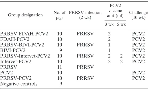
TABLE 1. Experimental design
Group designation No. of pigs
PRRSV infection (2 wk)
PCV2 vaccine
amt (ml) Challenge(10 wk)
3 wk 5 wk
PRRSV-FDAH-PCV2
10
PRRSV
2
PCV2
FDAH-PCV2
10
2
PCV2
PRRSV-BIVI-PCV2
10
PRRSV
1
PCV2
BIVI-PCV2
9
1
PCV2
PRRSV-Intervet-PCV2
10
PRRSV
2
2
PCV2
Intervet-PCV2
10
2
2
PCV2
PRRSV
11
PRRSV
PCV2
10
PCV2
PRRSV-PCV2
10
PRRSV
PCV2
Negative controls
9
on August 17, 2020 by guest
http://cvi.asm.org/
lymph nodes (mesenteric, superficial inguinal, tracheobronchial, and mediasti-nal), spleen, tonsil, and thymus using a rabbit polyclonal antiserum (42). PCV2 antigen scoring was done by a pathologist blinded to the treatment status of the animals, with scores ranging from 0 (no signal) to 3 (abundant amount of PCV2 antigen) as described previously (32).
Overall lymphoid lesion score.The overall microscopic lymphoid lesion score, which accounts for lymphoid depletion, histiocytic inflammation, and amount of PCV2 antigen, was calculated as previously described (32).
Statistical analysis.Statistical analysis of the data was performed using JMP software, version 8.0.1 (SAS Institute, Cary, NC). Summary statistics were cal-culated for all groups to assess the overall quality of the data, including normal-ity. Continuous data were analyzed using the one-way analysis of variance (ANOVA). APvalue of less than 0.05 was set as a statistically significant level throughout this study. If an ANOVA was significant, pairwise testing using Tukey’s adjustment was performed to assess specific group differences. Real-time PCR results (number of copies per ml of serum) were log10transformed prior to statistical analysis. Percent reduction for PCR data was measured as follows: 100⫺[(100⫻mean log10number of genomic copies/ml in the vaccinated group)/(mean log10number of genomic copies/ml in positive-control animals)]. Nonrepeated measures of necropsy and histopathology data were assessed using a nonparametric Kruskal-Wallis one-way ANOVA. If this nonparametric ANOVA test was significant (P⬍0.05), then pairwise Wilcoxon tests were used to assess differences between groups. In order to determine differences in prev-alence, a chi-square test was used.
RESULTS
ADWG.
There were no significant (
P
⬎
0.05) differences in
initial body weights among the groups of pigs at arrival in the
research facility. After PRRSV infection, between 2 and 3
weeks of age, the ADWG in PRRSV-infected pigs was
signif-icantly (
P
⬍
0.05) lower than that in noninfected pigs (0.25
⫾
0.01 kg versus 0.28
⫾
0.01 kg). Similarly, from 3 to 10 weeks of
age, the ADWG in PRRSV-infected pigs was significantly (
P
⬍
0.0001) lower than that in noninfected pigs (0.66
⫾
0.01 kg
versus 0.77
⫾
0.01 kg). PCV2 vaccination had no effect on
ADGW, as there was no significant (
P
⬎
0.05) difference
between the vaccinated and nonvaccinated groups from 3 to 10
weeks of age. The ADWG for the individual groups from the
time of PCV2 challenge until necropsy (10 to 13 weeks of age)
is summarized in Table 2. There was no significant (
P
⬎
0.05)
difference between PRRSV-infected and noninfected pigs or
between vaccinated and nonvaccinated pigs. However, the
ADWG in PCV2-infected pigs was significantly (
P
⬍
0.05)
lower than that in noninfected pigs (1.00
⫾
0.02 kg versus
1.08
⫾
0.05 kg).
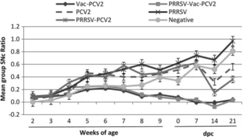
Anti-PCV2 IgG antibody levels.
At arrival, 95/99 pigs had
passively acquired PCV2 antibodies, with group mean SNc
ratios between 0.00 and 0.10 (Fig. 1), and no significant
differ-ences were observed among the different treatment groups.
The group mean PCV2 ELISA SNc ratios in vaccinated pigs
were significantly (
P
⬍
0.0001) lower than those in
nonvacci-nated pigs between 5 weeks of age (vaccinonvacci-nated pigs, 0.22
⫾
0.04; nonvaccinated pigs, 0.39
⫾
0.04) and 10 weeks of age
(vaccinated pigs, 0.06
⫾
0.02; nonvaccinated pigs, 0.51
⫾
0.04).
Significantly (
P
⬍
0.05) higher SNc ratios (lower levels of
antibody) were observed in the BIVI groups than in the FDAH
and Intervet groups during the growing period; however, this
was independent of PRRSV status. At the day of PCV2
chal-lenge at 10 weeks of age, 98% (58/59) of the vaccinated pigs
had detectable anti-PCV2 antibodies, whereas only 42.5% (17/
40) of the nonvaccinated pigs were seropositive. After PCV2
challenge (dpc 7, 14, and 21), vaccinated groups had
signifi-cantly (
P
⬍
0.001) lower SNc ratios than nonvaccinated
chal-lenge pigs, which were independent of the product used or
PRRSV infection status at the time of vaccination and at 14
and 21 dpc.
Prevalence and amount of PCV2 DNA in serum.
Pigs in the
negative-control group and in the PRRSV group remained
negative for PCV2 DNA throughout the study period. After
PCV2 challenge, PCV2 DNA was detected in the majority of
the nonvaccinated pigs at 7 (70%), 14 (100%), and 21 (100%)
dpc. Among the vaccinated pigs, PCV2 viremia was limited to
a few individual pigs (Table 3). Vaccination significantly (
P
⬍
0.05) reduced the amount of PCV2 DNA compared to that in
nonvaccinated pigs (Table 3). The mean reductions of PCV2
viremia in the serum at 21 dpc were 88.6% for FDAH-PCV2,
88.8% for BIVI-PCV2, 90.2% for Intervet-PCV2, 89.4% for
PRRSV-FDAH-PCV2, 66.8% for PRRSV-BIVI-PCV2, and
100% for PRRSV-Intervet-PCV2.
Anti-PRRSV IgG antibody levels.
All pigs were negative for
anti-PRRSV antibodies at the start of the study at 2 weeks of
age (data not shown). All pigs that were not experimentally
infected with PRRSV remained seronegative for PRRSV until
the termination of the study at 13 weeks of age. At 10 weeks of
age (dpc 0), the mean PRRSV ELISA S/P ratio values were
2.30
⫾
0.11 for PRRSV-infected, non-PCV2-vaccinated pigs
(
n
⫽
21) and 2.17
⫾
0.09 for PRRSV-infected,
PCV2-vacci-nated pigs (
n
⫽
30), with no significant (
P
⬎
0.05) difference
FIG. 1. Mean anti-PCV2 IgG antibody responses in the different
treatment groups following PCV2 challenge. PRRSV inoculation, the
first vaccination (Vac), challenge, and necropsy were performed at 2,
3, 10, and 13 weeks of age, respectively. Samples with an SNc ratio of
0.5 or greater are considered negative. Error bars indicate standard
errors.
TABLE 2. ADWG from 0 to 21 dpc
Group ADWG⫾SE (kg)a
PRRSV-FDAH-PCV2...1.02
⫾
0.03
A,B,CFDAH-PCV2 ...0.96
⫾
0.03
B,CPRRSV-BIVI-PCV2 ...1.12
⫾
0.04
A,BBIVI-PCV2 ...1.03
⫾
0.03
A,B,CPRRSV-Intervet-PCV2 ...0.99
⫾
0.05
B,CIntervet-PCV2 ...1.00
⫾
0.05
A,B,CPRRSV...0.98
⫾
0.06
B,CPCV2 ...0.91
⫾
0.05
CPRRSV-PCV2 ...0.98
⫾
0.06
B,CNegative controls ...1.21
⫾
0.06
Aa
Different superscripts (A, B, and C) within the same column represent significant differences (P⬍0.05) between groups.
on August 17, 2020 by guest
http://cvi.asm.org/
existing between groups. Similarly, there was no significant
(
P
⬎
0.05) difference in mean group PRRSV S/P ratios on dpc
21 between PCV2-vaccinated (1.78
⫾
0.11) and nonvaccinated
(2.08
⫾
0.13) pigs or between PCV2-challenged (1.87
⫾
0.10)
and nonchallenged (2.04
⫾
0.17) pigs.
Prevalence and amount of PRRSV RNA in serum.
At the
time of PCV2 vaccination (7 days after PRRSV inoculation)
and 7 days after PCV2 vaccination (14 days after PRRSV
inoculation), 100% of the pigs in the PRRSV-challenged
groups were positive for PRRSV RNA in serum, with PRRSV
RNA loads ranging from 6.16 to 6.66 log
10copies/ml (Table 4).
From 0 to 21 dpc, low levels of PRRSV RNA were detected in
a few (1 to 3) pigs in all PRRSV-infected groups (Table 4). No
significant (
P
⬎
0.05) differences were observed for PRRSV
prevalence or RNA loads among the different treatment
groups.
Macroscopic and microscopic lesions and PCV2 antigen in
lesions.
Macroscopic lung lesions were characterized by failure
of the lungs to collapse and by focal-to-diffuse, mottled tan
areas of pneumonia. Vaccinated, PCV2-challenged pigs (
n
⫽
59) had significantly (
P
⬍
0.05) lower mean lung scores than
nonvaccinated PCV2-infected pigs (
n
⫽
20) (1.97
⫾
0.32 versus
3.95
⫾
1.00). Moreover, PRRSV-negative PCV2-challenged
pigs (
n
⫽
39) had significantly (
P
⬍
0.05) lower mean lung
scores than PRRSV-positive PCV2-challenged pigs (
n
⫽
40)
(1.46
⫾
0.28 versus 3.45
⫾
0.62). There was no difference in the
severity of macroscopic lung lesions among vaccinated groups
(
P
⬎
0.05). In the PRRSV-PCV2 group, 9/10 pigs had lymph
nodes of 2 to 3 times the normal size. In the remaining groups,
individual pigs (1 to 2 per group) had mildly enlarged lymph
nodes.
Microscopic lesions were characterized by mild to severe
lymphoid depletion and mild to moderate histiocytic
replace-ment in lymphoid tissues. Among vaccinated pigs, 83.3% (49/
59) had normal lymphoid tissues and 16.9% (10/59) had mild
lymphoid lesions (overall lymphoid score, 1 or 2).
Nonvacci-nated and PCV2-infected pigs however, had the following
le-sion distribution: 10% (2/20) had normal lymphoid tissues,
45% (9/20) had mild lymphoid lesions (score, 1, 2, or 3), 40%
(8/20) had moderate lymphoid lesions (score, 4, 5, or 6), and
5% (1/20) had severe PCV2-associated lesions (score, 7).
There was no difference in lesion severity between
PRRSV-infected and nonPRRSV-infected pigs.
PCV2 antigen was detected in 8/10 pigs in the
PRRSV-PCV2 group, 7/10 pigs in the PRRSV-PCV2 group, and 0/59 pigs in the
vaccinated groups; the differences were statistically significant
(
P
⬍
0.05).
DISCUSSION
Various commercially available killed PCV2 vaccines are
being used worldwide, and their efficacies have been
demon-strated under both experimental conditions (13, 25, 29, 30, 40)
and field conditions (5, 12, 17, 19, 39). PCV2 vaccination is
recommended for usage in weaned pigs at about 3 weeks of age
or older. However, at this age many pigs derived from
PRRSV-positive breeding herds are infected with PRRSV, which is
thought to negatively impact PCV2 vaccine efficacy. In the
TABLE 3. PCV2 DNA-positive pigs among all pigs in each group at different days after PCV2 challenge
Group
No. of pigs PCV2 DNA positive/no. of pigs in group on the following days after PCV2 challengea:
7 14 21
PRRSV-FDAH-PCV2
1/10 (0.42
⫾
0.4)
A0/10 (0.00
⫾
0.0)
A0/10 (0.00
⫾
0.0)
AFDAH-PCV2
1/10 (0.38
⫾
0.4)
A0/10 (0.00
⫾
0.0)
A0/10 (0.00
⫾
0.0)
APRRSV-BIVI-PCV2
2/10 (0.89
⫾
0.6)
A3/10 (1.56
⫾
0.8)
B2/10 (0.81
⫾
0.5)
ABIVI-PCV2
4/9 (1.98
⫾
0.8)
AB2/9 (0.92
⫾
0.6)
BA1/9 (0.41
⫾
0.4)
APRRSV-Intervet-PCV2
0/10 (0.00
⫾
0.0)
A0/10 (0.00
⫾
0.0)
A0/10 (0.00
⫾
0.0)
AIntervet-PCV2
0/10 (0.00
⫾
0.0)
A0/10 (0.00
⫾
0.0)
A1/10 (0.36
⫾
0.4)
APRRSV
0/11 (0.00
⫾
0.0)
A0/11 (0.00
⫾
0.0)
A0/11 (0.00
⫾
0.0)
APCV2
7/10 (3.67
⫾
0.8)
B10/10 (6.69
⫾
0.4)
C10/10 (5.96
⫾
0.3)
BPRRSV-PCV2
7/10 (3.94
⫾
0.9)
B10/10 (7.22
⫾
0.2)
C10/10 (7.23
⫾
0.3)
BNegative controls
0/9 (0.00
⫾
0.0)
A0/9 (0.00
⫾
0.0)
A,B0/9 (0.00
⫾
0.0)
Aa
Different superscripts (A, B, and C) within the same column represent significant differences (P⬍0.05) in the mean amount of log10PCV2 DNA between groups. Values in parentheses represent the mean log10number of PCV2 DNA copies⫾standard error for the group.
TABLE 4. PRRSV RNA-positive pigs among all pigs in each group for pigs experimentally inoculated with PRRSV 7 days prior to PCV2
vaccination and 8 weeks prior to PCV2 challenge
Group
No. of pigs PRRSV RNA positive/no. of pigs in group on the indicated days aftera:
PCV2 vaccination PCV2 challenge
0 7 0 7 14 21
PRRSV-FDAH-PCV2
10/10 (6.16
⫾
0.3)
10/10 (6.66
⫾
0.3)
0/10 (0.00
⫾
0.0)
1/10 (0.43
⫾
0.4)
2/10 (0.96
⫾
0.6)
0/10 (0.00
⫾
0.0)
PRRSV-BIVI-PCV2
10/10 (6.35
⫾
0.1)
10/10 (6.40
⫾
0.3)
0/10 (0.00
⫾
0.0)
1/10 (0.32
⫾
0.3)
2/10 (0.66
⫾
0.4)
2/10 (0.67
⫾
0.5)
PRRSV-Intervet-PCV2
10/10 (6.29
⫾
0.2)
10/10 (6.33
⫾
0.2)
2/10 (0.63
⫾
0.4)
1/10 (0.34
⫾
0.3)
3/10 (1.07
⫾
0.6)
2/10 (0.00
⫾
0.0)
PRRSV
11/11 (6.48
⫾
0.2)
11/11 (6.59
⫾
0.4)
0/11 (0.00
⫾
0.0)
1/11 (0.32
⫾
0.3)
1/11 (0.37
⫾
0.4)
0/11 (0.00
⫾
0.0)
PRRSV-PCV2
10/10 (6.44
⫾
0.3)
10/10 (6.29
⫾
0.4)
2/10 (0.69
⫾
0.5)
1/10 (0.28
⫾
0.3)
2/10 (0.83
⫾
0.6)
2/10 (0.79
⫾
0.5)
a
Values in parentheses represent the mean log10PRRSV RNA copies⫾standard error for the group.
on August 17, 2020 by guest
http://cvi.asm.org/
present study, we evaluated PCV2 vaccine efficacy in
PRRSV-positive conventional pigs by experimental inoculation with a
known virulent isolate of PRRSV (14, 24, 28). It is well-known
that PRRSV strains differ in virulence (28), and the results of
this study may be different with other PRRSV challenge
iso-lates.
Increased PCV2 replication and clinical PCVAD were
ob-served in pigs experimentally coinfected with PRRSV and
PCV2 (2, 16, 38). Although PRRSV-infected pigs had
signifi-cantly increased macroscopic lung lesions in the present study,
significant differences in the severity of microscopic lesions or
amount of PCV2 antigen were not observed in
PCV2-chal-lenged pigs. A possible explanation for the discrepancy in
ob-servations is the difference in timing of pathogen
administra-tion: in the current study, the pigs were infected with PRRSV
at 2 weeks of age, followed by PCV2 challenge at 10 weeks of
age, whereas previously, coinfections were conducted at the
same time (16) or within an interval of a few days (2, 38) and
were done in younger pigs (1 to 35 days of age).
Although PCV2 vaccines are in general very effective, cases
of apparent vaccine failures continue to occur. Vaccine failures
under field conditions may be due to many reasons, including
failure to follow the proper administration protocols and to use
the recommended dose of the vaccines (30). The field strains
might be different from the vaccine strains, and the breed of
the pigs can also influence the outcome of PCVAD (22, 30).
The presence of high levels of passively derived anti-PCV2
antibodies at the time of vaccination has also been associated
with decreased vaccine efficacy and vaccine failures.
Experi-mental proof of this is lacking to date, as available research
results indicate no adverse effect of maternally derived
immu-nity on PCV2 vaccine efficacy (13, 29, 40). Similarly, the results
of the current study indicate that PCV2 vaccination in the face
of high levels of passively derived anti-PCV2 immunity was
effective in inducing a humoral immune response. At 10 weeks
of age, a significant higher prevalence of seropositive pigs was
found in the vaccinated group than in the nonvaccinated
con-trol group.
Several studies have investigated the effect of PRRSV
infection on vaccine efficacy but provided conflicting results.
PRRSV infection at the time of classical swine fever virus
vaccination significantly inhibited the host immune response
and resulted in vaccination failure after subsequent exposure
to classical swine fever virus (7, 43). For pseudorabies virus
vaccination, although PRRSV infection affected the duration
of the T-lymphocyte response, it did not inhibit the efficacy of
the vaccine, which was capable of inducing protective
immu-nity (7). In the present study, our data revealed that all the
commercial vaccines used in this study were able to induce a
protective immunity against PCV2 in the face of PRRSV
in-fection at the time of vaccination. The difference in the
exper-imental results might be due to different timings of infection
with PRRSV, vaccination, and challenge. In the present study,
pigs were challenged 5 weeks (two dose administrations) to 7
weeks (one dose administration) after vaccination, in contrast
to a study where challenge occurred 3 weeks after initial
vac-cination (43).
In a previous study (26), we found that PCV2 infection had
adverse effects on the efficacy of a modified live virus (MLV)
PRRSV vaccine. The different effects of PRRSV and PCV2
infection on the efficacy of vaccines may be due to the different
mechanisms that these two viruses use to induce
immunosup-pression (replication in macrophages versus lymphoid
deple-tion), which are still not completely understood, and the types
of vaccine (attenuated live versus inactivated) tested.
All PCV2 vaccines used, regardless of PRRSV infection
status at the time of vaccination, were able to suppress PCV2
replication after PCV2 challenge, as demonstrated by
signifi-cantly reduced levels of PCV2 viremia and signifisignifi-cantly
re-duced amounts of PCV2 antigen in lymphoid tissues. The
results of our study demonstrated that by 21 dpc there was no
difference in the prevalence of PCV2 DNA in vaccinated
groups regardless of PRRSV infection status at the time of
vaccination or the vaccine product used. We conclude that
prior PRRSV infection does not have an adverse effect on
commercial PCV2 vaccination in conventional growing pigs
and that pigs can be effectively immunized against PCV2 in the
face of maternal antibodies.
ACKNOWLEDGMENTS
We thank Matthew Umphress for assistance with the animal work.
Funding for this study was provided by the National Pork Board
CheckOff Dollars.
REFERENCES
1.Albina, E., F. Madec, R. Cariolet, and J. Torrison.1994. Immune response and persistence of the porcine reproductive and respiratory syndrome virus in infected pigs and farm units. Vet. Rec.134:567–573.
2.Allan, G. M., F. McNeilly, J. Ellis, S. Krakowka, B. Meehan, I. McNair, I. Walker, and S. Kennedy.2000. Experimental infection of colostrum deprived piglets with porcine circovirus 2 (PCV2) and porcine reproductive and re-spiratory syndrome virus (PRRSV) potentiates PCV2 replication. Arch. Vi-rol.145:2421–2429.
3.Bierk, M. D., S. A. Dee, K. D. Rossow, S. Otake, J. E. Collins, and T. W. Molitor.2001. Transmission of porcine reproductive and respiratory syn-drome virus from persistently infected sows to contact controls. Can. J. Vet. Res.65:261–266.
4.Cavanagh, D.1997. Nidovirales: a new order comprising Coronaviridae and Arteriviridae. Arch. Virol.142:629–633.
5.Cline, G., V. Wilt, E. Diaz, and R. Edler.2008. Efficacy of immunising pigs against porcine circovirus type 2 at three or six weeks of age. Vet. Rec.
163:737–740.
6.Crowther, R. A., J. A. Berriman, W. L. Curran, G. M. Allan, and D. Todd.
2003. Comparison of the structures of three circoviruses: chicken anemia virus, porcine circovirus type 2, and beak and feather disease virus. J. Virol.
77:13036–13041.
7.De Bruin, M. G., J. N. Samsom, J. J. Voermans, E. M. van Rooij, Y. E. De Visser, and A. T. Bianchi.2000. Effects of a porcine reproductive and respi-ratory syndrome virus infection on the development of the immune response against pseudorabies virus. Vet. Immunol. Immunopathol.76:125–135. 8.Dorr, P. M., R. B. Baker, G. W. Almond, S. R. Wayne, and W. A. Gebreyes.
2007. Epidemiologic assessment of porcine circovirus type 2 coinfection with other pathogens in swine. J. Am. Vet. Med. Assoc.230:244–250. 9.Drew, T. W.2000. A review of evidence for immunosuppression due to
porcine reproductive and respiratory syndrome virus. Vet. Res.31:27–39. 10.Duan, X., H. J. Nauwynck, H. W. Favoreel, and M. B. Pensaert.1998.
Identification of a putative receptor for porcine reproductive and respiratory syndrome virus on porcine alveolar macrophages. J. Virol.72:4520–4523. 11.Duan, X., H. J. Nauwynck, and M. B. Pensaert.1997. Virus quantification
and identification of cellular targets in the lungs and lymphoid tissues of pigs at different time intervals after inoculation with porcine reproductive and respiratory syndrome virus (PRRSV). Vet. Microbiol.56:9–19.
12.Fachinger, V., R. Bischoff, S. B. Jedidia, A. Saalmuller, and K. Elbers.2008. The effect of vaccination against porcine circovirus type 2 in pigs suffering from porcine respiratory disease complex. Vaccine26:1488–1499. 13.Fort, M., M. Sibila, A. Allepuz, E. Mateu, F. Roerink, and J. Segale´s.2008.
Porcine circovirus type 2 (PCV2) vaccination of conventional pigs prevents viremia against PCV2 isolates of different genotypes and geographic origins. Vaccine26:1063–1071.
14.Halbur, P. G., P. S. Paul, M. L. Frey, J. Landgraf, K. Eernisse, X. J. Meng, M. A. Lum, J. J. Andrews, and J. A. Rathje. 1995. Comparison of the pathogenicity of two US porcine reproductive and respiratory syndrome virus isolates with that of the Lelystad virus. Vet. Pathol.32:648–660.
on August 17, 2020 by guest
http://cvi.asm.org/
15.Harding, J., and E. Clark.1997. Recognizing and diagnosing postweaning multisystemic wasting syndrome (PMWS). Swine Health Prod.5:201–203. 16.Harms, P. A., S. D. Sorden, P. G. Halbur, S. R. Bolin, K. M. Lager, I.
Morozov, and P. S. Paul.2001. Experimental reproduction of severe disease in CD/CD pigs concurrently infected with type 2 porcine circovirus and porcine reproductive and respiratory syndrome virus. Vet. Pathol.38:528– 539.
17.Horlen, K. P., S. S. Dritz, J. C. Nietfeld, S. C. Henry, R. A. Hesse, R. Oberst, M. Hays, J. Anderson, and R. R. Rowland. 2008. A field evaluation of mortality rate and growth performance in pigs vaccinated against porcine circovirus type 2. J. Am. Vet. Med. Assoc.232:906–912.
18.Kim, J., H. K. Chung, and C. Chae.2003. Association of porcine circovirus 2 with porcine respiratory disease complex. Vet. J.166:251–256. 19.Kixmo¨ller, M., M. Ritzmann, M. Eddicks, A. Saalmu¨ller, K. Elbers, and V.
Fachinger.2008. Reduction of PMWS-associated clinical signs and infec-tions by vaccination against PCV2. Vaccine26:3443–3451.
20.Lager, K. M., W. L. Mengeling, and S. L. Brockmeier.1997. Duration of homologous porcine reproductive and respiratory syndrome virus immunity in pregnant swine. Vet. Microbiol.58:127–133.
21.Lo´pez-Fuertes, L., E. Campos, N. Dome´nech, A. Ezquerra, J. M. Castro, J. Dominguez, and F. Alonso.2000. Porcine reproductive and respiratory syn-drome (PRRS) virus down-modulates TNF-alpha production in infected macrophages. Virus Res.69:41–46.
22.Lope´z-Soria, S., J. Segale´s, M. Nofrarías, M. Calsamiglia, H. Ramirez, A. Minguez, I. M. Serrano, O. Marin, and A. Callen.2004. Genetic influence on the expression of PCV disease. Vet. Rec.155:504.
23.Neumann, E. J., J. B. Kliebenstein, C. D. Johnson, J. W. Mabry, E. J. Bush, A. H. Seitzinger, A. L. Green, and J. J. Zimmerman.2005. Assessment of the economic impact of porcine reproductive and respiratory syndrome on swine production in the United States. J. Am. Vet. Med. Assoc.227:385–392. 24.Opriessnig, T., P. G. Halbur, K. J. Yoon, R. M. Pogranichniy, K. M.
Har-mon, R. Evans, K. F. Key, F. J. Pallares, P. Thomas, and X. J. Meng.2002. Comparison of molecular and biological characteristics of a modified live porcine reproductive and respiratory syndrome virus (PRRSV) vaccine (In-gelvac PRRS MLV), the parent strain of the vaccine (ATCC VR2332), ATCC VR2385, and two recent field isolates of PRRSV. J. Virol.76:11837– 11844.
25.Opriessnig, T., D. M. Madson, J. R. Prickett, D. Kuhar, J. K. Lunney, J. Elsener, and P. G. Halbur.2008. Effect of porcine circovirus type 2 (PCV2) vaccination on porcine reproductive and respiratory syndrome virus (PRRSV) and PCV2 coinfection. Vet. Microbiol.131:103–114.
26.Opriessnig, T., N. E. McKeown, K. L. Harmon, X. J. Meng, and P. G. Halbur.2006. Porcine circovirus type 2 infection decreases the efficacy of a modified live porcine reproductive and respiratory syndrome virus vaccine. Clin. Vaccine Immunol.13:923–929.
27.Opriessnig, T., X. J. Meng, and P. G. Halbur.2007. Porcine circovirus type 2 associated disease: update on current terminology, clinical manifestations, pathogenesis, diagnosis, and intervention strategies. J. Vet. Diagn. Invest.
19:591–615.
28.Opriessnig, T., F. J. Pallare´s, N. Dachrit, A. L. Vincent, E. L. Thacker, E. M. Vaughn, M. Roof, and P. G. Halbur.2005. Genomic homology of ORF 5 gene sequence between modified live vaccine virus and porcine reproductive and respiratory syndrome virus challenge isolates is not predictive of vaccine efficacy. J. Swine Health Prod.13:246–253.
29.Opriessnig, T., A. R. Patterson, J. Elsener, X. J. Meng, and P. G. Halbur.
2008. Influence of maternal antibodies on efficacy of porcine circovirus type 2 (PCV2) vaccination to protect pigs from experimental infection with PCV2. Clin. Vaccine Immunol.15:397–401.
30.Opriessnig, T., A. R. Patterson, D. M. Madson, N. Pal, and P. G. Halbur.
2009. Comparison of efficacy of commercial one dose and two dose PCV2 vaccines using a mixed PRRSV-PCV2-SIV clinical infection model 2-3-months post vaccination. Vaccine27:1002–1007.
31.Opriessnig, T., S. Ramamoorthy, D. M. Madson, A. R. Patterson, N. Pal, S. Carman, X. J. Meng, and P. G. Halbur.2008. Differences in virulence among porcine circovirus type 2 isolates are unrelated to cluster type 2a or 2b and prior infection provides heterologous protection. J. Gen. Virol.89:2482– 2491.
32.Opriessnig, T., E. L. Thacker, S. Yu, M. Fenaux, X. J. Meng, and P. G. Halbur. 2004. Experimental reproduction of postweaning multisystemic wasting syndrome in pigs by dual infection withMycoplasma hyopneumoniae and porcine circovirus type 2. Vet. Pathol.41:624–640.
33.Opriessnig, T., S. Yu, J. M. Gallup, R. B. Evans, M. Fenaux, F. Pallares, E. L. Thacker, C. W. Brockus, M. R. Ackermann, P. Thomas, X. J. Meng, and P. G. Halbur.2003. Effect of vaccination with selective bacterins on conven-tional pigs infected with type 2 porcine circovirus. Vet. Pathol.40:521–529. 34.Pallare´s, F. J., P. G. Halbur, T. Opriessnig, S. D. Sorden, D. Villar, B. H. Janke, M. J. Yaeger, D. J. Larson, K. J. Schwartz, K. J. Yoon, and L. J. Hoffman.2002. Porcine circovirus type 2 (PCV-2) coinfections in US field cases of postweaning multisystemic wasting syndrome (PMWS). J. Vet. Di-agn. Invest.14:515–519.
35.Pogranichniy, R. M., K. J. Yoon, P. A. Harms, S. D. Sorden, and M. Daniels.
2002. Case-control study on the association of porcine circovirus type 2 and other swine viral pathogens with postweaning multisystemic wasting syn-drome. J. Vet. Diagn. Invest.14:449–456.
36.Ramamoorthy, S., and X. J. Meng.2008. Porcine circoviruses: a minuscule yet mammoth paradox. Anim. Health Res. Rev.10:1–20.
37.Rossow, K. D.1998. Porcine reproductive and respiratory syndrome. Vet. Pathol.35:1–20.
38.Rovira, A., M. Balasch, J. Segale´s, L. Garcia, J. Plana-Duran, C. Rosell, H. Ellerbrok, A. Mankertz, and M. Domingo.2002. Experimental inoculation of conventional pigs with porcine reproductive and respiratory syndrome virus and porcine circovirus 2. J. Virol.76:3232–3239.
39.Segale´s, J., A. Urniza, A. Alegre, T. Bru, E. Crisci, M. Nofrarías, S. Lo ´pez-Soria, M. Balasch, M. Sibila, Z. Xu, H. J. Chu, L. Fraile, and J. Plana-Duran. 2009. A genetically engineered chimeric vaccine against porcine circovirus type 2 (PCV2) improves clinical, pathological and virological out-comes in postweaning multisystemic wasting syndrome affected farms. Vac-cine27:7313–7321.
40.Shen, H. G., N. Beach, Y. W. Huang, P. G. Halbur, X. J. Meng, and T. Opriessnig.2010. Comparison of commercial and experimental porcine cir-covirus type 2 (PCV2) vaccines using a triple challenge with PCV2, porcine reproductive and respiratory syndrome virus (PRRSV), and porcine parvo-virus (PPV). Vaccine28:5960–5966.
41.Sorden, S. D.2000. Update on porcine circovirus and postweaning multisys-temic wasting syndrome (PMWS). Swine Health Prod.8:133–136. 42.Sorden, S. D., P. A. Harms, P. Nawagitgul, D. Cavanaugh, and P. S. Paul.
1999. Development of a polyclonal-antibody-based immunohistochemical method for the detection of type 2 porcine circovirus in formalin-fixed, paraffin-embedded tissue. J. Vet. Diagn. Invest.11:528–530.
43.Suradhat, S., S. Kesdangsakonwut, W. Sada, S. Buranapraditkun, S. Wong-sawang, and R. Thanawongnuwech.2006. Negative impact of porcine repro-ductive and respiratory syndrome virus infection on the efficacy of classical swine fever vaccine. Vaccine24:2634–2642.
44.Tischer, I., H. Gelderblom, W. Vettermann, and M. A. Koch.1982. A very small porcine virus with circular single-stranded DNA. Nature295:64–66. 45.Todd, D., M. Bendinelli, P. Biagini, S. Hino, A. Mankertz, S. Mishiro, C.
Niel, H. Okamoto, S. Raidal, B. W. Ritchie, and G. Teo.2005.Circoviridae, p. 327–334. InC. M. Fauquet, M. A. Mayo, J. Maniloff, U. Desselberger, and L. A. Ball (ed.), Virus taxonomy: eighth report of the International Com-mittee on Taxonomy of Viruses/ Elsevier Academic Press, San Diego, CA. 46.Van Breedam, W., P. L. Delputte, H. Van Gorp, G. Misinzo, N. Vanderhei-jden, X. Duan, and H. J. Nauwynck.2010. Porcine reproductive and respi-ratory syndrome virus entry into the porcine macrophage. J. Gen. Virol.
91:1659–1667.