Scholarship@Western
Scholarship@Western
Electronic Thesis and Dissertation Repository
8-21-2017 2:00 PM
Mechanisms underlying executive function deficits
Mechanisms underlying executive function deficits
Sagar Jayawantrao Desai The University of Western Ontario
Supervisor
Dr. Raj Rajakuamr
The University of Western Ontario
Graduate Program in Anatomy and Cell Biology
A thesis submitted in partial fulfillment of the requirements for the degree in Doctor of Philosophy
© Sagar Jayawantrao Desai 2017
Follow this and additional works at: https://ir.lib.uwo.ca/etd
Part of the Behavioral Neurobiology Commons, and the Cognitive Neuroscience Commons
Recommended Citation Recommended Citation
Desai, Sagar Jayawantrao, "Mechanisms underlying executive function deficits" (2017). Electronic Thesis and Dissertation Repository. 4774.
https://ir.lib.uwo.ca/etd/4774
This Dissertation/Thesis is brought to you for free and open access by Scholarship@Western. It has been accepted for inclusion in Electronic Thesis and Dissertation Repository by an authorized administrator of
I
Abstract
In our daily life, we come across situations where we meet unanticipated
challenges, we must take certain decisions, pay attention, be flexible and inhibit
impulsive actions to achieve goal directed behaviour. During these processes, we
unknowingly use sets of interdependent cognitive processes collectively called ‘executive
function’. Executive function is mainly regulated by the frontal lobe. Impaired executive function is associated with disorders such as schizophrenia, Alzheimer’s disease, autism
and attention deficit hyperactivity disorder (ADHD).
In this thesis, we investigated neurotransmitters and interactions among them
regulating executive function. Further, we investigated mechanisms underlying those
interactions mediating executive function in rats using an operant conditioning-based
set-shifting task, a common and validated test in animals to assess executive function. In our
first study, we identified for the first time that systemic injections of dopamine D1 and
glutamate N-methyl-D-aspartate (NMDA) receptor antagonists cause impaired
set-shifting and increased the occurrence of perseverative errors only after combined
administration at doses that failed to affect set-shifting following separate injections. The
discovery of this novel synergistic effect of glutamate and dopamine antagonists on
set-shifting prompted us to undertake our second study to determine if such synergy occurs
within the medial PFC (mPFC)- an important brain area associated with executive
function in rodents. Our results confirmed that mPFC is a site where seemingly mild
suppression of glutamate and dopamine activities, similar to that has been reported in
schizophrenia brains, may act cooperatively to manifest deficits in executive function via
increasing perseverative errors. Our third study was to identify molecular mechanisms
underlying such synergy. We found that protein kinase A (PKA) and extracellular
signal-regulated kinase (ERK1/2) signaling cascades transduce this effect, with ERK1/2
phosphorylation in mPFC neurons as an obligatory step for set-shifting.
The present results have substantially advanced our understanding of the
mechanisms underlying executive function. Our results also point to potential novel
II
Keywords:
executive function, set-shifting, prefrontal cortex, glutamate, dopamine,III
Co-Authorship Statement
Chapter 1 (introduction) and 5 (discussion), were written by Sagar J. Desai.
Chapter 2, entitled “Combination of behaviourally sub-effective doses of glutamate
NMDA and dopamine D1 receptor antagonists impairs executive function” was written
by Sagar J. Desai with input from Dr. N. Rajakumar and Dr. Brian Allman. Chapter 3,
entitled “Glutamate and dopamine abnormalities in the medial prefrontal cortex act
synergistically to cause executive dysfunction” was written by Sagar J. Desai with input
from Dr. N. Rajakumar and Dr. Brian Allman. Chapter 4 entitled “Molecular mechanisms
associated with D1-NMDA receptor interaction mediated set-shifting in rats” was written
by Sagar J. Desai.
IV
Acknowledgments
Foremost, I would like to express my sincere gratitude to Dr. Rajakumar for the
continuous support of my Ph.D. study and research, for his patience, motivation,
enthusiasm, and immense knowledge. His guidance helped me in all the time of research
and writing of this thesis. I could not have imagined having a better advisor and mentor
for my Ph.D. studies.
I would like to thank Dr. Brian Allman for his generous support of my thesis work. It
would never have been possible for me to take this work to the completion without his
incredible support and encouragement. I have greatly benefited from his continuous
insightful comments and suggestions regarding my project work as well as manuscripts
we wrote.
I thank my advisory committee: Drs. Shawn Whitehead, Walter Rushlow, Martin
Kavaliers, Raj Rajakumar and Brian Allman for their encouragement, insightful
comments, and hard questions.
I would like to offer my special thanks to all the labs in the department of ACB;
especially Allman lab (Ashley, Krystal, and Greg) who graciously allowed me to use all
the lab equipment and supplies. I also would like to thank Adrianna who took care of my
animals in my absence.
I owe my deepest gratitude to Mrs. Rajakuamar who supported me throughout my Ph.D.
Memories of those countless delicious meals will be with me forever. Words cannot
express my appreciation for everything you have done to keep me motivated and making
me feel like at home.
Last but not the least, I would like to thank my family: my parents, my brother Sameer,
my sister in law Sujata for their immeasurable love and care. I would like to thank my
wife Pooja who stayed back in India without me for almost a year and let me complete
V
Table of Contents
Abstract ... I
Co-Authorship Statement... III
Acknowledgments... IV
Table of Contents ... V
List of Tables ... X
List of Figures ... XI
List of Appendices ... XIV
List of Abbreviations ... XV
Chapter 1 ... 1
Introduction ... 2
1.1 Executive functions ... 2
Executive functioning: Components ... 3
Inhibition ... 3
Representative tasks used to assess inhibitory control ... 4
Working memory ... 4
Representative tasks used to assess working memory ... 5
Cognitive flexibility ... 5
Representative tasks used to assess cognitive flexibility ... 6
Neuroanatomical correlates of executive functioning in human ... 6
Executive functioning in rodents ... 7
Behavioural flexibility in rodents ... 8
Digging task ... 8
Maze task ... 11
VI
Error classification ... 13
Neural circuits subserving behavioural flexibility in rodents ... 13
Prefrontal Cortex (PFC) ... 14
Mediodorsal Thalamus (MDT) ... 14
Nucleus Accumbens (NAc) ... 15
Striatum ... 15
Neurotransmitters associated with behavioural flexibility ... 15
GABA ... 16
Dopamine ... 16
Glutamate ... 17
Dopamine-glutamate interactions ... 18
Mechanisms underlying D1-NMDA interactions ... 19
D1-NMDA receptor interactions through protein kinase A (PKA) signaling cascade ... 19
D1-NMDA receptor interactions through extracellular signal-regulated kinase (ERK) signaling cascade ... 20
Physical D1-NMDA receptor interactions ... 20
1.2 Thesis objectives and hypotheses ... 23
Study I ... 23
Study II... 24
Study III ... 25
1.3 References ... 27
Chapter 2 ... 49
Combination of behaviourally sub-effective doses of glutamate NMDA and dopamine D1 receptor antagonists impairs executive function1 ... 50
VII
2.2 Introduction ... 51
2.3 Materials and methods ... 53
Animals ... 53
Drugs ... 53
Apparatus ... 54
Set-shifting ... 54
Statistical analysis ... 58
2.4 Results ... 60
Effect of MK-801, SCH 23390 or combined drug treatment on visual-cue retrieval ... 60
Effect of MK-801, SCH 23390 or combined drug treatment on set-shifting ... 61
Response latencies ... 67
Omissions ... 67
Omissions ... 68
2.5 Discussion ... 69
2.6 Conclusions ... 72
2.7 References ... 73
Chapter 3 ... 81
Glutamate and dopamine abnormalities in the medial prefrontal cortex act synergistically to cause executive dysfunction ... 82
3.1 Abstract ... 82
3.2 Introduction ... 83
3.3 Materials and methods ... 85
Animals ... 85
VIII
Surgery ... 85
Micro-infusion procedure ... 86
Behavioural apparatus ... 86
Set-shifting task ... 87
Histology ... 88
Statistical analysis ... 88
3.4 Results ... 90
Effect of SCH 23390, MK-801 or combined antagonist infusion on visual-cue retrieval ... 90
Effect of SCH 23390, MK-801 or combined antagonist infusion on set-shifting... 91
Response latencies and omissions ... 96
Omissions ... 97
Effect of co-infusion of receptor antagonists on visual-cue learning ... 98
3.5 Discussion ... 100
3.6 References ... 104
Chapter 4 ... 113
Molecular mechanisms associated with D1-NMDA receptor interaction mediated set-shifting in rats ... 114
4.1 Abstract ... 114
4.2 Introduction ... 115
4.3 Materials and methods ... 119
Animals ... 119
Drugs ... 119
Surgery ... 120
IX
Behavioural apparatus ... 121
Set-shifting task ... 121
Histology ... 121
Statistical analysis ... 122
4.4 Results ... 123
Effect of re-testing rats on behavioural pattern during set-shifting. ... 123
Reduced Fyn kinase activity in the mPFC does not affect set-shifting .. 125
Increasing PKA activity in the mPFC rescues co-infusion of D1 and NMDA blockers-induced impairment in set-shifting ... 127
ERK phosphorylation in mPFC neurons is essential for set-shifting ... 130
Augmenting PKA activity does not affect the set-shifting deficits caused by the inhibition of ERK phosphorylation ... 135
4.5 Discussion ... 139
Effect of PKA elevation on dual combination-induced set-shifting deficits ... 139
Effect of inhibiting Src kinase on set-shifting ... 144
4.6 References ... 145
Chapter 5 ... 158
General discussion ... 159
5.1 Future directions ... 164
5.2 Limitations: ... 166
5.3 References ... 167
Appendices ... 176
X
List of Tables
XI
List of Figures
Figure 1.1 Flanker Task (Eriksen and Eriksen, 1974) ... 4
Figure 1.2 Schematic of the digging task used to assess behavioural flexibility in rodents.
... 10
Figure 1.3 Schematic of the maze task used to assess behavioural flexibility in rodents. 11
Figure 1.4 Schematic of operant-based task used to assess behavioural flexibility in
rodents. ... 12
Figure 2.1 Timeline of set-shifting task. ... 59
Figure 2.2 Effect of systemic administration of individual or combined antagonists on
visual-cue retrieval on set-shift day. ... 60
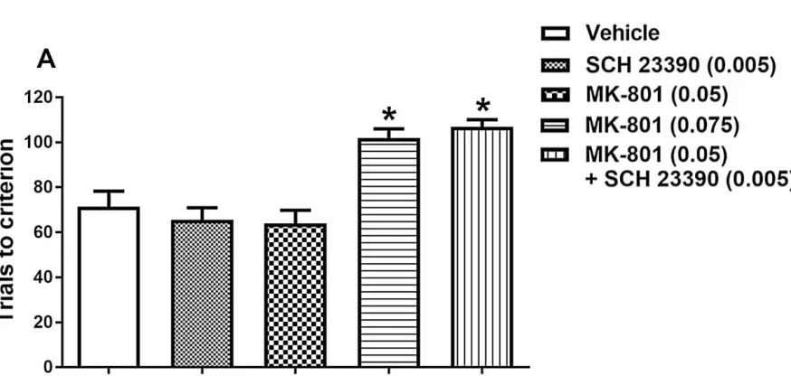
Figure 2.3 Effect of systemic administration of individual or combined antagonists on
trials to criterion during set-shifting. ... 62
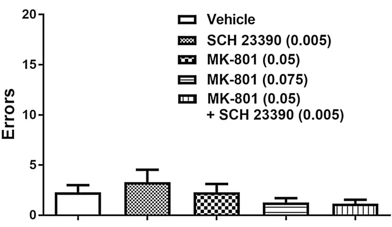
Figure 2.4 Effect of systemic administration of individual or combined antagonists on
errors to criterion during set-shifting. ... 64
Figure 2.5 Effect of drug treatments on different type of errors committed during
set-shifting. ... 66
Figure 2.6 Effect of drug treatments on latencies to response during different types of
trials... 67
Figure 2.7 Effect of drug treatments on number of omissions committed during
set-shifting. ... 68
Figure 3.1 Timeline of Experiment 1 and 2. ... 89
Figure 3.2 Effect of intra-mPFC infusion of SCH 23390 and MK-801 alone or in
XII
Figure 3.3 Effect of intra-mPFC infusion of SCH 23390 and MK-801 alone or in
combination on trials to criterion during set-shifting. ... 92
Figure 3.4 Effect of intra-mPFC infusion of SCH 23390 and MK-801 alone or in
combination on errors to criterion during set-shifting. ... 94
Figure 3.5 Intra-mPFC infusion of SCH 23390 and MK-801 alone or in combination
differentially affected the error profile during set-shifting. ... 95
Figure 3.6 Effect of intra-mPFC infusion of antagonists alone or in combination on
latencies to response during different types of trial. ... 96
Figure 3.7 Effect of intra-mPFC infusion of antagonists alone or in combination on the
number of omissions committed during set-shifting. ... 97
Figure 3.8 Intra-mPFC co-infusion of the lower, individually-ineffective doses of
antagonists did not impair learning. ... 98
Figure 3.9 Representative histology and reconstruction of infusion cannulae location in
mPFC. ... 99
Figure 4.1 Prefrontal cortical D1-NMDA receptor interactions and molecular signalling
cascades associated with them. ... 118
Figure 4.2 Effect of re-exposure to set-shifting on performance. ... 123
Figure 4.3 Effect of re-exposure to set-shifting on type of errors. ... 124
Figure 4.4 Effect of intra-mPFC infusion of PP2 on number of trials and errors to
criterion during set-shifting... 125
Figure 4.5 Effect of intra-mPFC infusion of PP2 on type of errors during set-shifting. 126
Figure 4.6 Effect of rolipram on dual combination-induced set-shifting impairments. . 128
XIII
Figure 4.8 Effect of PD98059 treatment on visual-cue learning and visual-cue retrieval
before set-shifting. ... 131
Figure 4.9 Effect of intra-mPFC infusion of PD98059 on trials and errors to criterion
during set-shifting. ... 132
Figure 4.10 Effect of PD98059 treatment on type of errors during set-shifting. ... 133
Figure 4.11 Effect of PD98059 treatment on number of omissions and latency to response
during set-shifting. ... 134
Figure 4.12 Effect of rolipram on PD98059-induced set-shifting impairments. ... 136
Figure 4.13 Effect of rolipram on PD98059-induced increased perseverance. ... 137
Figure 4.14 Representative histology and reconstruction of infusion cannulae tracks in
mPFC. ... 138
Figure 4.15 Possible mechanisms underlying D1-NMDA receptor interaction mediated
XIV
List of Appendices
Appendix A Formal License for material used with permission for Study I ... 176
Appendix B: Research Ethics and Approval Number for Study I ... 177
XV
List of Abbreviations
5CERT, 5 choice serial reaction time task
ADHD, attention deficit hyperactivity disorder
ANOVA, one-way analysis of variance
cAMP, cyclic AMP
CNS, central nervous system
CREB, cAMP Response Element-Binding Protein
dlPFC, dorsolateral prefrontal cortex
DMSO, dimethylsulfoxide
DREADD, designer receptors exclusively activated by designer drugs
ERK, extracellular signal-regulated kinase
GABA, gamma-Aminobutyric acid
GAD67, glutamic acid decarboxylase-67
LTP, long-term potentiation
MAPK, mitogen-activated protein kinase
MDT, mediodorsal thalamus
MRS, magnetic resonance spectroscopy
NAc, nucleus accumbens
NMDA, N-methyl-D-aspartate
OFC, orbitofrontal cortex
PCP, phencyclidine
PET, positron emission tomography
PFC, prefrontal cortex
PKA, protein kinase A
PKC, protein kinase C
SEM, standard error of mean
SPD, schizotypal personality disorder
SPOT, self-ordered pointing task
Introduction
1.1
Executive functions
While driving a car, upon reaching a traffic signal that has just turned amber, the
first thought that comes to our mind is about the distance of the car from the signal and
how much time remains before the light turns red. Depending on the assessment, if there
is sufficient time, we keep driving and get through the traffic signal safely. Alternatively,
if we think that the signal will turn red before we pass through it, we apply the brakes to
stop the car at the signal. In the above example, our brain works with enormous speed to
calculate our distance from the traffic signal, probable time left before light turns red, are
there any pedestrians looking for crossing the road, other vehicles around our car, etc.
Information from our current sensory environment and relevant past experiences are
considered and processed at very fast rate making it possible to drive a car safely. This is
an example of ‘executive function’ where multiple interdependent cognitive processes
work together synchronously to carry out a goal directed behaviour.
Executive function is a higher order cognitive ability comprising working memory,
behavioural flexibility (set-shifting and reversal learning), attention, problem solving
ability and inhibition (Gilbert and Burgess 2008; Diamond 2013; Miyake et al. 2000).
These processes facilitate us to think, plan, get engaged, learn from the outcomes and
tackle unfamiliar circumstances to carry out goal-directed behaviours in our day-to-day
life (Elliott, 2003). As described by Funahashi (2001), executive function is ‘a product of
the co-ordinated operation of various processes to accomplish a particular goal in a
flexible manner’. It would be difficult to perform daily activities without proper
executive functioning ability (Damasio, 1994). Various central nervous system (CNS)
disorders including schizophrenia, Alzheimer’s disease, autism and attention deficit
hyperactivity disorder (ADHD) show deficits in executive function (Millan et al., 2012).
Executive function impairments may affect cognition, behaviour and personality.
Behavioural component involve difficulty in pursuing goals, problem-solving ability and
switching between possible strategies i.e. behavioural flexibility. These impairments
affect a patient’s social life drastically and lead to immense economical loss to the
Executive functioning: Components
Conventionally, executive function was considered as a single construct, associated
with higher order cognitive skills (Shallice, 1988). Executive function has also been
conceptualized as a balanced association of multiple cognitive processes bringing about
higher order cognitive function (Stuss and Alexander, 2000). As per the latter concept,
there are three core components of executive functions: inhibition, working memory, and
behavioural flexibility; all of which are interrelated, interdependent and work together
(Miyake et al., 2000; Lehto et al., 2003).
Inhibition
In the example of executive functioning given in the first paragraph, when we see
a traffic light turns amber from green, our brain calculates the probable time left to pass
through the signal before it turns red. If that time window is too short, our brain
commands to apply the brakes and stop the car at the signal. This self-inhibition is one of
the components of executive functioning (Mäntylä et al., 2010). Inhibitory control
involves regulating one’s thoughts, attention and emotions. It helps us to focus on our
aim in the presence of distractions. Inhibitory control allows us to avoid impulsive
responses and to think before making a choice. Inhibition works with other functions like
attention or working memory, and from our previous learnings help us to avoid making
incorrect choices. Impaired inhibition can profoundly affect our daily activities causing
us to react impulsively. CNS disorders including schizophrenia and ADHD, or conditions
like addiction and mania display pathology of impaired inhibition (Gut-Fayand et al.,
2001; Li and Sinha, 2008; Dumais et al., 2011).
The frontal lobes are thought to be associated with the regulation of inhibition. It
was found that patients with frontal damage seized anything in front of them on the
clinician’s desk, as those objects provided affordances, and inhibition was needed to stop
motor system to avoid such reactions (Lhermitte, 1983). In a lab experiment, Wallis et al,
(2001) showed that monkeys with lateral PFC damage cannot control behaviour of
reaching for food kept behind a transparent barrier provided, that they were trained to
Representative tasks used to assess inhibitory control
Inhibitory control can be assessed using a number of behavioural tests such as the
Stroop test (Macleod, 2005), Flanker Task, and Go/No-Go tasks. In the Stroop test, a
subject sees a series of colour names (black, blue, yellow, green, red). These words
appear in different colours, sometimes as congruent (e.g., the word blue, written in blue),
and sometimes non-congruent (e.g., the word blue, written in red). The subject is
instructed to indicate, as quickly and accurately as possible, the colour in which the word
is written, regardless if that matches the word itself.
Our ability to suppress inappropriate responses in a particular context can be
assessed by computer-based Flanker Task (Eriksen and Eriksen, 1974). During each trial,
the subject is presented with a set of arrows on screen as shown below (arranged as A, B,
C, or D) and a correct response is made by choosing either a left or right button,
dependent on the direction of the central arrow. The subject’s ability to avoid incorrect
responses is assessed in this task.
A
B
C
D
In a separate type of test, Go/no-go task, subjects are required to press a button when a
stimulus appears; however, on a trial when a particular stimulus is presented, the subject
must inhibit from pressing the button (Cragg and Nation, 2008).
Working memory
Working memory is an important component of executive function, which is
characterized by the ability to hold information in the mind temporarily while processing
it (Baddeley, 2012). An example of working memory would be solving a math problem
without using paper and pen. Another example of working memory is finishing the
meaningful sentence while we are talking, as we need to keep track what has been said
already and what we should say in order to finish the sentence. Working memory helps us
to make sense of things which unfold over time as we require to hold in our mind what
happened earlier and relating that to what comes later (Lett et al., 2014).
Representative tasks used to assess working memory
Working memory tasks are designed such that the subject must keep the
information provided in mind temporarily and process it to make a correct response.
Asking a subject to reorder the objects they have read, heard, smelled or seen is an
excellent measure of working memory. For example, repeating the numbers flashed on
screen (2, 7, 8, 1, 9) in numerical order (1, 2, 7, 8, 9) or reordering the items just heard
based on their size (cat, dog, rat, elephant).
In Corsi block-tapping task, a tester taps the objects on the screen and following
that, the subject must tap those objects in the same or opposite sequence in order to get
the trial correct (Berch et al., 1998; Cragg and Nation, 2007). In the self-ordered pointing
task (SPOT), a subject sees 12 objects on the screen (drawings or abstract designs), and is
asked to click one item at a time in any order without repeating the choice until all items
are clicked. After each choice, feedback is given. In addition to the tasks mentioned,
researchers have used the N-Back task to assess inhibition (Owen et al., 2005; Kane et
al., 2007), although it requires high levels of executive function, such as selective and
sustained attention, thus not making the task selective for working memory (Miller et al.,
2009).
Cognitive flexibility
Cognitive flexibility, which is a core feature of executive function, builds on the
integration of working memory and inhibition. When we come across a problem which
could not be solved by the way we know, we take a step back and try to solve it using
different approaches (changing our point of view). A simple example of behavioural
flexibility occurs when attempting to open a door; we elect to either pull it inside or push
it outside. If pulling does not work, we change our approach and try to push the door
Representative tasks used to assess cognitive flexibility
To investigate behavioural flexibility, researchers use set-shifting and
task-switching paradigms (Monsell, 2003). The Wisconsin card sorting task (WCST) is one of
the oldest tasks to assess behavioural flexibility in patients suffering from CNS disorders
like schizophrenia (Braff et al., 1991; Gooding et al., 1999; Prentice et al., 2008). In
WCST, reference cards with stimuli of different coloured shapes are flashed on a screen.
For each trial, a new test card is offered below the reference cards. The subject must then
match the test card to one of the reference cards based on the colour, shape, or number of
the stimuli presented on it. Feedback is provided after each match, enabling the subject to
acquire the correct rule of sorting the cards. Following a certain number of correct
responses, the rule is changed without notice, and the subject must shift to a new mode of
classification to get the trials correct. In set-shifting, ability of subject to switch to new
strategy is assessed in presence of old strategy which is not valid anymore.
As its name implies, task switching involves two tasks and the subject’s ability to
switch from one task to the other. For example, subject is shown a coloured square with a
number in the middle. Both sides of screen have a set of response keys: a key asking if
the number flashed is odd or even, and the other is if the number is lower or higher than
10. Now based on the colour if it is black, the subject needs to answer higher or lower
and if the colour is white, answer should be even or odd.
In both set-shifting or task-switching, errors are due to difficulty in inhibiting
previously correct strategy, and this tendency is termed as ‘attentional inertia’ (Dick,
2012; Longman et al., 2014). It was found that the behavioural flexibility is developed in
children by age of 7-9 (Gupta et al., 2009); during adulthood, it functions at the best and
declines during old age (Kray and Lindenberger, 2000).
Neuroanatomical correlates of executive functioning in human
Historically executive functioning has been thought to be associated with frontal
lobes. Patients with frontal cortex damage have shown impaired planning, organizing and
behavioural disinhibition (Smith and Jonides, 1999; Stuss and Alexander, 2000; Roca et
impairments in WCST (Milner, 1963; Nelson, 1976; Stuss et al., 2000). In addition to
this, patient population suffering from schizophrenia, a disorder with a prefrontal
pathology, also show executive function impairments (Gooding et al., 1999; Everett et
al., 2001; Prentice et al., 2008). Further, brain imaging studies in healthy individuals have
shown that the dorsolateral prefrontal cortex (dlPFC) is one of the areas that is activated
while performing executive function tests (Funahashi, 2001; Monchi et al., 2001). These
observations over the years, led to the conclusion that executive function is strongly
associated with the PFC, and hence, ‘executive function’ and ‘frontal lobe function’
became almost interchangeable terms (Ardila, 2008).
Although the association between the PFC and executive functioning has been
well-documented, brain imaging studies have also identified the contribution of other
brain areas in executive functioning (Monchi et al., 2001; Collette and Van Der Linden,
2002; Salmon and Collette, 2005; Alvarez and Emory, 2006; Collette et al., 2006).
Neuronal system associated with executive function is complex and interrelated (Gilbert
and Burgess, 2008). The PFC is highly connected with almost all brain regions including
parietal, temporal, occipital lobes as well as subcortical and limbic regions, and they
function interdependently (Adcock, 2000; Manoach et al., 2000; Stuss and Alexander,
2000; Cole et al., 2012; Zhang et al., 2014). Because of this, areas involved in executive
functioning may be greatly expanded. Taken together, impaired executive functioning
may be an outcome of PFC pathology, or it may be related to network disconnections or
damage to brain regions including mediodorsal thalamus (MDT), striatum, accumbens
etc.
Executive functioning in rodents
For decades, scientists have routinely used rodents as a main model for brain
research. Numerous studies have used rats to study executive functions including
working memory, attention, behavioural flexibility (set-shifting and reversal learning)
and decision making (Birrell and Brown, 2000; Miller, 2000; Mirza and Bright, 2001;
Floresco et al., 2009; Auger and Floresco, 2014). Studies have revealed the role of
different brain nuclei including PFC, striatum, nucleus accumbens (NAc) and ventral
and Bowman, 2002; Dalley et al., 2004; Floresco et al., 2006a, 2006b, 2009;
Boulougouris et al., 2007; Brooks et al., 2012; Lindgren et al., 2013). In our experiments,
we focused on set-shifting as a measure of behavioural flexibility and studied
neurotransmitters and the mechanisms associated with set-shifting.
Behavioural flexibility in rodents
The ability to reverse a choice behaviour or the ability to shift a response rule
when a previously learned strategy does not work has been widely used as a measure of
behavioural flexibility. Analogous to clinical WCST used by Grant and Berg (1948),
set-shifting tasks have been commonly employed for assessment of cognitive flexibility in
rodents. These tasks involve training animals to follow a certain rule and once they
acquire that learning (i.e. form an ‘attentional set’), the rule is changed and ability of
animals to figure out the new rule in presence of old one is assessed. Rats are required to:
a) inhibit previously correct strategy which is no longer valid b) keep looking for a new
strategy which works and c) once figured out, keep using new strategy without going
back to original one (Floresco et al., 2008). The stimuli used in set-shifting are called
‘dimensions’ and traditionally they have been visual, related to texture, spatial or
olfactory in nature. Depending on the stimuli used, there are two versions of set-shifting:
a) Intra-dimensional and b) Extra-dimensional set-shifting. After forming an initial set, if
the same type of stimulus used during set-shifting (i.e. after rules are changed), then it is
called as intra-dimensional set-shifting, and if the different type of stimulus is used for
latter phase of set-shifting, then it is called as extra-dimensional set-shifting. In
accordance with clinical studies in patients with prefrontal pathology, rats with PFC
lesions face more difficulties while performing extra-dimensional set-shifting than
intra-dimensional set-shifting (Dias et al., 1997; Birrell and Brown, 2000). Different types of
paradigms have been used to assess set-shifting in rodents. The most common examples
are: a) digging task b) maze task c) operant conditioning-based task.
Digging task
Digging tasks have been used widely and successfully by researches to assess
set-shifting in rodents (Barense et al., 2002; Young et al., 2010; Kos et al., 2011; Heisler et
rats. As shown in Figure 1.2, diverse stimulus dimensions are used to distinguish between
bowls: digging media filling the bowls, olfactory cues added to the medium and the
texture of the bowls. Rats use tactile or olfactory cues to retrieve food rewards in this
task. For example, food restricted rats are trained to dig in a bowl with filling medium
smelling like mint to obtain food rewards during every trial associating mint odour of the
medium with reward. This is called initial set formation. Next, during the set-shifting,
rats must switch from olfactory cue to the type of texture of the bowl (sand paper) as a
strategy to get rewards. So, in latter phase of the task (set-shifting), rats must dig in the
bowl with sand paper texture regardless of olfactory stimulus.
Advantages of this task are that it requires simple set up of instruments, and the
same rat could be tested more than once using different stimulus dimensions (Wallace et
al., 2014). Although it also has certain disadvantage like more interference of researcher
during the task. Furthermore, training rats takes more time as compared to the operant
Figure 1.2 Schematic of the digging task used to assess behavioural flexibility in
rodents.
In the digging task, rats are trained to dig in bowls by discriminating them based on odour,
texture or the medium filled in bowls. In the given example, relevant dimension is odour
and rats must dig in bowl smelling like mint to get food rewards (initial set formation).
During extradimensional set-shifting, rats must dig in the bowl with sand paper like texture
Maze task
In the maze task, a plus maze is used where rats shift between two stimulus
dimensions i.e. visual-cue and spatial-based discrimination strategies (Ragozzino et al.,
2003; Stefani et al., 2003; Floresco et al., 2009). Food restricted rats are initially trained
(initial set formation) to retrieve food rewards by entering in an arm with distinctive
visual cue (brightened or darkened arm; Figure 1.3). For the set-shifting, rat must learn to
turn in specific direction (either left or right) regardless of visual-cue in order to retrieve
the food rewards. Simple experimental set up is an advantage of using maze task.
Drawback of maze task would be lack of automaticity as well as an experimenter has to
handle rats after every trial.
Figure 1.3Schematic of the maze task used to assess behavioural flexibility in rodents.
For maze-based extradimensional set-shifting task, rats are trained to use a visual-cue
discrimination strategy i.e., entering the arm with the visual cue by turning to right or left
(initial set formation). During set-shifting, to earn food rewards, rats must use a response
discrimination strategy, and make a 90° right turn to enter in an arm to get food reward
Operant-based task
Floresco et al. (2008) has adapted the same approach for set-shifting as that of the
aforementioned plus maze. In an operant box, food restricted rats are trained to press the
lever associated with an illuminated visual-cue stimulus light to earn food rewards as a
part of initial training (visual-cue discrimination; Figure 1.4). Once rats achieve the
criterion for visual-cue discrimination, on the next day, contingencies are altered and now
rats must respond on one particular lever (response discrimination) during every trial to
get food rewards regardless of location of illuminated visual-cue stimulus light.
Advantage of using this task is that operant conditioning is fully automated, thus
reducing the interference of an experimenter. Furthermore, parameters like the type of
errors, omissions and latency to response are precisely recorded in trial by trial manner.
In addition to this, the associated training procedure in this task is short and robust as
compared to maze or digging tasks. Moreover, operant conditioning does not have strong
spatial or olfactory demands (Bizon et al., 2012).
Figure 1.4 Schematic of operant-based task used to assess behavioural flexibility in rodents.
In operant-based extradimensional set-shifting, rats are trained to respond on the lever with
illuminated visual-cue stimulus light (i.e. rats use visual-cue discrimination strategy to discriminate
between the levers). On set-shifting, rats must follow a response discrimination and press one
Error classification
Errors committed during set-shifting can be classified in to three categories:
Perseverative errors: Inability of rats to switch to new strategy give rise to this type of
errors during set-shifting. Perseverative error is an index of how quickly animals can
learn use of the correct strategy.
Regressive errors: While performing set-shifting even though rats try a novel strategy that
results in a correct trial (food reward), they often go back to the original rule which is
irrelevant during set-shifting making regressive errors. These regressive errors are an
index of the rat’s ability to maintain the newly acquired strategy.
Never-reinforced errors: While performing set-shifting, animals try to explore new
response choices to figure out the novel strategies which could result in a correct
response. During this process, rats try certain approaches that they never learnt during the
initial acquisition, only to discover that it also does not yield a food reward during the
set-shifting task. This type of error is called a never-reinforced error. Never-reinforced
errors are an index of how readily rats can ignore certain strategies which do not result in
a correct response.
Neural circuits subserving behavioural flexibility in rodents
Behavioural flexibility is not a unitary phenomenon, rather, it has been
conceptualized as different functions working together. To finish set-shifting
successfully, animals must meet three requirements of the task: a) Inhibit a previously
correct strategy which is now irrelevant during set-shifting. b) Keep looking for a new
strategy which could give food rewards. c) Once a new strategy is found, keep using it,
and avoid going back to the previously correct rule. Because different brain nuclei are
associated with different components mentioned, a successful set-shifting performance is
thought to be the result of brain circuitry involving number of brain regions rather than
Prefrontal Cortex (PFC)
From the studies in human, non-human primates as well as rodents, it is well
known that PFC is strongly implicated in behavioural flexibility (Brown and Tait, 2016).
Inactivation or damaging medial prefrontal cortex (mPFC) in rat results in impaired
set-shifting performance without affecting initial learning (initial set formation) (Ragozzino
et al., 1999a; b; Floresco et al., 2008). Following inactivation/lesioning of mPFC,
perseverative errors were found to be increased (rats are not able to switch to new rule,
hence repeating the same incorrect response despite not receiving any food rewards). On
the other hand, inactivation of orbitofrontal cortex (OFC) caused impaired reversal
learning in rats (Ghods-Sharifi et al., 2008). Interestingly, inactivation of mPFC did not
affect reversal learning, and lesioning OFC did not have any effect on set-shifting;
findings which suggest that although set-shifting and reversal learning are components of
behavioural flexibility, they are regulated by different regions of the PFC. Furthermore,
increased perseverance is the abnormality shared by rats with mPFC damage and the
patients with schizophrenia with frontal lobe pathology in set-shifting and WCST
respectively, suggesting PFC has similar role in executive functioning across species (For
review, Brown and Tait, 2016).
Mediodorsal Thalamus (MDT)
Mediodorsal thalamus has reciprocal connectivity with the PFC (Groenewegen,
1988). This anatomical association with PFC suggest that MDT may contribute to
behavioural flexibility. The role of MDT in a simpler form of behavioural flexibility i.e.,
reversal learning is controversial as some studies report impairments in reversal learning
following MDT lesioning (Means et al., 1975; Chudasama et al., 2001; Parnaudeau et al.,
2015) while others did not see any impairments (Tigner, 1974; Beracochea et al., 1989).
On the other hand, lesioning MDT produced increased perseverance in rodents in a
set-shifting task which is a more complex behaviour than reversal learning (Hunt and
Aggleton, 1998). Furthermore, inactivating MDT did not affect the initial learning phase
in set-shifting, suggesting the MDT is associated with shifting strategy and not general
Clinical brain imaging studies have shown activation of MDT following incorrect
response in WCST suggesting it may be acting as a trigger to switch the strategy (Monchi
et al., 2001). Thus, across species, MDT is associated with behavioural flexibility, and
abnormal functioning of MDT results in increased perseverance.
Nucleus Accumbens (NAc)
Floresco et al. (2006a), investigated effect of inactivation of Core and
Shell on maze-based set-shifting task performance. It was found that inactivation of
NAc-Core did not affect initial acquisition but impaired set-shifting. Type of errors committed
during set-shifting were ‘regressive’ and ‘never-reinforced’; error profiles different than
perseverative error profile associated with mPFC or MDT lesioning. Increased regressive
errors meant that rats were not able to hold on to the new strategy and more often they
went back to the previously correct strategy suggesting that NAc-Core plays important
role in maintenance of a novel response. Never-reinforced errors are considered as an
index of how quickly rats can get rid of the strategy which would not give food pellet
(correct response) (Floresco et al., 2008). An increase in never-reinforced errors suggests
that NAc-Core mediates the inhibition of unsuitable response choices through learning of
a novel strategy. Inactivation of NAc-Shell neither affected initial acquisition nor the
set-shifting.
Striatum
Inactivation of striatum did not affect initial acquisition in rats but impaired
set-shifting. Errors committed were regressive in nature suggesting that striatum plays an
important role in maintaining newly learnt strategy (Ragozzino et al., 2002b; Haluk and
Floresco, 2009).
Neurotransmitters associated with behavioural flexibility
Various pharmacological studies in animals have provided evidence for
involvement of different neurotransmitter systems regulating set-shifting. Most
commonly, gamma-Aminobutyric acid (GABA), dopamine, glutamate and have been
GABA
Involvement of GABAergic neurotransmitter in cognition has been the topic of
discussion for last two decades. GABA has got the attention of researchers due to its
involvement in the prefrontal pathophysiology of schizophrenia and possible role in
cognitive impairments associated with schizophrenia (Coyle, 2004; Lewis et al., 2008;
Vinkers et al., 2010; Lett et al., 2014). Since then several studies in human as well as in
animals have investigated the role of GABA in executive functions including working
memory, attention and behavioural flexibility (Michels et al., 2012; Auger and Floresco,
2014; Banuelos et al., 2014; Chen et al., 2014).
Enomoto et al. (2011) studied effect of intra-mPFC infusion of bicuculline
(GABA-A receptor antagonist) on set-shifting. It was found that, bicuculline significantly
impaired set-shifting in rats. Impaired set-shifting resulted from increased perseverative
errors without any effect on regressive and never-reinforced errors; an error profile
similar to mPFC inactivation studies (Ragozzino et al., 1999a; Floresco et al., 2008).
Dopamine
Historically, the role of prefrontal dopamine has been examined for its
contribution in executive functioning. Evidence from human as well as animal studies
have confirmed that normal dopamine receptor functioning (D1/D2) is essential for
normal executive functions including working memory, attention and behavioural
flexibility (Sawaguchi and Goldman-Rakic, 1994; Müller et al., 1998; Misener et al.,
2004; Vijayraghavan et al., 2007; Floresco, 2013). Furthermore, decreased number of
dopamine D1 receptors in dlPFC of schizophrenic patients was correlated with the poor
performance on a working memory task (Okubo et al., 1997; Akil et al., 1999).
Considering the role of dopaminergic neurotransmitter in cognitive processes,
researchers studied the involvement of dopamine in behavioural flexibility using
pharmacological approaches in rodents. For example, intra-mPFC infusion of dopamine
D1 receptor antagonist impaired set-shifting without affecting initial acquisition
(Ragozzino, 2002a; Gauthier et al., 2014). Interestingly, stimulation of D1 receptors did
not improve set-shifting (Floresco et al., 2006b), a finding in line with Fletcher et al.
affect set-shifting but reversed set-shifting impairments in amphetamine sensitized rats.
Further, intra-mPFC infusions of dopamine D2 receptor antagonist impaired set-shifting
set-shifting by increasing perseverance whereas stimulation of D2 receptors in the mPFC
did not affect shifting. In addition to this, while D4 receptor agonist impaired
set-shifting performance, stimulation of D4 receptors by an agonist improved the set-set-shifting
above control level (Floresco et al., 2006b). These findings indicate that the “inverted-U”
shaped function underlying dopamine receptor regulating working memory does not
appear to hold true for set-shifting function mediated by the PFC. This finding is in
accordance with
Glutamate
In the brain, glutamate is a major excitatory neurotransmitter. Glutamate
N-methyl-D-aspartate (NMDA) receptors regulate synaptic plasticity i.e. long-term
potentiation (LTP) and cognition (Villarreal et al., 2002). NMDA receptor antagonists
including ketamine and phencyclidine (PCP) cause cognitive symptoms of schizophrenia
in healthy volunteers or exacerbate these impairments in schizophrenic patients (Malhotra
et al., 1996, 1997; Coyle and Tsai, 2004; Morgan et al., 2004; Morris et al., 2005). This
led to glutamatergic hypothesis of schizophrenia where NMDA altered functioning was
related to impaired cognition in schizophrenic patients (Coyle, 1996, 2006; Tsai and
Coyle, 2002; Paz et al., 2008).
Due to deep association of glutamate with cognition, researchers investigated if
glutamate is involved in controlling executive functions in humans as well as animals. In
a clinical study (Krystal et al., 2000) found that administration of sub-anesthetic dose of
ketamine in healthy volunteers impaired their performance in WCST. In animal studies,
normal functioning of NMDA receptors was found to be essential for working memory as
well as attention (Murphy et al., 2005; Baviera et al., 2008; Smith et al., 2011; Wang et
al., 2012b; Su et al., 2014; Auger and Floresco, 2017).
In animals, behavioural flexibility was found to be regulated by NMDA receptors.
Intra-mPFC infusion of NMDA receptor antagonist (MK-801) impaired set-shifting and
was replicated by acute or repeated systemic injections of either MK-801, PCP or
ketamine in animals (Rodefer et al., 2005; Egerton et al., 2008; McLean et al., 2008).
Dopamine-glutamate interactions
Both dopamine and glutamate abnormalities in the PFC are thought to be
associated with cognitive pathology of schizophrenia (Akil et al., 1999; Coyle, 2006).
Role of individual neurotransmitters regulating executive functions has been studied.
Although the interaction between dopamine and glutamate regulating different
behaviours has been investigated, its role in executive function is yet to be explored
sufficiently.
A number of studies have investigated interaction between D1 and NMDA
receptors regulating different behaviours. It was found that combined infusion of
sub-effective doses of D1 and NMDA receptor antagonists in the mPFC or NAc-Core
impaired instrumental learning in rats (Smith-Roe and Kelley, 2000; Baldwin et al.,
2002b). Similarly, co-infusion of NMDA and D1 receptor antagonists on contralateral
sides of amygdala impaired glucose-conditioned learning of flavor preference in rats;
co-infusion of one of the antagonists on one side and vehicle on the other side did not show
any effect on behaviour suggesting that, blockade of both D1 and NMDA receptors was
essential to cause impairment (Touzani et al., 2013). Furthermore, functional striatal
NMDA channels are needed to control D1-stimulated locomotor behaviour (Kreipke and
Walker, 2004). In addition to this, deletion of NR1 subunit of NMDA receptors in the
NAc was found to attenuate apomorphine-induced decrease in acoustic startle response in
rats suggesting an interaction between D1-NMDA receptors (Glass et al., 2013). Along
with behaviours mentioned above, D1-NMDA interactions were found to control higher
cognitive functions. Intra-medial dorsal striatum co-infusion of D1 and NMDA receptor
antagonists at respective ineffective dose significantly decreased accuracy of visual
discrimination in the 5 choice serial reaction time task (5CSRT) in rats (Agnoli and Carli,
2011). Moreover, Nai et al. (2010) reported that uncoupling of D1-NMDA receptor
interaction by using an interfering peptide (TAT-D1-t2 peptide) eliminates D1
receptor-induced upregulation of NMDA mediated LTP in rat hippocampal cultures. In addition to
working memory in mice. Intra-mPFC infusion of D1 receptor agonist rescued
set-shifting in amphetamine-sensitized rats (Fletcher et al., 2005) suggesting D1-NMDA
interaction controlling behaviour.
From the aforementioned literature, it seems that there are strong interactions
between D1 and NMDA receptors regulating different types of behaviours in animals.
These behavioural findings were supported by number of electrophysiology studies
showing synergistic interaction between dopamine D1 and glutamate NMDA receptors.
Kruse et al. (2009) reported that, D1 receptors and NMDA receptors are co-localized in
single pyramidal neuron as well as GABA interneurons in the adult rat PFC. In the same
study, activation of D1 receptors was found to potentiate NMDA receptor mediated
increase in cytosolic calcium ions. Activation of D1 receptors upregulated NMDA
receptor-mediated LTP in the hippocampus, PFC or striatum (Calabresi et al., 2000;
Gurden et al., 2000; Kerr and Wickens, 2001; Nai et al., 2010). Furthermore, both D1 and
NMDA receptors potentiated each other’s expression at synaptic membrane in PFC and
striatum (Jay, 2003; Hallett et al., 2006; Scott et al., 2006; Castner and Williams, 2007;
Gao and Wolf, 2008).
Although interactions between D1-NMDA receptors are studied in detail, direct
evidence of role of this interaction regulating set-shifting is still to be understood.
Mechanisms underlying D1-NMDA interactions
From the above-mentioned studies, it seems that interactions between D1-NMDA
receptors are important. A number of studies investigated the mechanisms underpinning
D1-NMDA receptor interactions which are complex in nature. These interactions either
facilitate or inhibit responses to receptor activation (Cepeda and Levine, 2006).
D1-NMDA receptor interactions through protein kinase A
(PKA) signaling cascade
D1-NMDA receptor interactions have been reported to be mediated by number of
molecular signaling cascades in the PFC as well as the striatum (Kerr and Wickens, 2001;
cAMP-PKA cascade which leads to a multitude of outcomes including phosphorylation
of NR1 subunits of NMDA receptors (Snyder et al., 1998) and activation of voltage-gated
Ca++ channels (Cepeda et al., 1998; Tseng and O’Donnell, 2004; Kruse et al., 2009). On
the other hand, antagonizing D1 receptors attenuated the PKA-mediated phosphorylation
of NR1 subunits (Edwards et al., 2002), and inhibiting PKA prevented D1-induced
NMDA-mediated calcium signaling (Kruse et al., 2009). Thus, PKA is an important
signaling molecule used by NMDA receptor interactions. Table 1.1 summarizes
D1-NMDA interactions mediated through PKA cascade.
D1-NMDA receptor interactions through extracellular
signal-regulated kinase (ERK) signaling cascade
Sarantis et al. (2009) showed that combined treatment with sub-effective doses of
D1 agonist and NMDA in the mPFC or hippocampus slices caused an increase in
phosphorylation of NR1 and NR2B subunits of NMDA receptors. Interestingly, the
signaling pathway associated with the synergism involved phosphorylation of ERK1/2.
However, unlike what was described in the striatum, D1 ERK1/2 cascade is
independent of DARPP-32 as an intermediary in the PFC. In the same study, authors
stated the possibility of an involvement of PKA and protein kinase C (PKC) which would
activate ERK1/2 signaling pathway as reported previously by Cahill et al. (2014). In
agreement with the study of Sarantis et al. (2009), involvement of ERK1/2 signaling in
D1-NMDA interaction was also reported by others in the hippocampus and NAc
(Papadeas et al., 2004; Haberny and Carr, 2005; Sarantis et al., 2012).
Physical D1-NMDA receptor interactions
Direct physical interaction between D1 and NMDA receptors has also been
reported in hippocampal neuronal cultures, PFC and the striatum (Lee et al., 2002; Pei et
al., 2004; Hu et al., 2010; Groveman et al., 2012). Protein-protein interactions between
C-terminals of D1 receptors and NMDA receptor subunits NR1 or NR2A allow direct
cross-talk between two receptors (Lee et al., 2002; Fiorentini et al., 2003; Pei et al.,
2004). Interaction of D1 with NR1 increases plasma membrane insertion of D1 receptors
through Fyn kinase, a family member of Src kinase, in PFC (Pei et al., 2004; Hu et al.,
2010). Table 1.1 summarizes D1-NMDA interactions mediated through direct
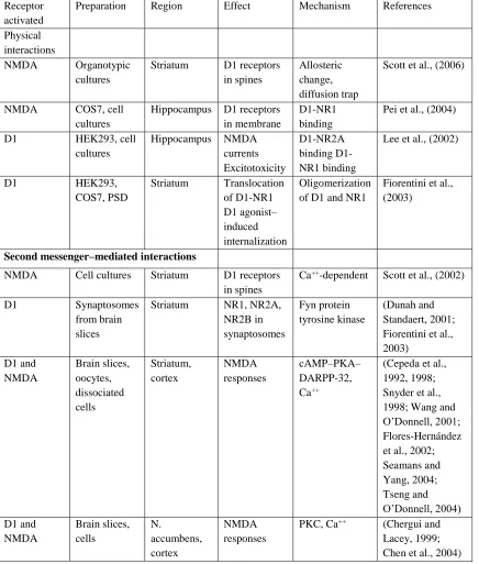
Table 1.1Summary of D1 and NMDA receptor interactions
Receptor activated
Preparation Region Effect Mechanism References
Physical interactions
NMDA Organotypic cultures
Striatum D1 receptors in spines
Allosteric change, diffusion trap
Scott et al., (2006)
NMDA COS7, cell cultures
Hippocampus D1 receptors in membrane
D1-NR1 binding
Pei et al., (2004)
D1 HEK293, cell cultures
Hippocampus NMDA currents Excitotoxicity
D1-NR2A binding D1-NR1 binding
Lee et al., (2002)
D1 HEK293,
COS7, PSD
Striatum Translocation of D1-NR1 D1 agonist– induced internalization
Oligomerization of D1 and NR1
Fiorentini et al., (2003)
Second messenger–mediated interactions
NMDA Cell cultures Striatum D1 receptors in spines
Ca++-dependent Scott et al., (2002)
D1 Synaptosomes from brain slices
Striatum NR1, NR2A, NR2B in synaptosomes Fyn protein tyrosine kinase (Dunah and Standaert, 2001; Fiorentini et al., 2003) D1 and NMDA Brain slices, oocytes, dissociated cells Striatum, cortex NMDA responses cAMP–PKA– DARPP-32, Ca++
(Cepeda et al., 1992, 1998; Snyder et al., 1998; Wang and O’Donnell, 2001; Flores-Hernández et al., 2002; Seamans and Yang, 2004; Tseng and O’Donnell, 2004) D1 and NMDA Brain slices, cells N. accumbens, cortex NMDA responses
PKC, Ca++ (Chergui and Lacey, 1999; Chen et al., 2004)
1.2
Thesis objectives and hypotheses
Based on the overview of the literature, deficits in the behavioural flexibility and
increased propensity of perseverative errors are core cognitive abnormalities in certain
CNS disorders, including, schizophrenia, and are associated with abnormal functioning of
interconnected brain areas, including the PFC. Post-mortem findings and previous animal
studies showed marked decreases of glutamate and dopamine levels in the PFC
individually and is sufficient to cause increased perseverative errors leading to
impairment in behavioural flexibility. However, recent in vivo imaging studies have
shown that, in living patients the loss of these neurotransmitters is minimal.
Consequently, our overall hypothesis was that the minimal loss of glutamate or
dopamine function is sufficient to produce impaired behavioural flexibility by
increased perseverative errors provided those losses co-occur in the brain. This also
suggests that there may by synergy between glutamate and dopamine neurotransmitter
abnormalities. In order to test our overall hypothesis, an operant conditioning-based
set-shifting paradigm was used. Following three studies were undertaken to test our overall
hypothesis and to elucidate associated molecular signaling mechanisms mediating
proposed synergy between deficiencies in neurotransmitter actions and how they are
relevant to behavioral flexibility.
Study I
The role of dopamine D1 and glutamate NMDA receptors is well-recognized in
the regulation of memory, cognition and executive functioning (Okubo et al., 1997;
Nieoullon, 2002; Volk et al., 2015; Thomas et al., 2017; Akil et al., 2013). Contrary to
the notion based on post-mortem studies, recent in vivo imaging studies have shown that
the changes in the dopamine and glutamate levels in the PFC of living schizophrenic
patients are very subtle (Laruelle et al., 2003; Théberge et al., 2007; Ohrmann et al.,
2007; Galińska et al., 2009; Rowland et al., 2009; Aoyama et al., 2011; Seese et al., 2011;
Szulc et al., 2011; Goto et al., 2012; Kegeles et al., 2012; Slifstein et al., 2015; Howes et
al., 2015). It is possible that these subtle alterations in neurotransmitter systems
ultimately result in noticeable cognitive impairments. In animal studies, the individual
and results showed that large decreases in individual transmitters in different brain areas
could impair set-shifting performance (Stefani et al., 2003; Haluk and Floresco, 2009;
Tait et al., 2014; Gauthier et al., 2014). Functional neuroimaging studies have indicated
that a number of cortical and subcortical areas including dlPFC, anterior cingulate cortex,
striatum, hippocampus and the MDT are associated with successful performance of the
WCST (Stratta et al., 1997; Rüsch et al., 2007; Wilmsmeier et al., 2010; Pedersen et al.,
2012; Young et al., 2000; Kemether et al., 2003). In agreement with this, several animal
studies have shown that, inactivation of number of brain areas or connections among
them could lead to impaired set-shifting. Since, the hypothesized interaction between
dopamine-glutamate neurotransmitters could occur in several potential areas as shown in
animal studies (Block et al., 2007; Floresco et al., 2008; 2009), we decided to use
systemic route of drug administration to target brain circuitry rather than specific nuclei.
We tested effect of combined systemic administration of dopamine D1 and glutamate
NMDA receptor antagonists on set-shifting behaviour in normal rats.
The following is the specific aim of the study I:
To determine the effect of systemic combined administration of “behaviourally
sub-effective” doses of D1 and NMDA receptor antagonists on set-shifting in normal
rats.
Study II
Across species, PFC is associated with executive functioning (Abbruzzese et al.,
1996; Ragozzino et al., 199b; Floresco et al., 2008; Moore et al., 2009). Patients with
prefrontal pathology of schizophrenia or accidental frontal lobe damage show impaired
executive functions (Milner, 1963; Nelson, 1976; Smith and Jonides, 1999; Stuss and
Alexander, 2000; Stuss et al., 2000; Roca et al., 2010; Sira and Mateer, 2014). In clinical
studies, prefrontal cortical dopamine as well as glutamate neurotransmitters were
proposed to regulate cognition and abnormal functioning of these neurotransmitters may
impair cognition in schizophrenia (Krystal et al., 2000; Coyle, 2006; Akil et al., 2013). In
line with the clinical studies, findings from animal studies confirm essential role of
executive functioning (Ragozzino, 2002a; Stefani and Moghaddam et al., 2003; Gauthier
et al 2014). In addition, several behaviours other than executive function, are regulated by
interaction between D1 and NMDA receptors (Smith-Roe and Kelley, 2000; Baldwin et
al., 2002; Fletcher et al., 2005; Nai et al., 2010; Agnoli and Carli, 2011; Touzani et al.,
2013; Glass et al., 2013).
From our first study, it was evident that the systemic combined administration of
behaviourally sub-effective doses of D1 and NMDA receptor antagonists caused
set-shifting impairments and produced error profile similar to inactivation or lesioning
studies in mPFC (Everett et al., 2001; Block et al., 2007; Floresco et al., 2008; Roca et
al., 2010). Therefore, we hypothesized that, PFC is one of the brain sites where
synergism between dopamine and glutamate abnormalities could occur. In the second
study, we sought to find out if the mPFC is associated with synergism between
D1-NMDA receptor antagonists. To test our hypothesis, we infused mPFC of normal rats
with D1 or NMDA receptor antagonists individually to determine their dose-response
profile in set-shifting. The highest but ineffective doses of these antagonists were then
combined to determine whether there is a synergism between them causing increased
perseverance in set-shifting.
The following is the specific aim of study II:
To determine the effect of intra-mPFC co-infusion of ‘behaviourally sub-effective’
doses of D1 and NMDA receptor antagonists on set-shifting in normal rats.
Study III
From the literature, it was evident that in different areas of the brain, interactions
between D1-NMDA receptors may use mainly three molecular signaling pathways: PKA,
ERK or direct protein-protein interactions between D1 and NMDA receptor complexes
through Fyn kinase. Interestingly, all main three pathways (PKA, ERK and Fyn kinase
mediated protein-protein interaction) have been implicated in the pathology of
schizophrenia (Tardito et al., 2001; Rybakowski et al., 2007; Yuan et al., 2010; Hahn,
In the mPFC, D1-NMDA receptors may interact via PKA signaling cascade to
regulate learning in rats (Baldwin et al., 2002). Furthermore, PKA was also found to
regulate synergistic effect of D1-NMDA receptors on excitability of pyramidal neuron in
the mPFC (Wang and O’Donnell, 2001). Along with PKA, ERK signaling was also found
to regulate D1-NMDA receptor interaction in the mPFC (Sarantis et al., 2009; Nagai et
al., 2007). Moreover, physical interaction between D1-NMDA receptors seems to
potentiate each other’s function (Gao and Wolf, 2008; Hu et al., 2010).
From results obtained in the study II, it appears that mPFC is one of the nuclei
where D1-NMDA receptors are interacting with each other to regulate set-shifting
behaviour. Thus, we hypothesized that molecular mechanisms underlying D1-NMDA
receptor interaction will use one or more of the following pathways in mPFC neurons to
regulate set-shifting behaviour: a) PKA b) ERK1/2 c) Fyn kinase-mediated interaction
between D1-NMDA receptors. To test our hypothesis, we performed a series of the
following experiments: a) effects of elevating PKA levels in the mPFC in animals
receiving intra mPFC infusion of a combination of D1 and NMDA antagonists that
disrupted set-shifting performance in study II; b) effects of inhibiting ERK1/2
phosphorylation on set-shifting performance in naïve rats; and c) effect of inhibiting Fyn
kinase in the mPFC on set-shifting behaviour.
Following specific aims were tested in this study:
1. To determine the effect of elevating PKA in the mPFC on D1-NMDA receptor
antagonist combination (from study II)-induced impaired set-shifting.
2. To determine the effect of inhibiting Fyn kinase and hence to verify if direct D1
NMDA receptor cross-talk facilitates set-shifting in normal rats.
3. To determine the effect of inhibiting ERK1/2-phosphorylation in the mPFC using
MAPK (mitogen-activated protein kinase) inhibitors on set-shifting in normal
1.3
References
Abbruzzese M, Ferri S, Scarone S (1996) Performance on the Wisconsin Card Sorting
Test in schizophrenia: Perseveration in clinical subtypes. Psychiatry Res 64:27–33.
Adcock RA (2000) Functional neuroanatomy of executive processes involved in
dual-task performance. Proc Natl Acad Sci 97:3567–3572.
Agnoli L, Carli M (2011) Synergistic interaction of dopamine D1 and glutamate
N-methyl-d-aspartate receptors in the rat dorsal striatum controls attention.
Neuroscience 185:39–49.
Akil M, Pierri JN, Whitehead RE, Edgar CL, Mohila C, Sampson AR, Lewis DA (1999)
Lamina-specific alterations in the dopamine innervation of the prefrontal cortex in
schizophrenic subjects. Am J Psychiatry 156:1580–1589.
Alvarez JA, Emory E (2006) Executive function and the frontal lobes: A meta-analytic
review. Neuropsychol Rev 16:17–42.
Aoyama N, Théberge J, Drost DJ, Manchanda R, Northcott S, Neufeld RW, Menon RS,
Rajakumar N, Pavlosky WF, Densmore M, Schaefer B, Williamson PC (2011) Grey
matter and social functioning correlates of glutamatergic metabolite loss in
schizophrenia. Br J Psychiatry 198:448–456.
Ardila A (2008) On the evolutionary origins of executive functions. Brain Cogn 68:92–
99.
Auger ML, Floresco SB (2014) Prefrontal cortical GABA modulation of spatial reference
and working memory. Int J Neuropsychopharmacol 18:1–11.
Auger ML, Floresco SB (2017) Prefrontal cortical GABAergic and NMDA glutamatergic
regulation of delayed responding. Neuropharmacology 113:10–20.
Baddeley A (2012) Working memory: theories, models, and controversies. Annu Rev
Baldwin AE, Sadeghian K, Kelley AE (2002) Appetitive instrumental learning requires
coincident activation of NMDA and dopamine D1 receptors within the medial
prefrontal cortex. J Neurosci 22:1063–1071.
Banuelos C, Beas BS, McQuail JA, Gilbert RJ, Frazier CJ, Setlow B, Bizon JL (2014)
Prefrontal cortical GABAergic dysfunction contributes to age-related working
memory impairment. J Neurosci 34:3457–3466.
Barense MD, Fox MT, Baxter MG (2002) Aged rats are impaired on an attentional
set-shifting task sensitive to medial frontal cortex damage in young rats. Learn Mem
9:191–201.
Baviera M, Invernizzi RW, Carli M (2008) Haloperidol and clozapine have dissociable
effects in a model of attentional performance deficits induced by blockade of
NMDA receptors in the mPFC. Psychopharmacology (Berl) 196:269–280.
Beninger RJ, Gerdjikov T V. (2005) Dopamine-glutamate interactions in reward-related
incentive learning. Dopamine and Glutamate in Psychiatric Disorders, pp 319–354.
Totowa, NJ: Humana Press.
Beracochea DJ, Jaffard R, Jarrard LE (1989) Effects of anterior or dorsomedial thalamic
ibotenic lesions on learning and memory in rats. Behav Neural Biol 51:364–376.
Berch DB, Krikorian R, Huha EM (1998) The Corsi block-tapping task: methodological
and theoretical considerations. Brain Cogn 38:317–338.
Birrell JM, Brown VJ (2000) Medial frontal cortex mediates perceptual attentional set
shifting in the rat. J Neurosci 20:4320–4324.
Bizon JL, Foster TC, Alexander GE, Glisky EL (2012) Characterizing cognitive aging of
working memory and executive function in animal models. Front Aging Neurosci 4–
19.
Block AE, Dhanji H, Thompson-Tardif SF, Floresco SB (2007) Thalamic-prefrontal