1556-6811/08/$08.00⫹0 doi:10.1128/CVI.00164-08
Copyright © 2008, American Society for Microbiology. All Rights Reserved.
Immune Responses of Mice with Different Genetic Backgrounds to
Improved Multiepitope, Multitarget Malaria Vaccine Candidate
Antigen FALVAC-1A
䌤
S. A. Kaba,
1† A. Price,
1,2Z. Zhou,
1,2V. Sundaram,
1P. Schnake,
1I. F. Goldman,
1A. A. Lal,
1V. Udhayakumar,
1and C. W. Todd
1,2*
Division of Parasitic Diseases, Centers for Disease Control and Prevention, Atlanta, Georgia 30341,1and
Atlanta Research and Education Foundation, Decatur, Georgia 300332
Received 7 May 2008/Returned for modification 11 June 2008/Accepted 29 August 2008
FALVAC-1A is a second-generation multitarget, multiepitope synthetic candidate vaccine against Plasmo-dium falciparum, incorporating elements designed to yield a stable and immunogenic molecule. Characteristics of the immunogenicity of FALVAC-1A were evaluated in congenic (H-2b
, H-2k
, andH-2d
) and outbred strains of mice. The influences of four adjuvants (aluminum phosphate, QS-21, Montanide ISA-720, and copolymer CRL-1005) on different aspects of the immune response were also assessed. FALVAC-1A generated strong antibody responses in all mouse strains. The highest mean enzyme-linked immunosorbent assay (ELISA) antibody concentrations against FALVAC-1A were observed in the outbred ICR mice, followed by B10.BR, B10.D2, and C57BL/6 mice, though this order varied for the different adjuvants, with no statistical differences between mouse strains. In all mouse strains, the highest anti-FALVAC-1A antibody titers in ELISAs were induced by FALVAC-1A in copolymer and ISA-720 formulations, followed by QS-21 and AlPO4. These anti-bodies were of all four subclasses, though immunoglobulin G1 (IgG1) predominated, with the exception of FALVAC-1A with the QS-21 adjuvant, which induced predominantly IgG2c responses. Both sporozoites and blood stages ofP. falciparumwere recognized by anti-FALVAC-1A sera in the immunofluorescence assay. In addition to antibody, cellular immune responses were detected; these responses were studied by examining spleen cells producing gamma interferon and interleukin-4 in enzyme-linked immunospot assays. In summary, FALVAC-1A was found to be highly immunogenic and elicited functionally relevant antibodies that can recognize sporozoites and blood-stage parasites in diverse genetic backgrounds.
One approach to an effective vaccine against Plasmodium
falciparummalaria may be an artificial molecule comprised of
key antigens and/or epitopes from different stages of the life cycle that induces both humoral and cellular immune effector mechanisms capable of mediating protection (9, 14, 15). The development of such a multicomponent subunit vaccine may also circumvent the problems associated with host genetic re-striction and parasite antigenic variability sometimes noted with vaccines based on a single antigen. This approach also allows a number of distinct epitopes to be assembled to a size sufficient to be immunogenic (12, 15, 16, 36, 40). A further advantage is the ability to manipulate the construct, as new information regarding protective epitopes and antigen diver-sity becomes available.
Our laboratory previously developed a multistage, multiva-lent antigen, FALVAC-1, which comprised 21 different B-cell, T-cell, and cytotoxic T-lymphocyte (CTL) epitopes from seven different antigens of P. falciparum and a universal helper epitope from tetanus toxoid (37). Although FALVAC-1 showed promising results by inducing antibodies in both rab-bits and mice that had in vitro antiparasitic activity (33, 37, 38),
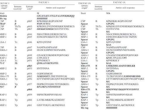
it was unsuitable for further clinical development because of concerns about the stability of the molecule, potential homol-ogy of an epitope with human sequences, and product yield. Thus, in the context of a vaccine development program, the molecule was redesigned to redress these limitations. The new molecule, termed FALVAC-1A, retained most of the FALVAC-1 epitopes and their order in the molecule but in-cluded some replaced or modified epitopes and, most critically, two types of spacer sequences intended (i) to promote molec-ular conformation and folding and (ii) to facilitate antigen processing (23, 42). The design and construction of the FALVAC-1A gene, as well as the expression, purification, and physicochemical characteristics of FALVAC-1A and its immu-nogenicity in rabbits, have been described previously (45). The amino acid sequence of FALVAC-1A, including the origin of the epitopes, is shown in Table 1 (45).
Because a vaccine construct must be immunogenic in genet-ically diverse populations, analysis of major histocompatibility (MHC)-restricted immune responses in experimental animal systems is of interest to characterize novel antigens. Thus, in the present study, FALVAC-1A was tested for its immunoge-nicity against different genetic backgrounds of bothH-2 (H-2b,
H-2d, and H-2k) congenic and outbred (ICR) mice. Subunit
antigens will almost certainly require adjuvants to maximize their potential; therefore, FALVAC-1A was formulated with four different adjuvants: aluminum phosphate (AlPO4), the saponin QS-21, Montanide ISA-720, and copolymer CRL-1005 in a water-in-oil (w-o) emulsion. Because both humoral and
* Corresponding author. Mailing address: Division of Parasitic Dis-eases, 4770 Buford Highway, MS F-36, Centers for Disease Control and Prevention, Atlanta, GA 30341. Phone: (770) 488-4526. Fax: (770) 488-4253. E-mail: [email protected].
† Present address: Division of Malaria Vaccine Development, Walter Reed Army Institute of Research, Silver Spring, MD 20910.
䌤Published ahead of print on 10 September 2008.
1674
on August 17, 2020 by guest
http://cvi.asm.org/
cellular responses are believed to be important to the devel-opment of immunity to malaria, antibody responses to the molecule were determined by enzyme-linked immunosorbent assays (ELISAs) and immunofluorescence assays (IFAs), and T-cell responses were determined by enzyme-linked immuno-spot assays (ELISPOTs) measuring interleukin-4 (IL-4) and gamma interferon (IFN-␥).
MATERIALS AND METHODS
Adjuvant preparations.FALVAC-1A was adsorbed onto aluminum phos-phate (AlPO4) (ADJU-PHOS; Accurate Chemicals & Scientific Corporation,
Westbury, NY) in the proportions 10g FALVAC-1A per 40g aluminum by
overnight incubation at 4°C on a rocker (6). Greater than 90% of the antigen was bound (data not shown). The aqueous saponin adjuvant QS-21 (a gift from Antigenics, Inc., Lexington, MA) and FALVAC-1A were appropriately diluted and mixed in phosphate-buffered saline (PBS) (pH 6.8). Equal volumes of the oil
phase adjuvant Montanide ISA-720 (a gift from SEPPIC SA, Paris, France) and FALVAC-1A in PBS (pH 7.2) were emulsified using two glass syringes and a double-hubbed needle to yield a w-o emulsion. The nonionic block copolymer CRL-1005 (43) (a gift from Robert Hunter) was formulated in an adjuvant oil phase consisting of 89% squalene, 10% sorbitan monooleate (Span 80; Sigma), and 1% CRL-1005 by weight. Equal volumes of the CRL-1005 oil phase and FALVAC-1A in PBS were similarly emulsified to give a w-o emulsion. FALVAC-1A was also administered without adjuvant in PBS. All formulations delivered a 10-g dose of FALVAC-1A in a 100-l volume.
Mice and immunizations.H-2congenic mice, C57BL/6 (H-2b
), B10.BR (
H-2k
), and B10.D2 (H-2d
) (Jackson Laboratory, Bar Harbor, ME), and an outbred ICR strain (Harlan Sprague Dawley, Indianapolis, IN) were maintained and used in AAALAC-accredited Centers for Disease Control and Prevention (CDC) facilities in accordance with the guidelines in the National Research Council manual (30). The study protocol was approved and monitored by the CDC Animal Care and Use Committee. For analysis of antibody responses, mice
were immunized subcutaneously on days 0, 14, and 28 with 10 g of
FALVAC-1A at each time point. For each of the four mouse strains, five groups
TABLE 1. Comparison of the components and amino acid sequences of FALVAC-1 and FALVAC-1Aa
FALVAC-1 epitope
originb
FALVAC-1 FALVAC-1A
epitope
originb
FALVAC-1A
Immune
reactivityc
Epitope
coded Amino acid sequencee
Immune
reactivity Amino acid sequence
Start M Start MA
Melittin GIGAVLKVLTTGLPALISWIKRKRQQ
His tag HHHHHH
CSP B p865 KPKHKKLKQPGDGNP CSP B KPKHKKLKQPGDGNP
TRAP B WSPCSVTCG Spacer GPGPG
CSP Th/Tc p843 KPKDELDYENDIEKKICKMEKCS CSP Th/Tc KPKDELDYENDIEKKICKMEKCS
CSP Th p867 DIEKKICKMEKCSSVFNVVNS CSP Th SVFNVVNS
Spacer KG
MSP-1 B p844 NSGCFRHLDEREECKCLL MSP-1 B NSGCFRHLDEREECKCLL
MSP-1 B p845 EDSGSNGKKITCECTKPDS MSP-1 B EDSGSNGKKITCECTKPDS
Spacer GPGPG
LSA-1 Tc p846 KPIVQYDNF LSA-1 Tc KPIVQYDNF
Spacer KAA
CSP B p847 NANPNANPNANP CSP B NANPNANPNANP
AMA-1 B p848 DGNCEDIPHVNEFSAIDL AMA-1 B DGNCEDIPHVNEFSAIDL
Spacer GPGPG
AMA-1 B p849 GNAEKYDKMDEPQHYGKS AMA-1 B GNAEKYDKMDEPQHYGKS
RAP-1 B p866 LTPLEELY RAP-1 B KNTLTPLEELYPT
LSA-1 Tc p851 KPNDKSLY LSA-1 Tc KPNDKSLY
P2-T Th QYIKAANSKFIGITEL Spacer KAA
p863 EBA-175 B LMIKEHILAIAIYESRILKR
MSP-2 B p853 SNTFINNA MSP-2 B SNTFINNA
Spacer GPGPG
MSP-2 B p854 GQHGHMGH MSP-2 B GQHGHMGH
EBA-175 B p862 NEREDERTLTKEYEDIVLK EBA-175 B TLTKEYEDIVLKSHMNRESDD
AMA-1 Tp p856 EFTYMINFGRGQNYWEHPYQKS AMA-1 Tp EFTYMINFGRGQNYWEHPYQKS
Spacer KG
AMA-1 Tp p857 DQPKQYEQHLTDYEKIKEG AMA-1 Tp DQPKQYEQHLTDYEKIKEG
Pf27 B KPLDKFGNIYDYHYEH Spacer KG
p864 EBA-175 B RDEWWKVIKKDVWNVISWVF
Spacer KG
RAP-1 Tp p859 SSPSSTKSSPSNVKSAS RAP-1 Tp SSPSSTKSSSPSNVKSAS
Spacer KG
RAP-1 Tp p860 LATRLMKKFKAEIRDFF RAP-1 Tp LATRLMKKFKAEIRDFF
Spacer KG
MSP-1 Tp p861 GISYYEKVLAKYKDDLE MSP-1 Tp GISYYEKVLAKYKDDLE
His tag HHHHHH
aAdapted from reference 45 with permission of the publisher. Sequences deleted during the design of FALVAC-1A are shown in bold italic type; sequences added
are shown in boldface type.
bThe origin of the epitope is shown as follows: CSP, circumsporozoite protein; TRAP, thrombospodin-related protein; MSP-1, merozoite surface protein-1; LSA-1,
liver stage antigen-1; AMA-1, apical membrane antigen-1; RAP-1, rhoptry-associated protein-1; P2-T, tetanus toxoid universal helper; MSP-2, merozoite surface
protein-2; EBA-175, erythrocyte binding antigen-175; Pf27,P.falciparumgametocyte 27-kDa antigen.
cThe immune reactivity of each epitope (synthetic peptide) is shown as follows: B, B cell; Th/Tc, T helper and cytotoxic T cell; Th, T helper cell; Tc, cytotoxic T cell;
Tp, T proliferative cell.
dCode number assigned to each epitope (synthetic peptide).
eThe sequences of each epitope and spacer are indicated. The entire sequence runs continuously down each row in this column.
on August 17, 2020 by guest
http://cvi.asm.org/
of 10 female mice, 6 to 8 weeks old, received the antigen either in PBS or
formulated with one of the adjuvants, AlPO4 (40g/mouse), QS-21 (20g/
mouse), Montanide ISA-720 (50:50), or copolymer CRL-1005 (50:50), respec-tively. Blood was collected from the retro-orbital plexus on days 0, 21, 42, 63, 84,
and 147. Serum samples were stored at⫺20°C until used for serological assays.
To evaluate the T-cell responses, groups of four mice were immunized on days
0 and 14 with 10g of FALVAC-1A at each time point in the same formulations
as mentioned above, and spleen cells were harvested and assayed on either day 21 or 23.
ELISA.Antibody concentrations were determined by ELISAs (34). Briefly, Immulon 2HB plates (Thermo Electron, Franklin, MA) were coated overnight at
4°C with either 1g/ml FALVAC-1A or 10g/ml of a component peptide in
PBS, blocked with 5% nonfat dried milk in PBS containing 0.05% Tween 20 (diluent), and washed with PBS containing 0.05% Tween 20. Sera were diluted in binary series with diluent from 1:200 to 1:1,638,400 for anti-FALVAC-1A and
from 1:100 to 1:204,800 for antipeptide antibody. Aliquots (100l) of these
dilutions were added per well, and the plates were incubated overnight at 4°C. The plates were washed between additions with PBS containing 0.05% Tween 20. To determine the titers of total anti-FALVAC-1A immunoglobulin G (IgG), horseradish peroxidase (HRP)-conjugated goat anti-mouse IgG (Southern Bio-tech, Birmingham, AL) was used at a dilution of 1:5,000. IgG subclasses were measured using 1:5,000 dilution of HRP-conjugated goat anti-mouse IgG1, IgG2b, IgG2c, and IgG3, respectively (Caltag Laboratories, Burlingame, CA).
Following washing, the final color reaction was developed using 3,3⬘,5,5⬘
-tetra-methylbenzidine (TMB) peroxidase substrate solution (Kirkegaard & Perry,
Gaithersburg, MD) for 5 min. The reaction was stopped by the addition of 50l
of 1.0 M phosphoric acid, and the optical density at 450 nm (OD450) was read on
a Spectramax 340 PC microplate reader (Molecular Devices, Sunnyvale, CA). Serum titers were determined as the geometric mean of the reciprocal dilution
where the OD450 of the titration curve equaled 0.1 (the maximum value of
preimmune sera at 1:100).
IFA.An immunofluorescence assay was used to determine anti-FALVAC-1A
reactivity with sporozoites (P. falciparum, Santa Lucia strain) and asexual
blood-stage parasites (P. falciparumFVO strain). Sera from individual mice in each
group were pooled before IFA determination. Methanol-fixed parasites on mul-tispot slides were incubated (30 min, room temperature, 100% relative humidity)
with 10-l aliquots of serial twofold serum dilutions in PBS, rinsed three times
with PBS, and incubated for 30 min with 1:100 dilution of fluorescein isothio-cyanate-conjugated goat anti-mouse IgG antibody (Kirkegaard & Perry Labora-tories [KPL], Gaithersburg, MD). Following three further PBS washes, the slides
were dried, mounted under buffered glycerin (pH 9), and examined at⫻40 with
a fluorescence microscope. Mouse preimmune serum was used as a negative
control. Reactions to the parasites were scored as follows:⫹4 (extremely bright),
⫹3 (yellow-green bright intensity),⫹2 (medium intensity),⫹1 (less intense but
with fluorescence),⫾(pale yellow green), and⫺(no fluorescence). Serum titers
were determined as the reciprocal of the highest serum dilution giving a positive
reading (⫹1). The slides were read independently by two individuals.
ELISPOT.An enzyme-linked immunospot assay was used to detect spleen
cells producing FALVAC-1A-specific IFN-␥and IL-4. Mice were immunized on
days 0 and 14, two mice from each group were sacrificed on either day 21 or 23, and spleen mononuclear cells were isolated at each time point (4). The ELISPOT procedure described in the BD ELISPOT set instruction manual (BD Biosciences Pharmingen, San Diego, CA) was followed. Briefly, the wells of Immunospot M200 96-well plates (BD Biosciences Pharmingen, San Diego, CA)
were coated with the capture antibody (purified anti-mouse IFN-␥or anti-mouse
IL-4; BD Biosciences Pharmingen) at a final concentration of 5g/ml in PBS and
incubated overnight at 4°C. The following day, the plates were washed once with complete RPMI medium (RPMI with 10% fetal bovine serum) and blocked with the same medium for 2 h at room temperature. The blocking solution was
removed, and the cultures were set up as follows: 106 spleen mononuclear
cells/well in 200l complete RPMI medium containing 10% heat-inactivated
fetal bovine serum, 50 U/ml gentamicin, 0.1 mM nonessential amino acids, 0.1
mM minimal essential medium vitamins, 2 mML-glutamine, and 2 mM
-mer-captoethanol. Experimental wells contained FALVAC-1A at a final
concentra-tion of 1g/ml. Concanavalin A (ConA) (Sigma, St. Louis, MO) at 0.5g/ml was
used as a nonspecific positive control. The cultures were incubated for 20 to 24 h
(for IFN-␥) or 40 to 48 h (for IL-4) at 37°C, 5% CO2, and 100% relative
humidity. Following incubation, the plates were washed three times with 200l
of distilled water and four times with PBS containing 0.1% Tween 20. Detection
antibody (biotinylated anti-IFN-␥or anti-IL-4 antibody; BD Biosciences
Phar-mingen) in 100l of dilution buffer (PBS containing 10% fetal bovine serum)
was added at a final concentration of 2g/ml, and the plates were incubated at
room temperature for 2 h. After three washes with PBS containing 0.1% Tween
20, 100l of conjugate (streptavidin-HRP; BD Biosciences Pharmingen) in
dilution buffer was added, and the plates were incubated at room temperature for 1 h. Following four washes with PBS containing 0.1% Tween 20 and two
washes with PBS, the spots were developed with 100l of substrate solution
(3-amino-9-ethyl-carbazole [AEC] substrate plus 20l of AEC chromogen per
ml) (BD ELISPOT AEC substrate set; BD Biosciences) for 15 min at room temperature. The substrate reaction was stopped by washing the wells with distilled water. The plates were air dried, and the spots corresponding to the
number of IFN-␥- or IL-4-secreting cells were enumerated using the automated
ELISPOT reader system ELR02 (AID Autoimmun Diagnostika GmbH, Strass-berg, Germany).
Statistical analysis.Geometric mean titers were calculated using the logarith-mic transformation of the reciprocal antibody titers. Statistical analysis was
performed by Student t tests, with aPvalue of⬍0.05 considered significant.
RESULTS
Antibody responses to FALVAC-1A in different strains of mice with different adjuvant formulations.To investigate an-tibody responses to FALVAC-1A and evaluate the effects of MHC differences, three inbred strains (H-2b,H-2k, andH-2d)
and one outbred strain of mice were immunized on days 0, 14, and 28 with 10g FALVAC-1A formulated with PBS, AlPO4, QS-21, Montanide ISA-720, and copolymer (CRL-1005), re-spectively. The kinetics of the mean anti-FALVAC-1A IgG responses measured in ELISAs are shown in Fig. 1. The ma-jority of the responses peaked at day 42, for all adjuvant/strain combinations, followed by a slow decline. All the adjuvants enhanced antibody responses significantly compared to FALVAC-1A used without an adjuvant. Responses stimulated by the two w-o emulsion adjuvants, Montanide ISA-720 and copolymer CRL-1005, were the highest, and equivalent in all
FIG. 1. Antibody responses to FALVAC-1A vaccine formulations in congenic mice. Mice were immunized with 10g FALVAC-1A/dose formulated in copolymer CRL-1005 (Copol/FAL), Montanide ISA-720 (ISA-ISA-720/FAL), QS-21 (QS-21/FAL), aluminum (AlPO4/FAL), or PBS (FALVAC-1A) on days 0, 14, and 28. Total IgG antibody titers (geometric mean of 10 mice) were determined as the geometric mean of the reciprocal dilution where the OD450 of the titration curve
equaled 0.1 (the maximum value of preimmune sera at 1:100).
on August 17, 2020 by guest
http://cvi.asm.org/
four mouse strains, followed by QS-21 and AlPO4 (Fig. 1). Within a given mouse strain, peak anti-FALVAC-1A IgG titers elicited by CRL-1005 and ISA-720 formulations were statisti-cally different from antibody titers obtained from mice immu-nized with QS-21 and AlPO4 formulations (P⬍0.05). When responses were compared between mouse strains for the same adjuvant, there were no significant differences between the responses, indicating minimal genetic restriction on the anti-body response at this level.
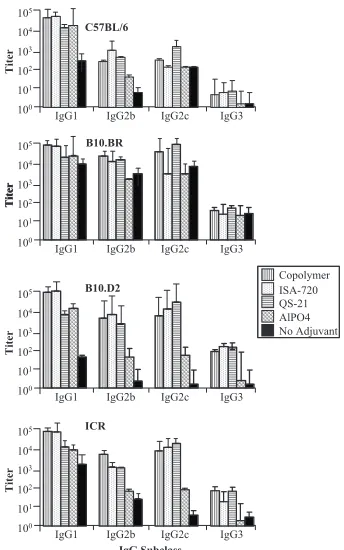
Profiles of the IgG subclass responses to different FALVAC-1A vaccine formulations.The profiles of various IgG subclasses of anti-FALVAC-1A antibodies are illustrated in Fig. 2. In this study we measured IgG2c production rather than IgG2a. Mice with the B6 background lack the Igh-1a allele that codes for IgG2a but instead express IgG2c from the Igh-1b allele (18, 26). In general, IgG1 was the predominant IgG subclass in all mouse strains tested in this study followed by IgG2c, IgG2b,
and IgG3 (IgG1⬎IgG2c⬎IgG2b⬎IgG3) as shown in Fig.
2. However, QS-21 adjuvant induced predominantly IgG2c in all strains, except for C57BL/6 mice. Mice immunized with FALVAC-1A alone also elicited anti-FALVAC-1A antibodies of different subclasses but at consistently lower antibody levels (Fig. 2). Although both copolymer CRL-1005 and Montanide ISA-720 adjuvants predominantly elicited higher levels of IgG1 in all mouse strains, these adjuvants also modulated other
subclasses of antibodies. The adjuvant AlPO4 was the least effective adjuvant in influencing the development of other IgG subclass antibodies, except for IgG1. The outbred ICR mice elicited high levels of various IgG subclasses followed by B10.D2 and B10.BR. In general, C57BL/6 mice were the least responders, regardless of the IgG subclass or adjuvant used.
The ratios between different subclasses were determined to better understand the relative levels of these antibodies. The ratios differed between outbred and inbred mice. The most notable change in the ratios of IgG subclasses was observed with QS-21 adjuvant, which induced antibodies with the high-est IgG2c/IgG1 and IgG2c/IgG2b ratios in all strains of mice. The IgG2b/IgG1 ratio was the least in all the mouse strains irrespective of the adjuvant used: (IgG1/IgG3⬎IgG2c/IgG3⬎
IgG1/IgG2c ⬎ IgG2b/IgG3⬎ IgG2c/IgG2b ⬎ IgG2b/IgG1).
The IgG1/IgG3 ratio was highest in C57BL/6 mice followed by outbred ICR, B10.D2, and B10.BR mice. AlPO4 induced an-tibodies with the highest IgG1/IgG3 ratio in all strains except for the B10.BR strain. These results indicate that both adju-vant and genetic background influenced the levels of various IgG subclasses to FALVAC-1A.
Antibody responses to vaccine components (peptide ELISA).
Analyses of epitope-specific antibody responses by ELISAs showed that the vaccine-induced antibodies recognized several epitopes, including both B- and T-cell epitopes incorporated into FALVAC-1A (Table 2). Six of the B-cell epitopes (mero-zoite surface protein-1 [MSP-1]/p844, MSP-1/p845, apical membrane antigen-1 [AMA-1]/p848, AMA-1/p849, circum-sporozoite protein [CSP]/p865, and rhoptry-associated pro-tein-1 [RAP-1]/p866) induced epitope-specific antibody re-sponses in all the mouse strains irrespective of the adjuvant used, and three B-cell epitopes (MSP-2/p853, erythrocyte bind-ing antigen-175 [EBA-175]/p863, and EBA-175/p864) com-pletely failed to induce antibody responses in any of the mouse strains (Table 2). As was observed in antibody responses to the whole-vaccine antigen, the antibody titers against the individ-ual epitopes were also higher in outbred ICR mice (1⫻102to 1⫻105) than those in the inbred mice (1⫻102to 5.1⫻104). ICR mice also elicited antibodies to at least 17 of the 21 epitopes in the vaccine, whereas inbred mice generated anti-bodies to only 13 of the epitopes. ICR mice responded to all but 1 of the 9 T-cell epitopes and 9 out of 12 B-cell epitopes, while inbred mice responded to only 5 (CSP/p843, liver stage antigen-1 [LSA-1]/p846, AMA-1/p857, RAP-1/p859, and CSP/ p867) of the T-cell epitopes and 8 B-cell epitopes. Outbred ICR mice were the best responders, followed by B10.BR and B10.D2 mice and then by C57BL/6 mice, regardless of the adjuvant formulation used. The highest antibody titers were seen in the copolymer CRL-1005 vaccine formulation group, fol-lowed by Montanide ISA-720, QS-21, and AlPO4 vaccine formu-lation groups. CRL-1005, ISA-720, and QS-21 vaccine formula-tion groups induced moderate to high anti-FALVAC-1A antibody titers to 5 of 12 B-cell epitopes (CSP/p865, MSP-1/p844, AMA-1/p848, AMA-1/p849, and RAP-1/p866) and only 1 of 9 T-cell epitopes compared to the low to moderate antibody titers induced by the AlPO4 vaccine formulation for the same epitopes. All the adjuvant formulations failed to induced antibodies to 3 (EBA-175/p863, EBA-175/p864, and MSP-1/p853) of 15 blood-stage epitopes, 1 (CSP/p867) of 4 sporozoite blood-stage epitopes, and 1 (LSA-1/p851) of 2 liver stage epitopes.
IgG Subclass
Copolymer ISA-720 QS-21 AlPO4 No Adjuvant IgG1 IgG2b IgG2c IgG3
100 101 102 103 104 105
Titer
C57BL/6
B10.BR
Titer
IgG1 IgG2b IgG2c IgG3 100
101 102 103 104 105
Titer
IgG1 IgG2b IgG2c IgG3 100
101 102 103 104 105
Titer
B10.D2
ICR
IgG1 IgG2b IgG2c IgG3 100
101 102 103 104 105
Titer
FIG. 2. FALVAC-1A-specific serum IgG subclass profiles induced by different adjuvant vaccine formulations. Mice were immunized with 10g FALVAC-1A/dose formulated in copolymer, Montanide ISA-720, QS-21, aluminum (AlPO4), or PBS as indicated in the legend to Fig. 1. Sera collected on day 42 (peak antibody titer time point) were used for IgG subclass profile determination by ELISAs. Subclass IgG antibody titers (geometric mean of 10 mice) were determined as the geometric mean of the reciprocal dilution where the OD450of the
titration curve equaled 0.1.
on August 17, 2020 by guest
http://cvi.asm.org/
TABLE 2. Mouse anti-FALVAC-1A peptide titers found in ELISAs a Epitope origin b Peptide Immune reactivity c Anti-FALVAC-1A peptide titer of pooled sera (10 mice) d C57BL/6 B10.BR B10.D2 ICR CRL- 1005 ISA- 720 QS-21 AlPO 4 No adj. CRL- 1005 ISA- 720 QS-21 AlPO 4 No adj. CRL- 1005 ISA- 720 QS-21 AlPO 4 No adj. CRL- 1005 ISA-720 QS-21 AlPO 4 No adj. CSP p865 B 6,400 3,200 3,200 800 100 51,200 6,400 12,800 3,200 200 6,400 6,400 6,400 200 100 25,600 12,800 12,800 3,200 1,600 CSP p843 Th/Tc 800 400 – – 100 25,600 25,600 51,200 1,600 1,600 12,800 25,600 6,400 – – 102,400 25,600 51,200 12,800 1,600 CSP p867 Th –––– – –––– – ––– – – – – –– – MSP-1 p844 B 3,200 6,400 3,200 3,200 100 51,200 3,200 25,600 6,400 1,600 12,800 12,800 6,400 100 100 6,400 12,800 6,400 100 100 MSP-1 p845 B 800 400 800 400 100 – 12,800 200 1,600 400 6,400 6,400 3,200 1,600 400 51,200 25,600 25,600 6,400 1,600 LSA-1 p846 Tc – – – – 6,400 1,600 100 – 400 800 800 – – 100 – 100 – 800 100 CSP p847 B –––– – – 2 0 0 – ––– 4 0 0 – – 1 0 0 8 0 0 – – – 1 0 0 AMA-1 p848 B 6,400 1,600 6,400 1,600 100 6,400 800 25,600 – – 1,600 800 1,600 800 100 12,800 12,800 6,400 800 100 AMA-1 p849 B 3,200 3,200 12,800 3,200 100 6,400 6,400 25,600 6,400 – 1,600 6,400 6,400 – – 12,800 12,800 12,800 6,400 1,600 RAP-1 p866 B 12,800 6,400 12,800 6,400 800 25,600 6,400 51,200 25,600 1,600 25,600 25,600 51,200 6,400 – 102,400 25,600 12,800 25,600 1,600 LSA-1 p851 Tc –––– – –––– – ––– – – – – –– – EBA-175 p863 B –––– – –––– – ––– – – – – –– – MSP-2 p853 B –––– – –––– – ––– – – – – –– – MSP-2 p854 B 100 – – – 100 –––– – ––– – – 1 0 0 – –– – EBA-175 p862 B –––– – – – – – – – – – – – 6,400 400 3,200 – 100 AMA-1 p856 Tp –––– – – – – – – – – – – – 1,600 1,600 – 1,600 100 AMA-1 p857 Tp 1,600 1,600 12,800 1,600 100 25,600 3,200 3,200 3,200 1,600 6,400 6,400 3,200 400 100 51,200 12,800 12,800 25,600 1,600 EBA-175 p864 B –––– – –––– – ––– – – – – –– – RAP-1 p859 Tp 400 – – – 100 – – 100 – – – – – – – 12,800 800 1,600 100 100 RAP-1 p860 Tp –––– – –––– – ––– – – 1 0 0 2 0 0 –– – MSP-1 p861 Tp –––– – – – – – – – – – – – 100 100 100 – – aFour strains of mice were immunized on days 0, 14, and 28 with 10 g FALVAC-1A formulated with the indicated adjuvants. Sera collected on day 42 (serum with peak antibody titer) were used for peptide ELISA. ELISA titers against each epitopic peptide were determined by titration. The order of epitopes is the same as in Table 1. In CSP, the first epitope, Th/Tc , KPKDELDYENDIEKKICKMEKCS runs continuously into the second Th epitope, SVFNVVNS. Antibody responses were determined jointly against the peptide KPKDELDYENDIEKKICKMEKCSSVFNVVNS. bThe origin of the epitope is shown as follows: CSP, circumsporozoite protein; MSP-1, merozoite surface protein-1; LSA-1, liver stage antigen-1; AMA -1, apical membrane antigen-1; RAP-1, rhoptry-associated protein-1; EBA-175, erythrocyte binding antigen-175; MSP-2, merozoite surface protein-2. cThe immune reactivity of each epitope (synthetic peptide) is shown as follows: B, B cell; Th/Tc, T helper and cytotoxic T cell; Th, T helper cell; Tc, cyt otoxic T cell; Tp, T proliferative cell. dFour strains of mice (C57BL/6, B10.BR, B10.D2, and ICR) were immunized with 10 g FALVAC-1A formulated with adjuvants (copolymer CRL-1005, Montanide ISA-720, QS-21, and AlPO 4 ) or with no adjuvant (No adj.). Dashes indicated that the titer was ⬍ 100.
on August 17, 2020 by guest
http://cvi.asm.org/
Reactivity of FALVAC-1A antisera with parasite antigens in IFAs.To determine whether FALVAC-1A-induced antibodies can recognize native antigens expressed on the parasites, IFA studies were performed with sporozoites and blood-stage par-asites using pooled sera collected on day 42 after primary immunization. Sera from all immunized groups showed reac-tivity with both sporozoites and blood-stage parasites (Fig. 3). In general, IFA titers of the serum pools against both stages were correlated with the ELISA group mean titers against the whole molecule. Sera from mice immunized with FALVAC-1A alone had the lowest IFA titers (4⫻101to 1.6⫻ 102) in all the mouse strains (Fig. 3). No correlation was found between the peptide ELISA titers and IFA titers.
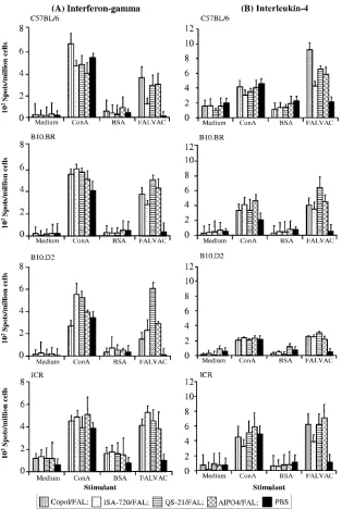
Cellular immune responses to FALVAC-1A assayed by ELISPOTs. In order to evaluate the induction of T-cell re-sponses to FALVAC-1A in mice, splenic IFN-␥- and IL-4-secreting cells were assayed by ELISPOTs. As shown in Fig. 4, T-cell responses against FALVAC-1A, as determined by the number of IFN-␥- and IL-4-secreting cells, were detected in all mouse strains, irrespective of the adjuvant used. The frequency of cytokine-secreting splenocytes from mice immunized with FALVAC-1A vaccine plus the different adjuvant formulations ranged from 222 to 609 (for IFN-␥) and 216 to 914 (for IL-4) spot-forming cells/106cells in comparison to 11 to 35 IFN-␥ spot-forming cells or 50 to 100 IL-4 spot-forming cells/106cells in the PBS control group (Fig. 4). IFN-␥and IL-4 secretion detected in this assay was antigen specific, since very few spots (or cells) could be observed when these immune cells were incubated with medium only (4 to 100 IFN-␥spots/106 cells and 7 to 88 IL-4 spots/106 cells) or stimulated with bovine serum albumin BSA (a nonspecific antigen) (27 to 100 IFN-␥ spots/106 cells and 19 to 100 IL-4 spots/106 cells (Fig. 4). A mitogen (ConA) that has the ability to activate all T-cell types was used as a positive control. ConA induced 269 to 657 IFN-␥ spot-forming cells/106 cells and 207 to 590 IL-4 spot-forming cells/106cells.
Outbred ICR mice responded with the highest mean num-ber of antigen-specific IFN-␥-secreting splenocytes (448.3 ⫾ 54.8 spots/106cells), followed by B10.BR (396.3⫾81.3 spots/ 106cells), B10.D2 (320.8⫾178.6 spots/106cells), and C57BL/6
(264.8⫾ 86.5 spots/106 cells) mice. Interestingly, the group receiving the QS-21 formulation exhibited the highest number
of FALVAC-1A-specific IFN-␥-secreting spleen cells in
B10.D2 mice and showed a similar trend in B10.Br mice (Fig. 4). In contrast, C57BL/6 mice were the best responders with regard to induction of IL-4-secreting cells, with an average of 643.8⫾ 176.4 spots/106cells, followed by outbred ICR mice with 584⫾ 118.7 spots/106 cells, B10.BR mice with 463.3 ⫾ 111.1 spots/106cells, and B10.D2 mice with 258.8⫾33.1 spot-forming cells/106cells.
Adjuvants were critical to the induction of T-cell responses, as the numbers of IFN-␥or IL-4 spot-forming cells/106cells in groups vaccinated with FALVAC-1A/adjuvant were signifi-cantly higher (P ⫽ 0.02) than in the groups vaccinated with FALVAC-1A alone (data not shown).
The influence of adjuvants on the overall response measured by ELISPOTs varied in different mouse strains. As measured by the number of IL-4 spot-forming cells/106cells in ELISPOTs, the influence of adjuvant in the different mouse strains was ranked as follows: for C57BL/6 mice, CRL-1005⬎QS-21⬎AlPO4⬎ ISA-720; for B10.BR mice, QS-21⬎AlPO4⬎CRL-1005⬎ISA-720; and for ICR mice, AlPO4⬎QS-21⬵CRL-1005⬎ISA-720. In B10.D2 mice, no significant differences were observed among the numbers of IL-4 spot-forming cells/106cells for the different ad-juvants used in formulating FALVAC-1A. Although copolymer CRL-1005 and Montanide ISA-720 had the greatest effect in eliciting anti-FALVAC-1A antibodies, ISA-720 had the least ef-fect on induction of IL-4-secreting cells in the ELISPOT. With respect to IFN-␥response, QS-21 generated the highest number of IFN-␥spot-forming cells/106cells in both B10.BR and B10.D2 mice, followed by AlPO4. ISA-720 generated the lowest IFN-␥ response in B10.BR mice, whereas CRL-1005 generated the low-est IFN-␥response in B10.D2 mice. In C57BL/6 mice, CRL-1005 induced the highest number of IFN-␥spot-forming cells, followed by AlPO4, QS-21, and ISA-720. In contrast, ISA-720 generated the highest number of IFN-␥ spot-forming cells/106 cells and AlPO4 generated the lowest number in the outbred ICR mice. No significant differences were observed in the number of cyto-kine-secreting cells generated among the different mouse strains (P⫽0.06). No positive correlation was observed between anti-FALVAC-1A titers in ELISAs and the numbers of IL-4-secreting cells in ELISPOTs.
DISCUSSION
FALVAC-1A is a synthetic recombinant malaria vaccine candidate antigen that contains 21 epitopes fromP. falciparum. The antigen was redesigned from its parental molecule, FALVAC-1, for the purpose of improving its safety, immuno-genicity, and stability. This antigen is fundamentally different from FALVAC-1 in two aspects. First, it contains several spacer sequences between the epitopes, inserted with the in-tent of facilitating antigen processing and increasing the sta-bility of this molecule. Second, three epitopes were replaced with new ones (45). Because of these substantial differences, it requires immunological reassessment before it can be taken to clinical trial. The basic characteristics of FALVAC-1A have been recently reported (45), and in this study we systematically evaluated the immunogenicity of this antigen in various
adju-FIG. 3. Reactivity of mice anti-FALVAC-1A sera withP. falcipa-rumsporozoites and blood-stage parasites. Day 42 sera were tested by IFA. Each bar represents the geometric mean titer of the total IgG antibody titers for 10 mice induced by the given adjuvant formulation.
on August 17, 2020 by guest
http://cvi.asm.org/
vant formulations in three inbred strains (C57BL/6, B10.BR, and B10.D2) and one outbred ICR strain of mice.
It is evident from this study that FALVAC-1A is a highly immunogenic protein capable of eliciting strong T-cell and antibody responses in both inbred and outbred mouse strains. FALVAC-1A induced antibody responses in all the strains of mice tested with some variation in the level of response. In general, the responses in outbred ICR mice were higher than in inbred (H-2b,H-2k, andH-2d) mice. Important differences in
the response pattern were noticed in the specificity of anti-body responses to individual peptide epitopes. Among the inbred strains, the C57BL/6 strain showed lower responses to peptides compared to those of the B10.BR and B10.D2 strains, and ICR mice showed the highest response. The antibodies showed functional reactivity to both sporozoites and blood-stage parasites, demonstrating that this multiepitope vaccine is capable of inducing antibodies that can recognize native anti-genic determinants on the parasite surface. Overall, the
re-FIG. 4. Detection of antigen-specific IFN-␥-producing (A) and IL-4-producing (B) T cells in the spleen cells of mice immunized with FALVAC-1A formulated in different adjuvants. Mice were immunized with 10g FALVAC-1A/dose formulated in copolymer (Copol/FAL), Montanide ISA-720 (ISA-720/FAL), QS-21 (QS-21/FAL), aluminum (AlPO4/FAL), or PBS on days 0 and 14. Seven days after the last immunization, spleen cells were harvested, and ELISPOT was performed on the same day. A total of 106spleen mononuclear cells/well were
incubated in vitro for 24 h (for IFN-␥) or 48 h (IL-4) with FALVAC-1A. Cells incubated with ConA were used as positive control, while bovine serum albumin (BSA) and medium only served as negative controls. Each bar represents the mean number of spots plus standard error (error bars) from three separate experiments. In each experiment, quadruplicate wells per treatment were set up.
on August 17, 2020 by guest
http://cvi.asm.org/
sponse to FALVAC-1A has improved compared to that of FALVAC-1, and this may be due to the recent modifications made to FALVAC-1A using spacers. The spacer sequences have been used to facilitate epitope processing (24, 42), to preserve conformation-dependent immunogenicity (23), and to overcome reduced immunogenicity by junctional epitopes (23). We have shown that the FALVAC-1A is a more stable molecule than FALVAC-1 is (45).
As expected, adjuvant influenced the antibody responses significantly. The FALVAC-1A-elicited antibody titers among the various adjuvant formulations within each mouse strain were statistically different (CRL-1005⬵ ISA-720⬎ QS-2⬎ AlPO4), suggesting that the adjuvant influenced the magnitude of the anti-FALVAC-1A antibody responses in these mice. These observations are consistent with our earlier observations (33, 37, 45) that copolymer CRL-1005 and Montanide ISA-720 adjuvants induced higher and long-lasting antibody titers to FALVAC-1A than QS-21 and AlPO4 adjuvants did. These adjuvants, CRL-1005 and Montanide ISA-720 (both water-in-oil), have been shown to induce both Th1 and Th2 responses and possibly CTL responses (2, 11, 19, 25, 27, 31, 32, 35, 39, 44). Outbred ICR mice induced epitope-specific antibodies with higher antibody titers than inbred mice did, and CRL-1005 and ISA-720 adjuvants were better at inducing epitope-specific antibodies in mice than QS-21 and AlPO4 adjuvants were. Besides the quantitative difference in the overall anti-body response, adjuvants qualitatively influenced the antianti-body responses. CRL-1005 and ISA-720 adjuvants induced predom-inantly subclass IgG1 antibody, while QS-21 was the best ad-juvant at inducing IgG2c subclass antibodies in all the mouse strains and had the highest IgG2c/IgG2b ratio in all strains of mice except for C57BL/6 mice. These observations are also consistent with the results obtained from our previous investi-gations showing that QS-21 modulated production of higher levels of IgG2a (analogous to IgG2c in other mouse strains) (33, 37, 38, 45). This property of QS-21 adjuvant has been generally attributed to the ability of this adjuvant to elicit higher levels of IFN-␥response.
We do not know why some epitopes failed to elicit antibody responses. It is possible that subtle differences between the mouse and human MHC molecules could partly explain this observation, since most of the epitopes incorporated into FALVAC-1A were discovered in human-based studies. In gen-eral, only some segments of a protein are recognized as epitopes, and this recognition pattern varies for each protein, apparently influenced to a large extent by the MHC complex. It remains to be determined whether FALVAC-1A could in-duce antibody responses in humans to the epitopes nonimmu-nogenic in mice.
In mice, IgG1 responses are usually associated with Th2 responses, whereas high levels of IgG2a, sometimes associated with IgG2b and IgG3, are thought to reflect Th1 responses (1). Several parameters influence the IgG subclass responses to proteins, including the dose of antigen used, use of adjuvants or delivery systems, and the intrinsic immunogenicity of the protein itself (1, 3, 7, 10, 17, 21, 29, 34). The response elicited by FALVAC-1A with high levels of multiple IgG subclasses is thus likely to be a combined effect of several parameters. Nevertheless, adjuvants appeared to be stronger regulators of immune responses (in this study) and induced high IgG1 as
well as considerable IgG2b, IgG2c, and IgG3 levels, reflecting adjuvant-dependent, mixed Th1/Th2 responses. The modula-tion of IgG subclass responses induced by FALVAC-1A sug-gests that T-cell responses to FALVAC-1A can, to some ex-tent, override other factors determining the IgG subclass response, possibly reflecting a polarization toward Th1. Fur-ther studies of FALVAC-1A-induced cytokine response are required to confirm this hypothesis. This may also explain the decreased IgG1 (the predominant subclass generated by most adjuvant treatment groups within each mouse strain) levels observed in the inbred mice. IgG1 has been implicated in protection againstPlasmodium yoelii(1, 13, 41). In addition to the modulatory effects of mouse strains and adjuvants on the Th1/Th2 balance of immune responses, evidence that intrinsic structures in the proteins themselves, termed modulotypes, may also contribute to this balance is accumulating (10).
IFN-␥plays a critical role in protection against preerythro-cytic stages of malaria, while IL-4 response has been impli-cated in an antibody-dependent Th cell protection against the blood stages (9, 14, 28). Our results show that FALVAC-1A induced strong IFN-␥and IL-4 responses in all immunized mice. Spleen cells from mice immunized with the various FALVAC-1A-adjuvant formulations upon stimulation with FALVAC-1A secreted significantly larger amounts of IFN-␥ and IL-4 (determined by the number of IFN-␥- or IL-4-secret-ing cells/106cells) than cells from mice immunized with PBS or medium or the negative-control mice. Each adjuvant group performed differently in each mouse strain with no clear pat-tern, as was observed in antibody responses with respect to the influence of adjuvant in T-cell responses to FALVAC-1A. QS-21 is known to induce predominantly IFN-␥response (5). However, in this study it induced distinctly high levels of IFN-␥ only in B10.D2 mice. This adjuvant also induced higher IL-4 response, indicating that this adjuvant can also activate both Th1 and Th2 pathways; this observation is also consistent with the pattern of antibody subclasses observed in this study. Al-though copolymer CRL-1005 and Montanide ISA-720 facili-tated predominantly IgG1 subclass production, they did not elicit high levels of IL-4 responses. Since we measured cytokine responses cross-sectionally, it may not reflect the full dynamics of in vivo cytokine responses, and this, in turn, may explain why we have failed to find direct correlations between cytokine responses and IgG subclass response patterns. It is of interest to note that whereas AlPO4 was not very potent in inducing antibody responses (Fig. 1), it was effective in inducing T-cell responses to FALVAC-1A compared to the other adjuvants studied here (Fig. 4). Classical cell-mediated immunity mea-sured by delayed-type hypersensitivity responses and induction of CD8⫹CTL responses to a range of polypeptide and protein antigens is poorly induced by alum, because of a lack of cross-priming. However, proliferative responses of CD4⫹T cells and Th2 cytokine production have been found to be enhanced in several murine and human studies, suggesting that alum boosts humoral immunity by providing Th2 cell help to follicular B cells (reviewed in reference 20). Although they have been used as a trusty aide to improve antibody responses to vaccines, the mechanism of alum adjuvanticity was only recently unveiled (20). Kool et al. (20) found that alum caused the production of uric acid, which stimulates dendritic cells. They reported that in response to uric acid, inflammatory monocytes and dendritic
on August 17, 2020 by guest
http://cvi.asm.org/
cells migrated to the injection site, took up antigens, and pro-cessed them down into T-cell-stimulating epitopes. These monocytes then migrated to lymph nodes where they matured into dendritic cells and activated CD4⫹T cells. The NLRP3 inflammasome also plays a role during development of im-mune responses elicited by alum-enhanced vaccination (8, 22). In conclusion, FALVAC-1A is a highly immunogenic vaccine candidate antigen capable of eliciting humoral and cellular re-sponses in diverse genetic backgrounds. FALVAC-1A-elicited antibodies recognize sporozoites and blood-stage asexual para-sites. FALVAC-1A immune responses are influenced by the choice of adjuvant: copolymer CRL-1005 and Montanide ISA-720 were the best adjuvants, followed by QS-21 and AlPO4. AlPO4 was effective in inducing T-cell responses to FALVAC-1A. Overall, the FALVAC-1A antigen showed considerable im-provement in eliciting strong cellular and antibody responses compared to FALVAC-1. The success of this vaccine to induce protective immunity against malaria remains to be evaluated in human clinical trials.
ACKNOWLEDGMENTS
This work was supported financially by the Malaria Vaccine Initiate (MVI) at PATH, Seattle, WA, and the U.S. Centers for Disease Control and Prevention (CDC), Atlanta, GA. We acknowledge At-lanta Research and Education Foundation for the support of this project. The Joint American Society for Microbiology (ASM)/National Center for Infectious Diseases (NCID) Post Doctoral Fellowship Pro-gram sponsored Stephen A. Kaba.
This work was supported technically by the Malaria Vaccine Initiate at PATH, Seattle, WA, and the U.S. Centers for Disease Control and Prevention, Atlanta, GA. We thank Michael Aidoo (HIV/AIDS Im-munology & Diagnostic Unit, CDC, Atlanta, GA) and Yvette Caro-Augular (Yerkes Vaccine Center, Emory University, Atlanta, GA) for their invaluable assistance in setting up the ELISPOT assay and using their facilities to read and analyze the plates. We thank Sara Crawford, Malaria Branch, Centers for Disease Control and Prevention, Atlanta, GA, for performing the statistical analysis. We are grateful to Robert M. Wohlhueter of the Scientific Resources Program, Centers for Dis-ease Control and Prevention (Atlanta, GA), for his assistance in the computer modeling and analysis of FALVAC-1A. We also thank An-tigenics, Inc. (Lexington, MA), for providing us with the adjuvant, QS-21, used in this study.
The opinions in this paper are those of the authors and do not reflect the official policy of the Centers for Disease Control and Prevention, the Department of Health and Human Services, the U.S. Public Health Services, or the government of the United States of America.
REFERENCES
1.Ahlborg, N., I. T. Ling, A. A. Holder, and E. M. Riley.2000. Linkage of
exogenous T-cell epitopes to the 19-kilodalton region ofPlasmodium yoelii
merozoite surface protein 1 (MSP119) can enhance protective immunity against malaria and modulate the immunoglobulin subclass response to
MSP119. Infect. Immun.68:2102–2109.
2.BenMohamed, L., G. Bertrand, C. D. McNamara, H. Gras-Masse, J. Hammer, S. L. Wechsler, and A. B. Nesburn. 2003. Identification of novel
immunodom-inant CD4⫹Th-1 type T-cell peptide epitopes from herpes simplex virus
glyco-protein D that confer protective immunity. J. Virol.77:9463–9473.
3.Cai, Q. L., F. Wei, Y. H. Lin, D. D. Shao, and H. Wang. 2004. Immunoge-nicity of polyepitope libraries assembled by epitope shuffling: an approach to
the development of chimeric gene vaccination against malaria. Vaccine23:
267–277.
4.Carvalho, L. H., J. C. R. Hafalla, and F. Zavala. 2001. ELISPOT assay to
measure antigen-specific murine CD8⫹T cell responses. J. Immunol.
Meth-ods252:207–218.
5.Cribbs, D. H., A. Ghochikyan, V. Vasilevko, M. Tran, I. Petrushina, N. Sadzikava, D. Babikyan, P. Kesslak, T. Kieber-Emmons, C. W. Cotman, and M. G. Agadjanyan. 2003. Adjuvant-dependent modulation of Th1 and Th2
response to immunization with-amyloid. Int. Immunol.15:505–514.
6.de Oliveira, G. A., P. Clavijo, R. S. Nussenzweig, and E. H. Nardin. 1994. Immunogenicity of an alum-adsorbed synthetic multiple-antigen peptide
based on B- and T-cell epitopes ofPlasmodium falciparum CS protein:
possible vaccine application. Vaccine12:1012–1017.
7.De Souza, J. B., I. T. Ling, S. A. Ogun, A. A. Holder, and J. H. Playfair. 1996. Cytokines and antibody subclass associated with protective immunity against blood-stage malaria in mice vaccinated with the C terminus of merozoite
surface protein 1 plus a novel adjuvant. Infect. Immun.64:3532–3536.
8.Eisenbarth, S. C., O. R. Colegio, W. O’Connor, Jr., F. S. Sutterwala, and R. A. Flavell. 2008. Crucial role for the Nalp3 inflammasome in the
immu-nostimulatory properties of aluminum adjuvants. Nature453:1122–1126.
9.Good, M. F., H. Xu, M. Wykes, and C. R. Engwerda.2005. Development and regulation of cell-mediated immune responses to the blood stages of malaria:
implications for vaccine research. Annu. Rev. Immunol.23:69–99.
10.Guy, B., T. Krell, V. Sanchez, A. Kennel, C. Manin, and R. Sodoyer.2005. Do Th1 and Th2 sequence motifs exist in proteins? Identification of amphipathic
immunomodulatory domains inHelicobacter pyloricatalase. Immunol. Lett.
96:261–275.
11.Habjanec, L., B. Halassy, and J. Tomasic.2008. Immunomodulatory activity of novel adjuvant formulations based on Montanide ISA oil-based adjuvants
and peptidoglycan monomer. Int. Immunopharmacol.8:717–724.
12.Haro, I., and M. J. Gomara.2004. Design of synthetic peptidic constructs for the vaccine development against viral infections. Curr. Protein Pept. Sci.
5:425–433.
13.Hirunpetcharat, C., J. H. Tian, D. C. Kaslow, N. van Rooijen, S. Kumar, J. A. Berzofsky, L. H. Miller, and M. F. Good.1997. Complete protective immu-nity induced in mice by immunization with the 19-kilodalton
carboxyl-ter-minal fragment of the merozoite surface protein-1 (MSP119) ofPlasmodium
yoeliiexpressed inSaccharomyces cerevisiae: correlation of protection with
antigen-specific antibody titer, but not with effector CD4⫹T cells. J.
Immu-nol.159:3400–3411.
14.Hisaeda, H., K. Yasutomo, and K. Himeno.2005. Malaria: immune evasion
by parasites. Int. J. Biochem. Cell Biol.37:700–706.
15.Hoffman, S. L., and L. M. Miller.1996. Perspectives on malaria vaccine
development, p. 1–13.InS. L. Hoffmann (ed.), Malaria vaccine
develop-ment: a multi-immune response approach. American Society for Microbiol-ogy, Washington, DC.
16.Hoover, D. M., and J. Lubkowski.2002. DNAWorks: an automated method for designing oligonucleotides for PCR-based gene synthesis. Nucleic Acids
Res.30:e43.
17.Hui, G. S., and C. N. Hashimoto.1998. Pathways for potentiation of
immu-nogenicity during adjuvant-assisted immunizations withPlasmodium
falcip-arummajor merozoite surface protein 1. Infect. Immun.66:5329–5336.
18.Jouvin-Marche, E., M. G. Morgado, C. Leguern, D. Voegtle, F. Bonhomme, and P. A. Cazenave.1989. The mouseIgh-1aandIgh-1bH chain constant regions are
derived from two distinct isotypic genes. Immunogenetics29:92–97.
19.Kenney, R. T., N. R. Rabinovich, S. Pichyangkul, V. L. Price, and H. D. Engers.2002. 2nd meeting on novel adjuvants currently in/close to human clinical testing. World Health Organization—Organisation Mondiale de la
Sante´ Fondation Me´rieux, Annecy, France, 5–7 June 2000. Vaccine20:2155–
2163.
20.Kool, M., T. Soullie, M. van Nimwegen, M. A. M. Willart, F. Muskens, S. Jung, H. C. Hoogsteden, H. Hammad, and B. N. Lambrecht.2008. Alum adjuvant boosts adaptive immunity by inducing uric acid and activating
inflammatory dendritic cells. J. Exp. Med.205:869–882.
21.Lee, E. A. M., D. R. Palmer, K. L. Flanagan, W. H. H. Reece, K. Odhiambo, K. Marsh, M. Pinder, M. B. Gravenor, W. A. Keitel, K. E. Kester, C. Diggs, D. Kaslow, V. Apostolopoulos, W. R. Ballou, A. V. S. Hill, U. Krzych, and M. Plebanski.2002. Induction of T helper type 1 and 2 responses to 19-kilodal-ton merozoite surface protein 1 in vaccinated healthy volunteers and adults
naturally exposed to malaria. Infect. Immun.70:1417–1421.
22.Li, H., S. B. Willingham, J. P.-Y. Ting, and F. Re.2008. Inflammasome activation by alum and alum’s adjuvant effect are mediated by NLRP3.
J. Immunol.181:17–21.
23.Livingston, B., C. Crimi, M. Newman, Y. Higashimoto, E. Apella, J. Sidney, and A. Sette.2002. A rational strategy to design multiepitope immunogens
based on multiple Th lymphocyte epitopes. J. Immunol.168:5499–5506.
24.Livingston, B. D., M. Newman, C. Crimi, D. McKinney, R. Chesnut, and A. Steele.2001. Optimization of epitope processing enhances immunogenicity
of multiepitope DNA vaccines. Vaccine19:4652–4660.
25.Lyon, J. A., E. Angov, M. P. Fay, J. S. Sullivan, A. S. Girourd, S. J. Robinson, E. S. Bergmann-Leitner, E. H. Duncan, C. A. Darko, W. E. Collins, C. A. Long, and J. W. Barnwell.2008. Protection induced byPlasmodium
falcip-arumMSP142 is strain-specific, antigen and adjuvant dependent, and
cor-relates with antibody responses. PLoS One3:e2830.
26.Martin, R. M., J. L. Brady, and A. M. Lew.1998. The need for IgG2c specific antiserum when isotyping antibodies from C57BL/6 and NOD mice. J.
Im-munol. Methods212:187–192.
27.Mata, E., A. M. Carcaboso, R. M. Herna´ndez, M. Igartua, G. Corradin, and J. L. Pedraz.2007. Adjuvant activity of polymer microparticles and
Mon-tanide ISA 720 on immune responses toPlasmodium falciparumMSP-2 long
synthetic peptides in mice. Vaccine25:877–885.
28.Matsumoto, S., H. Yukitake, H. Kanbara, and T. Yamada.1999. Long-lasting protective immunity against rodent malaria parasite infection at the
on August 17, 2020 by guest
http://cvi.asm.org/
blood stage by recombinant BCG secreting merozoite surface protein-1.
Vaccine18:832–834.
29.Morokata, T., J. Ishikawa, and T. Yamada.2000. Antigen dose defines T helper 1 and T helper 2 responses in the lungs of C57BL/6 BALB/c mice
independently of splenic responses. Immunol. Lett.72:119–126.
30.National Research Council.1996. Guide for the care and use of laboratory animals. National Academy Press, Washington, DC.
31.Osorio, Y., J. Cohen, and H. Ghiasi.2004. Improved protection from primary ocular HSV-1 infection and establishment of latency using multigenic DNA
vaccines. Investig. Ophthalmol. Vis. Sci.45:506–514.
32.Pink, J. R., and M.-P. Kieny.2004. 4th meeting on novel adjuvants currently in/close to human clinical testing. World Health Organization–Organisation
Mondiale de la Sante´ Fondation Me´rieux, Annecy, France, 23–25 June 2003.
Vaccine22:2097–2102.
33.Rafi-Janajreh, A., J. E. Tongren, C. Kensil, C. Hackett, F. Candal, A. A. Lal, and V. Udhayakumar.2002. Influence of adjuvants in inducing immune response to different epitopes included in a multiepitope, multivalent,
mul-tistagePlasmodium falciparumcandidate vaccine (FALVAC-1) in outbred
mice. Exp. Parasitol.101:3–12.
34.Rogers, P. R., and M. Croft.2000. CD28, Ox-40, LFA-1, and CD4 modula-tion of Th1/Th2 differentiamodula-tion is directly dependent on the dose of antigen.
J. Immunol.164:2955–2963.
35.Sachdeva, S., G. Ahmad, P. Malhotra, P. Mukherjee, and V. S. Chauhan.
2004. Comparison of immunogenicities of recombinantPlasmodium vivax
merozoite surface protein 1 19- and 42-kilodalton fragments expressed in Escherichia coli. Infect. Immun.72:5775–5782.
36.Sadler, K., W. Zeng, and D. C. Jackson.2002. Synthetic peptide epitope-based polymers: controlling size and determining the efficacy of epitope
incorporation. J. Pept. Res,60:150–158.
37.Shi, Y. P., P. Das, B. Hollway, V. Udhayakumar, J. E. Tongren, F. Candal, S. Biswas, R. Ahmad, S. E. Hasnain, and A. A. Lal.2000. Development,
expression, and murine testing of a multistagePlasmodium falciparum
ma-laria vaccine candidate. Vaccine18:2902–2914.
38.Shi, Y. P., S. E. Hasnain, J. B. Sacci, B. P. Hollway, H. Fujioka, N. Kumar,
R. Wohlhueter, S. L. Hoffman, W. Collins, and A. A. Lal.1999.
Immunoge-nicity and in vitro protective efficacy of a recombinant multistage
Plasmo-dium falciparumcandidate vaccine. Proc. Natl. Acad. Sci. USA96:1615– 1620.
39.Sugauchi, F., R. Y.-H. Wang, Q. Qi, B. Jin, H. J. Alter, and J. Y.-H. Richard, Q. Q. Wang, J. Bo, A. Harvey, and J. W.-K. Shih.2006. Vigorous hepatitis C
virus-specific CD4⫹and CD8⫹T cell response induced by protein
immu-nization in the presence of Montanide ISA-720 plus synthetic oligode-oxynucleotides containing immunostimulatory cytosine-guanine dinucleotide
motifs. J. Infect. Dis.193:563–572.
40.Theisen, D. M., F. B. Bouche, K. C. El Kasmi, I. von der Ahe, W. Ammerlaan, S. Demotz, and C. P. Muller.2000. Differential antigenicity of recombinant polyepitope-antigens based on loop-and helix-forming B and T cell epitopes.
J. Immunol. Methods242:145–157.
41.Tian, J. H., L. H. Miller, D. C. Kaslow, J. Ahlers, M. F. Good, D. W. Alling, J. A. Berzofsky, and S. Kumar.1996. Genetic regulation of protective im-mune response in congenic strains of mice vaccinated with a subunit malaria
vaccine. J. Immunol.157:1176–1183.
42.Velders, M. P., S. Weijzen, G. L. Eiben, A. G. Elmishad, P. M. Kloetzel, T. Higgins, R. B. Cicarelli, M. Evans, S. Man, L. Smith, and W. M. Kast.2001. Defined flanking spacers and enhanced proteolysis is essential for eradica-tion of established tumors by an epitope string DNA vaccine. J. Immunol.
166:5366–5373.
43.Vogel, F. R., and M. F. Powell.1995. A compendium of vaccine adjuvants and
excipients, p. 154–155,InM. F. Powell and M. J. Newman (ed.), Vaccine
design: the subunit and adjuvant approach. Plenum Press, New York, NY. 44.Xiao, L., A. Rafi-Janajreh, P. Patterson, Z. Zhou, and A. A. Lal.2002.
Adjuvants and malaria vaccine development. Chem. Immunol.80:343–365.
45.Zhou, Z., C. W. Todd, R. Wohlhueter, A. Price, L. Xiao, P. Schnake, P. Cullison, A. M. Martin, I. F. Goldman, C. R. Kensil, P. De La Vega, V. Udhayakumar, and A. A. Lal.2006. Development, characterization and immunogenicity of a
mul-tistage, multivalentPlasmodium falciparumvaccine antigen (FALVAC-1A)
ex-pressed inEscherichia coli. Hum. Vaccines2:14–23.