1556-6811/08/$08.00⫹0 doi:10.1128/CVI.00071-08
Copyright © 2008, American Society for Microbiology. All Rights Reserved.
Seroreactivity against Streptococcal DRS (Distantly Related to SIC)
Protein Is a Predictor for End-Stage Renal Failure
䌤
Virginia A. F. Boon,
1,3* James L. Munro,
1# George W. Kan,
2James Burnell,
3and Richard Speare
1School of Public Health, Tropical Medicine and Rehabilitation Sciences, James Cook University, Townsville, Queensland 4814, Australia1;
The Renal Unit, The Townsville Hospital, Townsville, Queensland 4810, Australia2; and School of Pharmacy and
Molecular Sciences, James Cook University, Townsville, Queensland 4810, Australia3
Received 25 February 2008/Returned for modification 29 May 2008/Accepted 25 July 2008
We hypothesized that immunoreactivity against antigens from nephritic strains ofStreptococcus pyogenesmay
be elevated in patients with end-stage renal failure (ESRF). Additionally, we investigated whether a difference in seroreactivity exists between nonindigenous and indigenous (Aboriginal/Torres Strait Islander) patients. To examine these possibilities, antibodies against potentially nephritogenic proteins, streptokinase (Ska1) (from M1), streptococcal pyrogenic exotoxin type B (SpeB) (from M1), the streptococcal inhibitor of complement-mediated cell lysis (SIC) (from M1) and its two variants, closely related to SIC (CRS) (from M57) and distantly related to SIC (DRS) (from M12) were determined in 66 patients and 31 healthy controls by enzyme-linked immunosorbent assays. A significantly higher proportion of patients compared to controls were seropositive to
Ska1 (Pⴝ0.004), DRS (Pⴝ0.0003), CRS (Pⴝ0.001), and SIC (Pⴝ0.018). Regression analysis showed that
seroreactivity to DRS (r2
ⴝ0.85,Pⴝ0.001) predicted the development of ESRF and that being diabetic was
positively associated with being an ESRF patient (r2
ⴝ0.37,P< 0.0001) and being indigenous (r2
ⴝ0.47,P<
0.0001). These results suggest that these ESRF patients were exposed to strains ofS.pyogenesthat secrete Ska1,
DRS, CRS, and SIC and may have pathological significance. No significant difference was observed between the indigenous patients and nonindigenous patients.
Poststreptococcal glomerulonephritis (PSGN) is an autoim-mune sequela that occurs in a minority of patients following infection withStreptococcus pyogenes. Only strains ofS.pyogenes
with certain M types have historically been associated with PSGN, and much research has been conducted to identify the nephrito-genic antigens responsible. Several antigens are suspected to be nephritogenic:streptococcal inhibitor of complement-mediated cell lysis (SIC) and its variants,closelyrelated toSIC (CRS), and
distantlyrelated toSIC (DRS); extracellular products, including M protein; nephritis strain-associated protein; preabsorbing anti-gen; and nephritis-associated plasmin receptor and a zymogen encoded by streptococcal pyrogenic exotoxin type B gene (5, 12, 13, 15, 16, 18). However, further large-scale epidemiological re-search is required to establish cause-and-effect relationships for candidate nephritogenic antigens.
PSGN has been shown to be associated with long-term renal complications (17) and is a risk factor for end-stage renal failure (ESRF). However, the exact number of cases of ESRF that can be attributed to PSGN is unknown. To date, all re-search conducted on nephritogenic antigens has focused on animal models or studies in those exhibiting acute PSGN. Therefore, in this study we investigated responses to several putative nephritogenic antigens in patients treated with hemo-dialysis for ESRF and focused on two main questions: (i)
antibody responses to the antigens in those with established disease compared with healthy controls and (ii) the difference in antibody response between indigenous (Aboriginal/Torres Strait Islander) and nonindigenous patients. Five antigens were investigated in this study; streptokinase (Ska1), the strep-tococcal pyrogenic exotoxin type B in its zymogen form (SpeB), and the SIC protein and its two variants (CRS and DRS). All of these antigens have been implicated in the patho-genesis of PSGN.
Streptokinase is produced by all strains ofS.pyogenesand group C and G streptococci; however, it has been shown that different alleles of the gene,ska, are associated with differing disease manifestations (12).ska1is a nephritic allele of strep-tokinase from the M1 type strain which is associated with outbreaks of PSGN, and it has been shown in a mouse model that the development of PSGN is largely dependent on the allele present (12).
Interest in SpeB’s role in the pathogenesis of PSGN is due to three observations. First, nephritis-associated strains ofS. pyo-genespreferentially secrete SpeB (14). Second, there is sero-logical reactivity against SpeB in the sera from PSGN patients (5, 13). Third, the cationic protein, SpeB, has been detected in a large proportion of PSGN biopsy specimens (5).
Thesicgene is present in themgaregulon of M1 strains, the M57 strains possess a homologous protein, CRS, located else-where in the genome, and a gene with partial homology tosic, calleddrs(distantly related tosic) has been found in M12 and M55 strains (2). While SIC interferes with complement func-tion in vitro by inhibiting binding of the membrane attack complex to the cell membrane (7), DRS binds C6 and C7 but has no significant effect on complement activity (3). Despite this fact, a possible relationship has been suggested between * Corresponding author. Mailing address: School of Public Health,
Tropical Medicine and Rehabilitation Sciences, James Cook University, Sir Angus Smith Drive, Douglas, Townsville 4812, Queensland, Australia. Phone: 61 07 47815959. Fax: 61 07 47815254. E-mail: Virginia.Boon@jcu .edu.au.
# Present address: AquaBounty Technologies, 6395 Camino Santa Fe, San Diego, CA 92131.
䌤Published ahead of print on 6 August 2008.
1699
on August 17, 2020 by guest
http://cvi.asm.org/
antibodies against DRS and PSGN (16). The proteins SIC, CRS, and DRS are expressed only by M types ofS.pyogenes
suggested to be nephritogenic.
First, given the link between PSGN and ESRF, we hypoth-esized that patients being treated for ESRF would have ele-vated levels of specific antistreptococcus antibodies. Second, since streptococcal skin infections and PSGN are widespread among indigenous communities (6) and rates of ESRF are as much as 10 times that of nonindigenous Australians, it was hypothesized that this population would have elevated levels of antibodies than the levels in nonindigenous patients. A finding of elevated antibody levels in indigenous patients would reflect repeated exposure to nephritogenic antigens, resulting in pro-gressive renal disease and ultimately ESRF.
MATERIALS AND METHODS
Patients and controls.Patients currently being treated for ESRF with hemo-dialysis at The Townsville Hospital (Queensland, Australia) were recruited for participation in the trial. Sera from 66 patients (age range, 18 to 68 years) being treated with hemodialysis were investigated. For these patients, there was a male/female ratio of 1.28, and 56% of patients were identified as being of Aboriginal and/or Torres Strait Islander (i.e., indigenous) descent. Serum
sam-ples were collected prior to the commencement of dialysis and stored at⫺80°C
until used. Antibodies were also measured in sera collected from 31 age-matched (based on mean) (range, 20 to 75 years) healthy controls with no known history of kidney disease. For the controls, there was a male/female ratio of 0.94, and 48%were identified as being of Aboriginal and/or Torres Strait Islander descent. Four of the indigenous controls were diabetic, 27 of the indigenous ESRF patients were diabetic, and 6 nonindigenous ESRF patients were diabetic. This study was based on informed consent and received ethical approval (James Cook University ethical approval reference number, H2394; Townsville Hospital eth-ical approval reference number, 03-04).
Bacterial strains and DNA.S.pyogenesstrain 2031 (emm1) was obtained from the Group A Streptococcus Reference Laboratory in Prague, Czech Republic.
NS488 (emm12) and BSA5 (emm57) are Northern Territory isolates and were
obtained from the Queensland Institute of Medical Research. All group A streptococcus strains were routinely propagated at 37°C on horse blood agar
(BioMe´rieux) or in liquid cultures of Todd-Hewitt broth (Oxoid), supplemented
with 1% (wt/vol) yeast extract.Escherichia coliBL21 strains were cultured at
37°C on Luria-Bertani (LB) agar or in LB broth with agitation at 200 rpm. Where
appropriate,E.colistrains were grown in the presence of kanamycin (25g/ml)
and ampicillin (100g/ml). Template preparation for PCR was performed using
the alkali lysis procedure previously described (8).
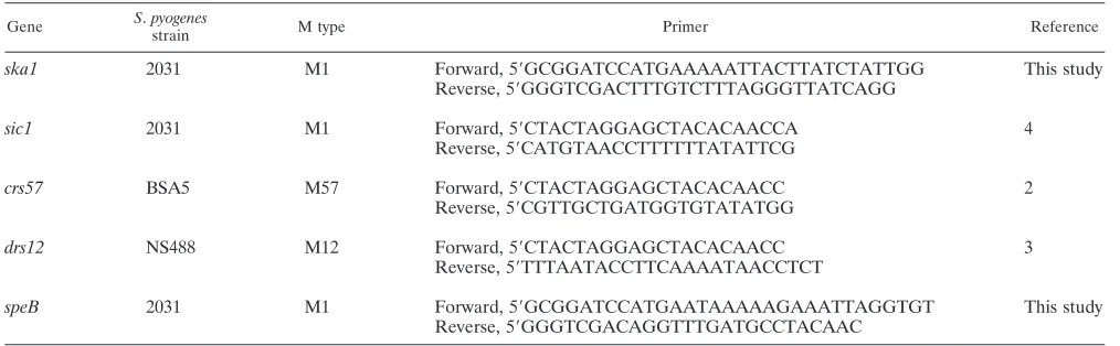
Cloning, expression, and purification of recombinant proteins.Using PCR, theska1,sic1,crs57,drs12, andspeBgenes were amplified with specific primers (Table 1). The amplified products obtained with the SIC, DRS, and CRS primers were subsequently cloned into the pBAD-TOPO-TA (thiofusion) expression system (Invitrogen, Australia), while the amplified Ska1 and SpeB products were
cloned into the pQE30 vector (Qiagen) upstream of the His6tag. The pQE30
vector was used to produce Ska1 and SpeB, because use of the pBAD-TOPO-TA vector resulted in the formation of inclusion bodies during protein production.
The pQE30 constructs were transformed intoE.coliBL21 cells harboring the
pREP4repressor plasmid (Qiagen). All transformants were screened by PCR,
and sequence analysis was performed (Macrogen, Korea) to confirm positive clones.
One-liter cultures ofE.coliBL21 containing the recombinant plasmid were grown
until the optical density at 600 nm (OD600) reached 0.5. Protein production was
induced over 4 h using 0.02%L-arabinose in the pBAD-TOPO-TA vector and 1 M
isopropyl--D-thiogalactopyranoside (IPTG) in the pQE30 vector.
To purify the recombinant thioredoxin fusion proteins and SpeB protein, under nondenaturing conditions, cell pellets from induced cultures were soni-cated to lyse the cells and centrifuged to remove the insoluble cellular debris. The expressed recombinant proteins were isolated from the resultant cleared lysate by using a column with Ni-nitrilotriacetic acid matrix (Qiagen). The re-combinant proteins were eluted with an imidazole (ICN Biomedical) gradient (20 to 300 mM) and dialyzed against phosphate-buffered saline. Clones express-ing only the thioredoxin fusion protein were also obtained as controls, and the protein was purified as described above.
Ska1 was purified under denaturing conditions. Briefly, following centrifuga-tion of the cultured broth, the insoluble pellet was solubilized in 8 M urea for 1 h,
and the resulting lysate was centrifuged at 10,000⫻gfor 20 min. The
superna-tant was mixed with the Ni-nitrilotriacetic acid matrix at room temperature for 1 h, eluted using urea at pH gradients (6.3 to 4.5), and dialyzed against phos-phate-buffered saline.
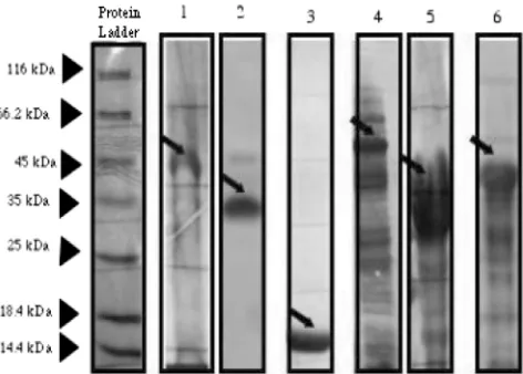
Analysis of proteins.Sodium dodecyl sulfate (SDS)-polyacrylamide gel elec-trophoresis was performed on all protein eluates as previously described (9). The majority of the SIC protein and SIC variant thioredoxin fusion proteins were intact and migrated between 30 and 50 kDa. Ska1 was observed at a band corresponding to approximately 47 kDa, and SpeB was purified in its inactive zymogen form at a band of approximately 37 kDa. Figure 1 shows the expression of the recombinant thioredoxin fusion proteins and the recombinant Ska1 and SpeB proteins on the Coomassie blue-stained gel (Fig. 1).
Western blot.Following SDS-polyacrylamide gel electrophoresis analysis, the fractionated proteins were transferred to polyvinylidene difluoride membrane (Millipore) by using a wet Western transfer apparatus (Bio-Rad) at 50 V for 2 h. The membrane was then removed from the transfer tank and allowed to air dry overnight at room temperature, thus preventing the requirement for a blocking step. For the identification of the SIC and SIC variant recombinant proteins, the blotted membrane was probed with CRS- and DRS-specific rabbit antiserum at a dilution of 1 in 1,000 (Institute of Medical and Veterinary Sciences, Gilles Plains, South Australia, Australia) by immunizing rabbits with recombinant M1 SIC (pQE30; Qiagen) from strain 2031 (anti-CRS) or recombinant M12 DRS (pQE30) from isolate NS488 as previously reported (4). Goat anti-rabbit (heavy and light chain) horseradish peroxidase (HRP)-conjugated immunoglobulin G (IgG) (Pierce Biotechnology, Australia) was used as the secondary antibody at a
dilution of 1 in 2,000. For the identification of Ska1 and SpeB, mouse anti-His4
antibody (Qiagen) was used at a dilution of 1 in 1,000 to detect the His tag. Goat anti-mouse (heavy and light chain) HRP-conjugated IgG (Pierce Biotechnology) was used as the secondary antibody diluted 1 in 2,000. Membranes were washed
⬘
on August 17, 2020 by guest
three times between each step and prior to visualization using the 3,3⬘ -diamino-benzidine tetrahydrochloride (DAB) chromogenic substrate (Fig. 2).
ELISA studies.An indirect ELISA was used to detect streptococcal antibodies
in sera. Unless otherwise stated, all steps were performed using 100l of each
reagent with a reaction period of 1 h at room temperature (28°C⫾2°C), and
between the steps, the plate was washed five times with wash buffer (TropBio; Australia). The wells of U-bottom immunoplates (Sarstedt, Australia) were
coated by adding recombinant protein (100g/ml) in ELISA diluent (TropBio)
to each well and incubating overnight. After the wells were blocked with 150l
of postcoating buffer (TropBio) for 2 h, the bound proteins were allowed to react with a dilution of human serum in ELISA diluent of 1 in 300. After treating the proteins with a secondary goat anti-human IgG (heavy and light chains)-HRP
conjugate (Pierce Biotechnologies), the reactions were detected with 2,2⬘
-azino-di-3-ethylbenzthiazoline-6-sulfonic acid (TropBio). The optical density was
mea-sured using dual absorbance of 414 and 492 nm (OD414,492) after incubation in
the dark for 1 h.
Statistical methods.The healthy population seroreactivity for the investigated proteins has not been established; therefore, we used a cutoff for positive serol-ogy based on three times the average of the lowest quartile of the studied population’s absorbance values, as previously described (16). Fisher’s exact test (two tailed) was used to calculate significance levels for relationships between positive serology to various streptococcal proteins and having ESRF. The data in this study were transformed to ensure a Gaussian distribution. For the ELISA studies comparing the level of antibodies among different groups of the popu-lation (indigenous controls, nonindigenous controls, indigenous patients, and nonindigenous patients), one-way analysis of variance was used followed by
Tamhane’s posthoc test with an␣of 0.05. The null hypothesis assumes that all
groups have similar levels of antibodies against each of the streptococcal anti-gens. Pearson’s rank correlation test was used for analysis of correlations. All the statistical calculations were done using the Statistical Package for Social Sciences (SPSS Inc., Chicago, IL).
RESULTS
Comparative antibody levels between ESRF patients (n ⴝ
66) and healthy controls (n ⴝ 31). A large proportion of
patients and healthy controls had detectable antibodies against Ska1, DRS, CRS, SIC, and SpeB. In general, patients had higher antibody levels than the healthy controls did (Fig. 3). The proportion of ESRF patients with a seropositive result for
Ska1 (P⫽ 0.004), DRS (P⫽0.0003), CRS (P⫽0.001), and SIC (P ⫽ 0.018) was significantly greater compared to the proportion seropositive in the controls, but no significant dif-ference was observed for SpeB (P⫽0.0538).
Antibodies against streptococcal proteins between groups.
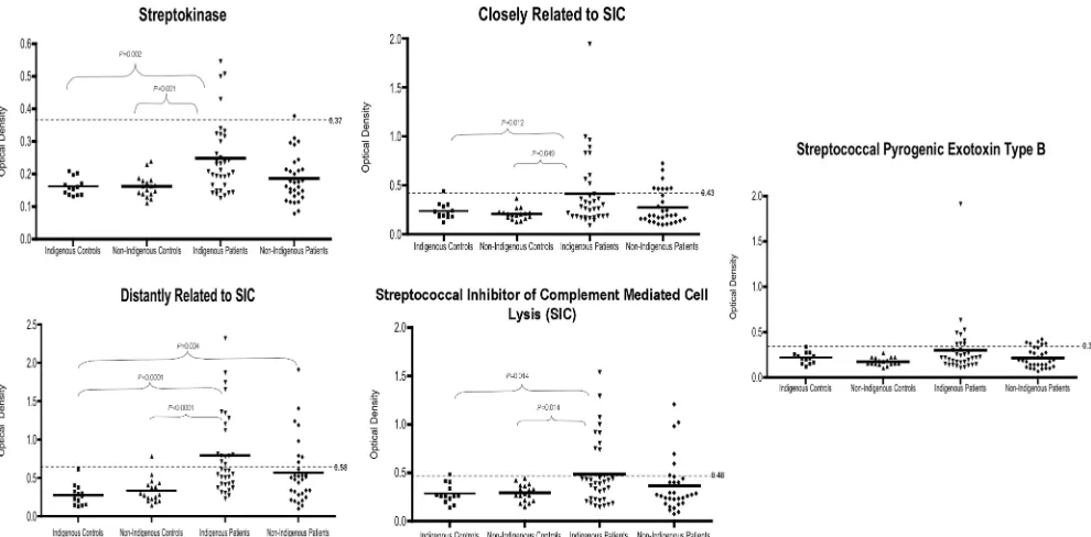
As predicted, indigenous patients (n⫽37) had elevated levels of antibodies to each of the streptococcal proteins compared to the nonindigenous patients (n ⫽ 29); however, these differ-ences were not statistically significant (Fig. 4). Overall, for all patients (n⫽ 97), there was a significant difference between the groups for Ska1 (P⫽0.001), SIC (P⫽0.024), CRS (P⫽
0.016), and DRS (P ⬍ 0.0001), but not SpeB (P ⫽ 0.16). Indigenous patients had significantly higher antibodies than both the indigenous (n ⫽ 15) and nonindigenous (n ⫽ 16) controls for Ska1, SIC, CRS, and DRS.
Correlations between antibodies in the different groups.
Re-lationships between antibody levels were investigated for the patient group (n ⫽ 66) (Table 2). In the patient group, all antibodies were closely correlated and were significant at the
␣ ⫽0.0001 except SIC and SpeB. In the control group (n ⫽
31), only antibody levels to CRS correlated with those of SIC, Ska1, and SpeB, and antibody levels to SIC correlated with those against DRS and SpeB.
Relationship between antibody levels, diabetes, and ESRF.
Being diabetic was positively associated with being an ESRF patient (r2⫽0.37,P⬍0.0001) and being indigenous (r2⫽0.47,
P⬍ 0.0001). Regression analysis showed antibodies to DRS (r2⫽0.85,P⫽0.001) predicted ESRF development. No other
associations was found between streptococcal antibodies and ESRF at␣ ⫽0.05 level.
FIG. 2. Western blot of antibodies against several recombinant streptococcal proteins. The antigens run in each lane are as follows: streptokinase (Ska1) (lane 1), streptococcal pyrogenic exotoxin type B (SpeB) in its zymogen form (lane 2), CRS (lane 3), DRS (lane 4), and SIC (lane 5). His4antibody was used as a positive control for the
detection of Ska1 and SpeB, polyclonal rabbit SIC antiserum was used to detect CRS and SIC, and anti-DRS rabbit polyclonal antibodies were used to detect DRS. Note that the black arrows denote the reaction of the antibody with the protein of interest (SIC, CRS, and DRS). The positions of the molecular mass standards (in kilodaltons) of the protein ladder used (catalog no. SM0431; Fermentas, Australia) are shown by the black arrowheads to the left of the gels.
FIG. 1. Expression of several recombinant streptococcal proteins on a Coomassie blue-stained SDS-polyacrylamide gel. The antigens run in each lane are as follows: streptokinase (lane 1), streptococcal pyrogenic exotoxin type B (lane 2), thioredoxin (lane 3), CRS (lane 4), DRS (lane 5), and SIC (lane 6). Note that the black arrows denote the purified protein band of interest (SIC, CRS, and DRS). The positions of the molecular mass standards (in kilodaltons) of the protein ladder used (catalog no. SM0431; Fermentas, Australia) are shown by the black arrowheads to the left of the gels.
on August 17, 2020 by guest
http://cvi.asm.org/
DISCUSSION
Four M types ofS.pyogenesare classified as nephritic based on an association with epidemics of acute glomerulonephritis. Recent studies have shown that certain streptococcal proteins are associated with the development of PSGN as demonstrated by elevated titers and glomerular antigen deposition in PSGN patients. These findings suggest that these antigens play a role in the development of acute glomerulonephritis. Although it has been proposed that exposure to these proteins is associated with long-term renal complications, the specific serological re-sponses in ESRF patients remain uncertain. In the present study, we used an ELISA to evaluate the level of serum anti-bodies against five nephritic streptococcal antigens in patients with established ESRF. We observed that ESRF patients had specific elevated antibodies to Ska1, SIC, CRS, and DRS com-pared to healthy controls. Further, no significant difference was observed between indigenous and nonindigenous patients. This is the first study to indicate that exposure to these strep-tococcal proteins is associated with long-term renal complica-tions.
We hypothesized that compared with nonindigenous pa-tients, indigenous patients would have significantly higher antibody levels. In the current study, indigenous patients had elevated levels of antibodies to all five streptococcal antigens; however, the difference was not significant. It is possible that in a larger sample a difference would be observed because on posthoc analysis indigenous patients differed from all controls for Ska1, DRS, CRS, and SIC. Nonindigenous patients had a significant difference only for DRS compared to indigenous controls. Additionally, compared to indigenous controls, indig-enous patients had significantly elevated antibody levels. In this study, indigenous patients and controls were from the same
geographical area, where streptococcal infections are endemic; therefore, it is unlikely that this difference represents an in-creased incidence of streptococcal skin infections in childhood but more likely represents an increased infection rate with nephritic strains of group A streptococcus.
This study provides evidence to support the hypothesis that the current epidemic of renal failure in the Australian indige-nous population may be a result of recurrent streptococcal skin infections and sporadic episodes of PSGN. It is postulated that complications of PSGN may manifest clinically later in life as progressive renal disease and ultimately ESRF. Furthermore, the results are not limited to the Australian indigenous popu-lation, as a proportion of nonindigenous patients in this study had specific elevated streptococcal antibodies, which may be implicated in their disease pathology. This finding was ex-pected, given the fact that the research was conducted in a tropical location where streptococcal skin infections, such as impetigo, are common (10). The M types isolated from strep-tococcal skin infections are associated with the development of PSGN.
Despite the fact that antibody titers to SpeB in its zymogen form are currently the best-known marker forS. pyogenes in-fections associated with acute PSGN (12), in this study no difference was observed between any of the groups. Further-more, high titers to SpeB are also associated with group A streptococcal infections in general (1). Therefore, it is possible that SpeB titers are a marker of acute infections and glomer-ulonephritis, but the antibodies are not long lasting. It has previously been shown that anti-SpeB titers tend to increase and peak within the first 2 weeks of onset of disease and then decrease with time (13). The group of patients ana-lyzed in this study were adults with established ESRF, and FIG. 3. Graph showing mean optical densities obtained for each streptococcal protein analyzed according to group. Using an ELISA, the sera from 31 healthy controls and 66 ESRF patients were compared for seroreactivity (measured in OD414,492units) to five streptococcal recombinant
antigens. Values shown are the mean optical densities⫾standard errors of the means (error bars).
on August 17, 2020 by guest
therefore, it would be years since a possible childhood epi-sode of PSGN.
Unlike streptokinase and SpeB, the SIC protein and SIC variant proteins are secreted only by M types that are histori-cally associated with PSGN. Thus, the fact that patients exhibit higher antibody levels to these antigens suggests that they have been infected with these so-called nephritic M types of S.
pyogenes. The findings of this study support the proposal by Sriprakash et al. (16) that seroreactivity to DRS may be of pathological significance. However, the patients in the current paper had clearly established renal disease, whereas in previ-ous papers determination of a history of PSGN has been con-ducted retrospectively using scarce medical records and there-fore include bias. In the present study, the control group was
healthy, and therefore, it is possible that the serological changes may reflect nonspecific factors associated with kidney failure and/or dialysis treatment. However, the statistical anal-ysis used to determine a cutoff for seropositivity incorporated all sample data and not just healthy controls, therefore allow-ing renal failure patients to be either seropositive or seroneg-ative depending upon the ELISA result.
This research has demonstrated that ESRF patients have significantly higher prevalence of seropositivity against Ska1, SIC, CRS, and DRS compared to healthy controls and shown that elevated antibodies to DRS is a predictor for ESRF. This is the first study to suggest elevated antibodies to Ska1, SIC, and SIC variant proteins might be of pathological significance in ESRF. This is possible via antigen-antibody deposition or FIG. 4. Scatterplots of sera from indigenous patients show greater reactivity to Ska1, DRS, CRS, and SIC streptococcal antigens than other experimental groups. Using an ELISA, the sera from 15 indigenous controls, 16 nonindigenous controls, 37 indigenous patients, and 29 nonindigenous patients were compared for seroreactivity (measured in OD414,492units) to five recombinant streptococcal antigens. The significant
results of statistical analyses of the mean serological responses are shown (calculated using Tamhane’s posthoc test). The solid horizontal lines represent the means for the groups, and the dotted horizontal lines show the cutoff for seropositivity (three times the overall mean of the lowest quartile).
TABLE 2. Pearson correlation coefficients between antibodies to streptococcal antigens in ESRF patients and healthy controls
Group (n) Streptococcal
antigen
Pearson correlation coefficient (r2) between antibodies to streptococcal antigens in patients
and controlsa
SIC CRS DRS SpeB
Healthy controls (31) Ska1 0.17 (0.079) 0.17(0.024) 0.067 (0.17) 0.12 (0.056)
SIC 0.24(0.0062) 0.16 (0.027) 0.24(0.0065)
CRS 0.00057 (0.90) 0.35(0.0006)
DRS 0.0034 (0.76)
ESRF patients (66) Ska1 0.29(⬍0.0001) 0.64(⬍0.0001) 0.80(⬍0.0001) 0.28(⬍0.0001)
SIC 0.16(0.0009) 0.34(⬍0.0001) 0.044 (0.09)
CRS 0.80(⬍0.0001) 0.67(⬍0.0001)
DRS 0.45(⬍0.0001)
aSignificant correlation coefficients (r2) are shown in bold type. The associatedPvalues are shown in parentheses.
on August 17, 2020 by guest
http://cvi.asm.org/
2.Binks, M., D. McMillan, and K. S. Sriprakash.2003. Genomic location and variation of the gene for CRS, a complement binding protein in the M57
strains ofStreptococcus pyogenes. Infect. Immun.71:6701–6706.
3.Binks, M., and K. S. Sriprakash.2004. Characterization of a
complement-binding protein, DRS, from strains ofStreptococcus pyogenescontaining the
emm12andemm55genes. Infect. Immun.72:3981–3986.
4.Binks, M. J., B. A. Fernie-King, D. J. Seilly, P. J. Lachmann, and K. S. Sriprakash.2005. Attribution of the various inhibitory actions of strepto-coccal inhibitor of complement (SIC) to regions within the molecule. J. Biol.
Chem.280:20120–20125.
5.Cu, G. A., S. Mezzano, J. D. Bannan, and J. B. Zabriskie.1998. Immuno-histochemical and serological evidence for the role of streptococcal
protein-ase in acute post-streptococcal glomerulonephritis. Kidney Int.54:819–826.
6.Fernie-King, B. A., D. J. Seilly, C. Willers, R. Wurzner, A. Davies, and P. J. Lachmann.2001. Streptococcal inhibitor of complement (SIC) inhibits the membrane attack complex by preventing uptake of C567 onto cell
mem-branes. Immunology103:390–398.
7.Goodfellow, A. M., W. E. Hoy, K. S. Sriprakash, M. J. Daly, M. P. Reeve, and
14.Poon-King, R., J. Bannan, A. Viteri, G. Cu, and J. B. Zabriskie.1993. Identification of an extracellular plasmin binding protein from a
nephrito-genic streptococci. J. Exp. Med.178:759–763.
15.Skattum, L., P. Akesson, L. Truedsson, and A. G. Sjo¨holm.2006. Antibodies
against four proteins from aStreptococcus pyogenesserotype M1 strain and
levels of circulating mannan-binding lectin in acute poststreptococcal
glo-merulonephritis. Int. Arch. Allergy Immunol.140:9–19.
16.Sriprakash, K. S., J. Hartas, and A. White.2002. Antibodies to streptococcal inhibitor of complement function and M peptides in a post-streptococcal
glo-merulonephritis endemic region of Australia. J. Med. Microbiol.51:589–594.
17.White, A. V., W. E. Hoy, and D. A. McCredie.2001. Childhood post-strep-tococcal glomerulonephritis as a risk factor for chronic renal disease in later
life. Med. J. Aust.174:492–496.
18.Yamakami, K., N. Yoshizawa, K. Wakabayashi, A. Takeuchi, T. Tada-kuma, and M. D. P. Boyle.2000. The potential role for nephritis-associ-ated plasmin receptor in acute poststreptococcal glomerulonephritis.
Methods21:185–197.