(PCV2) and Porcine Reproductive and Respiratory Syndrome Virus
(PRRSV) in a PCV2-PRRSV Challenge Model
Changhoon Park, Yeonsu Oh, Hwi Won Seo, Kiwon Han, Chanhee Chae
Department of Veterinary Pathology, College of Veterinary Medicine, Seoul National University, Seoul, Republic of Korea
The objective of the present study was to determine the effects of porcine circovirus type 2 (PCV2) and porcine reproductive and
respiratory syndrome virus (PRRSV) vaccinations in an experimental PCV2-PRRSV challenge model, based on virological
(viremia), immunological (neutralizing antibodies [NAs], gamma interferon-secreting cells [IFN-
␥
-SCs], and CD4
ⴙCD8
ⴙdou-ble-positive cells), and pathological (lesions and antigens in lymph nodes and lungs) evaluations. A total of 72 pigs were
ran-domly divided into 9 groups (8 pigs per group): 5 vaccinated and challenged groups, 3 nonvaccinated and challenged groups, and
a negative-control group. Vaccination against PCV2 induced immunological responses (NAs and PCV2-specific IFN-
␥
-SCs) and
reduced PCV2 viremia, PCV2-induced lesions, and PCV2 antigens in the dually infected pigs. However, vaccination against
PCV2 did not affect the PRRSV immunological responses (NAs and PRRSV-specific IFN-
␥
-SCs), PRRSV viremia,
PRRSV-in-duced lesions, or PRRSV antigens in the dually infected pigs. Vaccination against PRRSV did not induce immunological
re-sponses (PRRSV-specific IFN-
␥
-SCs) or reduce PRRSV viremia, PRRSV-induced lesions, or PRRSV antigens in the dually
in-fected pigs. In addition, vaccination against PRRSV increased PCV2 viremia, PCV2-induced lesions, and PCV2 antigens in the
dually infected pigs. In summary, vaccination against PCV2 reduced PCV2 viremia, PCV2-induced lesions, and PCV2 antigens
in the dually infected pigs. However, vaccination against PRRSV increased PCV2 viremia, PCV2-induced lesions, and PCV2
anti-gens in the dually infected pigs. Therefore, the PCV2 vaccine decreased the potentiation of PCV2-induced lesions by PRRSV in
dually infected pigs. In contrast, the PRRSV vaccine alone did not decrease the potentiation of PCV2-induced lesions by PRRSV
in dually infected pigs.
P
orcine circovirus type 2 (PCV2), which is a single-stranded
circular DNA virus within the family
Circoviridae
, is now
rec-ognized as the primary etiological agent of porcine
circovirus-associated diseases (PCVAD) such as postweaning multisystemic
wasting syndrome (PMWS), porcine respiratory disease complex
(PRDC), and other clinical diseases (
1
,
2
). Porcine reproductive
and respiratory syndrome virus (PRRSV), which is the causative
agent of PRRS, is a single-stranded, positive-sense, enveloped
RNA virus in the family
Arteriviridae
and the order
Nidovirales
(
3
). PRRSV infection in swine is characterized by late-term
abor-tion in gilts and sows and severe respiratory diseases in neonatal
and nursing pigs (
4
). PRDC is a serious problem in the pig
indus-try. PRRSV and PCV2 are the most commonly associated primary
pathogens in PRDC. Epidemiological analyses have revealed that
coinfection with PCV2 and PRRSV is most commonly observed in
field cases (
5
–
7
).
Several studies have previously demonstrated the relationship
between PCV2 and PRRSV. PCV2 does not affect PRRSV
replica-tion or lesions, but PRRSV increases PCV2 DNA loads in the sera
of coinfected pigs (
8
,
9
) and increases the levels of PCV2 antigens
in tissues (
10
), which results in more severe PCV2-associated
le-sions (
11
). Based on these results, one possible way to minimize
the effects of the PRRSV-associated enhancement of the
replica-tion of PCV2 and the inducreplica-tion of PMWS may be the use of a
PRRSV-based vaccine in preweaned pigs. However, there are no
reports in the literature describing the effects of PCV2 and PRRSV
challenges on pigs that have been immunized with either PCV2 or
PRRSV vaccines. In the absence of such a study, the PCV2
vaccine-PCV2-PRRSV and PRRSV vaccine-vaccine-PCV2-PRRSV interactions
have not been elucidated completely. Therefore, the objective of
the present study was to determine the effects of PCV2 and PRRSV
vaccinations in an experimental PCV2-PRRSV challenge model,
based on virological (viremia), immunological (neutralizing
anti-bodies [NAs], gamma interferon-secreting cells [IFN-
␥
-SCs], and
CD4
⫹CD8
⫹double-positive cells), and pathological (lesions and
antigens in lymph nodes and lungs) evaluations.
MATERIALS AND METHODS
Commercial vaccine.The inactivated chimeric PCV1-2 vaccine (Fostera PCV vaccine; Pfizer Animal Health, New York, NY) and modified live PRRS vaccine (Ingelvac PRRS MLV; Boehringer Ingelheim Animal Health, St. Joseph, MO) were used in this study. The inactivated chimeric PCV1-2 vaccine contains the genomic backbone of the nonpathogenic PCV1 with the PCV2 ORF2 capsid gene in place of the PCV1 capsid gene (12). The modified live PRRS vaccine (Ingelvac PRRS MLV) originated from the American isolate ATCC VR-2332 and was attenuated by serial passages in cell culture. The vaccine contained at least 1⫻104.950% tissue culture infective doses (TCID50) in 2 ml. Forty pigs were vaccinated with 2.0-ml doses of either the PCV2 or PRRSV vaccine or both intramuscu-larly at 3 weeks of age (Table 1). All of the vaccines that were used in this study were administered according to the manufacturer’s instructions (1 dose, intramuscular route).
Received5 August 2012 Returned for modification8 September 2012 Accepted2 January 2013
Published ahead of print9 January 2013
Address correspondence to Chanhee Chae, swine@snu.ac.kr.
Copyright © 2013, American Society for Microbiology. All Rights Reserved.
doi:10.1128/CVI.00497-12
on August 17, 2020 by guest
http://cvi.asm.org/
Animals and housing.A total of 72 colostrum-fed, cross-bred, con-ventional piglets were weaned and purchased at 18 days of age from a PRRSV-free commercial farm. They were all negative for PCV2, PRRSV, swine influenza virus, andMycoplasma hyopneumoniae, according to rou-tine serological testing. PCV2 and PRRSV were not detected in serum samples by real-time PCR (13,14). Individual piglets from 7 days of age were uniquely identified by their ear notches.
All of the pigs were housed in an environmentally controlled building with pens over completely slatted floors throughout the experiment. To avoid environmental contamination, the building was completely emp-tied, cleaned three times with hot (⬎95°C) water, and disinfected with a 2% potassium peroxymonosulfate- and sodium chloride-based product (Virkon S; Antec International, Sudbury, Suffolk, United Kingdom) for 3 days. The building was emptied for an additional 21 days before the pigs were introduced, and each pig was housed separately within the facility.
Experimental design.A total of 72 pigs were randomly divided into 9 groups (8 pigs per group): 5 vaccinated and challenged (VC) groups (1, 2, 3, 4, and 5), 3 nonvaccinated and challenged (NVC) groups (6, 7, and 8), and a negative-control group (9).
At 21 days of age (⫺28 days postchallenge [dpc]), pigs in groups 1 and 2 were immunized with the PCV2 vaccine, pigs in groups 3 and 4 were immunized with the PRRSV vaccine, and pigs in group 5 were immunized with both the PCV2 and PRRSV vaccines. At 49 days of age (0 dpc), pigs in the VC and NVC groups were intratracheally administered an intranasal 3-ml dose of PCV2b (strain SNUVR000463; 5th passage) containing 1.2⫻ 105TCID
50/ml and/or a 3-ml dose of PRRSV (strain SNUVR090851; North American genotype; 5th passage) containing 1⫻105TCID
50/ml. Pigs in groups 1 and 6 were challenged with PCV2. Pigs in groups 3 and 7 were challenged with PRRSV. Pigs in groups 2, 4, 5, and 8 were challenged with both PCV2 and PRRSV (Table 1).
Serology.Blood samples from each pig were collected by jugular ve-nipuncture at⫺28, 0, 10, and 21 dpc, and the sera were stored at⫺20°C. The serum samples were tested using a commercial PRRSV enzyme-linked immunosorbent assay (ELISA) (HerdCheck PRRS 2XR; Idexx Lab-oratories Inc., Westbrook, ME) and a PCV2 ELISA (Synbiotics, Lyon, France). A serum virus neutralization (SVN) test for PCV2 was performed as previously described (15). NA titers were expressed as the reciprocal of the highest serum dilution that completely blocked the infection in PK15 cells compared with the virus control. An SVN test for PRRSV was also performed as previously described (16).
Quantification of PCV2 DNA in blood.DNA was extracted from the serum samples obtained at⫺28, 0, 10, and 21 dpc by use of a QIAamp DNA minikit (Qiagen Ltd., Crawley, United Kingdom). The DNA ex-tracts were used to quantify the PCV2 genomic DNA copy numbers by real-time PCR as previously described (13).
Quantification of PRRSV RNA in blood.RNA was extracted from the serum samples obtained at⫺28, 0, 10, and 21 dpc for all of the pigs that
were used in this study, as previously described (14,17). Real-time PCRs for the PRRSV and vaccine strains were used to quantify the PRRSV genomic cDNA copy numbers following RNA extraction of the serum samples, which was performed as previously described (14,17).
Preparation of PCV2 and PRRSV antigens.The same PCV2 strain (or PRRSV strain) that was used for the challenge in the pigs was propagated in PCV-free PK15 cells (or MARC-145 cells for the PRRSV strain) to a titer of 104TCID
50/ml and then treated with two freeze-thaw cycles. The PCV2 antigen was prepared by concentrating the virus that was present in the cell culture by ultracentrifugation at 100,000⫻gat 4°C for 3 h. The virus pellet was resuspended in phosphate-buffered saline (PBS). The concen-trated PCV2 (or PRRSV) was inactivated by exposure to an 8-W germi-cidal UV lamp at a distance of 15 cm for 1 h. Inactivation was confirmed by the absence of the virus antigen from the PK15 cells (or MARC-145 cells for the PRRSV strain) as determined by an immunoperoxidase assay, as previously described (18,19).
ELISPOT assay.The numbers of PCV2- and PRRSV-specific IFN-␥ -SCs were determined for peripheral blood mononuclear cells (PBMCs) obtained at⫺28, 0, 10, and 21 dpc as previously described (20). Briefly, 100l containing 2⫻106PBMCs in RPMI 1640 medium that was sup-plemented with 10% fetal bovine serum (HyClone Laboratories, Inc., Se-lectScience, Bath, United Kingdom) was seeded into plates that were pre-coated overnight with anti-porcine IFN-␥monoclonal antibody (5g/ ml) (Mabtech, Mariemont, OH) and incubated with 100l of PCV2 antigen (20g/ml), PRRSV antigen (20g/ml), and phytohemagglutinin (10g/ml) (Roche Diagnostics GmbH, Mannheim, Germany) as positive controls or with PBS as a negative control for 40 h at 37°C in a 5% humid-ified CO2atmosphere. The wells were then washed five times with PBS (200l per well). Thereafter, the procedure was conducted according to the manufacturer’s instructions, using a commercial enzyme-linked im-munosorbent spot (ELISPOT) assay kit (Mabtech, Mariemont, OH). The spots on the membranes were read by an automated ELISPOT reader (AID ELISpot reader; AID GmbH, Strassberg, Germany). The results were expressed as numbers of IFN-␥-SCs per million PBMCs.
Flow cytometry.Phenotypic analyses of the CD4⫹CD8⫹ double-positive cells in PBMCs were performed using flow cytometry with two monoclonal antibodies, i.e., CD4a–R-phycoerythrin (CD4a–R-PE) and CD8a-fluorescein isothiocyanate (CD8a-FITC) (SouthernBiotech, Bir-mingham, AL), and a FACSCalibur flow cytometer (Becton, Dickinson, San Jose, CA) as previously described (21).
Immunohistochemistry.Superficial inguinal lymph nodes that had previously shown consistent and intense labeling for PCV2 and typical granulomatous lesions (22) were collected at necropsy for immunohisto-chemical analyses. A rabbit polyclonal anti-PCV2 antibody (23) (1:200 in PBS containing 0.1% Tween 20) and the SR30 monoclonal antibody (Ru-ral Technologies Inc., Brookings, SD) (1:10,000 in PBS containing 0.1% Tween 20) were used as the primary antibodies. Immunohistochemical analyses for PCV2 and PRRSV were performed as previously described (24,25).
Morphometric analyses.For morphometric analyses of the micro-scopic pulmonary and lymph lesion scores, lung sections were examined blindly and assigned estimated scores ranging from 1 (mild interstitial pneumonia) to 4 (severe interstitial pneumonia), as previously described (26). Superficial inguinal lymph node sections were also examined blindly, and their scores ranged from 0 (normal, i.e., no lymphoid deple-tion or granulomatous replacement) to 5 (severe lymphoid depledeple-tion and granulomatous replacement), as previously described (27).
For immunohistochemistry morphometric analyses, 3 sections were cut from each of three blocks of tissue from one entire pulmonary lobe and from the superficial inguinal lymph nodes of each pig. The slides were analyzed using the NIH Image J 1.43m program (http://rsb.info.nih.gov /ij) to obtain the quantitative data. For each slide, 10 fields were randomly selected, and the number of positive cells per unit area (0.95 mm2for the pulmonary lobe and 0.25 mm2for the lymph node) was determined as previously described (22,28). The mean values were also calculated. TABLE 1Study design with vaccination and challenge statuses for
PCV2 and PRRSVa
Group no.
Vaccination (⫺28 dpc) Challenge (0 dpc)
PCV2 PRRSV PCV2 PRRSV
1 ⫹ ⫺ ⫹ ⫺
2 ⫹ ⫺ ⫹ ⫹
3 ⫺ ⫹ ⫺ ⫹
4 ⫺ ⫹ ⫹ ⫹
5 ⫹ ⫹ ⫹ ⫹
6 ⫺ ⫺ ⫹ ⫺
7 ⫺ ⫺ ⫺ ⫹
8 ⫺ ⫺ ⫹ ⫹
9 ⫺ ⫺ ⫺ ⫺
aThere were eight animals in each group, and necropsy was performed at 21 dpc in all
cases.
on August 17, 2020 by guest
http://cvi.asm.org/
Statistical analyses.Summary statistics were calculated for all of the groups to assess the overall quality of the data, including normality. For single comparisons, analysis of variance (ANOVA) with apost hocTukey’s test was used to compare the primary variables (immunohistochemical scores) among groups. The continuous data for PCV2 and PRRSV serol-ogy and PCV2 DNA and PRRSV cDNA quantifications were analyzed using an ANOVA for each time point. When one-way ANOVA revealed a significance level with aPvalue of⬍0.05, Tukey’s honestly significant difference test was used to determine the significance of individual be-tween-group differences. Discrete data (histopathologic lung and lym-phoid lesion scores) were analyzed by the chi-square and Fisher’s exact tests.Pvalues of⬍0.05 were considered to be significant.
RESULTS
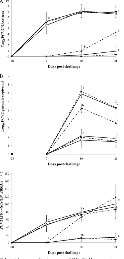
Serology of PCV2.
At challenge, all pigs vaccinated against PCV2
were seropositive, and all nonvaccinated pigs were negative by
ELISA and SVN tests. Pigs which received the PCV2 vaccine
fol-lowed by PCV2 challenge only (group 1), pigs which received the
PCV2 vaccine followed by dual challenge (group 2), and pigs
which received both the PCV2 and PRRSV vaccines followed by
dual challenge (group 5) had significantly higher anti-PCV2 IgG
antibody levels (
P
⬍
0.001) and NA titers (
P
⬍
0.01) (
Fig. 1A
) than
those of pigs which received the PRRSV vaccine followed by dual
challenge (group 4), pigs which were challenged with PCV2 only
(group 6), and pigs which were challenged with both PCV2 and
PRRSV (group 8) at 10 and 21 dpc. Pigs in group 6 had
signifi-cantly higher anti-PCV2 IgG antibody levels (
P
⬍
0.01) than those
of pigs in groups 4 and 8 at 21 dpc.
Serology of PRRSV.
At challenge, all pigs vaccinated against
PRRSV were seropositive, and all nonvaccinated pigs were
nega-tive by ELISA. Pigs which received the PRRSV vaccine followed by
PRRSV challenge only (group 3), pigs which received the PRRSV
vaccine followed by dual challenge (group 4), and pigs which
re-ceived both the PCV2 and PRRSV vaccines followed by dual
chal-lenge (group 5) had significantly higher anti-PRRSV IgG antibody
levels (
P
⬍
0.001) than pigs which received the PCV2 vaccine
followed by dual challenge (group 2), pigs which were challenged
with PRRSV only (group 7), and pigs which were challenged with
both PCV2 and PRRSV (group 8) at 10 dpc. Pigs which received
PRRSV followed by PRRSV challenge only (group 3) and pigs
which received both the PCV2 and PRRSV vaccines followed by
dual challenge (group 5) had significantly higher anti-PRRSV IgG
antibody levels (
P
⬍
0.05) than pigs in the other 4 groups (2, 4, 7,
and 8) at 21 dpc (
Fig. 2A
). Low PRRSV NA titers (
⬍
2 log
2) were
detected in all 8 groups throughout the experiment.
Quantification of PCV2 DNA in blood.
At challenge, no
genomic copies of PCV2 were detected in any of the serum
sam-ples from all 8 groups. Pigs which received the PRRSV vaccine
followed by dual challenge (group 4) and pigs which were
chal-lenged with both PRRSV and PCV2 (group 8) had significantly
more genomic copies of PCV2 in the serum than pigs which
re-ceived the PCV2 vaccine followed by PCV2 challenge only (group
1), pigs which received the PCV2 vaccine followed by dual
chal-lenge (group 2), pigs which received both the PCV2 and PRRSV
vaccines followed by dual challenge (group 5), and pigs which
were challenged with PCV2 only (group 6) at 10 and 21 dpc (
P
⬍
0.05). Pigs which were challenged with PCV2 only (group 6) had
significantly more genomic copies of PCV2 in the serum than pigs
which received the PCV2 vaccine followed by PCV2 challenge
only (group 1), pigs which received the PCV2 vaccine followed by
dual challenge (group 2), and pigs which received both the PCV2
and PRRSV vaccines followed by dual challenge (group 5) at 10
and 21 dpc (
P
⬍
0.05) (
Fig. 1B
). No genomic copies of PCV2 were
detected in any of the serum samples from nonchallenged pigs
(groups 3, 7, and 9) throughout the experiment.
FIG 1(A) Mean serum NA titers against PCV2. (B) Mean genomic copy numbers of PCV2 DNA in serum samples. (C) Mean numbers of PCV2-spe-cific IFN-␥-SCs in PBMCs., pigs which received the PCV2 vaccine followed by PCV2 challenge only (group 1);䊐, pigs which received the PCV2 vaccine followed by dual challenge (group 2);〫, pigs which received the PRRSV vac-cine followed by dual challenge (group 4);Œ, pigs which received both the PCV2 and PRRSV vaccines followed by dual challenge (group 5);Œ, pigs which were challenged with PCV2 (group 6);, pigs which were challenged with PCV2 and PRRSV (group 8). Different letters (a, b, and c) indicate significant (P⬍0.05) differences between groups.
on August 17, 2020 by guest
http://cvi.asm.org/
Quantification of PRRSV cDNA in blood.
At challenge, no
genomic copies of PRRSV were detected in any of the serum
sam-ples from all 8 groups. Pigs which received the PCV2 vaccine
fol-lowed by dual challenge (group 2), pigs which received the PRRSV
vaccine followed by dual challenge (group 4), pigs which were
challenged with PRRSV only (group 7), and pigs which were
chal-lenged with both PCV2 and PRRSV (group 8) had significantly
more genomic copies of PCV2 in the serum than pigs which
re-ceived the PRRSV vaccine followed by PRRSV infection only
(group 3) and pigs which received both the PCV2 and PRRSV
vaccines followed by dual challenge (group 5) at 10 and 21 dpc
(
P
⬍
0.05) (
Fig. 2B
). No genomic copies of PRRSV were detected
in any of the serum samples from nonchallenged pigs (groups 1, 6,
and 9) throughout the experiment.
PCV2-specific IFN-
␥
-SCs.
No PCV2-specific IFN-
␥
-SCs
were detected in PBMCs at
⫺
28 dpc for any of the pigs.
PCV2-specific IFN-
␥
-SCs were detected in PBMCs from the
PCV2-vaccinated pigs (groups 1, 2, and 5) at 0 dpc. Pigs which
re-ceived the PCV2 vaccine followed by PCV2 challenge only
(group 1), pigs which received the PCV2 vaccine followed by
dual challenge (group 2), pigs which received both the PCV2
and PRRSV vaccines followed by dual challenge (group 5), and
pigs which were challenged with PCV2 only (group 6) had
significantly more PCV2-specific IFN-
␥
-SCs than pigs which
received the PRRSV vaccine followed by dual challenge (group
4) and pigs which were challenged with both PCV2 and PRRSV
(group 8) at 10 and 21 dpc (
P
⬍
0.01) (
Fig. 1C
).
PRRSV-specific IFN-
␥
-SCs.
No PRRSV-specific IFN-
␥
-SCs
were detected in PBMCs at
⫺
28 dpc for any of the pigs.
PRRSV-specific IFN-
␥
-SCs were detected in PBMCs from the
PRRSV-vaccinated pigs (groups 3, 4, and 5) at 0 dpc. Pigs which received
the PRRSV vaccine followed by PRRSV infection only (group 3)
or followed by dual challenge (group 5) had significantly more
PRRSV-specific IFN-
␥
-SCs than pigs which received the PCV2
vaccine followed by dual challenge (group 2), pigs which received
the PRRSV vaccine followed by dual challenge (group 4), pigs
which were challenged with PRRSV only (group 7), and pigs
which were challenged with both PCV2 and PRRSV (group 8) at
10 and 21 dpc (
P
⬍
0.01). Pigs which received the PCV2 vaccine
followed by dual challenge (group 2) and pigs which were
chal-lenged with PRRSV only (group 7) had significantly more
PRRSV-specific IFN-
␥
-SCs than the pigs in 2 groups (groups 4 and 8) at 21
dpc (
P
⬍
0.01) (
Fig. 2C
).
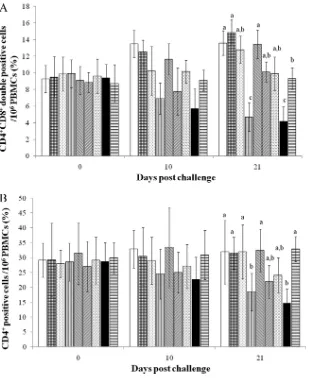
Identification of CD4
ⴙCD8
ⴙdouble-positive cells.
Pigs
which received the PCV2 vaccine followed by PCV2 challenge
only (group 1), pigs which received the PCV2 vaccine followed by
dual challenge (group 2), and pigs which received both the PCV2
and PRRSV vaccines followed by dual challenge (group 5) had
significantly larger proportions of CD4
⫹CD8
⫹double-positive
cells than pigs which received the PRRSV vaccine followed by dual
challenge (group 4), pigs which were challenged with both PCV2
and PRRSV (group 8), and negative-control pigs (group 9) at 21
dpc (
P
⬍
0.05). Negative-control pigs had significantly larger
pro-portions of CD4
⫹CD8
⫹double-positive cells than pigs which
received the PRRSV vaccine followed by dual challenge (group 4)
and pigs which were challenged with both PCV2 and PRRSV
(group 8) at 21 dpc (
P
⬍
0.05) (
Fig. 3A
).
Pigs which received the PCV2 vaccine followed by PCV2
chal-lenge only (group 1), pigs which received the PCV2 vaccine
fol-lowed by dual challenge (group 2), pigs which received the PRRSV
vaccine followed by PRRSV infection only (group 3), pigs which
received both the PCV2 and PRRSV vaccines followed by dual
challenge (group 5), and negative-control pigs (group 9) had
sig-nificantly larger proportions of CD4
⫹cells than pigs which
re-ceived the PRRSV vaccine followed by dual challenge (group 4)
FIG 2(A) Mean anti-PRRSV IgG antibody levels in serum samples. (B) Mean genomic copy numbers of PRRSV DNA in serum samples. (C) Mean numbers of PRRSV-specific IFN-␥-SCs in PBMCs.䊐, pigs which received the PCV2 vaccine followed by dual challenge (group 2);}, pigs which received the PRRSV vaccine followed by PRRSV challenge (group 3);〫, pigs which re-ceived the PRRSV vaccine followed by dual challenge (group 4);Œ, pigs which received both the PCV2 and PRRSV vaccines followed by dual challenge (group 5);o, pigs which were challenged with PRRSV (group 7);, pigs which were challenged with PCV2 and PRRSV (group 8). Different letters (a, b, and c) indicate significant (P⬍0.05) differences between groups. S/P, sample to positive ratio ofⱖ0.4 is considered to be positive.
on August 17, 2020 by guest
http://cvi.asm.org/
and pigs which were challenged with both PCV2 and PRRSV
(group 8) at 21 dpc (
P
⬍
0.03) (
Fig. 3B
).
Histopathologic lesion scores.
The typical granulomatous
in-flammatory reaction and lymphoid depletion that are associated
with PCV2 infection in pigs, which are consistent with the
histo-pathologic lesions in PCVAD, were observed in the lymph nodes
from pigs which received the PRRSV vaccine followed by dual
challenge (group 4) (
Fig. 4A
) and pigs which were challenged with
both PCV2 and PRRSV (group 8). Pigs in these groups (4 and 8)
had significantly higher lymphoid lesion scores than pigs in the
other 6 groups (1, 2, 3, 5, 6 [
Fig. 4B
], and 7) (
P
⬍
0.03). Pigs which
were challenged with PCV2 only (group 6) had significantly
higher lymphoid lesion scores than pigs in the other 5 groups (1, 2,
3, 5, and 7) (
P
⬍
0.03). No histopathologic lymph node lesions
were observed in the negative-control pigs (group 9).
The pulmonary histopathologic lesions were characterized by
alveolar proteinaceous and karyorrhectic debris that was
inter-spersed with macrophages. The alveolar septa in the pigs which
were challenged with both PCV2 and PRRSV (group 8) were
thickened with increased numbers of interstitial macrophages and
lymphocytes. Pigs which received the PCV2 vaccine followed by
dual challenge (group 2), pigs which received the PRRSV vaccine
followed by dual challenge (group 4) (
Fig. 5A
), pigs which were
challenged with PRRSV only (group 7), and pigs which were
chal-lenged with both PCV2 and PRRSV (group 8) had significantly
higher pulmonary lesion scores than pigs in the other 4 groups (1,
3 [
Fig. 5B
], 5, and 6) (
P
⬍
0.05) at 21 dpc (
Table 2
). The lungs of
the negative-control pigs (group 9) were normal.
Immunohistochemical scores.
Pigs which received the
PRRSV vaccine followed by dual challenge (group 4) (
Fig. 4A
) and
pigs which were challenged with both PCV2 and PRRSV (group 8)
had significantly more PCV2-positive cells per unit area in the
lymph node and lung than pigs which received the PCV2 vaccine
followed by PCV2 challenge only (group 1), pigs which received
the PCV2 vaccine followed by dual challenge (group 2), pigs
which received both the PCV2 and PRRSV vaccines followed by
FIG 3Analyses of CD4⫹CD8⫹double-positive cells (A) and CD4⫹cells (B) in PBMCs from the different groups. From left to right, the bars indicate the following groups: pigs which received the PCV2 vaccine followed by PCV2 challenge only (group 1), pigs which received the PCV2 vaccine followed by dual challenge (group 2), pigs which received the PRRSV vaccine followed by PRRSV challenge (group 3), pigs which received the PRRSV vaccine followed by dual challenge (group 4), pigs which received both the PCV2 and PRRSV vaccines followed by dual challenge (group 5), pigs which were challenged with PCV2 (group 6), pigs which were challenged with PRRSV (group 7), pigs which were challenged with PCV2 and PRRSV (group 8), and negative-control pigs (group 9). Different letters (a, b, and c) indicate significant (P⬍0.05) differences between groups.
on August 17, 2020 by guest
http://cvi.asm.org/
dual challenge (group 5), and pigs which were challenged with
PCV2 only (group 6) (
Fig. 4B
) (
P
⬍
0.05). Pigs in group 6 had
significantly more PCV2-positive cells per unit area in the lymph
node and lung than pigs in groups 1, 2, and 5 (
P
⬍
0.05).
Pigs which received the PRRSV vaccine followed by dual
chal-lenge (group 4) and pigs which were chalchal-lenged with both PCV2
and PRRSV (group 8) had significantly more PRRSV-positive cells
per unit area in the lung than pigs which received the PCV2
vac-cine followed by dual challenge (group 2), pigs which received the
PRRSV vaccine followed by PRRSV challenge only (group 3) (
Fig.
5B
), pigs which received both the PCV2 and PRRSV vaccines
fol-lowed by dual challenge (group 5), and pigs which were
chal-lenged with PRRSV only (group 7) (
P
⬍
0.02).
DISCUSSION
The goal of the present study was to determine the effects of PCV2
and PRRSV vaccinations in pigs that were challenged with
PCV2, PRRSV, or both. Our study focused on the PRRSV
vaccine-PCV2-PRRSV and PCV2 vaccine-vaccine-PCV2-PRRSV interactions.
With the single-challenge model, the PCV2 and PRRSV vaccines
reduced PCV2 and PRRSV viremia, respectively, in pigs.
How-ever, it was surprising that the reduction of those viruses in the
blood by each of the vaccines was different in the dual-challenge
model than in the single-challenge model. Theoretically, one way
to minimize the effect of the PRRSV-associated enhancement of
the replication of PCV2 and the induction of PMWS may be the
PRRSV-based vaccination of preweaned pigs in
PCV2-PRRSV-coinfected herds. Interestingly, pigs which received the PRRSV
vaccine followed by dual challenge showed significantly increased
PCV2 viremia, PCV2-induced lesions, and PCV2 antigens
com-pared to pigs challenged with PCV2 only but did not significantly
differ from pigs challenged with both PCV2 and PRRSV. Hence,
the PRRSV vaccine-PCV2-PRRSV model suggests that the PRRSV
vaccine and/or PRRSV itself could potentially result in the
en-hancement of PCV2 replication rather than in its control.
Pigs have a prominent CD4
⫹CD8
⫹double-positive cell
pop-ulation (reportedly up to 60% of total lymphocytes) in the
periph-eral blood (
29
). These CD4
⫹CD8
⫹double-positive cells are
memory and effector cells and have the capacity to produce large
amounts of IFN-
␥
. Hence, these double-positive cells might play a
role in protective immunity against viral diseases (
30
). Although
depletion of CD4
⫹CD8
⫹double-positive cells by PCV2 was not
demonstrated directly, enhancement of PCV2 replication may
impair the immune system. Once the replication of PCV2 reached
high levels, the numbers of CD4
⫹CD8
⫹double-positive cells and
PRRSV-specific IFN-
␥
-SCs were significantly reduced at 10 dpc in
pigs which received the PRRSV vaccine followed by dual
chal-FIG 4Immunohistochemistry for detection of PCV2 antigens in lymph nodes. (A) PCV2 antigens (arrows) were detected at 21 dpc in granulomatous inflammations of lymph nodes from pigs which received the PRRSV vaccine followed by dual challenge (group 4). (B) PCV2 antigens (arrow) were occa-sionally detected at 21 dpc in macrophages of lymph nodes from pigs which were challenged with PCV2 (group 6).
FIG 5Immunohistochemistry for detection of PRRSV antigens in the lung. (A) PRRSV antigens (arrows) were detected at 21 dpc in macrophages in se-verely thickened alveolar septa of lungs from pigs which received the PRRSV vaccine followed by dual challenge (group 4). (B) PRRSV antigens (arrow) were occasionally detected at 21 dpc in macrophages in mildly thickened alve-olar septa of lungs from pigs which received the PRRSV vaccine followed by PRRSV challenge (group 3).
on August 17, 2020 by guest
http://cvi.asm.org/
lenge. Decreased numbers of CD4
⫹CD8
⫹double-positive cells
and PRRSV-specific IFN-
␥
-SCs are significant because
PRRSV-specific IFN-
␥
-SCs are predominantly CD4
⫹CD8
⫹double-pos-itive cells (
31
) and play key roles in protective cell-mediated
im-munity against PRRSV infection (
16
,
32
).
In the present study, pigs which received the PRRSV vaccine
followed by dual challenge showed significantly increased PRRSV
viremia, PRRSV-associated lesions, and PRRSV antigens
com-pared to pigs challenged with PRRSV only. The impairment of the
protective cell-mediated immunity against PRRSV by PCV2 in the
PRRSV vaccine-PCV2-PRRSV model may have allowed for
in-creases in PRRSV viremia, PRRSV-associated lesions, and PRRSV
antigens rather than their reduction following the PRRSV vaccine.
Because these 3 parameters were significantly reduced in pigs
which received the PRRSV vaccine followed by PRRSV challenge
only compared with pigs challenged with PRRSV only, our results
ruled out its possible limited efficacy against a heterologous virus.
Regarding the efficacy, it is clear that the modified live PRRS
vac-cine offers protection against reinfection with homologous and, in
some cases, heterologous viruses (
33
).
Regardless of the PRRSV challenge, PCV2 vaccination induces
high levels of NAs and PCV2-specific IFN-
␥
-SCs, which are
im-portant protective immune parameters for the control of PCV2
infection (
31
,
34
–
36
). The induction of protective immunity
sig-nificantly reduced PCV2 viremia, PCV2-induced lesions, and
PCV2 antigens in both pigs which received the PCV2 vaccine
fol-lowed by PCV2 challenge only and pigs challenged with both
PCV2 and PRRSV compared with pigs challenged with PCV2
only. Our results suggest that PCV2 vaccination is able to reduce
the levels of PCV2 viremia in pigs regardless of the PRRSV
infec-tion status, as reported in a previous study (
9
). Our results may
explain why the PCV2 vaccine is effective under field conditions
where PCV2 and PRRSV are widespread in the pig population,
and accordingly, it is likely that most herds in which PCV2
vac-cines are being used are infected with PRRSV (
37
). In contrast, the
levels of PRRSV viremia were significantly higher in pigs which
received the PRRSV vaccine followed by dual challenge than in
pigs which were challenged with PRRSV only. These results
sug-gest that the reduction of PRRSV viremia by the PRRSV vaccine is
affected by the pig’s PCV2 infection status. Our results agree with
those of previous similar studies, in which PRRSV infection at the
time of PCV2 vaccination had no impact on the efficacy of the
vaccine but PCV2 infection at the time of PRRSV vaccination did
affect the efficacy (
38
,
39
). Our results are clinically meaningful,
indicating that swine practitioners should check PCV2 infection
statuses in pigs before PRRSV vaccines are used to control PRRSV
infection in swine herds. Therefore, the control of PCV2
infec-tions by the PCV2 vaccine may positively affect the full induction
of immunity by the PRRSV vaccine in herds that are suffering
from a coinfection with PCV2 and PRRSV.
In summary, vaccination against PCV2 reduced PCV2 viremia,
PCV2-induced lesions, and PCV2 antigens in dually infected pigs.
However, vaccination against PRRSV increased PCV2 viremia,
PCV2-induced lesions, and PCV2 antigens in dually infected pigs.
Therefore, the PCV2 vaccine decreased the potentiation of
PCV2-induced lesions by PRRSV in dually infected pigs. In contrast, the
PRRSV vaccine alone did not decrease the potentiation of
PCV2-induced lesions by PRRSV in dually infected pigs.
ACKNOWLEDGMENTS
This research was supported by contract research funds from the Research Institute for Veterinary Science (RIVS) of the College of Veterinary Med-icine and by the Brain Korea 21 Program for Veterinary Science in the Republic of Korea.
REFERENCES
1.Chae C.2004. Postweaning multisystemic wasting syndrome: a review of aetiology, diagnosis and pathology. Vet. J.168:41– 49.
2.Chae C.2005. A review of porcine circovirus 2-associated syndromes and diseases. Vet. J.169:326 –336.
3.Cavanagh D.1997. Nidovirales: a new order comprising Coronaviridae and Arteriviridae. Arch. Virol.142:629 – 633.
4.Zimmerman JJ, Benfield DA, Dee SA, Murtaugh MP, Stadejek T, Stevenson GW, Torremorell M.2012. Porcine reproductive and respira-tory syndrome virus (porcine arterivirus), p 461– 486.InZimmerman JJ, Karriker LA, Ramirez A, Schwartz KJ, Stevenson GW (ed), Diseases of swine, 10th ed. Wiley-Blackwell, West Sussex, United Kingdom. 5.Kim J, Chung HK, Chae C.2003. Association of porcine circovirus 2 with
porcine respiratory disease complex. Vet. J.166:251–256.
6.Kim J, Chung HK, Jung T, Cho WS, Choi C, Chae C.2002. Postweaning multisystemic wasting syndrome of pigs in Korea: prevalence, micro-scopic lesions and coexisting microorganisms. J. Vet. Med. Sci.64:57– 62. 7.Pallarés FJ, Halbur PG, Opriessnig T, Sorden SD, Villar D, Janke BH, Yaeger MJ, Larson DJ, Schwartz KJ, Yoon KJ, Hoffman LJ. 2002. Porcine circovirus type 2 (PCV-2) coinfections in US field cases of postweaning multisystemic wasting syndrome (PMWS). J. Vet. Diagn. Invest.14:515–519.
8.Rovira A, Balasch M, Segalés J, García L, Plana-Durán J, Rosell C, Ellerbrok H, Mankertz A, Domingo M.2002. Experimental inoculation of conventional pigs with porcine reproductive and respiratory syndrome virus and porcine circovirus 2. J. Virol.76:3232–3239.
TABLE 2Histopathologic lesions and antigen levels for PCV2 and PRRSV in different groups at 21 dpca
Group no.
Histopathology score PCV2 antigen level PRRSV antigen level
Lung Lymph node Lung Lymph node Lung
Lymph node
1 0.5⫾0.53b 0.37⫾0.51c 7⫾5.09b 17⫾6.05c
2 2.12⫾0.64a 0.62⫾0.74c 6.5⫾5.44b 20⫾6.87c 26.5⫾5.44b 4.5⫾2.03
3 0.87⫾0.64b 0.5⫾0.53c 14.75⫾4.03c 5.5⫾1.29
4 2.75⫾0.7a 3.87⫾0.83a 44⫾5.29a 61⫾8.2a 40.5⫾3.69a 6.5⫾2.08
5 0.4⫾0.23b 0.45⫾0.42c 8⫾6.91b 22⫾10.28c 16.5⫾4.38c 4⫾2.63
6 0.75⫾0.7b 2.12⫾0.64b 13.5⫾5b 43.5⫾8.69b
7 2.25⫾0.7a 0.5⫾0.53c 27.75⫾3.77b 8.5⫾2.64
8 2.87⫾0.64a 4.25⫾0.7a 53⫾4.24a 67.5⫾8.38a 41.75⫾5.9a 7.5⫾2.64
9 0.25⫾0.46b 0.25⫾0.46c
aData are means⫾standard deviations. The antigen level in the lung means the number of positive cells per unit area (0.95 mm2). The antigen level in the lymph node means the
number of positive cells per unit area (0.25 mm2
). Different letters (a, b, and c) indicate significant (P⬍0.05) differences between groups.
on August 17, 2020 by guest
http://cvi.asm.org/
9.Opriessnig T, Madson DM, Prickett JR, Kuhar D, Lunney JK, Elsener J, Halbur PG.2008. Effect of porcine circovirus type 2 (PCV2) vaccina-tion on porcine reproductive and respiratory syndrome virus (PRRSV) and PCV2 coinfection. Vet. Microbiol.131:103–114.
10. Allan GM, McNeilly F, Ellis J, Krakowka S, Meehan B, McNair I, Walker I, Kennedy S.2000. Experimental infection of colostrum de-prived piglets with porcine circovirus 2 (PCV2) and porcine reproductive and respiratory syndrome virus (PRRSV) potentiates PCV2 replication. Arch. Virol.145:2421–2429.
11. Harms PA, Sorden SD, Halbur PG, Bolin SR, Larger KM, Morozov I, Paul PS.2001. Experimental reproduction of severe disease in CD/CD pigs concurrently infected with type 2 porcine circovirus and porcine re-productive and respiratory syndrome virus. Vet. Pathol.38:528 –539. 12. Fenaux M, Opriessnig T, Halbur PG, Elvinger F, Meng XJ.2004. A
chimeric porcine circovirus (PCV) with the immunogenic capsid gene of the pathogenic PCV type 2 (PCV2) cloned into the genomic backbone of the nonpathogenic PCV1 induces protective immunity against PCV2 in-fection in pigs. J. Virol.78:6297– 6303.
13. Gagnon CA, del Castillo JR, Music N, Fontaine G, Harel J, Tremblay D. 2008. Development and use of a multiplex real-time quantitative poly-merase chain reaction assay for detection and differentiation ofPorcine circovirus-2genotypes 2a and 2b in an epidemiological survey. J. Vet. Di-agn. Invest.20:545–558.
14. Wasilk A, Callahan JD, Christopher-Hennings J, Gay TA, Fang Y, Dammen M, Reos ME, Torremorell M, Polson D, Mellencamp M, Nelson E, Nelson WM.2004. Detection of U.S., Lelystad, and European-like porcine reproductive and respiratory syndrome viruses and relative quantitation in boar semen and serum samples by real-time PCR. J. Clin. Microbiol.42:4453– 4461.
15. Pogranichnyy RM, Yoon KJ, Harms PA, Swenson SL, Zimmerman JJ, Sorden SD.2000. Characterization of immune response of young pigs to porcine circovirus type 2 infection. Viral Immunol.13:143–153. 16. Zuckermann FA, Garcia EA, Luque ID, Christopher-Hennings J, Doster
A, Brito M, Osorio F.2007. Assessment of the efficacy of commercial porcine reproductive and respiratory syndrome virus (PRRSV) vaccines based on measurement of serologic response, frequency of gamma-IFN-producing cells and virological parameters of protection upon challenge. Vet. Microbiol.123:69 – 85.
17. Han K, Seo HW, Shin JH, Oh Y, Kang I, Park C, Chae C.2011. Effect of the modified live porcine reproductive and respiratory syndrome virus (PRRSV) vaccine on European and North American PRRSV shedding in semen from infected boars. Clin. Vaccine Immunol.18:1600 –1607. 18. Bautista EM, Molitor TW.1997. Cell-mediated immunity to porcine
reproductive and respiratory syndrome virus in swine. Viral Immunol. 10:83–94.
19. Rodríguez-Arrioja GM, Segalés J, Balasch M, Rosell C, Quintant J, Folch JM, Plana-Durán J, Mankertz A, Domingo M. 2000. Serum antibodies to porcine circovirus type 1 and type 2 in pigs with and without PMWS. Vet. Rec.146:762–764.
20. Diaz I, Mateu E.2005. Use of ELISPOT and ELISA to evaluate IFN-␥, IL-10 and IL-4 responses in conventional pigs. Vet. Immunol. Immuno-pathol.106:107–112.
21. Oh Y, Seo HW, Han K, Park C, Chae C.2012. Protective effect of the maternally derived porcine circovirus type 2 (PCV2)-specific cellular im-mune response in piglets by dam vaccination against PCV2 challenge. J. Gen. Virol.93:1556 –1562.
22. Kim J, Choi C, Chae C.2003. Pathogenesis of postweaning multisystemic wasting syndrome reproduced by co-infection with Korean isolates of porcine circovirus 2 and porcine parvovirus. J. Comp. Pathol.128:52–59. 23. West KH, Bystrom JM, Wojnarowicz C, Shantz N, Jacobson M, Allan GM, Haines DM, Clark EG, Krakowka S, McNeilly F, Konoby C, Martin K, Ellis JA.1999. Myocarditis and abortion associated with
intra-uterine infection of sows with porcine circovirus 2. J. Vet. Diagn. Invest. 11:530 –532.
24. Han K, Seo HW, Oh Y, Kang I, Park C, Kang SH, Lee BH, Kwon B, Chae C.2012. Evaluation of monoclonal antibody-based immunohisto-chemistry for the detection of European and North American porcine reproductive and respiratory syndrome virus and a comparison with in situ hybridization and reverse transcription polymerase chain reaction. J. Vet. Diagn. Invest.24:719 –724.
25. Kim J, Chae C.2004. A comparison of virus isolation, polymerase chain reaction, immunohistochemistry, and in situ hybridization for the detec-tion of porcine circovirus 2 and porcine parvovirus in experimentally and naturally coinfected pigs. J. Vet. Diagn. Invest.16:45–50.
26. Halbur PG, Paul PS, Frey ML, Landgraf J, Eernisse K, Meng XJ, Lum MA, Andrews JJ, Rathje JA.1995. Comparison of the pathogenicity of two US porcine reproductive and respiratory syndrome virus isolates with that of the Lelystad virus. Vet. Pathol.32:648 – 660.
27. Kim J, Chae C.2004. Expression of monocyte chemoattractant protein-1 and macrophage inflammatory protein-1 in porcine circovirus 2-induced granulomatous inflammation. J. Comp. Pathol.131:121–126.
28. Halbur PG, Paul PS, Frey ML, Landgraf J, Eernisse K, Meng XJ, Andrews JJ, Lum MA, Rathje JA.1996. Comparison of the antigen distribution of two US porcine reproductive and respiratory syndrome virus isolates with that of the Lelystad virus. Vet. Pathol.33:159 –170. 29. Saalmuller A, Redehasse MJ, Buhring Jonjic H-JS, Koszinowski UH.
1987. Simultaneous expression of CD4 and CD8 antigens by a substantial proportion of resting porcine T lymphocytes. Eur. J. Immunol.17:1297– 1301.
30. Rodriques-Carreno MP, López-Fuertes L, Revilla C, Ezquerra A, Alonso F, Domínguez J.2002. Phenotypic characterization of porcine IFN-␥-producing lymphocytes by flow cytometry. J. Immunol. Methods 259:171–179.
31. Mateu E, Diaz I. 2008. The challenge of PRRS immunology. Vet. J. 177:345–351.
32. Meier WA, Galeota J, Osorio FA, Husmann RJ, Schnitzlein WM, Zuckermann FA. 2003. Gradual development of the interferon-␥ re-sponse of swine to porcine reproductive and respiratory syndrome virus infection or vaccination. Virology309:18 –31.
33. Lunney JK, Benfield DA, Rowland RRR.2010. Porcine reproductive and respiratory syndrome virus: an update on an emerging and re-emerging viral disease of swine. Virus Res.154:1– 6.
34. Chae C.2012. Commercial porcine circovirus type 2 vaccine: efficacy and clinical application. Vet. J.194:151–157.
35. Fort M, Sibila M, Perez-Martin E, Nofrarias M, Mateu E, Segalés J. 2009. One dose of a porcine circovirus 2 (PCV2) sub-unit vaccine admin-istered to 3-week-old conventional piglets elicits cell-mediated immunity and significantly reduces PCV2 viremia in an experimental model. Vac-cine27:4031– 4037.
36. Opriessnig T, Patterson AR, Madson DM, Pal N, Halbur PG.2009. Comparison of efficacy of commercial one dose and two dose PCV2 vac-cines using a mixed PRRSV-PCV2-SIV clinical infection model 2-3-months post vaccination. Vaccine27:1002–1007.
37. Fachinger V, Bischoff R, Jedidia SB, Saalmuller A, Elbers K.2008. The effect of vaccination against porcine circovirus type 2 in pigs suffering from porcine respiratory disease complex. Vaccine26:1488 –1499. 38. Opriessnig T, McKeown NE, Harmon KL, Meng XJ, Halbur PG.2006.
Porcine circovirus type 2 infection decreases the efficacy of a modified live porcine reproductive and respiratory syndrome virus vaccine. Clin. Vac-cine Immunol.13:923–929.
39. Sinha A, Shen HG, Schalk S, Beach NM, Huang YW, Halbur PG, Meng XJ, Opriessnig T.2010. Porcine reproductive and respiratory syndrome virus infection at the time of porcine circovirus type 2 vaccination has no impact on vaccine efficacy. Clin. Vaccine Immunol.17:1940 –1945.