Original Article
Laparoscopic radiofrequency
ablation assisted partial nephrectomy
versus laparoscopic partial nephrectomy for renal
angiomyolipomas: experience from a medical center
Wei Chen*, Xiao-Rong Wu*, Chen Jiang, Wen Kong, Ji-Wei Huang, Yong-Hui Chen, Jin Zhang, Wei Xue,
Dong-Ming Liu, Yi-Ran Huang
Department of Urology, Ren Ji Hospital, School of Medicine, Shanghai Jiao Tong University, Shanghai 200127, China. *Equal contributors.
Received October 15, 2016; Accepted March 18, 2017; Epub July 15, 2017; Published July 30, 2017
Abstract: Purpose: This study aimed to compare the functional outcome, safety and efficacy of laparoscopic radio-frequency ablation assisted partial nephrectomy (LRAPN) versus laparoscopic partial nephrectomy (LPN) for renal angiomyolipoma (AML). Methods: 103 patients treated with LRAPN or LPN were recruited into present study, and the perioperative characteristics were compared. Results: All operations were performed successfully without mas-sive hemorrhage or switch into open surgery. The baseline characteristics were comparable between two groups. In LRAPN group and LPN group, the median tumor size was 6.35 cm and 6.63 cm, mean operative time (OT) was 88.7 min and 100 min (P<0.05), mean warm ischemia time (WIT) was 0 min and 18.7 min (P<0.05); mean estimated blood loss (EBL) was 105.6 ml and 134 ml (P<0.05), mean retroperitoneal drainage lasted 3 days and 3.5 days, and mean postoperative hospital stay was 5.12 days and 5.6 days, respectively. The postoperative change in eGFR was significantly smaller in LRAPN group than in LPN group (P<0.05). No recurrence occurred in these patients during a median follow up duration of 43.6 months. Conclusion: LRAPN is an alternative, safe and feasible approach for the treatment of AML and may achieve better kidney function, less blood loss and shorter operative time as compared to LPN.
Keywords: Laparoscopic radiofrequency ablation assisted partial nephrectomy, angiomyolipoma, laparoscopic partial nephrectomy
Introduction
Renal angiomyolipoma (AML) is the most com-mon benign renal neoplasm, and composed of variable amounts of muscle, fat and vascular tissues. The incidence of AML has been report-ed to be 0.1% in males and 0.2% in females of a population without tuberous sclerosis com-plex (TSC) and be 20%-30% in TSC patients [1]. The diagnosis of AML is usually dependent on computerized tomography (CT) which may dis-play the appearance of adipose tissues with negative density (-20 Hounsfield units or less) in the lesion. However, adipose tissues can not be identified in 14% of AMLs by CT scan [2, 3]. With the growth of AMLs, they usually become more vascular and may develop tortuous ves-sels and aneurysms that are easy to rupture.
The main morbidity of AMLs is spontaneous life-threatening hemorrhage, which can be ret-roperitoneal or present with visible hematuria [4]. Though AMLs are often symptomatic and identified incidentally on imaging, they may cause flank pain, gross hematuria and hypoten-sion as a result of retroperitoneal hemorrhage, anorexia, nausea and vomiting. At presenta-tion, 14% of patients with sporadic renal AML and 44% of patients with TSC renal AMLs have hemorrhage [5]. AMLs larger than 4 cm are required treatment to reduce the risk of hemor-rhage or alleviate their symptoms such as pain [6].
as sirolimus and everolimus). Radiofrequency ablation (RFA) has effective hemostatic effect and is becoming a novel alternative treatment for RCC [7] and AMLs [5, 8]. However, few stud-ies have been conducted to investigate the out-come of laparoscopic radiofrequency ablation assisted laparoscopic partial nephrectomy (LRAPN). To the best of our knowledge, this was the first study comparing the outcomes of LRAPN versus LPN in AML patients. This article aimed to introduce LRAPN as a novel therapeu-tic method of AML.
Patients and methods
Patients
This study was approved by the Institutional Review Board. A total of 103 consecutive patients with AML who received surgical treat-ment for renal AMLs in our institution between February 2008 and December 2014 were ret-rospectively assessed. Written informed con-sent was obtained before study. Among them, LRAPN was performed in 41 patients (15 men and 26 women) with a median age of 49 years (range: 26-75 years) and LPN in 62 patients (25 men and 37 women) with a median age of 51.3 years (range: 25-78 years) by two experienced surgeons (YHC and JZ). All patients were preop-eratively evaluated by urine analysis, detection of serum creatinine (Scr), renal B-ultrasono- graphy and computed tomography (CT) and/or magnetic resonance imaging (MRI).
Patients with bilateral lesions or concomitant TSC were excluded from this study. Patients’ demographics, intra-operative parameters and post-operative outcomes including the findings from follow up were recorded and retrospec-tively analyzed. Hypertension, diabetes and cardiovascular disease were defined as comor-bidities potentially affecting the renal function. Surgical treatment
In this study, LRAPN (n=41) and LPN (n=62) were employed for treatment of renal AMLs in our hospital in the same period. All of the 103 patients received surgery via a retroperitoneal approach. After induction of general anesthe-sia, patients were placed in the left or right lat-eral decubitus position at 90°. Laparoscopy was performed with a 3- or 4-trocar technique. In LRAPN group, a ureteral stent was placed for
retrograde injection of methylene blue-saline mixture to assist the collection system closure in special patients. The renal artery was care-fully dissected for unintended clamping if seri-ous bleeding occurred. Then, the kidney was exposed and the tumors were localized. Intra-operative ultrasound was used to assess the depth of the lesion and its relation with major vessels. Thermal sensors were placed at the tumor periphery. Multiple biopsies were taken before RFA, and a Cool-TipTM water-perfused
ablation probe (Covidien Inc., Boulder, CO, USA) was inserted into the tumor afterward. The fre-quency, direction and depth of ablation probe depended on the base and depth of the tumor. After probe deployment, the cold circulation pump and the generator were started. Initial power was low and then increased with an increment of 10W, according to the manufac-turer’s instructions, and the final temperature was higher than 65°C. The output power was elevated to maintain the probe temperature at 90-100°C, which carbonized the needle track for hemostasis. After RFA, the tumor was col-lected with the electric scissor or ultrasound knife, and the tumor bed was carefully checked. A retroperitoneal drainage tube and urinary catheter were placed in all patients.
In LPN group, conventional LPN was performed in all the patients. After the construction of a retroperitoneal cavity, the paranephric fat was removed from the Gerota’s fascia, which was subsequently opened and incised away from the tumor to facilitate the excision and sutur-ing. The renal vessels were carefully separated, and the renal artery was completely clamped with laparoscopic bulldog clamps. After a tran-sient occlusion, the AML lesion was sharply excised with cold scissors in an almost blood-less field, and the suction device usually accom-panied the scissors to aid the separation from the normal kidney. Afterwards, hemostasis was achieved by suturing. Then, the vascular clamp was removed, and hemostasis was evaluated. Subsequently, the tumor was collected with an endobag, and a drainage tube was placed in the retroperitoneal cavity.
Data analysis
hospital visit or telephone. The Scr and eGFR were detected before and after surgery and at 6-month follow-up. Descriptive data are pre-sented as numbers and percentages, and con-tinuous data as means ± standard deviation (SD). The descriptive and continuous variables were compared using χ2 test or independent t
test. Statistical analysis was performed with SPSS version 17.0 (Statistical Package for Social Sciences, USA), and a value of P<0.05 was considered statistically significant.
Results
Patients’ characteristics
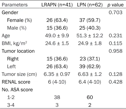
All operations were performed successfully without switch to open surgery. The detailed patients’ characteristics are show in Table 1. No significant differences were observed in these characteristics between two groups. One patient in LRAPN group and two patients in LPN group received intra-operative blood transfu-sion. Preoperative CT showed the mean tumor size was 6.35 ± 0.97 cm in LRAPN group and 6.63 ± 1.2 cm in LPN group (P=0.128).
Surgical outcomes
[image:3.612.90.304.96.282.2]Intra-operative variables and postoperative outcomes are shown in Table 2. In LRAPN group, the mean operative time (OT) was 100.7 ± 9.8 min, and only one patient required 5-min warm ischemia time (WIT) for renal artery clamping due to uncontrollable bleeding. The mean estimated blood loss (EBL) was 105.6 ±
Table 1. Baseline characteristics of patients in LRAPN group and LPN group
Parameters LRAPN (n=41) LPN (n=62) p value
Gender 0.703
Female (%) 26 (63.4) 37 (59.7) Male (%) 15 (36.6) 25 (40.3)
Age 49.0 ± 9.9 51.3 ± 12.2 0.231
BMI, kg/m2 24.6 ± 1.5 24.9 ± 1.8 0.115
Tumor location 0.958
Right 15 (36.6) 23 (37.1) Left 26 (63.4) 39 (62.9)
Tumor size (cm) 6.35 ± 0.97 6.63 ± 1.2 0.128
RENAL score 6 (4-10) 6.4 (4-10) 0.428
No. ASA score
1-2 38 60
3-4 3 2
51.3 ml, the mean retroperitoneal drainage lasted for 3.0 ± 0.6 days, and the mean pos- toperative hospital stay was 5.1 ± 1.0 days. In LPN group, the mean OT was 115 ± 19 min, the mean WIT was 22 ± 3.3 min, the mean EBL was 134 ± 86.2 ml, the mean retroperitoneal drainage lasted for 3.5 ± 0.8 days and the mean postoperative hospital stay was 5.6 ± 1.2 days. Significant differences were observed in the OT, WIT and EBL between both groups (P<0.05).
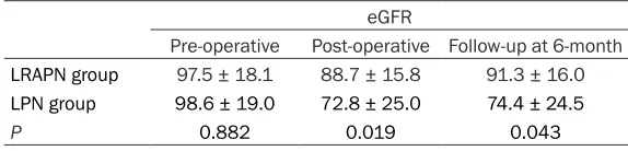
The mean preoperative eGFR was 97.5 ± 18.1 ml/min/1.73 m2 in LRAPN group and 98.6 ±
19.0 ml/min/1.73 m2 in LPN group, and no
sig-nificant difference was observed between them. However, the post-operative eGFR and eGFR at 6month follow-up were 88.7 ± 15.8 and 91.3 ± 16.0 ml/min/1.73 m2, respectively
in LRAPN group, which were significantly higher than in LPN group (72.8 ± 25.0 and 74.4 ± 24.5 ml/min/1.73 m2, P<0.05) (Table 3).
Complications
Intraoperative bleeding (Clavien II) was found in 1 patient and urine leakage (Clavien I) in 2 patients of LRAPN group; while two patients developed intraoperative bleeding (Clavien II), two had urine leakage (Clavien I) and one had transient hematuria (Clavien I) in LPN group. Significant differences were not observed in these complications between two groups (P>0.05). None had radiographic evidence of recurrent AML at a median follow-up duration of 43.6 months (range: 6-84 months) in both groups.
Discussion
Table 3. eGFR before and after surgery and at 6-month follow up in patients of LRAPN group and LPN group
eGFR
Pre-operative Post-operative Follow-up at 6-month
LRAPN group 97.5 ± 18.1 88.7 ± 15.8 91.3 ± 16.0
LPN group 98.6 ± 19.0 72.8 ± 25.0 74.4 ± 24.5
[image:4.612.88.375.302.371.2]P 0.882 0.019 0.043
Table 2. Perioperative characteristics of patients in LRAPN group and LPN group
Parameters LRAPN (n=41) LPN (n=62) p value
Operating time (min) 100.7 ± 9.8 115 ± 19.1 0.004
Warm ischemia time (min) 0 22 ± 3.3 0.000
Intraoperative Transfusions 1 (36.6) 2 (40.3)
Estimated blood loss (ml) 105.6 ± 51.3 134.0 ± 86.2 0.023
Drainage (days) 3.0 ± 0.6 3.5 ± 0.8 0.104
Hospitalization (days) 5.12 ± 1.0 5.6 ± 1.2 0.161
Intra-operative Complications 0.816
Hemorrhage 1 (2.4%) 2 (3.2%)
Postoperative Complications 0.992
Urine leakage 2 (4.9%) 2 (3.2%)
Hematuria 0 1 (1.6%)
PN for sporadic renal AML is able to preserve the kidney function and has acceptable compli-cation and low local recurrence rate [11, 12], and may improve kidney function when com-pared with RN in case of small renal AML [13]. In this study, we summarized our experience in the treatment of renal AML larger than 4 cm with LRAPN and LPN. To our knowledge, our study represents the largest series with the longest follow-up duration reported to date. The median size of AML at LRAPN was 6.35 cm (range: 4-10 cm), which was larger than the pre-viously reported [5, 8, 14]. During the follow-up period, none developed symptom recurrence or required dialysis.
The technique combining laparoscopic radio-frequency coagulation (RF) and PN (RF-LPN) for the treatment of renal tumor was firstly report-ed by Jacomides et al [15]. The advantages of this technique are that it allows not only tumor excision without ischemia time, but may elimi-nate the need for laparoscopic suturing and decrease the blood loss [7]. In our cohort, when LRAPN was performed for the treatment of renal AML, the findings were comparable to previously reported: the median OT, WIT and EBL in LRAPN group decreased significantly
as compared to conventional LPN group (P=0.004, 0.000 and 0.023, respectively). There was a significant decline in kidney function after RN as compared to PN [16]. The ischemia time of LPN within 30 min was generally accept-ed, but a recent study indicat-ed that every minute of isch-emia might affect the kidney function [17]. In our cohort, the mean WIT was 0 min in LRAPN group (one patient had 5-min WIT due to renal artery clamping for uncontrollable bleeding) which was much sig-nificantly shorter than in con-ventional LPN group (P= 0.000). Our previous study showed that laparoscopic ra-dio frequency ablation assist-ed tumor dissection was not only associated with a smaller decrease in GFR of the affect-ed kidney as shown by radionuclide scintigra-phy at 3 months and 12 months, but was able to better preserve kidney function as compared to LPN [18], which was also confirmed by the present study. During the follow-up period, none required permanent or transient dialysis. In the present series, 41 patients with renal AMLs were successfully treated with LRAPN. The incidence of procedure-specific complica-tions was 7.3% (3/41), only one received renal artery clamping due to uncontrollable bleeding, and two had transient urine leakage. The inci-dence of complications in LRAPN group was similar to that in LPN group. Although renal arterial embolization has been used more fre-quently than PN in the treatment of renal AML and it may reduce the risk of perioperative hemorrhage, it was not recommended to pa- tients in our series for its high recurrence rate and high incidence of complications unless the patients developed serious bleeding before surgery [19, 20].
and patients were not randomly assigned. Thus, further prospective studies are required to confirm our results and provide more evi-dence on the role and importance of LRAPN in the treatment of renal AMLs.
Conclusion
Compared with traditional LPN, LRAPN was demonstrated to have advantages in protect-ing renal function. Besides, there were also sig-nificant differences in several perioperative indicators, including operation time, warm isch-emia time, and safety. LRAPN offers favorable perioperative outcomes and survival rate, and is associated with low recurrence rate. Our results support LRAPN as an alternative, safe and feasible approach for the treatment of renal AMLs.
Acknowledgements
This study was supported by Grant Number 81472378 from National Natural Science Foundation of China and PYZY16-006 from the Incubating Program of Ren Ji Hospital, Shanghai, China.
Disclosure of conflict of interest
None.
Address correspondence to: Jin Zhang and Yong-Hui Chen, Department of Urology, Ren Ji Hospital, School of Medicine, Shanghai Jiao Tong University, 160 Pujian Road, Pudong District, Shanghai 200127, China. Tel: +86-021-68383776; E-mail: [email protected] (JZ); [email protected] (YHC)
References
[1] Fujii Y, Ajima J, Oka K, Tosaka A and Takehara Y. Benign renal tumors detected among healthy adults by abdominal ultrasonography. Eur Urol 1995; 27: 124-127.
[2] Lemaitre L, Claudon M, Dubrulle F and Maze-man E. Imaging of angiomyolipomas. Semin Ultrasound CT MR 1997; 18: 100-114. [3] Kim JK, Park SY, Shon JH and Cho KS.
Angio-myolipoma with minimal fat: differentiation from renal cell carcinoma at biphasic helical CT. Radiology 2004; 230: 677-684.
[4] Ewalt DH, Diamond N, Rees C, Sparagana SP, Delgado M, Batchelor L and Roach ES. Long-term outcome of transcatheter embolization of renal angiomyolipomas due to tuberous
sclerosis complex. J Urol 2005; 174: 1764-1766.
[5] Castle SM, Gorbatiy V, Ekwenna O, Young E and Leveillee RJ. Radiofrequency ablation (RFA) therapy for renal angiomyolipoma (AML): an alternative to angio-embolization and neph-ron-sparing surgery. BJU Int 2012; 109: 384-387.
[6] Oesterling JE, Fishman EK, Goldman SM and Marshall FF. The management of renal angio-myolipoma. J Urol 1986; 135: 1121-1124. [7] Zeltser IS, Moonat S, Park S, Anderson JK and
Cadeddu JA. Intermediate-term prospective re-sults of radiofrequency-assisted laparoscopic partial nephrectomy: a non-ischaemic coagula-tive technique. BJU Int 2008; 101: 36-38. [8] Prevoo W, van den Bosch MA and Horenblas S.
Radiofrequency ablation for treatment of spo-radic angiomyolipoma. Urology 2008; 72: 188-191.
[9] Mittal V, Aulakh BS and Daga G. Benign renal angiomyolipoma with inferior vena cava throm-bosis. Urology 2011; 77: 1503-1506.
[10] Park HK, Zhang S, Wong MK and Kim HL. Clini-cal presentation of epithelioid angiomyolipo-ma. Int J Urol 2007; 14: 21-25.
[11] Boorjian SA, Frank I, Inman B, Lohse CM, Chev-ille JC, Leibovich BC and Blute ML. The role of partial nephrectomy for the management of sporadic renal angiomyolipoma. Urology 2007; 70: 1064-1068.
[12] Msezane L, Chang A, Shikanov S, Deklaj T, Katz MH, Shalhav AL and Lifshitz DA. Laparo-scopic nephron-sparing surgery in the man-agement of angiomyolipoma: a single center experience. J Endourol 2010; 24: 583-587. [13] Lucas SM, Stern JM, Adibi M, Zeltser IS,
Ca-deddu JA and Raj GV. Renal function outcomes in patients treated for renal masses smaller than 4 cm by ablative and extirpative tech-niques. J Urol 2008; 179: 75-79; discussion 79-80.
[14] Sooriakumaran P, Gibbs P, Coughlin G, Attard V, Elmslie F, Kingswood C, Taylor J, Corbishley C, Patel U and Anderson C. Angiomyolipomata: challenges, solutions, and future prospects based on over 100 cases treated. BJU Int 2010; 105: 101-106.
[15] Jacomides L, Ogan K, Watumull L and Caded-du JA. Laparoscopic application of radio fre-quency energy enables in situ renal tumor ab-lation and partial nephrectomy. J Urol 2003; 169: 49-53; discussion 53.
[17] Thompson RH, Lane BR, Lohse CM, Leibovich BC, Fergany A, Frank I, Gill IS, Blute ML and Campbell SC. Every minute counts when the renal hilum is clamped during partial nephrec-tomy. Eur Urol 2010; 58: 340-345.
[18] Huang J, Zhang J, Wang Y, Kong W, Xue W, Liu D, Chen Y and Huang Y. Comparing zero isch-emia laparoscopic radio frequency ablation as-sisted tumor enucleation and laparoscopic partial nephrectomy for clinical T1a renal tu-mor: a randomized clinical trial. J Urol 2016; 195: 1677-1683.
[19] Kothary N, Soulen MC, Clark TW, Wein AJ, Sh-lansky-Goldberg RD, Crino PB and Stavropou-los SW. Renal angiomyolipoma: long-term re-sults after arterial embolization. J Vasc Interv Radiol 2005; 16: 45-50.