DNA-MEDIATED
HOLE AND ELECTRON TRANSPORT
Thesis by
Fangwei Shao
In Partial Fulfillment of the Requirements
for the Degree of
Doctor of Philosophy
California Institute of Technology
Pasadena, California
2008
(Defended June 26, 2007)
2008
ACKNOWLEGEMENTS
My five-year studies at Caltech have been one of the most important and
memorable periods in my life. I have always felt lucky and honored to become a member of this unique and world-class research institute. I have made acquaintance with so many intelligent and conscientious people who have inspired my enthusiasm for both science and life. They have been the role models in my professional development, as well as friends in my life. I owe an enormous debt of gratitude to each one of them.
First and foremost, I must thank my research advisor, Professor Jacqueline K. Barton. She has always been a great mentor to me. She is the person who led me into the fascinating world of DNA-mediated charge transport. During the first year in the lab, she had great patience to educate and transform me from a theoretical chemist to a handy bioinorganic experimentalist. I am amazed by her undying unyielding enthusiasm for chemistry and science in general. Her encouragement and brilliant ideas about research kept my spirits high as I struggled through the failures in experiments. I have always remembered the exciting spirit fulfilling me after every time that I discussed my projects with her. I admire her high standards for scientific researches and from her I learned to be a better critical thinker. She is truly an inspirational leader who brings together such a wonderful group of intelligent people who have contributed to my thesis work in various ways. Lastly, and perhaps mostly importantly, I thank you for the tremendous help you have provided for my scientific career. I eagerly look forward to our future interaction.
and for that I think her immensely. Tom Dunn is able to fix almost everything when I asked for help and revived many instruments over the years. I also thank Dian Buchness who was the first person I talked to in Chemistry department and always a source of a smile. With Lura Howe, two of them made the administrative tasks of my graduate studies so easy.
I must also acknowledge my thesis committee. I thank Professor Dennis
Dougherty for serving as chairman of my committee and for presiding over my candidacy and proposal defense. I also value highly the numerous discussions with my committee, including Professor Rudolph A. Marcus and Professor Harry Gray. Their insightful advice during my candidacy, fourth-year meetings and proposal defense are brilliant and inspire my thesis work a lot. I want to also thank my committee for taking time to read my proposal and research reports for all the exams.
Many Barton group members, both past and present, have provided me
tremendous help in various ways during my time at Caltech. Dr. Tashica Williams was the person coaching my experimental technique when I first joined the Barton lab. She was extremely nice and patient with me and tolerated my “first-experiment curse” unconditionally. I owe a big thank you to her. I thank Professor Chikara Dohno who kindly shared his wisdom on the cyclopropylguanine project. I would also like to thank Professor Melanie O’Neill and Dr. Katherine Augustyn with whom I had a great
Amazingly, they actually made me enjoy synthesizing transition metal complexes and cyclopropyl-modified bases. I must thank Dr. MiHee Lim for the intelligent discussion about both research and my future career, as well as being such a fantastic friend to me. She offered huge comforts to me, and made the tough graduation time and postdoc hunting endurable. I also thank her for introducing me to the authentic Korean food, and I enjoy that a lot. I am also looking forward to attending her wedding in Soul. Last but not the least, I want to thank rest of the Barton group for making the lab such a happy place to work.
I must present my appreciation to the Caltech Chinese community too. The Chinese students and scholars at Caltech constitute a caring family for me. Their considerations healed my homesickness and helped me go through the loneliness being away from my family. In particular, I want to thank Jie Yang, Yan Chen, and the cell group members, including Yanshun Liu, Rong Cai, Ke Wang and Jing Yang.
ABSTRACT
Since the elucidation of the double helical structure of DNA, it has been proposed that the dynamic -stacking base pair array may mediate charge migration, hole transport
(HT), and electron transport (ET). In this thesis work, both DNA-mediated HT and ET are investigated to explore their mechanisms by using kinetically fast electron/hole traps: cyclopropylamine-substituted bases, especially N4-cyclopropylcytosine (CPC), and N2 -cyclopropylguanine (CPG). Both biochemical reaction with a variety of photooxidants and electrochemistry show that the modified bases, CPC and CPG, have similar redox properties as the natural DNA bases and are irreversible kinetic traps by ring opening on the
picosecond time scale.
In DNA assemblies containing either [Rh(phi)2(bpy’)]3+ (Rh) or an anthraquinone derivative (AQ), two high energy photooxidants, appreciable oxidative damage at a distant CP
C is observed, which shows that hole migration must involve also the higher energy pyrimidine bases. The damage yield is modulated by lower energy guanine sites on the same or complementary strand. Significantly, the efficiency in trapping at CPC is similar to that at flanking CPG. Thus, HT is not simply a function of the relative energies of the isolated bases, but instead may require orbital mixing among the bases. Hole migration through DNA involves occupation of all the DNA bases with radical delocalization.
within the DNA assembly is seen to govern CPC oxidation, not simply the bases intervening between CPC and the tethered photooxidant.
These data are reconciled in a mechanistic model of conformationally gated hole transport through delocalized DNA domains. Oxidation of CPG separated from a tethered photooxidant by A-tracts with a series of lengths over 50 Å exhibits a nonmonotonically periodic distance dependence and shows that the domain sizes in the A-tract is 45 base
pairs. Sequence-dependent DNA structure and dynamics are essential to the transient formation of the domains and hole propagation among the domains. This dynamic, delocalized model provides a basis to reconcile and exploit DNA HT chemistry.
Jus as long-range hole transport through DNA has now been established, DNA-mediated electron transport has not been as well characterized. Three iridium complexes have therefore been designed in order to initiate both photooxidative and photoreductive reaction of DNA and allow direct comparison between the two. Redox potentials of excited Ir complexes are determined by both triplet energy (E0-0) and ground state redox potentials. Two of the iridium complexes prepared have excited state potentials that are suffcient to oxidize purines, but not pyrimidines. The excited state oxidation potentials of three Ir complexes are around 1.0 V and would be able to reduce DNA pyrimidines.
Both CPC and CPG in DNA can be decomposed by photoirradiation with the
noncovalently bound iridium complexes. In particular, two of the complexes have the potential to probe oxidation of purines and reduction of pyrimidines in DNA.
transport. In the Ir-tethered DNA assemblies, a metal complex stabilizes the DNA duplex through its intercalative, functionalized dppz ligand. Cyclopropylamine-substituted bases, CP
C and CPG, are used as kinetic fast electron and hole traps to probe the resulting charge migration processes after direct photoirradiation of the assemblies. Reductive
decomposition of CPC via ET as well as the oxidation of CPG via HT is observed. Thus, the iridium tethered DNA containing cyclopropylamine-substituted bases provides a unique model system to explore the two DNA-mediated charge transport processes through the same DNA bridges. For the first time, ET and HT can be initialized by the same photoredox probe employing the identical electronic interaction mode with DNA.
A flash quench technique was also applied to Iridium-tethered DNA in order to generate the ground state photoreductant and initiate photoreduction using 5’-bromo-uridine (BrU) as the electron trap. Efficiencies of BrU reduction in Ir-DNA upon flash quench technique was found to be comparable to that of CPG oxidation upon direct photoirradiation of Ir-DNA. Furthermore, in Ir-tethered DNA assemblies containing CPG or BrU as either the hole or electron trap, the sequence dependence of HT versus ET through an A-tract was examed. When CPG and BrU are placed in either purine or
pyrimidine strands in A-tract, decomposition of both modified bases are observed. Thus, transient electron occupancy during ET, as well as hole occupancy during HT, are distributed onto both purine and pymidine strands in A-tract. Additionally, BrU
Chapter 1
1.1. Introduction
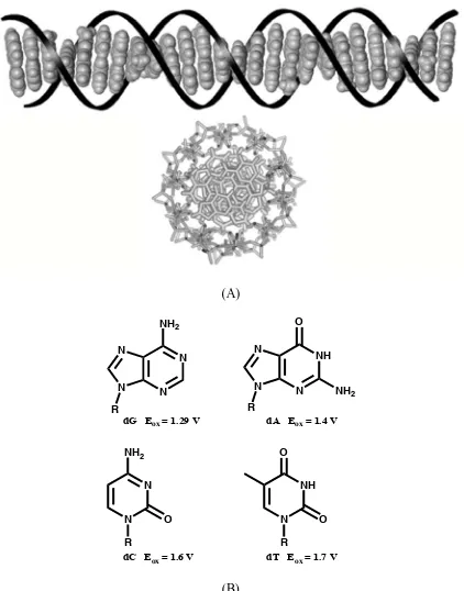
The first observation of right-handed double helical structure in B-form DNA was published more than 50 years ago.1 The salient structure of DNA is simply described as an array of heterocyclic aromatic base pairs, stacked at a distance of 3.4 Å and wrapped within a negatively charged sugar phosphate backbone as shown in Figure 1.1.2 About 10 years later, Eley and Spivey hypothesized the possibility that DNA can be a charge conduit based upon similarity of DNA structure to one-dimensional aromatic crystals.3 Generally speaking, DNA-mediated charge transport (CT) includes two types of charge migration processes.4 One is hole transport (HT), in which DNA is in an electron deficient status and the charge carrier is a radical cation.5 On the other hand, in electron transport (ET), DNA is reduced and an excess electron is propagated through DNA.6 The two processes are basically the same except for the involvement of the electronic orbitals of DNA oligonucleotides.
(A)
N N N
N
NH2
NH N N
N
O
NH2
N N NH2
O
NH N O
O
R R
R R
dG Eox = 1.29 V dA Eox = 1.4 V
dC Eox = 1.6 V dT Eox = 1.7 V
(B)
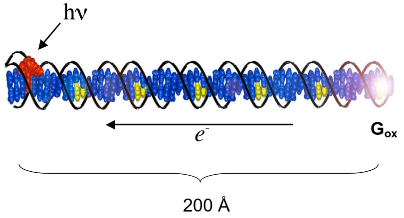
Figure 1.2. Schematic illustration of DNA-mediated hole transport through a 63-mer DNA duplex with covalently tethered Rh complex as photooxidant. Irradiation of Rh intercalator leads to oxidation of 5’-G in six guanine doublets over 200 Å. Figure is adapted from reference 10.
200 Å
h
-systems for exploiting DNA CT have been established by using a decade of biochemical and spectroscopic assays.10,13-17 Given the substantial observations on DNA CT, scientists now are interested in the biological relevant applications of this process and also the fundamental mechanisms governing the features of DNA CT.
1.2. Model systems and experimental assays for DNA-mediated CT
1.2.1. Photoredox probes serve as charge donors and acceptors in model
systems Charge transport through DNA is studied in vitro using external photoredox probes as photooxidants and photoreductants, as well as sometimes charge traps (Figure 1.2). These redox probes, which process well-characterized and varied redox,
photophysical and photochemical properties, include metallointercalators, organic intercalators and modified bases.
Several transition metal complexes are used as photooxidants in the study of DNA HT. In particular, phenanthrequinone diimine (phi) ligand of Rh(III) complexes (e.g., [Rh(phi)2(bpy’)]3+) and dipyridophenazine (dppz) ligand of Ru(II) complexes (e.g., [Rh(phen)(bpy’)(dppz)]2+) facilitate tight binding of the octahedral complexes by intercalating into DNA base pair stacking.18 The interaction modes of these complexes have been well characterized by spectroscopy and crystallography. High-resolution NMR shows the complexes are major groove binders and the ancillary, non-intercalating ligands of the complexes define the sequence specificity of the DNA binding.19 A 1.2 Å crystal structure of --[Rh(R,R,-dimethyltrien)(phi)]3+ with a DNA octamer
Metallointercalators Ru N N N N N N N N O H N 2+ H N O O R 9 Rh N HN HN
N H NH N O H N HN
O O
R
3+
9
[Rh(phi)2(bpy')]3+
[Ru(dppz)(phen)(bpy')]2+ Organic intercalators
N
H2N N
H O O N H NH R 3 9 Ethidium (Et) O O N H
OPO3OR O Anthraquinone (AQ) S N N N Cl
-Methylene Blue (MB)
O O OCH3 OH OH O O OH
H2N
O OH Daunomycin (DM) Modified bases N N N N NH2 R NH N N N O R NH N N N O N H R N N N N N R NH N N O NH2 R N R
2-Aminopurine (Ap) Inosine (I) N2-Cyclopropylguanine (CPG)
1-N6-Ethenoadenine (A)
7-Deazaguanine (zG)
Methylindole (M)
dppz complexes are well known as a “DNA light-switch” due to their intercalative contact mode with DNA.21 Protonation of the dppz imine in these complexes results a luminescence quench in aqueous solution. Intercalation within the DNA -stack protects
the phenazine nitrogens from water and restores the luminescence of the complex. Organic intercalators often have optical or electrochemical properties that can be used to exploit either DNA HT or DNA ET. As shown in the middle of Figure 1.3, Ethidium (Et), intercalating into DNA with the similar association constant as metallointercalators (K ~ 106 M-1),22 exhibits strong fluorescence in the presence and absence of DNA,23 which provides extensive application of Et to spectroscopic studies of DNA CT.24,25 The excited Et can only promote reaction with reagent of low oxidation potentials, such as 7-deazaguanine, which is due to insufficient potential of Et (E(Et*/0 ~ 1.2 V versus NHE) compared to natural bases.26 Another type of organic intercalators, such as methylene blue (MB) and daunomycin (DM) have reversible reduction at
negative potentials.27 Whether covalently cross-linked or non-covalently bound to DNA, these molecules can intercalate tightly to DNA and are used as electrochemical probes in the studies of ground-state DNA-mediated ET on an electrode film.28 In particular, DM is readily covalently attached to the guanine residues in double stranded DNA and the structure of DM-DNA conjugates are well characterized by crystallography.29
Nature generates modified DNA bases by methylation and oxidation of natural DNA bases.32 In the study of DNA CT, we take advantage of modified bases since they have minimal perturbation on structures and dynamics upon incorporation into DNA and meanwhile modulate the redox properties, chemical reactivity and photophysics of nucleic acid bases. For example, adenine analogues, 2-aminopurine (Ap) and 1-N6-ethenoadenine (A), are fluorescent bases that can serve as photooxidants (Ap: E*/- ~ 1.5 V; A: E*/- ~ 1.4 V versus NHE).33 The fluorescence quenching of the two bases in DNA
was applied to characterize the features of DNA HT. Guanine analogues, such as 7-deazaguanine (ZG) and methylindole (M), are two unnatural bases which have lower potentials than guanine and thus can serve as a thermal hole sink in model system for DNA HT.26,34 In addition, since methylindole generates a signature radical cation signal upon oxidation, M makes an ideal probe for investigation of CT by transient absorption spectroscopy.34
1.2.2. Spectroscopic, biochemical, and electrochemical assays for exploiting
DNA-mediated CT
1.2.2.1. Probe DNA-mediated CT by optical spectroscopy A variety of spectroscopic techniques are applied to investigate the optical property variations of either the charge donors and/or acceptors in model systems via DNA CT.34-36 Time-resolved techniques combined with steady-state spectroscopy allow us to probe DNA CT from femtosecond to millisecond time scales and measure both rate constants and
quenched. Hole injection to oxidize distant guanine provides an extra HT pathway to quench Ap*. Fractional fluorescence quenching (Fq) of Ap* via HT can be distinguished by comparing the fluorescence of redox-active duplex containing guanine to an identical reference duplex where G is substituted by inosine (I),33 a guanine analogue with higher oxidation potential (E+/0 ~ 1.5 V versus NHE). Thus, HT yield is deduced from the fluorescence quantum yield () as Fq =1 G/I. In DNA assemblies with Ap and G intervened by 0 to 9 AT base pairs, efficient hole injection over above 30 Å was observed.38
In addition to the initial steps of DNA HT, the whole process of HT including hole migration and trapping can be probed by fluorescence and transient absorption spectroscopy. In DNA containing methylindole, flash-quenched generated Ru(III) intercalator (vide infra) initialized HT to oxidize distant M. The resultant reaction intermediate, M radical cation, was detected by both transient absorption and EPR spectroscopy over long molecular distance (>20 Å).17
1.2.2.2. Biochemical observation of long-range oxidative damage. A
Figure 1.5. Photolysis and DNA-mediated CT initialized by irradiation of covalently tethered [Rh(phi)2(bpy’)]3+ and corresponding wavelengths. The complementary strand to Rh-tethered strand is 32P-radioactively labeled. Irradiation at 313 nm triggers direct strand cleavage and the intercalation site is revealed. DNA-mediated hole transport from
phi complexes makes them particularly useful probes in exploring DNA-mediated CT as shown in Figure 1.5. When radioactively labeled Rh-tethered DNA duplexes are excited by UV light ( = 313 nm), direct strand cleavage is promoted and indicates the
intercalation sites of the complex. Alternatively, visible light irradiation at 365 nm
generates a powerful excited state oxidant (E (Rh3+*/2+) ~ 2 V vs NHE)41 and initiates HT to oxidize guanine over a long distance. The HT yield as guanine damage can then be revealed by hot pipiridine treatment followed by gel electrophoresis. In a series of Rh-tethered DNA assemblies, proximal guanine doublets were positioned at fixed position to the photooxidant and distal guanine doublets were moved away up to 75 Å. Comparable oxidative damage yield via DNA HT were observed at both GG sites in all the
assemblies.10 A similar assay has been applied to Ru-tethered DNA assemblies, except that a flash quench technique is used because direct photoexcitation of Ru(II)
(E(Ru2+*/3+) ~ 0.6 V versus NHE) is not sufficient to oxidize DNA bases.42 Here, excited Ru(II) complex is quenched by electron transfer from a diffusion controlled oxidative quencher (e.g., Co(NH3)52+) and yields the ground state oxidant Ru(III) (E(Rh3+/2+) ~ 1.6 V vs NHE) in situ on the submicrosecond time scale (Figure 1.6).36 It has been
demonstrated that covalently bound Ru(III) complex generated by flash quench technique in situ can promote oxidation of guanine doublets via long-range HT over 37 Å.43
Figure 1.7. Electrochemistry assays for DNA ET. Upper: Schematic illustration of alkanethiol functionalized DNA duplexes fabricated on a gold surface for use in
characterized by scanning tunneling and scanning probe microscopy.45 A variety of redox-active species, such as MB and DM, intercalate in the base stack of individual DNA helices at a defined distance from the electrodes and act as electrochemical reporters for ET through the intervening DNA bridge.46-48 The reduction of the probes monitored by cyclic voltammetry or chronocoulometry should provide a measure of the efficiency of ET through DNA. The first observation of ET through a densely packed DNA film used covalently crosslinked daunomycin as a redox probe.44 Remarkably, efficient reduction of DM exhibited an distance independence on the position of DM up to 100 Å.48b Electron tunneling through the alkyl linker between the electrode and DNA monolayer is the rate-limiting step in ET through DNA films.47 The sensitivity of ET through DNA-modified films was further explored using electrocatalysis of noncovalent intercalator MB.46,49 In this study, the reduction signal of MB was significantly enhanced when coupled to the reduction of a freely diffusive ferricyanide as shown in figure 1.7. With electrocatalysis, all the single base mismatches, including the thermodynamically stable GT and GA mismatches, were detected by both cyclic voltammetry and
chronocoulometry.48a
1.3. Biology application of DNA-mediated CT.
Before starting to explore the answers for these questions, it is first necessary to demonstrate that CT over long distance can actually occur in DNA within the cell. Unlike typical in vitro studies with DNA is free in solution, in eukaryotic cells DNA is packaged and protected in nucleosome core particles (NCP).50 Would CT proceed through DNA with restricted motion and significant binding in NCP? Using a photoexcited Rh(III) intercalator, oxidative guanine damage within the NCP is observed up to 80 Å.51 Thus, oxidative damage via inherent DNA CT is not prevented in nucleosome although NCP may protect DNA from solution-mediated damage. Furthermore, the DNA-mediated CT within cell nuclei and mitochondrion is explored by using Rh(III) intercalator as
photooxidant.52,53 CT-charactoristic 5’-G specificity is observed at the oxidation damage
site in Hela genomic DNA.52 In original mitochrondrial DNA, base oxidations via CT is
revealed by a primer extension reaction. The oxidation sites in conserved sequence block
II overlap with known mitochondrial hot spots for cancers, which indicates the
correlation of CT with cancer-related genome damage.53
Since the evidence above shows that DNA-mediated CT occurs in cellular milieu,
redox reaction with a biological partner of DNA, such as protein, may be able to be
initiated by DNA CT. Many base-excision repair proteins, such MutY, possess
[4Fe-4S]2+ clusters that have no apparent catalytic or structural roles.54 DNA-mediated CT
leads to oxidation of a DNA-bound MutY.55 Oxidation of the [4Fe-4S]2+ cluster in MutY
was promoted by a DNA-tethered Ru(III) complex generated by the flash quench
technique and degradation of oxidized cluster was observed by EPR spectroscopy.56 In
have also detected that oxidation potential of the cluster was shifted when the repair
enzymes are bound to DNA.57 These exciting results of DNA-dependent redox activity
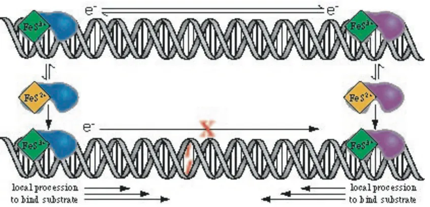
provoke the proposition that MutY, as well as other DNA repair enzymes, may employ
DNA-mediated CT to locate the lesion in DNA (Figure 1.8). When bound to DNA, the
repair enzymes become redox active and charge transfer through well-matched DNA to an
alternate bound repair protein can lead to the rapid redistribution of proteins onto genome
sites in the vicinity of DNA lesions. This redox-activation model provides a rationale to
explain the unbelievable coexistence of the low copy number and high performance of the
repair proteins.
1.4. Mechanistic considerations of CT through DNA.
DNA-mediated CT, Bixon and Jortner initially proposed a model which combines the two simple models above based upon only the energies of the isolated nucleic acid bases.59 In this model, charge sequentially hops between low energy guanine sites with tunneling through AT base pairs. However, none of the models above that are purely based upon energetic factors of isolated bases can account for the unique features of CT through DNA, especially for those results obtained by probing DNA CT on fast time scales (vide infra). This is presumably due to the ignorance of the signature
characteristics possessed by DNA -system.
1.4.2. Base -stack mediates CT. The analogy between base pair stack along
DNA helices and solid-state -systems provides the possibility of CT existence in DNA.
The interactions between the -electrons of base pairs generate the electron-coupling
requisite for CT to occur. One way to test that is to disturb the -stacking by mismatch
significant attenuation of damage over long distance occurs in presence of DNA-binding protein M.HhaI or TBP, which disrupt DNA base stacking by base flipping or
dramatically kinking the DNA.62 Furthermore, the suspicion that CT may undergo DNA sugar-phosphate backbone is eliminated by examining ET through self-assembled DNA films.63 Introduction of one or even two nicks on DNA backbones yields nondetectable effects on electron transfer, while single CA mismatch significantly diminishes ET efficiency. These results obtained by spectroscopic, biochemical and electrochemical assays, confirm that the pathway of CT through DNA is indeed the base pair stack, not via the sugar-phosphate backbone. The considerations of including the structural
dynamics of DNA base pair stacking, as well as the stacking of photoredox probes within the -stacking, into the mechanism of DNA CT, are invoked.
1.4.3. Stacking redox probes into DNA plays a role in CT. Given that DNA-mediated CT undergoes primarily through base pair stacking, it is not unexpected that the way photoredox probes interact with the DNA -system also could be crucial to the
charge migration. In fact we found it is true in many of the model systems for CT studies. The luminescence of Ru(II) intercalator was reported to be quenched by a Rh(III)
intercalator over a distance of 40 Å, while the quenching reaction was not observed with non-intercalating tethered Ru(II) and Rh(III) complexes.26 The unambiguous correlation between the strength of intercalative binding and yield of distant guanine damage was also observed when examining the oxidative damage induced by a series of Ru(II) complexes with similar redox potentials.64 Moreover, using both steady-state and
exhibited dramatically different rates and yields of CT.33 Ap, which can base pair with T and stack as a normal base in DNA helices, has a rapid CT rate (k ~ 10101011 s-1) with
shallow distance dependence. Conversely, CT involving A* is several orders of
magnitude slower and decreases steeply in both the rate constant and the yield over 13.6 Å. A high-resolution NMR structure of A-containing duplex shows a non-rigid
conformation of bulky A that prevents it from effective stacking with the flanking DNA
bases, which makes resulting CT efficiency of A* significantly differentiate from that of
Ap*. Thus, although CT is an intrinsic feature of DNA itself, it has to been also noted that the difference among the redox probes in the model systems of CT cannot be neglected. Distinctions in the interaction modes with DNA -stacking adopted by photoredox probes exert significant impacts on the rate and yield of CT; the diverse features of redox probes makes it difficult to reconcile the fundamental mechanism of CT.
Et* in DNA assemblies containing ZG showed two characteristic decay rate constants, 5 ps and 75 ps. The short component was assigned as the rate of direct CT between Et* and Z
G and, while the longer decay (75 ps) was proposed to be reorientational motion of Et* to a CT-favored conformation within its binding site. None of rate constants varied according to the distance between Et and ZG over 17 Å, while the yield of CT (as ZG damage) exhibited a shallow distance dependence. It was hypothesized that Et undergoes a conformation rearrangement within DNA -stacking (75 ps) when the favorable
conformation required for CT on 5 ps was not accessible. Although gating by the
dynamic motion of photooxidant within DNA base stack explains the constant CT rate, it cannot reconcile the shallow distance dependence of the CT yield. Dynamic motion of bases on the intervening DNA bridge should be also included. Femtosecond spectroscopy of Ap-containing 35-mer duplexes, in which Ap and the electron donor G are either flanked (ApG) or separated by one adenine (ApAG) exhibits multiple components of Ap* decay.66 Fast decay rate represents the CT rate (kCT)between Ap* and G. The
temperature dependence of kCT directly characterizes the modulation of DNA CT from dynamic motion of base pairs. Both kCT of ApG and ApAG increased weakly with temperature, resulting in less variation in kCT at higher temperature. The complete loss of kCT after DNA assemblies were denatured, confirmed again that the CT is an inherent
Figure 1.10. Schematic illustration of how fluctuations in the -stacking of DNA affects DNA charge transport. CT is rapid in a perfectly stacked DNA (top), is gated by
conformational alignment in an imperfectly stacked DNA (middle), and does not occur at all in a poorly stacked DNA (bottom), where CT-active conformation cannot be accessed within the lifetime of of the excited probe molecule. Figure is adopted from reference 66.
*
k
em
*
k
CT
We attentively propose that DNA CT needs to undergo a CT-active conformation: specific, well-coupled arrangements of DNA base achieved by dynamic motion of DNA bases with stacking. The yield of CT reflects the probability of DNA assemblies
1.4.5. Trapping rates of electron acceptors versus back electron transfer.
Except for several observations of intermediate reaction radicals by transient absorption spectroscopy,17,68 CT yield reported by the redox reaction over long molecular distances is based on the oxidative damage at a thermal hole trap, guanine or its analogues.13,26 However, the guanine radical has a relatively long lifetime (millisecond time scale)36 compared to the gating process of CT: the dynamic motion of DNA bases. Before being trapped as permanent guanine damage, charge can undergo many pathways, which inevitably convolute the concomitant CT process. In particular, fast back electron transfer (BET) within the initially generated radical ion/ion pair can severely diminish the amount of charge that remains. For instance, no detectable permanent guanine damage was observed when Ap or thionin (Th), a DNA intercalator, were used as covalently
incorporated photooxidants to DNA.69 Although the charge injection for the two oxidants occurs on picosecond time scale or even faster,35,70 BET, with a significantly faster rate than the trapping of guanine radial cation by water and/or oxygen, depletes the hole density in duplex DNA. A complementary experiment by using a kinetic fast hole trap, N2-cyclopropylguanine (CPG), supportes this rationale. The cyclopropyl ring on the exocyclic amine of guanine can undergo a rapid ring-opening reaction on the picosecond time scale upon one-electron oxidation.71 Indeed, oxidative decomposition of CPG was observed by photolysis of both Ap and Th containing DNA duplexes.69a,72 Thus, accurate evaluation of CT yield also requires the trapping rate of charge acceptor to match the time scale of DNA CT gated by the dynamic motion of DNA bases.
Figure 1.11. Illustration of conformationally gated domain model for DNA-mediated CT. A radical cation, hole, is injected into a domain over 4-5 base pairs and delocalizes
redox probes, as well as the trapping of electron acceptors, we proposed a mechanistic model for DNA-mediated hole transport: conformationally gated domain hopping model (Figure 1.11). A domain is a transiently extended -orbital over 45 base pairs (Chapter
1.5. References
1. Watson, J. D., Crick, F. C. H. A structure for deoxyribonucleic acid. Nature1953, 171, 737-738.
2. Sanger, W. Principles of Nucleic Acid Structure Cator, C. R., ed.; Springer-Verlag: New York. 1984.
3. Eley, D. D., Spivey, D. I. Semiconductivity of organic substances: Nucleic acids in the dry state. Trans. Faraday Soc.1962, 58, 411.
4. (a) Delaney, S., Barton, J. K. Long-range DNA charge transport J. Org. Chem.2003, 68, 6475-6483; (b) Rokita, S. E., Ito, T. Chemical probing of reductive electron transfer in DNA Charge Transfer in DNA: From Mechanism to Application, Wagenknecht, H. A., ed.; Wiley, 2005, 133-152.
5. O’Neill, M. A., Barton, J. K. Sequence-dependent DNA Dynamics: The regulator of DNA-mediated charge transport In Charge Transfer in DNA: From Mechanism to Application, Wagenknecht, H. A., ed.; Wiley, 2005, 27-75.
6. Carell, T., Meltzer, M. Excess electron transfer in DNA probed with flavin- and thymine dimer-modified oligonucleotides Charge Transfer in DNA: From Mechanism to Application, Wagenknecht, H. A., ed.; Wiley, 2005, 77-92.
7. (a) Topics in Current Chemistry: Long-Range Electron Transfer in DNA:1, Schuster, G. B., ed.; Springer-Verlag: New York, 2004, 236; (b) Schuster, G. B. Long-range charge transfer in DNA: transient structural distortions control the distance
dependence. Acc. Chem. Res.2000, 33, 253-260; (c) Giese, B. Long-distance electron transfer through DNA. Ann. Rev. Biochem.2002, 71, 51-70; (d) Lewis, F. D.,
Letsinger, R. L., Wasielewski, M. R. Dynamics of photoinduced charge transfer and hole transport in synthetic DANN hairpins. Acc. Chem. Res.2001, 34, 159-170. 8. Oxidation potentials of the four isolated bases are estimated as follows: G: Eox = 1.3
eV; A: Eox = 1.4 eV; C: Eox > 1.6 eV; T: Eox > 1.7 eV. See Steenken, S., Jovanovic, S.V. How easily oxidizable is DNA? One-electron reduction potentials of adenosine and guanosine radicals in aqueous solution J. Am. Chem. Soc. 1997, 119, 617-618. 9. Steenken, S., Telo, J. P., Novais, H. M., Candeias, L. P. One-electron-reduction
11.Dandliker, P. J., Holmlin, R. E., Barton, J. K. Oxidative thymine dimer repair in the DNA helix Science, 1997, 275, 1465-1468.
12.DeRosa, M. C., Sancar, A., Barton, J. K. Electrochemically monitoring DNA repair by photolyase Proc. Nat. Acad. Sci. USA2005, 102, 10788-10792.
13.Hall, D. B., Holmlin, R. E., Barton, J. K. Oxidative DNA damage through long range electron transfer Nature1996, 382, 731-735.
14.Takada, T., Kawai, K., Cai, X., Sugimoto, A., Fujitsuka, M., Majima, T. Direct observation of hole transfer through double-helical DNA over 100 Å J. Am. Chem. Soc. 2004, 126, 1125-1129.
15.Nakatani, K., Dohno, C., Saito, I. Modulation of DNA-mediated hole-transport efficiency by changing superexchange electronic interaction J. Am. Chem. Soc.2000, 122, 5893-5894.
16.Yoo, J., Delaney, S., Stemp, E., Barton, J. K. Rapid radical formation by DNA charge transport through sequences lacking intervening guanines J. Am. Chem. Soc.2003, 125, 6640-6641.
17.Delaney, S., Yoo, J., Stemp, E. D. A., Barton, J. K. Charge equilibration between two distinct sites in double helical DNA Proc. Nat. Acad. Sci. USA2004, 101, 10511-10516.
18.Núñez, M. E., Barton, J. K. Probing DNA charge transport with metallointercalators Curr. Opin. Chem. Biol. 2000, 4, 199-206.
19.(a) David, S. S., Barton, J. K. NMR evidence for specific intercalation of -[Rh(phen)2phi]3+ in [d(GTCGAC)]2 J. Am. Chem. Soc. 1993, 115, 2984-2985; (b) Hudson, B. P., Barton, J. K. Solution structure of a metallointercalator bound site-specifically to DNA J. Am. Chem. Soc.1998, 120, 6877-6888.
20.Kielkopf, C. L., Erkkila, K. E., Hudson, B. P., Barton, J. K., Rees, D. C. Structure of a photoactive rhodium complex intercalated into DNA Nature Struc. Biol. 2000, 7, 117-121.
21.Jenkins, Y., Friedman, A. E., Turro, N. J., Barton, J. K. Characterization of
dipyridophenazine complexes of ruthenium (II): the light switch effect as a function of nucleic acid sequence and conformation Biochemistry, 1992, 31, 10809-10816. 22.Waring, M. J. Complex formation between ethidium bromide and nucleic acids J. Mol.
Biol. 1965, 13, 269-282.
24.Wan, C. Z., Fiebig, T., Kelley, S. O., Treadway, C. R., Barton, J. K., Zewail, A. H. Femtosecond dynamics of DNA-mediated electron transfer. Proc. Natl. Acad. Sci. USA1999, 96, 6014-6019.
25.Fiebig, T., Wan, C. Z., Kelley, S. O., Barton, J. K. Femtosceond dynamics of the DNA intercalator, eithidium and electron transfer with mononucleotides in water Proc. Natl. Acad. Sci. USA1999, 96, 1187-1192.
26.Kelley, S. O., Barton, J. K. DNA-mediated electron transfer from a modified base to ethidium: -Stacking as a modulator of reactivity Chem. Biol.1998, 5, 413-425.
27.Kelley, S. O., Barton, J. K., Jackson, N., Hill, M. G. Electrochemistry of methylene blue bound to a DNA-modified electrode Bioconjug. Chem. 1997, 8, 31-37.
28.(a) Norden, B, Tjerneld, F. Structure of methylene-blue DNA complexes studied by linear and circular-dichroism spectroscopy Biopolymers1982, 21, 1713-1734. (b) Tuite, E, Kelly, J. M. The interaction of methylene-blue with polynucleotides and DNA – a spectroscopic study J. Am. Chem. Soc.1994, 116, 7548-7556.
29.(a) Wang, A. H. J., Gao, Y. G., Liaw, Y. C., Li, Y. K. Formaldehyde cross-links daunorubicin and DNA efficiently – HPLC and X-ray-diffraction studies
Biochemistry1991, 30, 3812-3815. (b) Leng, F. F., Savkur, R., Fokt, I., Przewloka, T., Priebe, W., Chaires, J. B. Base specific and regioselective chemical cross-linking of daunorubicin to DNA J. Am. Chem. Soc.1996, 118, 4731-4738.
30.Schuster, G. B. Long-range charge transfer in DNA: Transient structural distortions control the distance dependence Acc. Chem. Res.2000, 33, 253-260.
31.Behrens, C. Burgdorf, L. T., Schwögler, A., Carell, T. Weak distance dependence of excess electron transfer in DNA Angew. Chem. Int. Ed. 2002, 41, 1763-1766.
32.Bestor, T. H. The DNA methyltransferases of mammals Human Molecular Genetics
2000, 9, 2395-2402.
33.Kelley, S.O., Barton, J. K. Electron transfer between bases in double helical DNA Science1999, 283, 375-381.
34.Delaney, J. Y., Stemp, E. D. A., Barton, J. K. Charge equilibration between two distinct sites in double helical DNA Proc. Nat. Acad. Sci. USA 2004, 101, 10511-10516.
35.Wan, C., Fiebig, T., Schiemann, O., Barton, J. K., Zewail, A. H. Femtosecond direct obaservation of charge transfer between bases in DNA Proc. Nat. Acad. Sci. USA
36.Stemp, E. D. A., Arkin, M. R., Barton, J. K. Oxidation of guanine in DNA by Ru(phen)2dppz2+ using the flash-quench technique J. Am. Chem. Soc.1997, 119, 2921-2925.
37.O’Neill, M. A., Barton, J. K. 2-Aminopurine: A probe of structural dynamics and charge transfer in DNA and DNA:RNA hybrids J. Am. Chem. Soc. 2002, 124, 13053-13066.
38.O’Neill, M.A., Barton, J. K. DNA charge transport: conformationally gated hopping through stacked domains J. Am. Chem. Soc.2004, 126, 11471-11483.
39.Steenken, S., Jovanovic, S. V. How easily oxidizable is DNA? One-electron
reduction potentials of adenosine and guanosine radicals in aqueous solution J. Am. Chem. Soc. 1997, 119, 617-618.
40.Sugiyama, H., Saito, I. Theoretical studies of GG-specific photocleavage of DNA via electron transfer: significant lowering of ionization potential and 5'-localization of HOMO of stacked GG bases in B-Form DNA J. Am. Chem. Soc.1996, 118, 7063-7068.
41.Turro, C., Evenzahav, A., Bossmann, S. H., Barton, J. K. Turro, N. J. Excited state properties of Rh(phi)2(phen)3+ and related complexes: A strong photooxidant Inorg. Chim. Acta.1996, 243, 101-108.
42.Murphy, C. J., Arkin, M. R., Ghatlia, N. D., Bossmann, S., Turro, N. J., Barton, J. K. Fast photoinduced electron transfer through DNA intercalation Proc. Nat. Acad. Sci. USA1994, 91, 5315-5319.
43.Arkin, M. R., Stemp, E. D. A., Pulver, S. C., Barton, J. K. Long-range oxidation of guanine by Ru(III) in duplex DNA Chem. Biol. 1997, 4, 389-400.
44.Kelley, S. O., Jackson, N. M., Hill, M. G., Barton, J. K. Long-range electron transfer through DNA film Angew. Chem. Int. Ed. 1999, 38, 941-945.
45.(a) Ceres, D. M., Barton, J. K. In situ scanning tunneling microscopy of DNA-modified gold surfaces: bias and mismatch dependence J. Am. Chem. Soc.2003, 125, 14964-14965. (b) Sam, M., Boon, E. M., Barton, J. K., Hill, M. G., Spain, E. M. Morphology of 15-mer duplexes tethered to Au(111) using scanning probe microscopy Langmuir, 2001, 17, 5727-5730.
46.Boon, E. M., Ceres, D. M., Drummond, T. G., Hill, M. G., Barton, J. K. Mutation detection by electrocatalysis at DNA-modified electrodes Nature Biotechnology2000,
47.Drummond, T. G., Hill, M. G., Barton, J. K. Electron transfer rates in DNA films as a function of tether length J. Am. Chem. Soc. 2004, 126, 15010-15011.
48.(a) Boon, E.M., Jackson, N. M., Wightman, M. D., Kelley, S. O., Hill, M. G., Barton, J. K. Intercalative stacking: A critical feature of DNA charge-transport
electrochemistry J. Phys. Chem. B2003, 107, 11805-11812. (b) Liu, T. thesis for Ph.D. Electrochemical studies of electron transfer in DNA films with covalently tethered intercalators. 2007, Caltech.
49.Kelley, S. O., Boon, E. M., Barton, J. K., Jackson, N. M., Hill, M. G. Single-base mismatch detection based on charge transduction through DNA Phys. Rev. E 1997, 55, 7390-7395.
50.Luger, K., Mäder, A., Richmond, R., Sargent, D., Richmond, T. J. Crystal structure of the nucleosome core particle at 2.8 Å resolution Nature 1997, 389, 251-260.
51.Núñez, M. E., Noyes, K.T., Barton, J. K. Oxidative charge transport through DNA in nucleosome particles J. K. Chem. Biol. 2002, 9, 403-415.
52.Núñez, M. E, Holmquist, G. P., Barton, J. K. Evidence for DNA charge transport in the nucleus Biochemstry2001, 40, 12465-12471.
53.Merino, E. J., Barton, J. K. Oxidation by DNA charge transport damages conserved sequence block II, a regulatory element in mitochondrial DNA Biochemstry2007, 46, 2805-2811.
54.(a) Cunningham, R. P., Asahara, H., Bank, J. F., Scholes, C. P.,Salerno, J. C., Surerus, K., Munck, E., McCracken, J., Peisach, J., and Emptage, M. H.
Endonuclease III is an iron-sulfur protein, Biochemistry1989, 28, 4450-4455. (b) Hinks, J. A, Evans, M. C. W., de Miguel, Y., Sartori, A. A., Jiricny, J., and Pearl, L. H. (2002) An iron-sulfur cluster in the family 4 uracil-DNA glycosylases, J. Biol. Chem. 2002, 277, 16936-16940. (c) Rebeil, R., Sun, Y., Chooback, L., Pedraza-Reyes, M., Kinsland, C., Begley, T. P., and Nicholson, W. L. Spore
photoproductlyase from Bacillus subtilis spores is a novel iron-sulfur DNA repair enzyme which shares features with proteins such as class III anaerobic ribonucleotide reductases and pyruvate-formatelyases J. Bacteriol. 1998, 180, 4879-4885. (d) Lee, C. H., Kim, S. H., Choi, J. I., Choi, J. Y., Lee, C. E., and Kim, J. Electron
paramagnetic resonance study reveals a putative iron-sulfur cluster in human rpS3 protein, Mol. Cells 2002, 13, 154-156.
55.Boal, A. K., Yavin, E., Lukianova, O. A., O’Shea, V. L., David, S. S., Barton, J. K. DNA-bound redox activity of DNA-repair glycosylases containing [4Fe-4S] clusters Biochemistry2005, 44, 8397-8407.
DNA repair protein by guanine radical Proc. Nat. Acad. Sci. USA2005, 102, 3546-3551.
57.Gorodesky, A. A., Barton, J. K. DNA-mediated electrochemistry of disulfides on graphite J. Am. Chem. Soc.2007, 129, 6074-6075.
58.Marcus, R. A., Sutin, N. Electron transfer in chemistry and biology Biochim. Biophys. Acta. 1985, 811, 265-
59.(a) Bixon, M., Jortner, J. Long-range and very long-range charge transport in DNA Chem. Phys.2002, 281, 393-408. (b) Jortner, J., Bixon, M., Voityuk, A. A., Rösch, N. Superexchange mediated charge hopping in DNA J. Phys. Chem. A.2002, 106, 7599-7606.
60.Kelley, S. O., Holmlin, R. E., Stemp, E. D. A., Barton, J. K. Photoinduced electron transfer in eithidium modified DNA duplexes: Dependence on distance and base stacking J. Am. Chem. Soc.1997, 119, 9861-9870.
61.Yoo, J., Delaney, S., Stemp, E. D. A., Barton, J. K. Rapid radical formation by DNA charge transport through sequences lacking intervening guanines J. Am. Chem. Soc.
2003, 125, 6640-6641.
62.(a) Rajski, S. R., Kumar, S., Roberts, R. J., Barton J. K. Protein-modulated DNA electron transfer J. Am. Chem. Soc.1999, 121, 5615-5616. (b) Rajski, S. R., Barton, J. K. How different DNA-binding proteins affect long-range oxidative damage to DNA Biochemistry2001, 40, 5556-5564.
63.Liu, T., Barton, J. K. DNA electrochemistry through the base pairs not the sugar-phosphate backbone J. Am. Chem. Soc.2005, 127, 10160-10161.
64.Delaney, S., Pascaly, M., Bhattacharya, P. K., Ham, K., Barton, J. K. Oxidative damage by ruthenium complexes containing the dipyridophenazine ligand or its derivatives: A focus on intercalation Inorg. Chem.2002, 41, 1966-1974.
65.(a) Mordlund, T. M., Andersson, S., Nilsson, L., Rigler, R., Graeslund, A.,
Mclaughlin, L. W. Structure and dynamics of a fluorescent DNA oligomer containing the EcoRI recognition sequence: Fluorescnece molecular dynamics, and NMR studies Biochemistry1989, 28, 9095-9013. (b) Giudice, E., Lavery, R. Simulations of nucleic acids and their complexes Acc. Chem. Res.2002, 35, 350-357.
66.O’Neill, M. A., Becker, H., Wan, C., Barton, J. K., Zewail, A. H. Ultrafast dynamics in DNA-mediated electron transfer: base gating and the role of temperature Angew. Chem. Int. Ed.2003, 42, 5896-5900.
68.Takada, T., Kawai, K., Fujistuka, M., Majima, T. Rapid long-distance hole transfer through consecutive adenine sequence. J. Am. Chem. Soc.2006, 128, 11012-11013.
69.(a) O’Neill, M. A., Dohno, C., Barton, J. K. Direct chemical evidence for charge transfer between photoexcited 2-aminopurine and guanine in duplex DNA J. Am. Chem. Soc.2004, 126, 1316-1317. (b) Dohno, C., Stemp, E. D. A., Barton, J. K. Fast back electron transfer prevents guanine damage by photoexcited thionine bound to DNA J. Am. Chem. Soc.2003, 125, 9586-9587.
70.Reid, G. D., Whittaker, D. J., Day, M. A., Turton, D. A., Kayser, V., Kelly, J. M., Beddard, G. S. Femtosecond electron-transfer reactions in mono- and polynucleotides and in DNA J. Am. Chem. Soc.2002, 124, 5518-5527.
71.Nakatani, K., Dohno, C., Saito, I. Design of a hole-trapping nucleobase: termination of DNA-mediated hole transport at N2-cylcopropyldeoxyguanosine J. Am. Chem. Soc.
2001, 123, 9681-9682.
Chapter 2
Long Range Oxidative Damage to Cytosines in Duplex DNA
Adapted from: Shao, F., O’Neill, M. A., and Barton, J. K. Proc. Nat. Acad. Sci. USA
2.1. Introduction.
Oxidative damage to DNA from a distance through long range migration of charge has now been established in many DNA assemblies using different pendant photooxidants through both biochemical and spectroscopic assays.1-8 The DNA base pair stack can mediate hole transport (HT) over at least 200 Å,2,3 and the reaction is
exquisitely sensitive to the dynamic structure and stacking within the DNA duplex.9,10 This sensitivity to perturbations in base pair stacking has been advantageous in the development of DNA-based sensors for mutational analysis,11 and may provide a role for DNA-mediated HT within the cell,11,12 but it has limited the application of physical techniques to explore HT mechanistically.
While not a robust molecular wire, the DNA duplex has in some experiments been characterized as a wide band gap semiconductor.13,14 More prevalent have been models of incoherent HT involving a mixture of localized charge hopping among low energy sites, guanines and sometimes adenines, and tunneling through higher energy pyrimidine bases.15-18 These mechanisms do not provide a rationale, however, for the sensitivity of HT to DNA structure. We have observed that DNA HT is gated by the dynamical motions of the DNA bases9,19 and have described DNA HTas confor-mationally gated hopping through transient, well-stacked DNA domains.20
establishing HT for those photooxidants that produce limited oxidative DNA damage owing to rapid back CT.24,25 Guanine, although a low energy site, provides a relatively slow (ms)26 hole trap through irreversible reaction with water and/or oxygen.27
Significantly, in experiments using CPA, HTinvolving adenines has been documented.23 Here, using the kinetically fast electron hole trap,N4-cyclopropylcytosine (CPC), we demonstrate that hole migration involves not only low energy purine sites but also the higher energy pyrimidine bases. We examine long range HT in DNA assemblies
containing two different high energy photooxidants, and we find appreciable oxidative damage at a distant CPC. The yield of CPC decomposition is modulated by lower energy guanine sites within the assembly and is comparable in magnitude to that for a
neighboring CPG. Thus, during the course of HT through DNA, appreciable hole density must reside also on high energy pyrimidine sites. HTthrough DNA involves all the DNA bases.
2.2. Materials and methods.
2.2.1. Modified bases. N4-Cyclopropylcytidine (r CPC) and N4-cyclopropyl-cytosine (CPC) were synthesized by stirring 4-thiouridine or 4-thiouracil, respectively in
1:2 cyclopropylamine:ethanol solution at 55 oC for 2 days. Following purification by
reverse phase HPLC, the product was confirmed by 1H NMR and ESI-TOF mass spectrometry. Cyclic voltammetry was carried out in aqueous solution (pH 7) using a glassy carbon electrode with Ag/AgCl as reference.
N N N O H R
- e- N
N N O H R N N N O H R N N N O H R OH O + N N NH2 O R
H2O
or O2
CPC HPC
5'-ACGATT YACCGAGTGAT-3' 3'-TGATAACPCTGGCTCACTA-5'
OX
hv
CT
5'-ACGATT Y ACCGAGTGAT-3' 3'-TGATAACPC
oxTGGCTCACTA-5' OX Digest HPLC 0 50 100 150
10 15 20 25
UV Intensity (a.u.)
Retention Time (min)
dC dI
dG dT
dA
dCPC
Rh N NH H N N H HN N O H N 3+ H N O HO 9 OX =
Rh(phi)2bpy'
O O N H OH O AQ
Figure 2.1. Experimental assay to test for hole occupation of pyrimidines during HT through duplex DNA. (top) Oxidation of CPC leads to rapid decomposition through ring scission and subsequent generation of two characteristic products. (bottom) Two potent photooxidants, [Rh(phi)2(bpy’)]3+, Rh, and an anthraquinone derivative, AQ, positioned remote from CPC (covalently appended to the DNA with a C9 linker) initiate hole
reverse-phase column; for trityl on 5% ~ 35% and for trityl off 5% ~ 20% MeCN in 50 mM NH4OAc over 30 min), and analyzed by mass ESI-TOF spectrometry. CPC- or CP G-containing strands were obtained by incubating oligonucleotides possessing 4-thiouridine,
or 2-fluoroinosine, respectively, in 6M aqueous cyclopropylamine at 60 oC for 16 hours
prior to HPLC purification. The photooxidants, Rh(phi)2(bpy’)3+ (phi =
9,10-phenanthrenequinone diimine; bpy’ = 4-methyl-4’(butyric acid)-2,2’-bipyridine) (Rh) and an anthraquinone derivative (anthraquinone-2-carboxylic acid (2-hydroxyethyl) amide) (AQ) were synthesized and tethered to the complementary strands as previously
described;25 Rh-tethered assemblies are purified by RP-HPLC (Rainin Dynamax 300 Å C4 column, same solvents as DNA purification, 5% ~ 15% over first 20 min followed by 15% ~ 30% over 30 min). AQ-tethered DNA is purified as regular oligonucleotides. Rh-tethered assemblies, only the -isomers (determined from CD spectroscopy) were used.
DNA oligonucleotides were quantitated by UV-visible spectroscopy. Duplex solutions (5 μM in 10mM sodium phosphate, 50 mM NaCl, pH 7) were prepared by
combining equimolar amounts of the desired DNA complements and annealing with
regulated cooling from 90 oC to ambient temperature over a period of 2 hours. Under our
experimental conditions the duplexes melt between 53 and 60 oC (2 μM duplexes in 10 mM sodium phosphate, 50 mM NaCl, pH 7), depending upon the DNA sequence. The
presence of CPC depresses the duplex melting temperature by 1 oC.
photooxidant. Interduplex controls were conducted by irradiating a mixture of duplexes containing both photooxidant without CPC and CPC without photooxidant. Experiments reported here were carried out aerobically, however similar results were obtained in the absence of oxygen. After irradiation (0-40 minutes), duplex samples were digested by 37
oC incubation with phosphodiesterase, S1 endonuclease and alkaline phosphatase for 4
hours in order to yield the free nucleosides, and the samples were analyzed by reverse phase HPLC (Chemcobond 5-ODS-H, 4.6 * 100 mm). Oxidation of r CPC (250 μM) by
Rh or AQ (25 μM) and subsequent HPLC analyses were carried out under the same conditions described for the DNA samples. The oxidation products of rCPC nucleoside were characterized by ESI-TOF MS.
2.3. Results.
2.3.1. The pyrimidine hole trap and its oxidation. We have prepared CPC by reaction of cyclopropylamine with 4-thiouracil. Just as the oxidation potential of CPG does not differ significantly from that of G,22,28 the oxidation potential of CPC is expected to be very similar to unmodified C. Cyclic voltammetry reveals no oxidation of CPC at potentials of <1.6 V versus NHE in aqueous solution (pH 7), while oxidation of G was observed at ~1.3 V versus NHE. Thus the cyclopropyl moiety does not significantly reduce the oxidation potential of C. For the isolated nucleosides, then, CPC, like C, is significantly harder to oxidize than G.
Table 2.1. Assemblies to test long range oxidative damage to CPC in duplex DNA
Duplexa Sequenceb % Decomposition of CP
Cc Rh-G-1
Cp-1
Rh - 5’-ACGATT GACCGAGTCAT-3’ 3’-TGCTAACP
CTGGCTCAGTA-5’ 36 Rh-I-1
Cp-1
Rh - 5’-ACGATT IACCGAGTCAT-3’
3’-TGCTAACPCTGGCTCAGTA-5’ 92
Rh-G-2 Cp-2
Rh – 5’-ACGACC GATTGAGTCAT-3’ 3’-TGCTGGCP
CTAACTCAGTA-5’ 5
Rh-I-2 Cp-2
Rh – 5’-ACGACC IATTGAGTCAT-3’ 3’-TGCTGGCP
CTAACTCAGTA-5’ 23 AQ-G-1
Cp-1
AQ - 5’-ACGATT GACCGAGTCAT-3’ 3’-TGCTAACP
CTGGCTCAGTA-5’ 10 d
AQ-I-1 Cp-1
AQ - 5’-ACGATT IACCGAGTCAT-3’ 3’-TGCTAACP
CTGGCTCAGTA-5’ 36 d
AQ-A-1 Cp-1
AQ - 5’-ACGATT AACCGAGTCAT-3’ 3’-TGCTAACP
CTGGCTCAGTA-5’ 15 d
AQ-I-3 Cp-3
AQ - 5’-ACIATT ITTACCIAITCAT-3’
3’-TICTAACPCAATGGCTCAITA-5’ 86
AQ-I-4 Cp-4
AQ - 5’-ACIATT ICTTACCIAITCAT-3’ 3’-TICTAACP
CGAATGGCTCAITA-5’ 93 AQ-I-4
Cp-5
AQ - 5’-ACIATTI CTTACCIAITCAT-3’ 3’-TICTAACCP
GAATGGCTCAITA-5’ 100 e
AQ-7 Cp-7
AQ - 5’-ACIATT I CTTACCIAITCAT-3’ 3’-TICTAACP
CCP
GAATGGCTCAITA-5’ 90 f
/100g AQ-8
Cp-8
AQ - 5’-ACIATT C ITTACCIAITCAT-3’ 3’-TICTAACP
GCP
CAATGGCTCAITA-5’ 86/98 a. See Figure 2.1.
b. The CPC or CPG substitutions alter the melting temperatures (Tm) by 1 oC, e.g., the Tms of 2 μM
G-1/Cp-1 and G-G-1/Cp-1/C-G-1/Cp-1 are both 58 oC in 10 mM sodium phosphate and 50 mM sodium chloride, pH 7.
c. After 10 min of irradiation unless indicated (see materials and methods). Relative uncertainties are ±5%. d. After 40 min of irradiation.
e. Percent decomposition of CPG.
Photoinduced electron transfer from N4-cyclopropylcytidine (rCPC) to each of these oxidants is observed in solution. The oxidation products are completely analogous to those found upon oxidation of CPG.22 Irradiation of either Rh or AQ in the presence of rCPC induces efficient decomposition of the nucleoside and leads exclusively to the products of one electron oxidation followed by ring scission and trapping with water and/or oxygen (Figure 2.1).
2.3.2. Assay for pyrimidine radical formation. Our experimental assay to test for hole occupation of pyrimidines is also shown schematically in Figure 2.1. This assay employs duplex DNA assemblies containing CPC positioned remotely from a covalently tethered photooxidant. Irradiation initiates hole injection and migration through the duplex; the presence of hole density on CPC can be detected by its decomposition
monitored via HPLC after enzymatic digestion. As with CPA and CPG,22,23 substitution of the cyclopropyl moiety onto C causes little structural perturbation in the duplex. It is readily accommodated in B-DNA, with little change in duplex melting temperature (1
oC) and no change in hypochromism.
In the DNA sequences employed (Table 2.1), CPC is positioned 5’ to either an AA or a GG site, and is base paired with G or inosine (I). Importantly, CPC is 4-7 base pairs away from the site of the tethered photooxidant; in order to oxidize CPC, a hole must first traverse this intervening low energy bridge. Based upon current mechanistic models involving hopping among low energy bases, one would expect essentially no reaction at CP
C in these duplexes.28
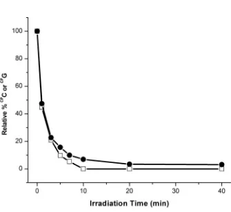
Figure 2.2. Long range oxidative damage to cytosines in duplex DNA. HPLC traces show the oxidative decomposition of CPC with increasing irradiation, 0-40 minutes for Rh-G-1 (top left) and 0 (black) versus 5 (red) minutes for Rh-I-1 (top right). Insets show the full HPLC traces. Plots of the amount of CPC as a function of irradiation time reveals the sequence dependence of CPC decomposition with Rh (bottom left) and AQ (bottom right): Rh-G-2 (closed circles), Rh-I-2 (open circles), Rh-G-1 (closed squares), Rh-I-1 (open squares), and in (d), AQ-G-1 (closed circles), AQ-I-1 (open circles), and AQ-I-3 (closed squares). For AQ, note the dramatic increase in efficiency of CPC photooxidation when the intervening guanines (AQ-I-1) are replaced with inosines (AQ-I-3).
Decomposition of CPC is an intraduplex reaction that requires light and photooxidant.
DNA duplex. Despite the significant energetic differences among the intervening DNA bases, decomposition of CPC is clearly observed upon irradiation of the Rh-tethered DNA assemblies (Table 2.1). This observation provides direct HT chemical evidence for the existence of hole density on the pyrimidines.
The extent of hole occupation on the pyrimidine, moreover, is modulated by the energetics of the full duplex. When CPC is base paired with G, limited oxidation of CPC (~5 %) is observed in the sequence context 3’-GGCPCT-5’ (Rh-G-2) after 10 minutes of irradiation at 365 nm. However, the yield of CPC photodecomposition after 10 minutes of irradiation increases by 7-fold when the sequence context around CPC is altered to 3’-AACPCT-5’ (Rh-G-1). This difference in yield of CPC decomposition is attributed to the sequence dependence of hole distribution; in 3’-GGCPCT-5’, the hole density is likely more localized on the GG doublet, while in 3’-AACPCT-5’ the hole density is more diffuse, and there is thus a greater distribution on the CPC.
modulation also establishes that the ring opening of the cyclopropyl-amine radical does not drive the reaction; variations in ring opening are instead dependent upon the sequence of the DNA assembly and its energetics.
Note that control experiments have also been conducted under parallel conditions where CPC-substituted assemblies lacking Rh are mixed with Rh-tethered assemblies lacking CPC. This control provides a test of whether any interduplex reaction occurs, as might be expected if a diffusible species were involved rather than long range DNA-mediated CT. In these control experiments, no photodecomposition of CPC is observed. Hence the oxidative reaction is intraduplex.
Sequence-dependent oxidation of CPC is an inherent feature of DNA CT,
irrespective of oxidant. Covalently tethered AQ is therefore also found to be effective at photooxidation of CPC from a distance (Table 2.1, Figure 2.2), albeit at a somewhat lower efficiency. Noteworthy here is the fact that in these duplexes AQ is restricted to capping the duplex terminus, while the Rh photooxidant intercalates predominantly between the third and fourth bases. Photooxidation of CPC by AQ is also found to be an intraduplex reaction.
It is also noteworthy that H-bonding to CPC is not required for long range oxidation of CPC. We still find significant oxidation of CPC in assemblies where CPC opposes A, creating a non-hydrogen bonded mismatch (Table 2.1, AQ-A-1/Cp-1). The extent of reaction at CPC is therefore governed by intrinsic sequence dependent variations in the distribution of hole density in double stranded DNA.
2.3.4. Comparisons in photooxidation of CPC and CPG, modified bases with
equal trapping rates but different energetics. To compare further the hole occupancy on purines and pyrimidines, we examined two duplexes of equivalent sequence, one containing CPC and the other containing CPG at the neighboring site (CPG in AQ-I-4/Cp-5 and CPC in AQ-I-4/Cp-4 in Table 2.1). Based upon a thermal equilibration of charge, given a conservative difference in potential between C and G of ~ 0.30.5 V, the relative hole occupancy on CPC versus CPG should be ~1:105108. As evident in Table 2.1,
however, CPG and CPC in these two duplexes are seen to decompose with similar efficiency.
As shown in Figure 2.3, we also examined photooxidation of duplexes containing both CPG and CPC in the same assembly. Here, then, trapping times for the two modified bases should be equal, and one would expect the relative energetics of the fast traps to determine their relative efficiencies in photodecomposition. Remarkably, however, in these duplexes (AQ-7/Cp-7 and AQ-8/Cp-8), when CPG and CPC are incorporated as neighboring bases on the same strand, their efficiency of photodecomposition is comparable, although not identical (Figure 2.3, Table 2.1).
Figure 2.3. Hole distribution on the DNA bridge does not reflect the relative energies of the individual bases. Shown is a plot of the amount of CPC (closed circles) or CPG (open squares) as a function of irradiation time in AQ-7/Cp-7. This duplexes contain CPC and CP
bridge does not reflect the relative energies of the isolated bases.
2.4. Discussion.
2.4.1. Mechanistic models for long range DNA CT. In many experiments characterizing DNA CT, a shallow distance dependence in the reaction has been demonstrated,1-8 and this shallow distance dependence has been reconciled through models primarily involving long range diffusive charge hopping.15,16 Models involving a mixture of hopping among low energy guanine sites and tunneling through AT tracts have provided a useful starting point for reconciling many experiments. Once
experiments demonstrated rapid, long range DNA HT across assemblies containing adenine tracts,5,7,34,35 however, the model was modified to also include hopping on low energy adenines.18 These models, however, consistently explain DNA HT in the context solely of base energetics. Yet many experiments have underscored the sensitivity of DNA HT to DNA structure and dynamics.9-11,19,20 Indeed it is now apparent that conformational motions of the DNA bases are required for effective CT; indeed increased rigidity of DNA assemblies limits CT.36
Figure 2.4. Schematized delocalized domain model for DNA charge transport. Charge migrates through the DNA bridge by hopping among domains, extended -orbitals
energies of bases on the same and opposite strand. Indeed theoretical predictions have been made indicating the modulation of baseionization potentials in DNA by sequence dependent structure37 and dynamics.38 In this context it is also interesting to consider the asymmetrical distribution of oxidative damage at the 5’-G of 5’-GG-3’ doublets,39 observed after relatively slow secondary reactions leading to trapping of the G radical and ultimately DNA strand cleavage. Yet these modulations do not provide sufficient energetic fluctuations to account for differences in oxidation potential between C and G. Here, experimentally, once the rate of trapping at C and G are made comparable by cyclopropyl substitution, the data indicate that the radical is delocalized comparably over the two neighboring bases. Thus, not only is there modulation, in fact there must
necessarily be orbital mixing among the purines and pyrimidines to generate extended -orbitals.
Models involving superexchange tunneling through pyrimidines are also
unsatisfactory, since these data establish that the hole occupies the full DNA bridge, both pyrimidines as well as purines.15,40 Models derived solely upon base energetics
furthermore do not provide a reasonable rationale for the sensitivity of HT to sequence-dependent structure and dynamics. An alternative mechanistic description is required.
2.4.2. Charge transport through transient, delocalized domains. As illustrated in Figure 2.4, we propose a model where charge migrates through the DNA bridge among delocalized domains. Our data require occupation of all bases of the DNA bridge,
although not necessarily simultaneously. These DNA domains can be described as extended -orbitals formed transiently depending upon DNA sequence and dynamics.
length and temperature have provided evidence for a domain size of ~ 4 base pairs,20 and distinguish a domain from a polaron.3,41 Recently, a variable-range hopping model was developed and it was found to that delocalized bridge states are required to account for experimental yields of DNA hole transport.42 Data here, where comparable efficiency of reaction at CPC and a neighboring CPG are observed, provide direct support for
delocalization of charge.
However, delocalization clearly does not occur over the entire duplex. The
transport is partly incoherent since we have found also that HT is gated by base motions.9 Thus these delocalized domains form and break up transiently, facilitating and limiting HT. This model accounts well for the attenuation in HT seen in AT-rich sequences, owing to their inherent flexibility, and tunneling through AT sequences need not be invoked.15,16 It also explains oxidative repair of thymine dimers at a distance, another fast trap.43,44 While this model is more challenging to test experimentally, and describe
theoretically, than superexchange or localized hopping, these and other data45,46 show that DNA-mediated HT requires a more complete treatment.
2.5. References
1. Hall, D. B., Holmlin, R. E., Barton, J. K. Oxidative DNA damage through long range electron transfer Nature1996, 382, 731-735.
2. Núñez, M. E., Hall, D. B., Barton, J. K. Long range oxidative damage to DNA: effects of distance and sequence Chem. Biol.1999, 6, 85-97.
3. Henderson, P. T., Jones, D., Hampikian, G., Kan, Y. Z., Schuster, G. Long-distance charge transport in duplex DNA: the phonon-assisted polaron-like hopping
mechanism Proc. Natl. Acad. Sci. USA1999, 96, 8353-8358.
4. Giese, B. Long-dstance electron transfer through DNA Annu. Rev. Biochem.2002, 71, 51-70.
5. Takada, T., Kawai, K., Cai, X., Sugimoto, A., Fujitsuka, M., Majima, T. Direct observation of hole transfer through double-helical DNA over 100 Å J. Am. Chem. Soc. 2004, 126, 1125-1129.
6. Nakatani, K., Dohno, C., Saito, I. Modulation of DNA-mediated hole-transport efficiency by changing superexchange electronic interaction J. Am. Chem. Soc.2000, 122, 5893-5894.
7. Yoo, J., Delaney, S., Stemp, E., Barton, J. K. Rapid radical formation by DNA charge transport through sequences lacking intervening guanines J. Am. Chem. Soc.2003, 125, 6640-6641.
8. Delaney, S., Yoo, J., Stemp, E. D. A., Barton J. K. Charge equilibration between two distinct sites in double helical DNA Proc. Nat. Acad. Sci. USA2004, 101