Scholarship@Western
Scholarship@Western
Electronic Thesis and Dissertation Repository
1-23-2018 12:30 PM
Influence of the Metastatic Microenvironment on Stem-like
Influence of the Metastatic Microenvironment on Stem-like
Human Breast Cancer Cells
Human Breast Cancer Cells
Ashkan Sadri
The University of Western Ontario
Supervisor Allan, Alison L.
The University of Western Ontario
Graduate Program in Anatomy and Cell Biology
A thesis submitted in partial fulfillment of the requirements for the degree in Master of Science © Ashkan Sadri 2018
Follow this and additional works at: https://ir.lib.uwo.ca/etd
Part of the Cancer Biology Commons
Recommended Citation Recommended Citation
Sadri, Ashkan, "Influence of the Metastatic Microenvironment on Stem-like Human Breast Cancer Cells" (2018). Electronic Thesis and Dissertation Repository. 5188.
https://ir.lib.uwo.ca/etd/5188
This Dissertation/Thesis is brought to you for free and open access by Scholarship@Western. It has been accepted for inclusion in Electronic Thesis and Dissertation Repository by an authorized administrator of
i
Ninety percent of breast cancer-related mortalities result from metastasis. We have previously demonstrated that stem-like ALDHhiCD44+ breast cancer cells are critical for metastasis, and preferentially target the lung and bone marrow (BM). We hypothesize organ tropism occurs through promotion of the ALDHhiCD44+ phenotype. Using a 2D ex vivo model, lung and BM conditioned media (CM) were utilized to assess their influence on stem-like phenotype and behavior. Exposure of human breast cancer cells to lung-CM significantly decreased the proportion of cells with a stem-like ALDHhiCD44+ phenotype, decreased expression of cancer stem cell (CSC)-related genes, and increased gene expression related to migration (p<0.05). Lung-CM also induced a viable non-adherent subpopulation that expressed significantly decreased CD44 expression and was unable to form mammospheres (p<0.05). Analysis of lung-CM revealed presence of proteins related to migration, adhesion, and stemness. Taken together, the lung microenvironment may promote metastasis of breast cancer cells in a CSC-independent manner.
Keywords
ii
Co-Authorship Statement
iii
Acknowledgments
To begin, I have been lucky enough to work every day with a fantastic group of fellow lab mates, who I’m happy to say have become my good friends. Thank you for your welcome
advice and invaluable support you have given me throughout our time together. To David, thank you for getting me started with lab techniques, teaching me animal handling skills that only master animal handlers come to learn, and educating me about all things European history. You have shown me great kindness and quirkiness in everything you have done for me, I really appreciate that. To Ying, it was always a pleasure coming into the lab and being greeted by you. You were constantly positive when my experiments go awry, providing a smile, and more importantly, a solution. I have learnt lab techniques that I didn’t even consider, and your guidance through everything was an enormous help in building me into a competent researcher. You were always willing to donate your time to accommodate me, and for that I am very thankful. To Lori, without your advice and guidance, the majority of my project would not have gone nearly as smooth as it did. You provided me with everything I needed to know about performing flow cytometry, and graciously helped troubleshoot a lot of my experiments. Whenever I needed help, your door was always open. To Corey, Jenna, and Sami, thank you guys for keeping me positive and providing much needed social interaction. It was a pleasure getting to know you guys and I hope we keep in touch over the years. I know my jokes are bad, but hopefully not to the point we lose communication. Overall, I could not have asked for a better support team and group of friends during my time in the lab.
Beyond my research, I must express my very profound gratitude to my parents, Shiva and Faramarz Sadri and to my partner Sabina Reis for providing me with unfailing support and continuous encouragement throughout my years of study and through the process of researching and writing this thesis. Maman and Baba, coming to a brand-new country at a young age is a daunting, unsettling task, but you have proven that with hard work and dedication, you can do anything. This accomplishment would not have been possible without them. I would also like to share my thanks to Jennifer and Brian Small for their delicious vegan meals and hospitable lodgings.
iv
and helped construct this meaningful thesis. Without your unique perspectives and scientific input, making sense of all things science would have been much more difficult.
Last, but certainly not least, my cup runneth over with gratitude for my supervisor, Dr. Alison Allan. You introduced me to the field of research and gave me a chance to exercise my curiosity. Your competency, intelligence, and genuine personality made this experience all the more enjoyable. Thank you for your patience and encouragement, you have taught me so much in many respects. I am gratefully indebted to you for making this thesis possible. Your constructive criticism has seen me through to becoming a better writer and researcher, and most importantly, a thinker. I consider it an honor to be working with someone as passionate about their research as you are. Even when science resists and provides unexpected results, I will always remain an optimist.
I would like to take a moment and touch on something I feel is important. Upon doing research for this thesis, I have discovered a renewed sense of appreciation for those affected by breast cancer. Their resilience and strength are the reason we do this research, however incremental it may seem in the larger scheme of things. An African proverb illustrates this perfectly; “If you think you are too small to make a difference, you haven’t spent the night with a mosquito”.
v
Table of Contents
Abstract ... i
Co-Authorship Statement... ii
Acknowledgments... iii
Table of Contents ... v
List of Tables ... viii
List of Figures ... ix
List of Appendices ... xi
List of Abbreviations, Symbols, Nomenclature ... xii
1 INTRODUCTION ... 1
2 LITERATURE REVIEW... 2
Cancer ... 2
Breast Cancer ... 3
Histopathology and Molecular Subtypes ... 4
Epithelial-to-Mesenchymal Transition ... 6
Metastasis ... 7
Organ Tropism of Metastasis ... 8
The “Seeds”: Stem-Like Breast Cancer Cells ... 9
Characterization of CSCs ... 9
Stem-Like Cancer Cells and Metastasis ... 13
Plasticity of Stem-Like Cancer Cells ... 15
The “Soil”: Organ Microenvironments ... 17
The Lung Microenvironment and Lung Metastasis ... 18
The Bone Microenvironment and Bone Metastasis ... 20
vi
3 HYPOTHESIS AND OBJECTIVES ... 24
Hypothesis... 24
Objectives ... 24
4 MATERIALS and METHODS ... 25
Cell Culture and Reagents ... 25
Lung and Bone Marrow Ex Vivo Model Systems ... 25
Organ Conditioned Media Generation ... 25
Flow Cytometry Analysis ... 29
RT-qPCR Analysis... 34
Analysis of ALDH1A1, ALDH1A3, and CD44 Gene Expression ... 34
Quantitative RT-PCR Human Cancer Stem Cell® Array ... 34
Cell Viability Assays ... 36
Trypan Blue Exclusion ... 36
LIVE/DEAD® Assay ... 36
Mammosphere Assay ... 38
Proteomic Analysis of Lung-CM ... 39
Statistical Analysis ... 39
5 RESULTS ... 40
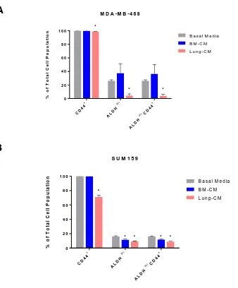
Exposure to lung-conditioned media decreased ALDH activity and CD44 expression in human breast cancer cells ... 40
Exposure to lung-conditioned media increased gene expression of ALDH1A3 in human breast cancer cells ... 40
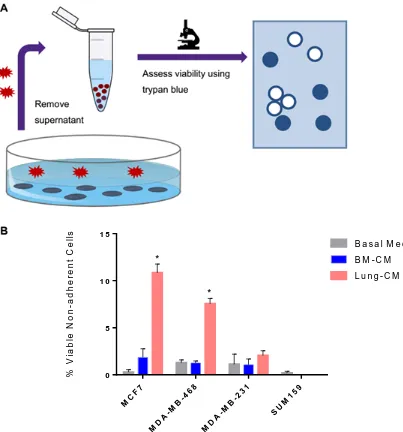
Treatment with lung-CM induced a viable, non-adherent breast cancer subpopulation ... 43
Lung-CM and BM-CM reduced CD44 expression, ALDH activity, and ALDH1A1 gene expression in non-adherent human breast cancer cells. ... 47
vii
Exposure to lung-CM increased mRNA expression related to migration and
decreased mRNA expression of CSC markers. ... 49
Lung-CM contained proteins related to migration, adhesion, and stemness ... 51
6 DISCUSSION ... 60
Summary of Key Experimental Findings ... 61
Lung-CM inhibits stem-like phenotype and behaviour in adherent human breast cancer cells ... 63
Lung-CM induced a viable, non-adherent breast cancer cell subpopulation with decreased stem-like phenotype and function ... 65
Lung-CM contained proteins related to migration, adhesion, and stemness ... 67
Osteopontin ... 69
Periostin ... 69
β-Catenin ... 70
Proteins that Influence Cell Detachment and Extracellular Transport ... 71
Potential Influence of Intracellular Proteins ... 72
Lung-CM increases gene expression related to migration and decreases expression of CSC markers ... 73
Possible Limitations of the Study ... 77
Future Directions ... 80
Final Conclusions... 82
7 References ... 83
viii
List of Tables
Table 1. Human Breast Cancer Cell Lines and Culturing Conditions ... 26
Table 2. Gene List and Primer Sequences ... 35
Table 3. CXCL8, CD24, MUC1, ATM and WEE1 emerge as key genes affected by
lung-CM treatment. ... 54
Table 4. Function of CXCL8, CD24, MUC1, ATM, and WEE1.. ... 55
Table 5. Function of proteins within lung-CM related to ALDH/RA signaling pathway and CD44 cleavage. ... 58
ix
List of Figures
Figure 1. The ALDHhiCD44+ phenotype in breast cancer cells enhances metastasis-initiating capacity. ... 23
Figure 2. Generation of organ-conditioned media. ... 28
Figure 3. Representative flow cytometry gating strategy for analysis of ALDH activity and/or CD44 cell surface expression in SUM159 human breast cancer cells. ... 31
Figure 4. Representative flow cytometry gating strategy for Analysis of ALDH activity and/or CD44 cell surface expression in adherent MDA-MB-468 human breast cancer cells. 32
Figure 5. Representative flow cytometry gating strategy for analysis of ALDH activity and/or CD44 cell surface expression in non-adherent MDA-MB-468 human breast cancer cells.. ... 33
Figure 6. Exposure to lung-conditioned media decreased ALDH activity and CD44
expression in human breast cancer cells. ... 41
Figure 7. Exposure to lung-conditioned media increased CD44 and ALDH1A3 mRNA expression in human breast cancer cells. ... 42
Figure 8. Exposure to lung-conditioned media supported the production of viable
non-adherent breast cancer cells assessed by trypan blue exclusion. ... 44
Figure 9. Exposure to lung-conditioned media supported the production of viable
non-adherent breast cancer cells assessed by Live/Dead® assays.. ... 46
Figure 10. Exposure to lung-conditioned media reduced CD44 expression, ALDH activity, and ALDH1A1 gene expression in MDA-MB-468 human breast cancer cells... 48
Figure 11. Exposure to lung-conditioned media impaired mammosphere formation by non-adherent human breast cancer cells. ... 50
x
Figure 13. Classification of intra- and extracellular proteins found in lung-CM. ... 56
Figure 14. Mass spectrometry analysis of lung-CM identifies key proteins related to the ALDH/RA and CD44 pathways. ... 59
xi
List of Appendices
Appendix 1. Approved animal use protocol ... 97
Appendix 2. Exposure to organ-CM did not affect protein expression of ALDH1A3 in human breast cancer cells. ... 98
Appendix 3. Relative mRNA expression levels of cell lines and cell subpopulations in
response to basal media. ... 99
xii
List of Abbreviations, Symbols, Nomenclature
7-AAD 7-Aminoactinomycin D
ABC ATP-Binding Cassette
ADAM10 A Disintegrin and Metalloproteinase Domain-Containing Protein 10
ADH1 Alcohol Dehydrogenase 1
ALDH Aldehyde Dehydrogenase
ANOVA Analysis of Variance
ATM Ataxia-Telangiectasia Mutated
BAA- BODIPY-aminoacetate
BAAA BODIPY-aminoacetaldehyde
BM Bone Marrow
BM-CM Bone Marrow-Conditioned Media
BMSC Bone Marrow Stromal Cells
Ca+2 Calcium Ion
Calcein-AM Calcein-acetoxymethyl
CAM Cell Adhesion Molecule
CAM Chick Chorioallantoic Membrane
CD24 Cluster of Differentiation 24
CD44 Cluster of Differentiation 44
xiii
CRABP2 Cytosolic Retinoic Acid Binding Protein 2
CSC Cancer Stem Cell
CTC Circulating Tumour Cell
CXCL8 C-X-C Motif Chemokine Ligand 8
CXCL12 C-X-C Motif Chemokine Ligand 12
CXCR4 C-X-C Motif Chemokine Receptor 4
DCIS Ductal Carcinoma in situ
DEAB Diethylaminobenzaldehyde
DMEM:F12 Dulbecco’s Modified Eagle Medium/Nutrient Mixture F-12
ECM Extracellular Matrix
EDTA Ethylenediaminetetraacetic Acid
EMT Epithelial-to-Mesenchymal Transition
ER Estrogen Receptor
ETHD-1 Ethidium Homodimer-1
Ex/Em Excitation/Emission
FABP5 Fatty Acid Binding Protein 5
FBS Fetal Bovine Serum
GAPDH Glyceraldehyde 3-Phosphate Dehydrogenase
HA Hyaluronic Acid
xiv
HER2 Human Epidermal Growth Factor Receptor 2
HSC Hematopoietic Stem Cell
HUVEC Human Umbilical Vein Endothelial Cells
LCIS Lobular Carcinoma in situ
IDT Integrated DNA Technologies
IE-MS Iterative Exclusion-Mass Spectrometry
IL8 Interleukin 8
MDCK Madin-Darby Canine Kidney
MMTV-PyMT Mouse Mammary Tumor Virus-Polyomavirus Middle T-Antigen
MS Mass Spectrometry
mtDNA Mitochondrial DNA
MUC1 Mucin 1
NEK2 NIMA-related Kinase 2
OPN Osteopontin
PBS Phosphate-Buffered Saline
PDLSC Periodontal Ligament Stem Cells
PE Phycoerythrin
POSTN Periostin
PR Progesterone Receptor
xv
RA Retinoic Acid
RBP Retinol Binding Protein
rhPOSTN Recombinant Human Periostin Protein
SEM Standard Error of the Mean
siRNA Small Interfering RNA
TGF-β Transforming Growth Factor β
TN Triple Negative
VCAM1 Vascular Cell Adhesion Molecule 1
VEGF Vascular Endothelial Growth Factor
VLA-4 Very Late Antigen-4
1
INTRODUCTION
Cancer is devastating. It is the source of physical, emotional, and mental stress that not only affects the immediate individual involved, but extends further to family and friends. In 2017 alone, it is estimated that 206,200 Canadians will be diagnosed with cancer1. These figures have increased by nearly 10,000 new diagnoses within the past two years2. Unfortunately, these figures are expected to rise by an additional 20% by 2020 largely due an aging and growing population3. Greater emphasis on cancer prevention, adopting a healthy lifestyle, and earlier detection is necessary to counter such undesirable outcomes3. Breast cancer is the most common malignancy in women worldwide4. Despite its high prevalence, patient prognosis is strong with 5-year survival rates nearing 99% when localized to the breast5. Once the cancer leaves the confines of the breast, the chances of
surviving this drastically decrease5. In fact, the dissemination of cancer from the primary
affected region to distant organs accounts for 90% of all cancer related deaths6. Recently,
it has been proposed that a rare population of tumour cells, commonly referred to as cancer stem cells, may be mediating metastasis and secondary tumour formation7. Therefore, mortality related to breast cancer rarely occurs as a result of the primary tumour, but rather the cascading effect of multiple organs becoming compromised, including the lung and bone 8-10.
2
LITERATURE REVIEW
Cancer
The term “cancer” encompasses a subset of diseases that are characterized by atypical cell
growth and proliferative patterns. As these aberrant cells develop, they acquire genetic disruptions that enable sustained proliferative signaling and evasion of growth suppressors11. Most often, the onset of this disease begins once genes involved in cell cycle
regulation become mutated and dysfunctional. These genes are most often classified either as oncogenes or tumour suppressor genes. Once mutated, oncogenes become activated, giving cells the ability to induce uncontrolled cell growth12. In contrast, mutations in tumour suppressor genes may render normal cells incapable of DNA repair required to regulate cell growth13. It is likely that oncogene activation and tumour suppressor gene inactivation occur simultaneously as cancer progresses, ultimately resulting in tumour formation14.
Tumours can be characterized by their benign or malignant nature, with the latter capable of invasive traits that represent the hallmarks of cancer. A growth that is neither invasive to surrounding or distant regions, such as a common wart, is considered benign and does not pose a significant health risk11. In rare cases, benign tumours may impinge on blood vessels or nerves that supply organ systems. These instances are considered higher risk and require resection, however they are not considered malignant tumours15. Malignant tumours tend to be life-threatening due to the capacity to leave the primary site and invade into surrounding tissues or distant secondary sites of the body via access to systemic circulation15. These tumours are referred to as malignant cancers and require early detection and subsequent treatment to provide the best prognosis for patients. In later stages of tumour progression, if the tumour has spread to distant sites, both primary and adjuvant treatments (e.g. tumour resection, radiation, hormonal therapy, or chemotherapy) become far less efficient at easing tumour burden16. Together, inefficiencies in treatment strategies
and efficacy leave both patients and the healthcare system in distress.
estimates that 1 in 2 Canadians will develop cancer within their lifetime, a value that is increased from a 1 in 2.5 frequency just two years ago1,2. Similarly, the overall 5-year
survival rates in cancer patients have also decreased from 63% in 2015, to 60% in 20171,2. Despite advances in targeted treatment and enhanced screening techniques, cancer is proving to be relentless. If these patterns persist, cancer-associated deaths will account for 30% of Canadian premature deaths in 2017, 1. Premature deaths are measured by potential years of life lost (PYLL) and account for deaths occurring at younger ages, a vital statistic when assessing economic health for any given country. Between 2010 and 2012, cancer represented the largest PYLL among Canadians, with 1.5 million years lost due to cancer1. Due to a loss in productivity associated with high PYLL values, not only is the growing Canadian economy impacted, but these patients also pose a significant financial burden on the healthcare system. In 2008, the Public Health Agency of Canada estimated that $3.8 billion was devoted to direct healthcare costs for cancer patients (hospitalization, treatments, etc.), while an additional $586 million was lost to indirect costs associated with decreased economic productivity17. Considering that cancer diagnoses have increased in recent years, the economic impact of cancer is expected to increase steadily with time.
Breast Cancer
The breasts are two prominences situated on the ventral torso of primates, morphologically identical in both male and female offspring. Once females enter puberty, secretion of sex and growth hormones (namely estrogen) promote mammary development. Subcutaneous adipose tissue within the breast supports a network of ductal and lobular tissue, that together, comprise the feeding mammary gland. The lobular epithelium of the breast serves a lactiferous function, producing and secreting milk down the ductal epithelium for expulsion out the nipple by contracting myoepithelial cells 18. Due to hormonal cycling involved in mammary development and throughout child-bearing years, the lobular and ductal cells are most susceptible to tumour formation, and cancers that arise are termed lobular or ductal carcinomas respectively16. So long as the tumour is confined to primary breast tissue, the 5-year survival rate is an exceptional 98.6% among females5.
aggressive cases of breast cancer involve secondary tumour formation at distant organs, resulting in significantly reduced organ function.
Histopathology and Molecular Subtypes
Recent findings regarding tumour heterogeneity suggest that each tumour is distinct and unique from patient-to-patient. This further extends into distinctions between multiple tumours identified within a single individual. Upon histopathological analyses of biopsied tumour samples from the breast, inter-tumour heterogeneity is evident19. Histopathology provides a rudimentary understanding of the cell subpopulations involved in tumour development, their morphology, and predicting aberrant growth patterns. The main purpose of this technique is to distinguish whether the breast tumour is originating from ductal or lobular tissue architecture20. At the time of detection, ductal or lobular carcinomas
in situ (DCIS or LCIS respectively) are non-invasive as they remain confined to epithelial-lined compartments within the mammary tissue20. Surgical resection and localized therapy strategies such as radiation are highly effective and demonstrate strong patient prognosis with a 99% survival outlook over five years21. Unfortunately, more than half of breast cancer incidences are invasive ductal or lobular carcinomas (IDC or ILC) upon initial diagnosis22. Tumours that have spread from epithelial-lined compartments of the mammary
tissue into the stroma are classified as invasive carcinomas through histopathological analyses20. The associated treatment strategies for patients with IDC or ILC are less
effective and become limited as the tumour spreads from the primary tissue. A lack of targeted therapies for invasive carcinomas is reflected in the 5-year survival rates dropping to near 25% once the tumour has acquired invasive potential21.
PR+), and lacking human epidermal growth factor receptor 2 (HER2-)24. Breast cancer
cells with a high proliferative (Ki67) index and/or expressing HER2, in addition to both ER+/PR+, are classified as the Luminal B subtype. Both luminal A and B breast cancers have been associated with positive survival outcomes24,25. Because both Luminal A and B subtypes are ER+, hormone therapy is highly effective to treat patients, particularly in the adjuvant setting19. Despite their similarities, Luminal B breast cancer cells are genetically altered from the Luminal A subtype, resulting in poorer prognosis than those affected by Luminal A breast cancer26. Further, the HER2+ breast cancer subtype lacks expression of both ER and PR, rendering these cells unresponsive to targeted hormone therapy24. Together with increased proliferation within this subtype, HER2+ breast cancer cells tend to metastasize and spread more readily to surrounding tissues, resulting in poorer patient prognosis relative to luminal breast cancers23. Inhibition of HER2+ ligand-receptor interactions using HER2-targeting agents such as trastuzumab and lapatinib has improved overall patient survival and time-to-disease progression, however many patients will acquire resistance to therapy over time27.
Perhaps the most difficult breast cancer subtype to treat is the TN breast cancer subtype which lacks the cell surface receptors found on the aforementioned breast cancer subtypes (ER-/PR-/and HER2-)24. Often the most aggressive subtype of breast cancer, TN breast
cancer patients are highly prone to metastases which results in the poorest prognosis28.
Despite some TN breast cancers being initially responsive to traditional chemotherapy, a hallmark of TN breast cancer is their high likelihood of distant recurrences within 3-years of initial diagnosis29. As endocrine treatment strategies targeting ER/PR/ or HER2 are ineffective on the TN subtype, central pathways involved in proliferation, growth and migration are being actively investigated as potential targets30.
modulate tumour progression through intrinsic factors such as migration and/or invasion19.
Epithelial-to-Mesenchymal Transition
Given the anatomical composition and functional nature of mammary tissues, primary tumours developing in the breast are epithelial-derived and termed carcinomas32. Understanding how healthy epithelial cells function to maintain homeostasis is necessary to predict changes in epithelial function in a diseased state. The epithelial cell utilizes networks of cell-cell and cell-extracellular matrix (ECM) interactions to provide apical-basal cell polarity. Desmosomes, adherens junctions and gap junctions are protein complexes that maintain physical association between adjacent epithelial cells (cell-cell), while cell-ECM interactions are necessitated by integrins and cell adhesion molecules (CAMs)33,34. In a dynamic and invasive tumour microenvironment, extracellular cues reduce characteristic epithelial cell-cell and cell-ECM interactions, and can induce a mesenchymal cell phenotype35. The epithelial-to-mesenchymal transition (EMT) and its
reverse process, mesenchymal-to-epithelial transition (MET) describe this transformation from one phenotype to another7. Once in the mesenchymal state, these cells lack
apical-basal polarity and possess increased migratory and invasive potential36,37. The role of EMT and MET were first documented in embryogenesis, but the importance of these processes further translates into cancer progression, and subsequently metastasis7,38. Cancer progression requires cells of the primary tumour to disassociate and invade into the surrounding stroma. This process of invasion is mediated in part by the loss of cell-cell adhesions, which enhances cellular motility, while deterioration of cell-ECM interactions allows catabolic cell secretions (e.g. matrix metalloproteinases; MMPs) to penetrate the basement membrane34,37. As tumourigenic cells penetrate the porous basement membrane and extend into the surrounding stroma, early stage carcinomas become invasive malignancies7. In order to support tumourigenic growth, invading cells secrete angiogenic factors (such as vascular endothelial growth factor [VEGF] and transforming growth factor beta [TGF-β]) to support vascular growth necessary for nutrient delivery towards the tumour34,37. Although recent studies document maintained tumour progression in the
Metastasis
The spread of cancer from a primary tumour to a distant secondary site is referred to as metastasis, or metastatic disease. Although treatment strategies targeting the primary tumour are highly efficient, nearly 30% of women diagnosed with early-stage breast cancer will ultimately develop metastatic lesions40-42. For tumourigenic cells to metastasize, they must leave the primary site and enter systemic circulation by directly invading the surrounding vasculature, or indirectly through the lymphatic system43. As previously mentioned, the invasive behaviour of cancer cells induced by EMT and other mechanisms allows cancer cells to invade the surrounding stroma. Together with enhanced vascular recruitment (angiogenesis and vasculogenesis) surrounding the primary tumour site, cancer cells further develop means to enter into the vasculature/lymphatics and leave the primary site43-45.
Millions of cells are capable of dissociating from the primary tumour and entering the vasculature every day. However, the relative incidence of metastatic tumour development is rare, suggesting that the metastatic process has inefficiencies42. As tumour cells
intravasate into the circulation, experimental studies have shown that >80% of these cells can survive the shear and compressive stress associated with the circulatory phase of metastasis41,42,46. The majority of these circulating tumour cells (CTCs) arrest in the first capillary bed they encounter, while others remain selective for specific organ microenvironments such as the bone, lung, and brain47-49. Only after successful extravasation, whereby the CTCs exit the circulation and invade the distant organ, can secondary tumour formation become possible43. Despite the large number of cells that survive the circulatory phase of metastasis and successfully extravasate at the secondary site, the inefficiencies associated with metastasis are highlighted when assessing tumour-initiating potential at the distant tissue. Experimental studies have shown that only ~2% of cancer cells that successfully reach the secondary tissue microenvironment have the capacity to initiate a new tumour, and <0.1% of cells can persist into the successful formation of macrometastases42. These inefficiencies suggest that aspects of the metastatic
Organ Tropism of Metastasis
In the event of metastasis, patterns of cancer dissemination to secondary sites are not random, but rather coordinated50. Many cancers have shown preferential metastatic capacity towards particular organs, a process referred to as organ tropism. Among the various cancer subtypes, the patterns of organ tropism are variable and dependent on the cancers’ origin. Some cancers predominantly metastasize to a specific organ (e.g. prostate
cancer metastasizing to the bone), while other cancers follow a sequential pattern of metastasis (e.g. colorectal cancer often forms secondary metastases in the bone lung brain)51. As each organ differs in anatomical position, blood/nutrient supply, and organ microenvironment composition, invading tumour cells face different demands based on the target organ. Clinically, breast cancer metastasis has demonstrated preferential metastasis to the lung, bone, liver, brain and lymph nodes52.
Two competing theories that attempt to elucidate mechanisms involved in organ-specific metastasis are Stephen Paget’s “seed and soil” hypothesis, and Ewing’s mechanical arrest
When considering a biologically relevant theory for organ tropism, the likely mechanism is a combination of both Paget’s and Ewing’s hypotheses. A complex system that delivers
metastatic cells to different organ tissues based on relative blood supply, and then initiation and maintenance of said tumour would be mediated by favourable interactions with the soluble and insoluble factors provided by the organ.59,60.
The “Seeds”: Stem-Like Breast Cancer Cells
Research conducted by Massagué and colleagues uncovered an association between the molecular characteristics of breast cancer cell (the “seed”) and the preferential tissue to which the breast cancer cells metastasize52. Using in vitro and in vivo studies with the triple-negative MDA-MB-231 human breast cancer cell line, they demonstrated that specific gene expression signatures can dictate a breast cancer cell’s preference to metastasize to
either the lung, the bone, or the brain47-49. However, this work did not take into
consideration the heterogeneous nature of primary metastatic breast cancer tumours. Subsequent limiting dilution analyses in vivo confirmed this notion by demonstrating that isolation and injection of low numbers of primary breast cancer cells into healthy immune deficient mice resulted in only a small fraction of cells harnessing the ability to initiate and produce a primary tumour61,62. These studies supported the idea that only a subset of cells within a primary tumor have tumour-initiating capacity, suggesting that this rare subpopulation may contain stem-like traits, often referred to as cancer stem cells (CSCs). The concept of a CSC subpopulation within tumours first originated in hematologic cancers, gaining credibility with evidence that only 1 to 4% of myeloma and leukemia cancer cells demonstrated enhanced proliferative and colony formation potential63-65. Although the cellular origins of cancer stem cells in some solid tumours remain controversial, recent studies conducted by Blanpain et al have successfully demonstrated that tumour populations in intestinal, prostate, and breast cancer can be traced back to a stem/progenitor origin, reinforcing the validity of CSC model66.
Characterization of CSCs
during their investigation of hematopoietic system regeneration in vivo, a true HSC must fulfill two requirements: the ability to self-renew and to maintain a multipotent state67.
Self-renewal refers to the cells’ ability to produce a sister HSC without losing multipotent
potential, while multipotency is the ability of a progenitor cell to differentiate into any functional cell within a given lineage67,68. Originating from these well-characterized HSC attributes, CSCs must be able to generate a heterogeneous tumour population (differentiation) while concomitantly maintaining their own population (self-renewal)68. Numerous studies have validated these stem cell characteristics to be true among a CSC subpopulation, and in addition, have demonstrated that CSCs also possess enhanced drug resistance, anchorage-independence, and increased migration relative to non-CSCs52,61,62,69,70. CSCs have also been associated with aggressive metastasis, and in many instances, found to express molecular markers of EMT7. Considering the inefficiencies previously mentioned regarding the metastatic process, it is reasonable to postulate that a rare CSC subpopulation of primary breast cancer cells may also be able to establish and drive distant secondary tumour development.
In light of their stem-like properties, CSCs can be isolated from a whole cell population using similar molecular screening techniques used with HSCs. In breast cancer, CSCs from patient tumours and various breast cancer cell lines have successfully been enriched for based on high aldehyde dehydrogenase (ALDH) enzymatic activity and the co-expression of the cluster of differentiation 44 (CD44) cell surface marker61,62,71.
2.6.1.1
ALDH
The ALDH family is made up of 19 evolutionarily conserved isoenzymes that are localized intracellularly in the cytoplasm, mitochondria, and nucleus72. The main function of ALDH is to catalyze the oxidation of aldehydes into carboxylic acids, along with other functions such as ester hydrolysis and scavenging for hydroxyl radicals72. Of particular interest are the isoenzymes (ALDH1A1, ALDH1A2, ALDH1A3, and ALDH8A1) involved in the conversion of vitamin D to retinoic acid (RA), as they have recently been implicated in cancer cell “stemness”73. The lipophilic RA molecule is capable of passive diffusion in a
the human cytosolic ALDH1A subfamily (ALDH1A1 and ALDH1A3) are highly expressed in early progenitor cells and have been documented to overlap with side population cells capable of excluding Hoechst 33342 stain, another modality for identifying stem-like cells61. Intrinsically high ALDH (ALDHhi) activity and Hoechst 33342 excluding stem-like side populations demonstrate increased expression of ABC transporters, a feature thought to provide CSCs with chemo-resistance75. This protective mechanism renders CSCs particularly resistant to conventional cancer therapies, permitting relapse over complete remission, and prolonging tumour longevity7,76. Much of the research concerning ALDH activity in cancer utilizes the metabolism of ALDEFLUOR™ substrate to isolate a subpopulation of tumourigenic cells with stem-like characteristics via flow cytometry72. It was initially predicted that ALDH1A1 was responsible for the majority of ALDEFLUOR™ metabolism, however recent evidence suggests ALDH1A3 is also involved72,74. Considering the metabolism of ALDEFLUOR™ is non-specific, it is likely that the ALDH activity detected in a cancer is due to the combined activity of two or more ALDH isoforms72.
Analyses of intracellular ALDH activity in liver, lung, esophageal, and breast cancer cells has been a useful tool to estimate how these tumourigenic cells may behave in vivo73. In
breast cancer, cells with elevated ALDH activity have demonstrated increased migratory capacity and the ability to form mammospheres in vitro70,77. The importance of ALDH
activity in vivo was emphasized by Ginestier et al. after transplantation of 50,000 ALDHlo
metastatic site (43.7%) compared to the primary tumour (28.7%), suggesting the importance of ALDH1 in metastatic disease and secondary tumour formation82. With
accumulating evidence supporting the tumorigenic role of ALDH in patients with metastatic breast cancer, a meta-analysis assessing 921 patients for elevated ALDH1A1 expression in breast cancer tumours concluded that ALDH1A1+ can be used as a biomarker for the prediction of tumour progression and poor patient outcome83.
2.6.1.2
CD44
Membrane-spanning CD44 is a glycosylated cell surface receptor that has well-defined roles in cell-cell and cytoskeletal cell-ECM interactions (via Rho GTPase signaling), promotion of cell survival and invasion (via PI3/Akt and MAPK-Ras pathways). Importantly, CD44 has a strong association with cell migration through interactions with matrix metalloproteases (MMPs) and other matrix remodeling enzymes which together coordinate cellular locomotion62,84. The principal ligand for CD44 is hyaluronic acid (HA), a major component of extracellular matrices, which has also been reported to maintain long term self-renewal85. Functional CD44 protein is encoded by a single gene with 20 exons, where exons 1-5 and 16-20 comprise the standard isoform (CD44s), while exons 6-15 are alternatively spliced to produce CD44 variants (CD44v)86. Although CD44s has been
implicated repeatedly in a variety of cancers, recent investigations have begun to examine specific splice variants and their association in cancer progression87. CD44v4 in human
subpopulation was unable to form tumours, even after injection of 500,000 cells62. These
findings suggest CD44 expression has an important role in tumour development.
Stem-Like Cancer Cells and Metastasis
Metastasis has been correlated with poor overall survival and mortality in several types of cancers, a major obstacle in cancer treatment. The involvement of CSCs in metastatic disease has received particular attention because they have been implicated in the initiation of the metastatic cascade through EMT processes91. Interestingly, both ALDH and/or CD44 have been used as markers for the identification of metastasis-prone cancer cell subpopulations. Previous investigation conducted in the Allan lab by Croker et al found breast cancer cells expressing the stem-like ALDHhiCD44+ phenotype demonstrated enhanced metastatic behavior in vitro, as their ability to migrate and invade was significantly increased relative to the non-CSC ALDHlowCD44- subpopulation70. Moreover, these stem-like breast cancer cells exhibited increased ability to form anchorage-independent colonies in vitro when compared to non-CSCs, suggesting these cells could potentially colonize in a distant organ microenvironment after detachment and dissemination70. Subsequent in vivo analyses confirmed the metastatic potential of
stem-like breast cancer cells after orthotopic injection into the mammary fat pad of NOD/SCID-IL2Rγ mice led to increased spontaneous metastases to the liver, spleen, and most notably the lung70. Not only did stem-like cells preferentially metastasize to these organs relative
with either knockout of ALDH1A1 or ALDH1A392. Collectively, ALDH activity among
CSC subpopulations has been demonstrated to be a critical factor in cancer dissemination through coordinated activity of ALDH1 isoenzymes.
The concerns revolving around CSCs and metastasis extend past their ability to disseminate and colonize distant organs. Tumour cells expressing stem-like characteristics present a significant barrier between effective cancer therapy and improved patient prognosis91. As CSC are postulated to have a slow rate of division and efficient efflux pumps capable of removing toxic agents, traditional chemo- and radiation therapies become inadequate in targeting CSC without off-target effects on healthy tissues91. Despite these limitations, conventional therapy remains the first line of treatment and often results in cancer recurrence due to a residual CSC subpopulation91. A subsequent study conducted by Croker
et al investigated the role of stem-like ALDHhiCD44+ breast cancer in therapy resistance. By inhibiting ALDH enzymatic activity, CSCs became transiently sensitized to chemotherapy (doxorubicin/paclitaxel) or radiotherapy measured by decreased cell viability and colony formation in vitro93. Moreover, therapy resistance was attributed to ALDH activity in part by the ALDH1A1 isoenzyme, and not ALDH1A3, demonstrated by siRNA knockdown in vitro92. These findings suggest ALDH activity is an important
mediator of CSC therapy resistance, and has since been supported by several studies reporting similar sensitization through ALDH inhibition94,95. Interestingly, a recent study
by Yang et al demonstrated that ALDH1A1 overexpression directly correlated with increased activity of multidrug efflux pumps through phosphorylation by NIMA-related kinase 2 (NEK2)96. An increase in efflux pump activity could support CSCs with high ALDH activity to remove therapeutic toxins and allow the tumourigenic cell to continue through to metastasis. Considering the importance of ALDH to drug resistance, without ALDH inhibition in CSC subpopulations, tumorigenic cells remain resistant to therapy and could be detrimental to patients as metastasis persists.
metastasis as CSCs become less likely to undergo apoptosis in response to therapy. Despite their supportive role in the cancer progression, transient activation and inactivation of cellular processes by CSCs is required to complete the metastatic cascade, suggesting CSC plasticity as a key contributor to metastasis.
Plasticity of Stem-Like Cancer Cells
It is generally believed that normal development is largely unidirectional, where slow-proliferating stem cell populations gives rise to highly proliferative progenitor cells, ultimately producing terminally differentiated mature cell types that regulate organ function97. The unidirectional nature of the cellular maturation process allows distinct cell types with varying specialties to be present within a single organ system and maintain functional homeostasis throughout the organisms lifespan97. This is especially clear in organs such as the heart, where the annual cardiac myocyte turnover rate is ~1% per year at age 20, drastically decreasing to ~0.4% after the age of 7598. That is not to say every post-mitotic mature cell is incapable of proliferation. Within the pancreas, mature pancreatic β-cells have been reported to expand their population through self-duplication as opposed to stem-cell differentiation99. Instances such as self-duplication suggest mature
cells may have alternative options related to cell fate, despite having undergone terminal differentiation. In particular, the concept of cell plasticity has received much attention recently as it supports the notion that a cell can alter its phenotype or behaviour in response to environmental queues100. The metabolic and epigenetic mechanisms required to induce phenotypic plasticity were first documented during early embryogenesis, but have been shown to re-activate during normal tissue regeneration, inflammation, and notably during tumour development101.
from one patient sample, yet represent two distinct subpopulations of breast CSCs expressing mesenchymal-like (EMT) and epithelial-like (MET) phenotypes respectively102. Interestingly, the EMT-CSCpopulation was associated with a quiescent state and preferentially localized to the invading tumour front, while the MET-CSC phenotype was more proliferative and centrally localized102. During tumour development, it was initially thought that EMT-CSC mediate invasion into surrounding tissues, while the MET-CSCs drive tumour growth internally. As the tumour progresses, CSCs change states in order to maintain invasion and proliferation accordingly. These findings suggest that plasticity between an epithelial or mesenchymal state in breast CSCs is a transient behaviour, rather than a fixed state. Thus, the role of CSC plasticity during tumour propagation, invasion, and metastasis is an important consideration in patient treatment.
Furthermore, plasticity among CSC populations is also evident during therapy. Initially, it was postulated that conventional therapeutic agents target and deplete non-CSCs, while CSCs evade treatment and expand their population to form a more aggressive tumour upon recurrence103. In a recent study conducted by Goldman et al, therapeutic treatment of taxanes or anthracyclines on human breast cancer cells in vitro and in vivo not only induced apoptosis in the majority of breast cancer cells, but also promoted the transition of non-CSCs towards a CSC state104. The therapy-resistant cells demonstrated increased
expression of breast CSC markers (CD44+CD24-) and augmented tumour growth, while
decreasing survival using patient derived xenograft models in mice. Indeed, these effects were not due to an enrichment for the CSC population but rather a transition from non-CSC to non-CSC state, demonstrated by the dose-dependent increase of the non-CSC population after acute low dose treatment104. Importantly, the plastic nature of CSCs was demonstrated as removal of the chemotherapeutic agent reverted newly generated CSCs back to a non-CSC state104. CSC plasticity is not specific to breast cancer as similar findings have been reported in prostate and ovarian cancer as well103. Nonetheless, the plasticity of the CSC state highlights several complexities with regards to the identification of tumourigenic cells and their subsequent response to conventional therapies.
several studies, the scientific community has faced difficulties in identifying a universal method for identification of elusive CSCs105. This is likely attributed to the search for a
specific CSC phenotype, rather than a highly plastic subpopulation of tumourigenic cells capable of transitioning from one state to another.
The “Soil”: Organ Microenvironments
Cancer metastasis follows an organ-specific pattern of dissemination53. If the metastatic site is compatible with the disseminated cancer cells, interactions between these cells and the microenvironment will likely promote colonization and secondary tumour formation106. Both clinical observation and experimental murine models suggest that organ-specific metastasis occurs independent of anatomical position, rate of blood flow, and number of cancer cells reaching the organ106. Using radiolabeled melanoma cells, cancer cell progression through systemic circulation and successful delivery to key organs was monitored after intravenous injection into murine models. Interestingly, radiolabeled melanoma cells were selective in colonizing specific organs, demonstrating that although tumour cells were capable of reaching the secondary organ, they required a congenial microenvironment to support extravasation and tumour development107. More recently, high expression of very late antigen 4 (VLA-4) on the endothelial cells surrounding the lung, bone, and brain tissue have been demonstrated to support homing of circulating breast cancer cells toward these organs through binding of their natural receptor, vascular cell adhesion molecule-1 (VCAM-1), aberrantly expressed on the surface of breast CTCs108. In our lab, research conducted by Chu et al has demonstrated the role of soluble organ-derived factors in promoting metastatic behaviour of breast cancer cells using a novel ex vivo model system52. Clinically relevant organs representing common sites of breast cancer metastasis (lung, bone marrow, liver, brain, LN) were harvested from female nude mice and cultured to produce organ-specific conditioned media (CM) for use in functional assays. The findings suggested that native soluble factors within organ-CM induced chemotactic and proliferative functions among the MDA-MB-231, MDA-MB-468, SUM149, and SUM159 human breast cancer cell lines analyzed52,109. Interestingly, these
of metastasis52.
The Lung Microenvironment and Lung Metastasis
The lungs are an indispensable organ involved in respiratory function, mediating the intake of oxygen and disposal of carbon dioxide during normal physiological conditions110. The functional anatomy of the lungs is divided into two zones. First air enters the conduction zone in the upper respiratory tract, travelling down the trachea and directed into each lung via the bronchi and terminal bronchioles110. The lower respiratory tract represents the respiratory zone, where air is shuttled past the terminal bronchioles and into the respiratory bronchioles, eventually reaching the alveolar ducts where individual alveoli necessitate gas exchange through an expansive network of capillaries110. Interestingly, the lungs are often implicated in various cancers, both as a direct cause from external factors (e.g. chemical pollutants) and/or dysregulation of normal physiology111. In particular, the lungs are a major site for tumour metastasis of breast and other cancers. A recent investigation of 1,088 medical records from non-metastatic breast cancer patients between 2004 and 2012 demonstrated that metastases to the lungs developed in 35% of patients, after a median follow-up time of 6.9 years112. Although incidence of breast cancer metastases are higher
in bone marrow than in lung tissue, tumour formation in respiratory organs severely impact the quality of life and are the leading cause of breast cancer related deaths113. Often, the more aggressive subtypes such as HER2+ and TN breast cancers metastasize to the lung where they largely contribute to impaired respiratory function, leading to intense pain, laboured breathing, and often hemoptysis114-117.
(POSTN), into the tumour-stroma microenvironment119,120. POSTN is a nonstructural
soluble protein that is present at low levels in healthy adults, but becomes significantly overexpressed at sites of inflammation and within the tumour stroma121. Findings by Malanchi et al demonstrate the necessity for POSTN in secondary tumour development as knockout of POSTN in PyMT mice had no effect on primary breast tumour size and volume, but significantly reduced incidence of pulmonary metastases120. More recently, treatment of human periodontal ligament mesenchymal stem cells (PDLSCs) with recombinant human periostin protein (rhPOSTN) accelerated migratory and proliferative capacity among treated hPDLSCs122.
Further investigation of the lung microenvironment and its relation to tumour formation has been modeled in vivo, however in vitro techniques for more detailed molecular characterization of the lung microenvironment are limited as it is difficult to adequately represent the complexities of native lung tissue in culture123. Utilizing the aforementioned
ex vivo model of organ-conditioned media, exposure to lung-CM has been shown to induce migration of human breast cancer cell lines, as well as increase proliferative capacity in two cell lines, MDA-MB-231 and MDA-MB-46852. In addition, the stem-like ALDHhiCD44+ breast cancer subpopulation was exposed to organ-specific CM (bone
marrow, lymph, liver, lung, and brain) to assess how this subset responded to soluble organ-derived factors. Interestingly, ALDHhiCD44+ breast cancer cells were found to
preferentially migrate towards lung-CM over all other organ conditions in vitro52. These
findings support observations by Croker et al, where ALDHhiCD44+ breast cancer cells were observed to preferentially metastasize to the lung in vivo70.
recruiting metastasis-initiating cells to the lung. However, little is known regarding the processes leading to organ-specific metastasis in the context of the CSC model.
The Bone Microenvironment and Bone Metastasis
to support a CSC niche133. In a glioblastoma model, inhibition of CXCR4/CXCL12
interactions led to decreased self-renewal and survival among CSC populations, emphasizing the importance of the bone marrow niche in supporting CSC populations134. It would be beneficial to achieve a stronger understanding related to the interactions that occur between HSC and the native bone marrow microenvironment as these interactions may be translatable to CSCs.
As is the case with the lungs, the complexities of the bone marrow microenvironment make it very difficult to accurately investigate its role in cancer metastasis and CSC maintenance
in vitro, resulting in the majority of research being performed in animal models135. Our preliminary findings utilizing the ex vivo organ-CM model demonstrate MDA-MB-231 and SUM159 breast cancer cell lines exposed to bone marrow-conditioned media (BM-CM) exhibit enhanced migratory potential in both stem-like ALDHhiCD44+ and whole cell populations136. Similar to the analysis of lung-CM protein content mentioned previously, protein array analysis of BM-CM highlighted potential mediators of metastasis, including the CD44 ligand OPN136. Notably, exposure of MDA-MB-231 breast cancer cells to BM-CM induced stem-like behavior including tumorsphere formation and colony-forming ability, mediated at least in part by OPN136. Although present in CM generated from bone
marrow stromal cells, the bone matrix and cancer cells themselves are also capable of producing soluble OPN137. The relevance of OPN to bone metastatic capacity is
emphasized by experiments utilizing murine models deficient in, or overexpressing OPN, where there was a strong correlation between OPN and the likelihood of skeletal metastasis137.
Study Rationale
Ninety percent of breast cancer-related mortalities result from metastasis, a process whereby the primary tumour disseminates and targets distant secondary organs. Interestingly, events leading up to secondary tumour formation have marked inefficiencies, with only a very small proportion of primary tumour cells able to reach, persist and grow into a secondary tumour138. We believe this rare subset of cells may be stem-like cancer cells. CSCs possess unique capabilities of self-renewal and differentiation, and help to potentiate the development of secondary tumours. Breast CSCs from patient tumours and cell lines have been successfully isolated based on high ALDH enzymatic activity and co-expression of the cell-surface glycoprotein CD44. These two CSC markers actively provide the cell with protective detoxifying mechanisms as well as enhanced metastatic capacity, respectively52,76,84. We have previously observed that ALDHhiCD44+ cells preferentially
migrate and/or metastasize to the lung and bone marrow microenvironments, where secondary tumours severely impact organ function. In addition, previous work in our lab has demonstrated that bone marrow-conditioned media can enhance the stem-like behavior of breast cancer cells. However, the specific role of the lung and bone microenvironments promoting metastasis of stem-like ALDHhiCD44+ cells remain poorly understood.
Figure 1. The ALDHhiCD44+ phenotype in breast cancer cells enhances
metastasis-initiating capacity. (A) In the primary breast tumor, ALDHhiCD44+ cells comprise a
3
HYPOTHESIS AND OBJECTIVES
Hypothesis
The lung and bone microenvironments promote stem-like and metastatic behavior of human breast cancer cells.
Objectives
4
MATERIALS and METHODS
Cell Culture and Reagents
Several genetically unique immortalized human breast cancer cell lines were used in this study. The cell lines MDA-MB-468 (TN subtype), SUM159 (TN subtype), MDA-MB-231 (TN subtype) and MCF-7 (Luminal A subtype) are epithelial in origin and have adherent culture properties. The metastatic capacity of these cell lines in vivo (from greatest to least) are SUM159 > MDA-MB-231 > MDA-MB-468 > MCF-747,52,139. Human breast cancer
culturing conditions are described in Table 1. Media was purchased from Invitrogen
(Burlington, ON, Canada). Fetal bovine serum (FBS) was purchased from Sigma (St. Louis, MO, USA) and VWR (Mississauga, ON, Canada). Tissue culture plastic was
purchased from NuncTM (Fisher Scientific, Ottawa, ON, Canada). Trypsin was purchased
from Invitrogen and used at a concentration of 0.25% in citrate saline. Ethylene diamine tetraacetic acid (EDTA) was purchased from Bioshop Canada Inc. (Burlington, ON, Canada) and used at a concentration of 2 mM in deionized water. Cells were cultured at low passage numbers (<10) for all experiments and maintained under normal culture
conditions of 37°C and 5% CO2.
Lung and Bone Marrow
Ex Vivo
Model Systems
Using a novel technique adapted by Chu et al, lung-CM and BM-CM were generated as described below and used to investigate the significance of organ-derived soluble factors and their influence on stem-like breast cancer phenotype and function 52,136.
Organ Conditioned Media Generation
Table 1. Human Breast CancerCell Lines and Culturing Conditions
Cell Line Culturing Conditions Source
MDA-MB-468
MDA-MB-231
MCF7
𝛼MEM + 10% FBS
DMEM:F12 + 10% FBS
DMEM + 10% FBS
MD Anderson Cancer Center
MD Anderson Cancer Center
Koropatrick Lab
SUM159 HAM F:12 + 5% FBS, 0.5% insulin,
0.1% Hydrocortisone, 1% HEPES
placed into pre-weighed 50-mL conical tubes with 30mL sterile phosphate-buffered saline (PBS).
4.2.1.1
Lung Conditioned Media (Lung-CM)
Harvested lungs were washed three times in ice cold PBS before being dissociated into ~1 mm3 fragments. Lung tissues were weight-normalized by resuspension in a 4:1 media to tissue (v/w) ratio in Dulbecco’s Modified Eagle Medium/Nutrient Mixture F-12 (DMEM:F12) supplemented with Mito+ serum extender (1X, BD Biosciences, Mississauga, Canada) and penicillin-streptomycin. Lung fragments and media were cultured at 37°C and 5% CO2 for 24 hours. Following culturing, conditioned media (CM)
was harvested, diluted by three volumes of media and centrifuged at 900g for 15 minutes at 4°C to remove residual cell debris. Lung-CM was passed through a 0.22μm syringe filter (Corning, Germany), aliquoted and stored at -80°C until use. To account for mouse-to-mouse variability, lung-CM from multiple mice was pooled prior to use in experimental studies (Figure 2A).
4.2.1.2
Bone Marrow Conditioned Media (BM-CM)
Isolated tibia and femurs from mice were trimmed clean of excess muscle tissue and epiphyses removed. Since the cellular content in bone marrow (BM) is lower than in other organs, a different approach was used to generate BM conditioned media (BM-CM). Using a 27-gauge x ½ inch needle, PBS was flushed through the shaft of each long bone. The collected BM cells which had been previously characterized by Chu et al to be bone marrow stromal cells (BMSC), were then centrifuged for 5 minutes at 1000g, resuspended in DMEM + 10% FBS + pen/strep and incubated at 37°C + 5% CO2. BMSCs were seeded
at a concentration of ~1 x 10⁷ cells/flask in T-75 flasks and cultured at 37°C and 5% CO2
Figure 2. Generation of organ-conditioned media. Healthy female nude mice were euthanized by CO2 inhalation and organs were removed aseptically. (A) Harvested lungs
were washed, minced into ~1 mm3 fragments, and resuspended in a 4:1 media to tissue (v/w) ratio for culturing at 37°C and 5% CO2 for 24 h. Following culture, lung-CM media
is collected and further diluted by three volumes of basal media. (B) Femurs and tibias were excised from female nude mice and subject to bone marrow extraction using a 27gauge x ½ inch needle to flush out the BM contents. Collected cellular bone marrow stromal cells (BMSC) were seeded and cultured for 2-3 passages before collection of BM-CM. All organ-CM is centrifuged to remove cellular debris and subjected to sterile filtration prior to use in experimental studies.
BM tissue was pushed out from the long bones and
mechanically disassociated using a 27-gauge x ½ inch needle,
followed by a 3-week culture period prior to collection. Lung tissue was mechanically disassociated using a
surgical scalpel and cultured in serum free media for 24
Flow Cytometry Analysis
Flow cytometry was used to identify the frequency of ALDHhi, CD44+, and ALDHhiCD44+cells from both the MDA-MB-468 and SUM159 breast cancer cell lines. Initial seeding densities were determined based on 60% tissue culture confluency at a 48-hour timepoint. MDA-MB-468 (4 x 10⁵ cells) and SUM159 (1.5 x 10⁵ cells) were seeded and grown on 60 mm tissue culture dishes in regular growth media for 48 hours. Cells were then washed with PBS and exposed to three different treatments: BM-CM, lung-CM, or basal media as negative control. Cultured breast cancer cells were harvested using trypsin (1x) after 24, 48, and 72 hours and labeled as described below.
The Aldefluor™ assay kit (StemCell Technologies, Vancouver, BC, Canada) was used to assess ALDH activity. The Aldefluor™ kit uses an uncharged fluorescent ALDH substrate
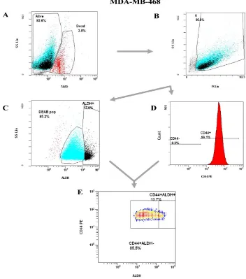
Figure 3. Representative flow cytometry gating strategy for analysis of ALDH activity
and/or CD44 cell surface expression in SUM159 human breast cancer cells. Whole cell populations of SUM159 breast cancer cells were harvested and labelled with 7-AAD, CD44-PE, and the Aldefluor™ assay kit. Analysis was performed using a three-colour multi-parameter gating strategy on a Beckman Coulter EPICS XL-MCL flow cytometer.
(A) Viable cells were identified based on 7-AAD exclusion, and (B) gated based on forward scatter. Cells satisfying viability criteria were then assessed for (C) ALDH activity relative to a DEAB control, and (D) CD44 expression relative to a cells only control. (E)
Figure 4. Representative flow cytometry gating strategy for Analysis of ALDH
activity and/or CD44 cell surface expression in adherent MDA-MB-468 human breast
cancer cells. Adherent whole cell populations of MDA-MB-468 breast cancer cell lines were harvested and labelled with 7-AAD, CD44-PE, and the Aldefluor™ assay kit. Analysis was performed using a three-colour multi-parameter gating strategy on a Beckman Coulter EPICS XL-MCL flow cytometer. (A) Viable cells were identified based on 7-AAD exclusion, and (B) gated based on forward scatter. Cells satisfying viability criteria were then assessed for (C) ALDH activity relative to a DEAB control, and (D)
Figure 5. Representative flow cytometry gating strategy for analysis of ALDH
activity and/or CD44 cell surface expression in non-adherent MDA-MB-468 human
breast cancer cells. Non-Adherent cell populations of MDA-MB-468 breast cancer cell lines generated after exposure to organ-CM were harvested and labelled with 7-AAD, CD44-PE, and the Aldefluor™ assay kit. Analysis was performed using a three-colour multi-parameter gating strategy on a Beckman Coulter EPICS XL-MCL flow cytometer.
(A) Viable cells were identified based on 7-AAD exclusion, and (B) gated based on forward scatter. Cells satisfying viability criteria were then assessed for (C) ALDH activity relative to a DEAB control, and (D) CD44 expression relative to cells only control. (E)
RT-qPCR Analysis
MDA-MB-468 and SUM159 cells exposed to lung-CM, BM-CM, or basal media for 24 hours were harvested (≤ 5 x 10⁵ cells) and lysed using RLT lysis buffer (Qiagen, Germany). Total RNA extraction was performed using a column based RNA purification method (RNeasy® Micro kit, Qiagen, Germany). RNA quality and concentration was determined using the NanoDrop One apparatus (Thermo Fisher Scientific, USA), and RNA was stored at -80°C.
Analysis of ALDH1A1, ALDH1A3, and CD44 Gene
Expression
Subsequent cDNA synthesis was completed (Invitrogen, USA), combined with Supergreen Mastermix (Wisent Bioproducts, CA) and custom primer sets that were designed to detect ALDH1A1, ALDH1A3, and CD44 mRNA expression (Table 2). Samples were loaded onto 96-well plates and loaded onto the Stratagene Mx3000p instrument. The thermal profile setup began at 95 °C for 5 min to allow cDNA to denature, followed by 40 cycles at 95 °C for 15 seconds and 60 °C for 30 seconds.
Relative quantification was performed using a standard curve method with serial dilutions (1, 1:10, 1:100, 1:1000, 1:1 x 10⁴). Data were analyzed using ΔCT values and transcript levels normalized to the internal control glyceraldehyde 3-phosphate dehydrogenase (GAPDH). MXPro software (Agilent, CA, USA) was used for qPCR data analysis.
Quantitative RT-PCR Human Cancer Stem Cell® Array
RNA samples from MDA-MB-468 human breast cancer cells were harvested (~5 x 10⁵
Table 2. Gene List and Primer Sequences
Gene Primer Sequence Source
ALDH1A1
ALDH1A3
CD44
GAPDH
Forward: 5’ – CGT TGG TTA TGC TCA TTT GGA A –3’
Reverse: 5’ – TGA TCA ACT TGC CAA CCT CTG T –3’
Forward: 5’ – ATG CGG ATT GCC AAA GAG GA –3’
Reverse: 5’ – AGC CAA CTT CAG GGC TTT GT –3’
Forward: 5’ – GGG TGT ACA TCC TCA CAT CCA A –3’
Reverse: 5’ – GCT CAC GTC ATC ATC AGT AGG G –3’
Forward: 5’ – TTG CCC TCA ACG ACC ACT TTG T–3’
Reverse: 5’ – AGG GGT CTT ACT CCT TGG AGG C–3’
Integrated DNA Technologies (IDT)
Integrated DNA Technologies (IDT)
Integrated DNA Technologies (IDT)
and concentration was determined using the NanoDrop One apparatus (Thermo Fisher Scientific, USA), and stored at -80°C. Subsequent cDNA synthesis was completed using the RT2 First Strand Kit (Qiagen, Germany), followed by RT-qPCR preparation using RT² SYBR Green ROX qPCR Mastermix (Qiagen, Germany). Samples were then loaded onto preset 96-well RT² Profiler™ PCR Array Human Cancer Stem Cells arrays (Qiagen, Germany). The thermal setup began at 95 °C for 5 min to allow cDNA to denature, followed by 40 cycles of 95 °C for 15 seconds and 60 °C for 30 seconds. RT-qPCR was
performed on the Stratagene Mx3000p instrument, with analysis of ΔCT values performed using MXPro software (Agilent, CA, USA) and Qiagen’s online Data Analysis Center. All transcript levels were normalized to the internal GAPDH control.
Cell Viability Assays
Trypan Blue Exclusion
MCF7, MDA-MB-468, MDA-MB-231, and SUM159 cells were cultured (37°C, 5% CO2)
and doubling time was analyzed to determine initial seeding densities that result in 60% tissue confluency after 48 hours incubation at 37°C (2 x 10⁵, 1 x 10⁵, 7.5 x 10⁴, and 7.5 x 10⁴ cells respectively). Cells were seeded on 6-well plates and grown for 48 hours, allowing cells to re-adhere and adjust to in vitro conditions. Cells were then washed with PBS x 2 and exposed to lung-CM, BM-CM, or basal media for an additional 48 hrs. Viability of resulting non-adherent breast cancer cell subpopulations was performed by collecting 10 µL of media and combining with an equal volume of Trypan Blue (1X, St. Louis, MO,
USA). Total number of cells (live and dead) was enumerated using a hemocytometer under
a light microscope and percentage of viable cells was determined. Representative images of floating and adherent cells were obtained using an inverted Olympus IX70 microscope.