_____________________________________________________________________________________________________
ISSN: 2231-0614, NLM ID: 101570965
Lipoprotein-associated Phospholipase A
2
, Lp-PLA
2
,
and Age, are Predictors for Future Cardiovascular
Events in Acute Coronary Syndrome Patients
Nguyen Van Khoi
1, Tran Thanh Vinh
2, Le Xuan Truong
3, Nguyen Chi Thanh
3,
Nguyen Quoc Tuan
4and Le Ngoc Hung
2,5*1
Department of Thoracic Diseases, Cho Ray Hospital, HCMC, Viet Nam.
2
Department of Biochemistry, Cho Ray Hospital, HCMC, Viet Nam.
3
Department of Biochemistry, The University of Medicine and Pharmacy, HCMC, Viet Nam.
4
Department of Interventional Cardiology, Cho Ray Hospital, HCMC, Viet Nam.
5
Department of Laboratory, Cho Ray-Phnom Penh Hospital, Cambodia.
Authors’ contributions
This work was carried out in collaboration between all authors. Author TTV designed the study, performed the statistical analysis, wrote the protocol and wrote the first draft of the manuscript. Author LNH designed the study, performed the final statistical analysis, wrote the protocol, and wrote the first draft of the manuscript and the final manuscript. Authors NVK and LXT managed the analyses of the study. Authors NCT and NQT managed the literature searches. All authors read and approved the final manuscript.
Article Information
DOI: 10.9734/JAMMR/2017/33587 Editor(s): (1)Alex Xiucheng Fan, Department of Biochemistry and Molecular Biology, University of Florida, USA.
(2)Pietro Scicchitano, Cardiology Department, Hospital “F. Perinei” Altamura (Ba), Italy. (3)Alexander D. Verin, Vascular Biology Center, Georgia Regents University Augusta, Georgia, USA. (4)Domenico Lapenna, Associate Professor of Internal Medicine, Department of Medicine and Aging Sciences, University “G. d’Annunzio” Chieti-Pescara, Chieti, Italy. Reviewers: (1) Bernadette Ngo Nonga, University of Yaounde I, Cameroon. (2)Abraham O. Samson, Bar Ilan University, Safed, Israel. (3)Takashi Ikeno, National Center of Neurology and Psychiatry, Japan. (4)Yunqing Chen, The Second Affiliated Hospital of Chongqing Medical University, China. (5)Andreja Sinkovic, University Clinical Centre Maribor, Slovenia Complete Peer review History:http://www.sciencedomain.org/review-history/19637
Received 21st April 2017 Accepted 16th June 2017 Published 21st June 2017
ABSTRACT
Aims: We investigated whether Lp-PLA2 activity of blood samples collected 24 h after hospital admission could serve as a predictor of future cardiovascular events in Vietnamese patients with acute coronary syndrome (ACS). In addition, we correlated Lp-PLA2 with common risk factors of
ACS, such as age, HDL, diabetes, BMI, etc. and compared Lp-PLA2 levels of ACS patients with those of a control group.
Methods and Results: Lp-PLA2 activity was measured in serum collected in fasting state within 24 h of hospitalization in 293 ACS patients. The mean [SD] of Lp-PLA2 activity (nmol/min/mL) was higher in ACS patients than in controls (212.7[57.8] versus 182.5[58], p<0.001), the mean difference was 14.2% of mean in ACS group. Both non-ST elevated myocardial infarction (NSTEMI) and STEMI had higher Lp-PLA2 compared to unstable angina (UA) (p<0.017). The 3
rd
tertile of Lp-PLA2 had a strong correlation with MI (NSTEMI/STEMI) showing ORs of 3.65 and 3.98 in logistic regression analysis compared to the 2 lower tertiles. There were only two factors, Lp-PLA2 and age, serving as best independent predictors for future CV events in multivariate HR analysis. However their effects strongly began to start around 50 days after acute phase of ACS. The future CV event rates had an increasing trend from 1st to 3rd tertile (ptrend=0.011). The HR between 2nd to 1st tertile was 1.71 (p=0.075). The 3rd tertile had nearly a double of adjusted hazard ratio (HR) as 1.92 compared to the 1st tertile (p=0.029). There was weak association of Lp-PLA2 activity with risk factors of CV diseases.
Conclusion: Lp-PLA2 in acute phase of ACS was different between 3 groups of ACS. Both Lp-PLA2 and age were independent predictors for future cardiovascular events in ACS patients.
Keywords: Lipoprotein-associated phospholipase A2; acute phase; acute coronary syndrome;
predictor.
1. INTRODUCTION
Lipoprotein-associated phospholipase A2 (Lp-PLA2) is a calcium-independent member of the A2 phospholipase superfamily [1], also known as platelet-activating factor acetylhydrolase (PAF-AH), which is produced mainly by macrophages and lymphocytes [2]. The oxidation of LDL to oxLDL yields oxidized phospholipids, that are hydrolyzed solely by Lp-PLA2 producing lysophosphatidylcholine and non-esterified fatty acids. These 2 potent pro-inflammatory and proatherogenic mediators up-regulate the expression of adhesion molecules activating leukocytes and recruiting macrophages and monocytes to atherosclerotic plaques [3]. Importantly, Lp-PLA2 has been found strongly expressed in the vicinity of macrophages of vulnerable and ruptured plaques [4,5]. Lp-PLA2 is a potential novel inflammatory risk factor for coronary artery disease and has been suggested to provide information related and additional to that obtained from traditional lipid analyses [6] and complementary to C-reactive protein [7].
Many epidemiological studies have suggested Lp-PLA2 as an independent predictor of cardiovascular events [7,8,9] and higher levels of Lp-PLA2 have also been associated with stable coronary artery disease in case-control studies [7,10,11]. Those studies worked on subjects with Lp-PLA2 collected at preexisting stable status of cardiovascular diseases. Few data are available concerning the clinical value of Lp-PLA2 collected
in acute phase of acute coronary syndrome (ACS). There were only 2 published studies on Lp-PLA2 in acute phase of ACS patients [12,13]. The levels of Lp-PLA2 in acute phase of ACS patients were slightly higher than that in healthy control subjects [12] or that collected at a delay time, i.e. 30 days, from the first days of ACS [13]. Thus it remains unclear whether Lp-PLA2 could be different between three clinical groups of ACS. In addition, the risk of future cardiovascular events or mortality was not related to Lp-PLA2 levels in ACS patients reported by those 2 studies. The biological role of Lp-PLA2 in ACS patients still remains unclear. The ethnic variations in ACS were suggested to be present [14].
We investigated whether Lp-PLA2 could represent both a biomarker for group classification in early ACS and a predictor for future cardiovascular events in Vietnamese ACS patients. We also examined the correlation between acute-phase Lp-PLA2 and other known risk factors in ACS patients and compared Lp-PLA2 levels of ACS patients with those of Vietnamese healthy control subjects.
2. MATERIALS AND METHODS
2.1 Study Population
In-patients were included in the study if diagnosed as first acute coronary syndrome (ACS). Emergency Department was the first department receiving patients, thereafter patients were referred to Cardiology Department or Cardiology Intervention. Patients were excluded if using lipid-lowering medicine about one year prior to hospitalization or having other
diseases except diabetes and/or arterial
hypertension.
Patients were classified according to three groups of ACS: unstable angina (UA), non-ST elevation myocardial infarction (NSTEMI) and STEMI. UA was defined as unexpected chest pain, usually at rest, no elevation in troponin, with or without ECG changes indicative of ischemia.
The diagnosis of NSTEMI was made when a patient had symptoms of unstable angina, had no ST-segment elevation on the ECG, and had an elevation in cardiac enzymes (troponin I and CK-MB).The ST-Elevation myocardial infarction (STEMI) was defined as the same as NSTEMI but "ST segment" on the ECG appeared "elevated,” reflecting coronary artery total occlusion.
Healthy control subjects were persons who came to the Internal General Clinics of Out-patient Department for health check-up visit for their own reasons. The healthy control group was matched with above ACS patients by age and sex. Only persons who did not have any clinical or history of atherosclerosis and no evidence of pathological electrocardiogram were recruited into study.
The cross-sectional study compared levels of
Lp-PLA2 activity in ACS patients to those in healthy
subject controls matched by age and gender. The cohort study was done in all ACS patients for recording the second cardiovascular events occurred within the maximum follow-up time of 2 years since the hospitalization.
All subjects gave written informed consent. The study was approved by the Medical Ethics Committee of Cho Ray Hospital.
2.2 Data Collection
During the time of hospitalization, patients were interviewed with a prepared questionnaire for
information about smoking status, alcohol behavior, any history of diabetes mellitus, hypertension, hyperlipidemia, current drug use, and family history of early MI.
In all patients directly follow-up was conducted by researchers during the hospitalization until the hospital discharge. Thereafter, patients were followed as out-patients at the Cardiology Intervention Clinics or Cardiology Clinics of Out-Patient Department, Cho Ray Hospital in their own routine visit schedules. Patients could also conduct the health follow-up at local hospitals at their locations. The monitoring of patient follow-up was routinely performed on telephone interview by researchers with patients or their family relatives. The follow-up time was completed when a secondary cardiovascular event occurred and recorded, or censored at 2 years as the maximum time of follow-up.
Secondary cardiovascular events were defined either as cardiovascular disease (CVD) as the main cause of death, nonfatal myocardial infarction (UA, NSTEMI, STEMI), coronary revascularization, or ischemic cerebrovascular event (stroke). The source of data were hospital case records during hospitalization, out-patient record book of each clinics in the out-patient department if patients performed the follow-up at Cho Ray Hospital, or hospital discharge certificate from local hospitals, or treatment sheets reported by authorized primary care private physicians. Other event data were excluded as invalid data.
2.3 Laboratory Methods
Blood samples were collected in fasting state within the first 24 hours after hospital administration. Patients fasted around 10-12 hours after the last meal before measurement of lipid blood panel and phenotype analysis of LDL cholesterol with gradient-gel electrophoresis. All serums samples were collected in standard procedure, thereafter transferred to plain tubes for storing. Serum samples were stored at -80°C until analysis for Lp-PLA2 activity. Other tests, CK-MB, troponin, lipid blood panel were done within day of blood collection as routine performance.
The serum Lp-PLA2 activity (nmol/min/mL) was
determined by the PLAC Test for Lp-PLA2
Activity enzyme assay on automated clinical
chemistry analyzers (Lp-PLA2 Activity;
diaDexusInc, South San Francisco, CA). This test was demonstrated to be equivalent to the legally marketed predicate device, diaDexus
PLAC Test, measuring Lp-PLA2 in concentration
(ng/mL). PLAC Test for Lp-PLA2 Activity
December 2014. The Siemens Advia 1800 Clinical Chemistry Analyzer (Siemens Healthcare Diagnostics, NY, USA) was used to run the
PLAC Test for Lp-PLA2 Activity enzyme assay.
Lp-PLA2 in serum hydrolyzes the sn-2 position
of the substrate,
1-myristoyl-2-(4-nitrophenylsuccinyl) phosphatidylcholine,
producing a colored reaction product, nitrophenol. The rate of formation of
4-nitrophenol was measured
spectrophotometrically for 8.5 minutes and the
Lp-PLA2 activity calculated from the rate of
change in absorbance at 410 nm. A set of five
Lp-PLA2 calibrators was used to generate a
standard curve fit of change in absorbance
versus Lp-PLA2 activity levels in nmol/min/mL.
The kit of PLAC Test for Lp-PLA2 Activity was
included two controls.
The clinical sensitivity (limit of quantification) of the assay was 10 nmol/min/mL with CV of 20%. The intra-assay and inter-assay variability, determined by testing five human pooled serum
samples and two controls with Lp-PLA2 activity in
the calibration range of the assay (65-289 nmol/min/mL) were 0.3-0.8% and 1.6-4.2%, respectively. Linearity was assessed with a dilution series from 3 high level samples, 249-308 nmol/min/mL) to low level samples (73-74
nmol/min/mL), all R2 were from 0.992 to 0.999.
CK-MB and troponin I (TnI-Ultra) in serum were
measured by chemiluminescent immunoassay
on Advia Centaur Immunoassay System
(Siemens Healthcare Diagnostics, NY, USA).
The blood lipid panel, including total cholesterol, triglycerides, HDL-C, LDL-C, and blood glucose
were measured by spectrophotometry on
Mindray BS-800 (Mindray, Shenzhen, China).
2.4 Statistical Methods
The body mass index (BMI) was calculated by dividing the patient’s weight in kilograms by the square of the patient’s height inmeters. Patients were classified as underweight (BMI <18.5 kg/m2), normal weight (BMI 18.5–24.9 kg/m2), overweight (BMI 25–29.9 kg/m2), and obese (BMI ≥ 30 kg/m2). Smoking was defined as current smoking or previously smoked. Alcohol behavior meant drinking alcohol almost everyday or being alcoholism. Family history of early MI was positive in the case of any documented CVD of one first-degree relative at aged less than 55 years. Diabetes mellitus was diagnosed if
patients had history of previously undergone dietary treatment, additional oral antidiabetic or insulin medication, or had laboratory results of fasting plasma glucose (FPG) equal or higher than 126 mg/dL (7 mmol/L). Hypertension was defined as systolic blood pressure ≥ 140 mmHg and/or diastolic blood pressure ≥ 90 mmHg and/or in using antihypertensive drugs.
The distribution of Lp-PLA2 activity was normal, z scores of Skewness and Kurtosis less than 2.0, therefore Lp-PLA2 activity was presented as mean (SD). Other quantitative data were also presented as mean (SD), else stated. Qualitative data were reported in number of cases and proportions. Sociodemographic, clinical, laboratory and Lp-PLA2 activity characteristics of ACS patients and healthy control subjects were presented as means (SD) or proportions, as appropriate.
The parametric tests, t-test and ANOVA, were used for comparing means between 2 or ≥ 3 independent quantitative groups, respectively. For ANOVA test, if p values were less than 0.05, the post hoc Bonferroni test was used to identify different paired groups with p = 0.05/n[n-1]/2.
The associations of sociodemographic data and cardiovascular risk factors with Lp-PLA2 activity were evaluated by Chi-quare χ2 test.
Partial Spearman correlation coefficients were calculated for Lp-PLA2 activity and age; blood lipids (total cholesterol, triglycerides, HDL-C, LDL-C).
The odds ratios, 2x2 tables, were used for univariate analysis of correlations between Lp-PLA2 in each of pairs of tertiles. Multinomial logistic regression was used for adjusted OR compared between 3rd tertile to 1st tertile in correlation with Lp-PLA2 by gender and age groups.
(yes/no), family history of early MI (yes/no), total cholesterol (mg/dL), HDL-C (mg/dL), triglycerides (mg/dL), LDL-C (mg/dL), was assessed in univariate analysis. Only those having the significant value (p<0.05) were included in multivariable analysis of hazard ratio (HR) Cox regression.
All statistical tests were two-sided and p-values of less than 0.05 were considered significant.
3. RESULTS
Overall, 323 ACS patients were enrolled into study. A total of 293 patients (90.7%) completed the follow-up after hospital discharge until the second cardiovascular event occurred or maximum of 2 year follow-up. There were 30 patients (9.3%) lost in follow-up including 9 who refused to be followed up, 13 who could not be contacted and 8 who did not obey a standard therapy. A total of 91 healthy control subjects were also included in the study.
Table 1 shows the sociodemographic, clinical, laboratory and Lp-PLA2 activity characteristics between ACS patients and healthy controls. Triglycerides and HDL-cholesterol, but not total- and LDL-cholesterol, were different between ACS patients and controls. Lp-PLA2 activity was statistically higher in ACS patients compared to controls, 212.7±57.8 versus 182.5±58 nmol/min/mL, respectively. The mean difference of 30 nmol/min/ml was lower than the SD, i.e. 58
nmol/min/mL, of data distribution of both groups. The mean difference was 14.2% compared to mean of Lp-PLA2 activity in ACS group.
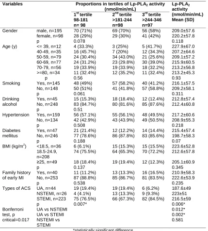
Table 2 shows the relationship of various cardiovascular risk factors (gender, age, smoking, drinking alcohol, BMI, hypertension, diabetes mellitus, family history of early MI) and 3 types of ACS with Lp-PLA2 activity. There was no association between Lp-PLA2 activity and any of cardiovascular risk factors. There was a strong and positive relationship between Lp-PLA2 and 3 types of ACS. Post-hoc analysis (Bonferroni test) showed that Lp-PLA2 activity was higher in both NSTEMI and STEMI than in UA (p<0.017).
Table 3 shows the weak correlations between lipid variables and LP-PLA2 activity. There was no correlation between Lp-PLA2 and triglycrides (p=0.213). Total cholesterol and LDL-cholesterol were positively associated with Lp-PLA2 (r=0.135 and r=0.145, respectively). HDL-cholesterol had a negative correlation. All correlations coefficients were statistically significant (p<0.02) but of weak intensity (r < 0.3).
Table 4 shows that 3rd tertile of Lp-PLA2 had a strong correlation with myocardial infarction MI (NSTEMI/STEMI) showing ORs of 3.65 in univariate analysis and 3.98 in multinominal logistic regression analysis adjusted by age and gender compared to that of the 2 lower tertiles. The sensitivity and specificity of 3rd tertile of Lp-PLA2 for MI was 36.5% and 86.4%, respectively.
Table 1. Sociodemographic, clinical, laboratory and Lp-PLA2 activity characteristics between
ACS patients and healthy control subjects
Parameters ACS patients
n = 293
Healthy control subjects n = 91
P!
Gender: men, n (%) 195 (66%) 54 (59%) 0.21
Age, y (µ, SD) 62.6±12.7 63.6±11.1 0.48
Smoking status: yes, n (%) 145 (50%) 37 (41%) 0.14
Alcohol drinking: yes, n (%) 45 (16%) 11 (12%) 0.44
Family history of early MI, n (%) 40 (14%) 0 (0%) NA
Arterial hypertension, n (%) 159 (54%) 0 (0%) NA
Diabetes mellitus, n (%) 47 (16%) 0 (0%) NA
BMI, kg/m2, n (%) <18.5 36 (12%) 8 (9%) 0.24
18.5-24.9 208 (71%) 77 (85%)
≥25 49 (17%) 6 (7%)
Total cholesterol, mg/dL (µ, SD) 190±50 183±39 0.21
Triglycerides, mg/dL (µ, SD) 211±61 189±54 0.002*
HDL-C, mg/dL (µ, SD) 34.1±10.0 39.9±9.6 0.001*
LDL-C, mg/dL (µ, SD) 115±43 116±37 0.78
Table 2. Lp-PLA2 activity in relation to baseline characteristics and types of ACS
Variables Proportions in tertiles of Lp-PLA2 activity
(nmol/min/mL)
Lp-PLA2
activity
(nmol/min/mL) Mean (SD) 1st tertile
98-181 n= 98
2nd tertile >181-244 n=98
3rd tertile >244-346 n=97
Gender male, n=195 70 (71%) 69 (70%) 56 (58%) 209.0±57.6
female, n=98 28 (29%) 29 (30%) 41 (42%) 220.2±57.8
p 0.078 0.118
Age (y) <= 39, n=12 4 (33.3%) 3 (25%) 5 (41.7%) 227.9±67.0 40-49, n=35 16 (45.7%) 7 (20%) 12 (34.3%) 207.2±64.6 50-59, n=79 24 (30.4%) 34 (43.0%) 21 (25.6%) 209.1±57.2 60-69, n=77 24 (31.2%) 23 (29.8%) 30 (39.0%) 215.9±60.5 70-79, n=56 19 (33.9%) 19 (33.9%) 18 (32.2%) 213.2±56.8 >=80, n=34 11 (32.4%) 12 (35.2%) 11 (32.4%) 213.2±45.3
p 0.56 0.93
Smoking Yes, n=145 48 (49%) 57 (58.2%) 40 (41.2%) 216.1±57.5 No, n=148 50 (51%) 41 (41.8%) 57 (58.8%) 209.2±58.1
p 0.061 0.311
Drinking alcohol
Yes, n=45 15 (15.3%) 18 (18.4%) 12 (12.4%) 212.8±57.4 No, n=248 83 (84.7%) 80 (81.6%) 85 (87.6%) 212.4±60.8
p 0.51
Hypertension Yes, n=159 56 (57.1%) 55 (56.1%) 48 (49.5%) 217.2±60.6 No, n=134 42 (42.9%) 43 (43.9%) 49 (50.5%) 208.9±55.3
p 0.508 0.218
Diabetes mellitus
Yes, n=47 21 (21.4%) 12 (12.2%) 14 (14.4%) 215.4±57.4 No, n=246 77 (78.6%) 86 (87.8%) 83 (85.6%) 198.7±58.3
p 0.188 0.07
BMI (kg/m2) <18.5, n=36 6 (6.1%) 15 (15.3%) 15 (15.5%) 223.6±52.8 18.5-24.9,
n=208
74 (75.5%) 64 (65.3%) 70 (72.2%) 212.6±57.8
≥25, n=49 18 (18.4%) 19 (19.4%) 12 (12.3%) 205.1±60.9
p 0.137 0.345
Family history of early MI
Yes, n=40 11 (11.2%) 13 (13.3%) 16 (16.5%) 210.9±58.3 No, n=253 87 (88.8%) 85 (86.7%) 81 (83.5%) 222.6±53.9
p 0.538 0.235
Types of ACS
Bonferroni test, p critical=0.017
UA, n=44 19 (19.4%) 19 (19.4%) 6 (6.2%) 187.6±49 NSTEMI, n=26 4 (4.1%) 13 (13.3%) 9 (9.3%) 223±51 STEMI, n=223 75 (76.5%) 66 (67.3%) 82 (84.5%) 216.5±59
p 0.007* 0.006*
UA vs NSTEMI 0.012*
UA vs STEMI 0.002*
NSTEMI vs STEMI
0.581
*statistically significant difference
Table 3. Spearman correlation coefficients (R) between lipid variables and Lp-PLA2 activity
Lipid variables R p
Total cholesterol, mg/dL 0.135 0.021* Triglycerides, mg/dL -0.073 0.213 HDL-cholesterol, mg/dL -0.158 0.007* LDL-cholesterol, mg/dL 0.145 0.013*
*
statistically significant difference
During a follow-up of 2.0 years, 86 major cardiovascular events occurred in 293 patients (the Kaplan–Meier estimated event rate was
29.4% at 2 years) including: 29 dead cases (5 death cases occurred during hospitalization),
21 with recurrent myocardial infarction, 5 with ischemic strokes, 7 with coronary
Table 5 shows the Cox proportional hazard (HR) in univariate analysis between each of risk factors with cardiovascular events and the adjusted HR in multivariate analysis for those having the significant value of HR (p < 0.05) in univariate analysis. There were 3 factors having positive HR in univariate analysis: age, gender and Lp-PLA2. The factor of gender was totally attenuated in multivariate analysis. The only 2 independent risk factors for cardiovascular events in 2-year follow-up were age (adjusted HR: 1.036, p=0.001) and Lp-PLA2 activity tertiles (adjusted HR: 1.376, p=0.027). This means that the increase of age of one year will increase HR 3.6%, and the change of one tertile will increase HR 37.6%.
Table 6 shows the future cardiovascular event rates having a trend to increase from 1st tertile to 3rd tertile of Lp-PLA2 (ptrend=0.011). The HR compared between 2nd tertile to 1st tertile (as reference) was so high as 1.71 (increased 71% HR of events) but not reaching the level of
statistical significance (p=0.075). The 3rd tertile had nearly double of risk, adjusted HR of 1.92 (increased 92% HR of events) when compared to the 1st tertile of Lp-PLA2 (p=0.029).
Fig. 1 shows the cumulative probability (cum hazard) of major cardiovascular events during 2-year follow-up according to tertiles of Lp-PLA2 levels in acute phase of ACS in 293 patients. The overall comparison between 3 levels of tertiles showed a significant difference (p=0.043, Log-Rank test). The pairwise comparisons between each pair of tertiles were as follows: 1st tertile vs. 2nd tertile (p=0.05); 1st tertile vs. 3rd tertile (p=0.014); and 2nd tertile vs. 3rd tertile (p=0.628). In Fig. 1, the curves showed an increase in the cumulative probability of a second cardiovascular event during 2-year follow up. This trend was particularly pronounced in the 2nd and 3rd tertiles of Lp-PLA2 activity after 50 days of follow up. However, before 50 days, the cumulative probability is independent of Lp-PLA2 activity.
Table 4. The correlation (OR) between 3 types of ACS and Lp-PLA2 activity
Tertiles of Lp-PLA2
activity
Types of ACS (n) Univariate analysis Multinomial logistic
regression analysis, adjusted by gender and age groups
UA MI
(NSTEMI/STEMI)
OR CI 95% p OR CI 95% p
Tertile 1st 19 79 1
Tertile 2nd 19 79 1 0.49-2.03 1
Tertile 3rd 6 91 3.65 1.39-9.60 0.006*
Tertile 1st + 2nd 38 158 1
Tertile 3rd 6 91 3.65 1.48-8.96 0.03* 3.98 1.6-9.8 0.003* *
statistically significant difference
Table 5. Association of Lp-PLA2 activity in acute phase of ACS with major cardiovascular
events during 2-year follow-up
Parameters Units Univariate analysis Multivariate analysis
HR (95% CI) p Adjusted HR (95% CI) p
Age year 1.031 (1.013-1.049) 0.001* 1.036 (1.015-1.057) 0.001* Gender male 0.613 (0.4-0.94) 0.025* 0.697 (0.403-1.207) 0.198 Smoking yes 0.84 (0.549-1.284) 0.421 1.222 (0.696-2.145) 0.484 Drinking alcohol yes 0.959 (0.531-1.729) 0.888 1.316 (0.676-2.561) 0.419 BMI ≥25 kg/m2 0.911 90.505-1.644) 0.757 1.079 (0.59-1.973) 0.806 Hypertension yes 0.916 90.6-1.4) 0.686 0.716 (0.446-1.150) 0.167 Diabetes mellitus yes 1.116 90.639-1.948 0.7 1.189 90.633-2.231) 0.59 Family history early
MI
yes 0.94 (0.499-1.772) 0.849 1.214 (0.623-2.367) 0.569
Total cholesterol mg/dL 1.0 90.995-1.004) 0.882 0.988 (0.973-1.004) 0.139 HDL-cholesterol mg/dL 1.002 90.981-1.023) 0.874 1.005 (0.98-1.031) 0.679 LDL-cholesterol mg/dL 1.001 (0.996-1.006) 0.694 1.014 (0.996-1.031) 0.126 Triglycerides mg/dL 0.998 (0.995-1.002) 0.29 1.0 (0.996-1.005) 0.877 Lp-PLA2 tertile 1.374 91.056-1.789) 0.018* 1.376 (1.037-1.826) 0.027*
*
Fig. 2 shows the cumulative probability (cum hazard) of major cardiovascular events during
2-year follow-up according to 5 age groups, <= 39, 40-49, 50-59, 60-69, 70-79 and ≥ 80 y, in
acute phase of ACS in 293 patients. The overall comparison between 5 age groups showed a
significant difference (p=0.021Log-Rank test). The pairwise comparisons between each pair of age groups showed only the difference between
age group ≥80 y versus age groups ≤ 59 y (p < 0.05).
Table 6. Kaplan-Meier event rates and adjusted HRs (95% CIs) by Lp-PLA2 activity tertiles at
2-year follow-up
Lp-PLA2
activity nmol/min/mL
N Event
rates by Tertile, %
Univariate analysis Multivariate analysis
n (%) Unadjusted HRs
(95% CI)
p Adjusted HRs
(95% CI)**
p
1st tertile 98-181
98 19 (19.4%) 1 reference
2nd tertile >181-244
98 32 (32.6%) 1.75 (0.992-3.008) 0.053 1.71 (0.947-3.091) 0.075
3rd tertile >244-346
97 35 (36.1%) 1.978 (1.131-3.458) 0.017* 1.92 (1.069-3.447) 0.029*
pChi-square 0.026
ptrend 0.011
*
statistically significant difference **
adjusted by age, gender, smoking, drinking alcohol, BMI ≥ 25 kg/m2, hypertension, diabetes mellitus, family history of early MI, cholesterol total, HDL-cholesterol, LDL-cholesterol and triglycerides
Fig. 1. Cumulative probability of second cardiovascular events during 2-year follow-up according to tertiles of Lp-PLA2 activity in acute phase of ASC in 293 patients (p=0.043 by
Fig. 2. Cumulative probability of second cardiovascular events during 2-year follow-up according to 5 groups of age in acute phase of ASC in 293 patients (p=0.021 by Log-Rank test)
4. DISCUSSION
Our study investigated Lp-PLA2 activity in acute phase of ACS in 293 Vietnamese patients and found some relevant results. Lp-PLA2 activity was a biomarker for group classification in ACS. Patients with Lp-PLA2 activity in the 3
rd
tertile had risk of STEMI nearly 4 times (3.98 [1.69-9.8]: OR [95% CI]) compared to the 2 lower tertiles (Table 4). Lp-PLA2 activity in acute phase was also an independent risk factor for future cardiovascular events in multivariate analysis, after adjustment for a wide range of routine risk factors, over 2-year follow-up after acute phase of ACS. There was about 2-fold increased risk, 1.92 [1.069-3.447] (adjusted HR [95% CI) (Table 6) of future CV events in patients with Lp-PLA2 activity in the 3rd tertile compared to those in the 1st tertile. Finally, Lp-PLA2 activity in acute phase of ACS patients was higher than that of healthy volunteer controls.
The Lp-PLA2 activity in ACS patients was significantly higher than that in healthy controls (p=0.001), but the difference was not so wide, 212 versus 182 nmol/min/mL. The mean
difference of 30 nmol/min/ml was lower than the SD, i.e. 58 nmol/min/mL, of data distribution of both groups. The slightly higher Lp-PLA2 activity in ACS patients compared to healthy persons observed in our study was similar to that previously reported by Oldgren et al. [12] In ACS patients. The same findings were reported in previous case-control studies in stable coronary disease [11,15,16]. In the study by Blankenberg et al. [16] patients with ACS had higher Lp-PLA2 activity than those with stable coronary disease. The mean levels of Lp-PLA2 activity were slightly lower at 30 days of follow-up than at baseline, 35.7 vs 40.9 nmol/min/mL (p<0.001) in PROVE IT-TMTI 22 as reported by O’Donoghue et al. [13]. These data suggest that Lp-PLA2 could not be as other acute-phase reactants (inflammatory markers) such as C-reactive protein (CRP) and interleulin-6 [6]. Lp-PLA2 was weakly associated with CRP and fibrinogen in the FRISC II study, indicating that it could be a marker of more chronic low-grade inflammation in ACS [6,12,15]. In our study, we found the strong relationship between Lp-PLA2 activity and the highest pathological type of ASC, i.e., STEMI. Patients with Lp-PLA2 activity in the 3
rd
STEMI nearly 4 times higher compared to that of the other 2 lower tertiles. Thus, Lp-PLA2 activity could be considered as a pathological chronic inflammatory marker for ACS.
Lp-PLA2 activity in acute phase of ACS was not related to the number of diseased vessels detected at coronary angiography and was considered to be not related with the severity of coronary artery disease in the study by Oldgren et al. [12]. Lp-PLA2 was not useful for risk stratification when measured early after ACS in PROVE IT-TIMI 22 Trial [13]. The result in our study was different from these 2 studies. PL-PLA2 activity was significantly different between the 3 types of ACS. Both NSTEMI and STEMI had higher PL-PLA2 activity than UA. In addition, the multinominal logistic regression showed that patients with Lp-PLA2 in the 3
rd
tertile had nearly 4 times STEMI compared to those in the 1st tertile. The reasons for difference could be due to the retrospective analyses in the study by Oldgren et al. [12], and the delay time for sample collection, around 7 days after the onset of ACS event, in the study by O’Donoghue et al. [13]. Another reason may be the exclusion criteria of no use of lipid-lowering substances at least one year before entering into our study. This matter could have been contributed to the higher study population homogeneity in our study compared to previously published studies. The level of Lp-PLA2 activity in STEMI patients was higher than that in UA group in our study. In contrast, the mean level of Lp-PLA2 was lowest in patients with STEMI in PROVE IT-TIMI 22 [13]. The Lp-PLA2 activity has been reported to be higher in patients with ACS than with stable angina [16]. Thus Lp-PLA2 activity may have clinical value in risk stratification in acute phase of ACS, as well as in stable coronary artery disease.
The other relevant finding of our study was the predictive value of Lp-PLA2 in acute phase of ACS for future cardiovascular events. No previous studies on Lp-PLA2 activity in ACS patients had the same conclusion as our study [12,13]. There was some explanation for our findings. Lp-PLA2 was not considered as an acute-phase reactant, since its levels in acute phase were only slightly higher than those occurring in stable condition (about 16.5% higher). Thus, Lp-PLA2 in acute phase of ACS may have the same role in prediction of future cardiovascular events compared to that in stable status of ACS. Our study showed that patients with 3rd tertile of Lp-PLA2 activity had around 2-fold increase of relative hazard of cardiovascular
events compared to patients with 1st tertile (p=0.029). Patients with Lp-PLA2 in 2
nd
tertile had 75% of increase of hazard ratio of CV events compared to patients of 1st tertile, but not yet reaching the level of statistical significance (p=0.075). The predictive value of Lp-PLA2 in stable coronary heart diseases had been reported in many previous studies [8,17,18,19]. Thus, our study suggests that Lp-PLA2 may have in acute phase of ACS the same predictive value as in stable coronary artery disease.
Besides Lp-PLA2, age was also an independent risk factor for future CV events (p=0.021, Log Rank test). However, the pairwise comparisons between each pair of age groups showed only the difference between age group ≥80 y versus age groups ≤ 59 y (p < 0.05).
Finally, Lp-PLA2 activity in acute phase of ACS had positive association with total cholesterol and LDL-cholesterol, and negative association with triglycerides and HDL-cholesterol (p< 0.05). However, the strength of association was weak with r < 0.3. This result was the same as reported with Lp-PLA2 collected in stable phase of ACS or coronary artery diseases [12,16,19] or in acute phase of ACS [12,13]. The weak relationships between Lp-PLA2 activity with other standard risk factors of cardiovascular diseases suggest that Lp-PLA2 is not a risk factor for ACS but a biomarker for severity classification of ACS.
Limitations to the present study included the use of questionnaire on investigation about history of taken lipid-lowering medication one year before entering into the study and the on-phone interview for cases lost from routine follow-up schedules. It could have a number of patients being used lipid-lowering drugs but not revealed. Furthermore, not all patients or their relatives had good knowledge on diseases as well as having proven documents for confirmation of cardiovascular events. However, researchers had to try all best ways to get the correct data and exclude all bias. Another limitation was the independence of CV events from Lp-PLA2 activity during early time of around 50 days from the first day of acute coronary syndrome (first day of hospitalization). Therefore the practical prognostic values of Lp-PLA2 activity in ACS patients will be continued to investigate.
5. CONCLUSION
heart disease in both risk stratification in acute phase of ACS and in prediction for future cardiovascular events.
CONSENT
As per international standard or university standard, patient’s written consent has been collected and preserved by the authors.
ETHICAL APPROVAL
As per international standard or university standard, written approval of Ethics committee has been collected and preserved by the authors.
COMPETING INTERESTS
Authors have declared that no competing interests exist.
REFERENCES
1. Tew DG, Southan C, Rice SQ, Lawrence MP, Li H, Boyd HF, Moores K, Gloger IS, Macphee CH. Purification, properties, sequencing, and cloning of a lipoprotein-associated, serine-dependent phospholipase involved in the oxidative modification of low-density lipoproteins. Arterioscler Thromb Vasc Biol. 1996;16:591–599.
2. Asano K, Okamoto S, Fukunaga K, Shiomi T, Mori T, Iwata M, Ikeda Y, Yamaguchi K. Cellular source(s) of platelet-activating factor acetylhydrolase activity in plasma. Biochem Biophys. Res. Commun. 1999; 261:511-514.
3. Caslake MJ, Packard CJ, Suckling KE, Holmes SD, Chamberlain P, Macphee CH. Lipoprotein-associated phospholipase a(2), platelet-activating factor acetylhydrolase: A potential new risk factor for coronary artery disease. Atherosclerosis. 2000;150:413– 419.
4. Epps KC, Wilensky RL. Lp-PLA2 – a novel risk factor for highrisk coronary and carotid artery disease. J. Intern. Med. 2011;269: 94-106.
5. Searle J, Danne O, Müller C, Möckel M. Biomarkers in acute coronary syndrome and percutaneous coronary intervention. Minerva Cardioangiol. 2011;59:203-23. 6. James SK, Oldgren J, Lindback J,
Johnston N, Siegbahn A, Wallentin L. An acute inflammatory reaction induced by
myocardial damage is superimposed on a chronic inflammation in unstable coronary artery disease. Am. Heart J. 2005;149: 619–626.
7. Tselepis AD, Dentan C, Karabina SA, Chapman MJ, Ninio E. PAF-degrading acetylhydrolase is preferentially associated with dense LDL and VHDL-1 in human plasma. Catalytic characteristics and relation to the monocyte-derived enzyme. Arterioscler. Thromb. Vasc. Biol. 1995;15:1764–1773.
8. Koenig W, Khuseyinova N, Lowel H, Trischler G, Meisinger C. Lipoprotein-associated phospholipase A2 adds to risk prediction of incident coronary events by C-reactive protein in apparently healthy middle-aged men from the general population: results from the 14-year follow-up of a large cohort from southern Germany. Circulation. 2004;110:1903– 1908.
9. Hakkinen T, Luoma JS, Hiltunen MO, Macphee CH, Milliner KJ, Patel L, Rice SQ, Tew DG, Karkola K, Yla-Herttuala S. Lipoprotein-associated phospholipase A(2), platelet-activating factor acetylhydrolase, is expressed by macrophages in human and rabbit atherosclerotic lesions. Arterioscler. Thromb. Vasc. Biol. 1999;19:2909–2917. 10. Oei HH, van der Meer IM, Hofman A,
Koudstaal PJ, Stijnen T, Breteler MM, Witteman JC. Lipoprotein-associated phospholipase A2 activity is associated with risk of coronary heart disease and ischaemic stroke: the Rotterdam study. Circulation. 2005;111:570–575.
11. Khuseyinova N, Imhof A, Rothenbacher D, Trischler G, Kuelb S, Scharnagl H, Maerz W, Brenner H, Koenig W. Association between Lp-PLA2 and coronary artery disease: Focus on its relationship with lipoproteins and markers of inflammation and hemostasis. Atherosclerosis. 2005; 182:181–188.
12. Oldgren J, James SK, Siegbahn A, Wallentin L. Lipoprotein – associated phospholipase A2 does not predict mortality or new ischaemic events in acute coronary syndrome patients. European Heart Journal. 2007;28:699–704.
acute coronary syndromes in the PROVE IT‐TIMI 22 (PRavastatin Orator Vastatin Evaluation and Infection Therapy‐Thrombolysis In Myocardial Infarction) Trial. Circulation. 2006;113: 1745–1752.
14. Lanza GA. Ethnic variations in acute coronary syndromes. Heart. 2004;90:595-597.
15. Oldgren J, Wallentin L, Grip L, Linder R, Norgaard BL, Siegbahn A. Myocardial damage, inflammation and thrombin inhibition in unstable coronary artery disease. Eur. Heart J. 2003;24:86–93. 16. Blankenberg S, Stengel D, Rupprecht HJ,
Bickel C, Meyer J, Cambien F, Tiret L, Ninio E. Plasma PAF-acetylhydrolase in patients with coronary artery disease: Results of a cross-sectional analysis. J. Lipid Res. 2003;44:1381–1386.
17. Koenig W, Twardella D, Brenner H, Rothenbacher D. Lipoprotein‐associated phospholipase A2 predicts future
cardiovascular events in patients with coronary heart disease independently of traditional risk factors, markers of inflammation, renal function, and hemodynamic stress. Arterioscler. Thromb. Vasc. Biol. 2006;26:1586–1593.
18. Ballantyne CM, Hoogeveen RC, Bang H, Coresh J, Folsom AR, Heiss G, Sharrett AR. Lipoprotein-associated phospholipase A2, high-sensitivity creactive protein, and risk for incident coronary heart disease in middle-aged men and women in the atherosclerosis risk in communities (ARIC) study. Circulation. 2004;109:837–842. 19. Packard CJ, O’Reilly DS, Caslake MJ,
McMahon AD, Ford I, Cooney J, Macphee CH, Suckling KE, Krishna M, Wilkinson FE, Rumley A, Lowe GD. Lipoprotein-associated phospholipase A2 as an independent predictor of coronary heart disease. West of Scotland Coronary Prevention Study Group. N. Engl. J. Med. 2000;343:1148–1155.
_________________________________________________________________________________ © 2017 Khoi et al.; This is an Open Access article distributed under the terms of the Creative Commons Attribution License (http://creativecommons.org/licenses/by/4.0), which permits unrestricted use, distribution, and reproduction in any medium, provided the original work is properly cited.
Peer-review history: