_____________________________________________________________________________________________________
British Journal of Medicine & Medical Research
5(2): 270-274, 2015, Article no.BJMMR.2015.027 ISSN: 2231-0614
SCIENCEDOMAIN international
www.sciencedomain.org
Left Ventricular Noncompaction: A Rare Cause of
Heart Failure in a HIV Patient
Dane D. Gruenebaum
1*, Jagadeesh K. Kalavakunta
2, Joel M. Cohn
2and Heather S. Laird-Fick
11
Michigan State University, Department of Internal Medicine, East Lansing, MI, USA.
2
Michigan State University/Sparrow Hospital, Department of Cardiology, East Lansing, MI, USA.
Authors’ contributions
All the four authors contributed to the conception, design, analysis and preparation of the manuscript. All authors read and approved the final manuscript.
Article Information
DOI:10.9734/BJMMR/2015/12983
Editor(s):
(1) Jimmy T. Efird, Department of Public Health, Director of Epidemiology and Outcomes Research East Carolina Heart Institute, Brody School of Medicine, Greenville, North Carolina, USA.
Reviewers:
(1) Anonymous, East Medical Center Higashi Municipal Hospital City of Nagoya, Japan. (2)Mra Aye, Medicine, Melaka Manipal Medical College, Melaka, Malaysia. Peer review History:http://www.sciencedomain.org/review-history.php?iid=661&id=12&aid=5999
Received 26th July 2014 Accepted 18th August 2014 Published 9th September 2014
ABSTRACT
Background: Heart failure in patients with human immunodeficiency virus (HIV) is often from dilated cardiomyopathy as a result of HIV itself, drug myotoxicity, secondary infections, or drug-induced atherosclerosis. Left ventricular noncompaction (LVNC) is a rare cardiac congenital abnormality which occurs due to early arrest of endomyocardial morphogenesis.
Case: A 47- year-old female patient with HIV presented with sudden onset shortness of breath and symptoms of congestive heart failure. Echocardiography showed noncompacted endocardium with reduced left ventricular function. She was subsequently diagnosed with LVNC.
Discussion: Multiple etiologies have been implicated in cardiomyopathy among HIV patients. LVNC is a rare cause of left ventricular failure, particularly in this population. Echocardiography plays a pivotal role in the diagnosis.
Conclusion: It is often challenging to identify the underlying cause of cardiomyopathy in a patient with HIV. While LVNC is a rare cause of left ventricular failure, typical findings on echocardiography can obviate the need for a more complex evaluative strategy.
Keywords: Left ventricular noncompaction; Human immunodeficiency virus infection; heart failure; Echocardiography.
1. BACKGROUND
The Joint United Nations Program estimated million people were living with
immunodeficiency virus (HIV) infection at the end of 2012 [1]. Heart failure in this population is often a result of dilated cardiomyopathy from HIV itself, drug myotoxicity, secondary infections, nutritional deficiencies or drug
atherosclerosis. Left ventricular noncompaction (LVNC) is a rare cardiac congenital
which occurs due to embryological arrest of endomyocardial morphogenesis between 4 weeks. We present a rare cause of cardiomyopathy due to LVNC in a 47
woman with HIV infection.
2. CASE
A 47 year-old Caucasian woman
year history of HIV infectionpresented to the emergency department withseven hours of worsening shortness of breath, paroxysmal nocturnal dyspnea, and orthopnea unreliev with albuterol nebulization. She also complained of malaise and palpitations, but denied fever, chills, chest pain, or diaphoresis.
Her past medical history was significant for HIV infection, hypertension (diagnosed a few months prior to the presentation), anemia, and history of poly-substance abuse. Medications included emtricitabine-tenofovir, darunavir,
metoprolol, promethazine, and amlodipine. She was a current half pack per day smoker,
regularly consumed three beers a week since age 21. Shelast used cocaine two days previously, and methamphetamine onemonth ago, (she reported these as sporadic usage, drug screens from other admissions were negative). Family history was negative for premature coronary artery disease or congestive heart failure.
Physical examination was significant for heart rate of 105 beats/minute, cachetic appearance conjunctival pallor, bibasilar crackles, and a 2/6 systolic murmur, most prominent in the mitral area.
Laboratory tests revealed elevated brain natriuretic peptide (BNP) at 492 pg/ml (normal <100 pg/ml), and negative cardiac enzymes with troponin levels consistently below 0.02 ng/ml (normal 0.00-0.03 ng/ml). Hemoglobin was 7.6 grams/dL, MCV of 65 fL, MCH 20.3 pg, MCHC
Gruenebaum et al.; BJMMR, 5(2): 270-274, 2015; Article no. BJMMR.20
The Joint United Nations Program estimated 35 million people were living with human (HIV) infection at the end Heart failure in this population is often a result of dilated cardiomyopathy from HIV itself, drug myotoxicity, secondary infections, nutritional deficiencies or drug-induced Left ventricular noncompaction (LVNC) is a rare cardiac congenital abnormality, embryological arrest of endomyocardial morphogenesis between 4-8 weeks. We present a rare cause of cardiomyopathy due to LVNC in a 47-year-old
with a seven infectionpresented to the emergency department withseven hours of worsening shortness of breath, paroxysmal nocturnal dyspnea, and orthopnea unrelieved She also complained of malaise and palpitations, but denied fever,
Her past medical history was significant for HIV infection, hypertension (diagnosed a few months prior to the presentation), anemia, and history of substance abuse. Medications included tenofovir, darunavir, ritonavir, metoprolol, promethazine, and amlodipine. She was a current half pack per day smoker, and had regularly consumed three beers a week since age 21. Shelast used cocaine two days previously, and methamphetamine onemonth sporadic usage, drug screens from other admissions were negative). Family history was negative for premature coronary artery disease or congestive heart
Physical examination was significant for heart rate of 105 beats/minute, cachetic appearance, conjunctival pallor, bibasilar crackles, and a 2/6 systolic murmur, most prominent in the mitral
Laboratory tests revealed elevated brain natriuretic peptide (BNP) at 492 pg/ml (normal <100 pg/ml), and negative cardiac enzymes with s consistently below 0.02 ng/ml 0.03 ng/ml). Hemoglobin was 7.6 grams/dL, MCV of 65 fL, MCH 20.3 pg, MCHC
31.1 g/dL and known iron deficiency from internal hemorrhoids for which she was not taking her iron. A lipid panel was within normal limi
count of 230 cells/uL. Electrocardiogram (ECG) revealed sinus tachycardia without any ST changes suggestive of ischemia or infarction. Chest radiograph showed cardiomegaly with pulmonary vascular congestion. In the emergency department, spiral co tomographic angiography was negative for pulmonary embolism.
A 2D-echocardiogram with perflutren lipid microsphere echo contrast (DEFINITY®
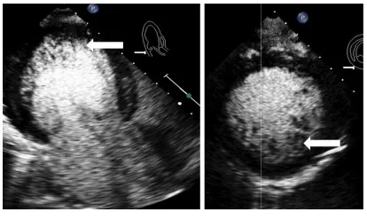
left ventricular (LV) ejection fraction of 25 with moderate to severe global hypokinesis of the left ventricle. The left ventricle appeared mildly dilated, with prominent sinusoidal culations to the apex suggestiveof LVNC (Figs 1, 2 and 3). Right ventricular function was normal. She was also incidentally noted to have mild mitral regurgitation.
Fig. 1. 2D echocardiogram of the left ventricle in the apical two chamber view with and
without color flow Doppler. This view demonstrates the flow of blood between the
trabeculae. [LV-left ventricle]
She was medically managed with an a
converting enzyme inhibitor, beta blocker, loop diuretic, fluid restriction and sodium restriction. Her HIV is well managed without history of opportunistic infections. HerECG, biomarkers did not suggest any ischemia or infarction. The diagnostic echocardiography findings obviated the need for an extensive evaluation for other causes of her cardiomyopathy. She was discharged within three days, and has not required additional hospitalizations.
; Article no. BJMMR.2015.027
31.1 g/dL and known iron deficiency from internal hemorrhoids for which she was not taking her iron. A lipid panel was within normal limits. CD4 count of 230 cells/uL. Electrocardiogram (ECG) revealed sinus tachycardia without any ST-T changes suggestive of ischemia or infarction. Chest radiograph showed cardiomegaly with pulmonary vascular congestion. In the emergency department, spiral computer tomographic angiography was negative for
echocardiogram with perflutren lipid DEFINITY®) showed left ventricular (LV) ejection fraction of 25-30% with moderate to severe global hypokinesis of left ventricle. The left ventricle appeared mildly dilated, with prominent sinusoidal trabe culations to the apex suggestiveof LVNC (Figs. ). Right ventricular function was normal. She was also incidentally noted to have
Fig. 1. 2D echocardiogram of the left ventricle in the apical two chamber view with and
without color flow Doppler. This view demonstrates the flow of blood between the
left ventricle]
Fig. 2. Apical three and two chamber view demonstrating noncompaction of the left ventricle. [LA-left atrium, LV-left ventricle]
Fig. 3. 2D echocardiogram with perflutren lipid microsphere echo contrast (DEFINITY®)
of the left ventricle in the apical three chamber and parasternal short axis view showing compacted and non-compacted
myocardium (arrow)
3. DISCUSSION
The HIV virus itself is asignificant cause of dilated cardiomyopathy, late in the course of infection.With the advent of highly active antiretroviral therapy, cardiomyopathy has become less common among patients with HIV, falling 30% [2] from a baseline annual rate of
15.9/1000 [3] patients annually in developed nations.
Other causes of cardiomyopathy in this population are myriad including: drug myotoxicity; secondary infection; coronary artery disease from medication-induced hyperlipidemia; and an autoimmune process triggered by HIV, coxsackievirus, cytomegalovirus, or Epstein virus [4]. Understandably, then, the dia work up for a patient with HIV and new onset congestive heart failure is often complex.
Left ventricular noncompaction (LVNC), previously known as spongy myocardium or hypertrabeculation syndrome, is a rare cause of
Gruenebaum et al.; BJMMR, 5(2): 270-274, 2015; Article no. BJMMR.20
Fig. 2. Apical three and two chamber view rating noncompaction of the left
left ventricle]
Fig. 3. 2D echocardiogram with perflutren lipid microsphere echo contrast (DEFINITY®)
of the left ventricle in the apical three chamber and parasternal short axis view
compacted myocardium (arrow)
The HIV virus itself is asignificant cause of dilated cardiomyopathy, late in the course of highly active antiretroviral therapy, cardiomyopathy has become less common among patients with HIV, falling 30% [2] from a baseline annual rate of
15.9/1000 [3] patients annually in developed
Other causes of cardiomyopathy in this e myriad including: drug myotoxicity; secondary infection; coronary artery induced hyperlipidemia; and an autoimmune process triggered by HIV, coxsackievirus, cytomegalovirus, or Epstein-Barr virus [4]. Understandably, then, the diagnostic work up for a patient with HIV and new onset congestive heart failure is often complex.
Left ventricular noncompaction (LVNC), previously known as spongy myocardium or hypertrabeculation syndrome, is a rare cause of
cardiomyopathy in the general population. One of several primary genetic cardiomyopathies, it is generally inherited in an autosomal dominant fashion [5]. It usually affects the left ventricle, but right ventricularinvolvement has been reported. It is an under-recognized phenomenon, but there has been greater interest in LVNC due to the technological advances and increased availability of the imaging modalities.
The pathophysiology of ventricular dysfunction in LVNCis not well understood. One group has suggested that subendocardial hypoperfusion and microcirculatory dysfunction related to the structural changes lead to ventricular dysfunction and arrhythmias [6]. Alternatively, the persistent presence of the trabecular layer may interfere with the effective ejection of blood from
ventricle because of changes in laminar flow [7,8]. LVNC has been associated with mitochondrial myopathy and othergenetic mutations that cause myofibrosis, which in turn may lead to LV dysfunction [9,10]. As one of our authors previously elucidated in their previous report the late presentation is mostly related to the process of myocardial fibrosis which takes several years leading to the LV dysfunction [11]. Patients with LVNC may be asymptomatic, or may present with heart failure or arrhythmia one case series, the most common manifestations at diagnosis were: dyspnea (79 %) and New York Heart Association class III or IV heart failure (35 %) [12].
Diagnosis is usually made based upon echocardiography. The most widely used criteria are noncompaction to compaction ratio (NC:C) > 2, and visualization of blood flowing directly from the ventricular cavity into the deep inter trabecular recess [12]. Computed tomography, positron emission tomography, and magnetic resonance imaging may also be use
LVNC has a poor prognosis. Early studies reported 35% mortality with mean follow 3.5 years [12]. More recently, mortality has been reported at 2-15% annually [18]. Patients should be treated aggressively according to standard heart failure guidelines, including the use of implantable cardiac defibrillators (ICD) to prevent sudden cardiac death. While there have been severalreports of unsuccessful defibrillation with ICDs in LVNC, it is unclear if this is related to the underlying disease [19,20]. Heart transplant may be considered if medical therapies fail.
; Article no. BJMMR.2015.027
pulation. One of several primary genetic cardiomyopathies, it is generally inherited in an autosomal dominant fashion [5]. It usually affects the left ventricle, but right ventricularinvolvement has been reported. It recognized phenomenon, but lately there has been greater interest in LVNC due to the technological advances and increased availability of the imaging modalities.
The pathophysiology of ventricular dysfunction in LVNCis not well understood. One group has ial hypoperfusion and microcirculatory dysfunction related to the structural changes lead to ventricular dysfunction and arrhythmias [6]. Alternatively, the persistent presence of the trabecular layer may interfere with the effective ejection of blood from the left ventricle because of changes in laminar flow [7,8]. LVNC has been associated with mitochondrial myopathy and othergenetic mutations that cause myofibrosis, which in turn may lead to LV dysfunction [9,10]. As one of our ed in their previous report the late presentation is mostly related to the process of myocardial fibrosis which takes several years leading to the LV dysfunction [11]. Patients with LVNC may be asymptomatic, or may present with heart failure or arrhythmias. In one case series, the most common manifestations at diagnosis were: dyspnea (79 %) and New York Heart Association class III or
Gruenebaum et al.; BJMMR, 5(2): 270-274, 2015; Article no. BJMMR.2015.027
Our patient received optimalmedical therapy. On follow up, she did not meet criteria for ICD placement.
4. CONCLUSION
It is often challenging to identify the underlying cause of cardiomyopathy in a patient with HIV. While LVNC is a rare cause of left ventricular failure, typical findings on echocardiography can obviate the need for a more complex evaluative strategy.
CONSENT
All authors declare that ‘written informed consent was obtained from the patient (or other approved parties) for publication of this case report and accompanying images.
ETHICAL APPROVAL
Not applicable.
COMPETING INTERESTS
Authors have declared that no competing interests exist.
REFERENCES
1. UNAIDS report on the global aids epidemic 2013. UNAIDS, 1 Nov 2013. Web. 14 Apr 2014.
Available:http://www.unaids.org/en/media/ unaids/contentassets/documents/epidemiol ogy/2013/gr2013/UNAIDS_Global_Report_ 2013_en.pdf.
2. Bijl M, Dieleman JP, Simoons M, van der Ende ME. Low prevalence of cardiac abnormalities in an hiv-seropositive population on antiretroviral combination therapy. Journal of Acquired Immune Deficiency Syndrome. 2001;27:318-320. 3. Barbaro G. Cardiovascular manifestations
of hiv infection. Circulation. 2002;106:1420-1425. doi: 10.1161/ 01.CIR.0000031704.78200.59
4. Barbaro G. HIV-associated
cardiomyopathy etiopathogenesis and clinical aspects. Herz. 2005;30:486–492. 5. Maron BJ, Towbin JA, Thiene G,
Antzelevitch C, Corrado D, Arnett D, Moss AJ, Seidman CE, Young JB. Contemporary definitions and classification of the cardiomyopathies: an American
Heart Association Scientific Statement from the Council on Clinical Cardiology, Heart Failure and Transplantation Committee; Quality of Care and Outcomes Research and Functional Genomics and Translational Biology Interdisciplinary Working Groups; and Council on Epidemiology and Prevention. Circulation.
2006;113:1807–1816. doi:
10.1161/CIRCULATIONAHA.106.174287. 6. Jenni R, Wyss CA, Oechslin EN,
Kaufmann PA. Isolated ventricular noncompaction is associated with coronary microcirculatory dysfunction. J Am Coll Cardiol. 2002;39:450–454. doi: 10.1016/S0735-1097(01)01765-X.
7. Henderson DJ, Anderson RH. The development and structure of the ventricles in the human heart. PediatrCardiol. 2009;30(5):588.
8. Pastore G, Zanon F, Baracca E, Piva M, Bernardi A, Piergentili C, Rigatelli G, Roncon L, Barold SS. Failure of transvenous ICD to terminate ventricular fibrillation in a patient with left ventricular noncompaction and polycystic kidneys. Pacing Clin Electrophysiol. 2012;35(2):e40.
9. Finsterer J, Stollberger C, Feichtinger H. Histological appearance of left ventricular hypertrabeculation/noncompaction. Cardiology. 2002;98:162-164
10. Burke A, Mont E, Kutys R, Virmani R. Left ventricular noncompaction: a pathological study of 14 cases. Hum Pathol.
2005;36:403–411. doi:
10.1016/j.humpath.2005.02.004.
11. Kalavakunta JK, Tokala H, Gosavi A, Gupta V. Left ventricular noncompaction and myocardial fibrosis: a case report. Int Arch Med. 2010;3:20.
12. Oechslin EN, Attenhofer Jost CH, Rojas JR, Kaufmann PA, Jenni R. Long-term follow-up of 34 adults with isolated left ventricular noncompaction: a distinct cardiomyopathy with poor prognosis. J Am Coll Cardiol. 2000;36:493–500. doi: 10.1016/S0735-1097(00)00755-5.
13. Conces DJ Jr, Ryan T, Tarver RD. Noncompaction of ventricular myocardium: CT appearance. Am J Roentgenol. 1991;156:717–718.
Gruenebaum et al.; BJMMR, 5(2): 270-274, 2015; Article no. BJMMR.2015.027
15. Hany TF, Jenni R, Debatin JF. MR appearance of isolated noncompaction of the left ventricle. J Magn Reson Imaging.
1997;7:437–438. doi:
10.1002/jmri.1880070231.
16. Bax JJ, Atsma DE, Lamb HJ, Rebergen SA, Bootsma M, Voogd PJ, Somer S, Schalij MJ, de Roos A, vander Wall EE. Noninvasive and invasive evaluation of noncompaction cardiomyopathy. J Cardiovasc Magn Reson. 2002;4:353–357. doi: 10.1081/JCMR-120013302.
17. Jenni R, Oechslin E, Schneider J, Attenhofer Jost C, Kaufmann PA. Echocardiographic and pathoanatomical characteristics of isolated left ventricular non-compaction: a step towards classification as a distinct cardiomyopathy. Heart. 2001;86:666–671. doi: 10.1136/heart.86.6.666.
18. Murphy RT, Thaman R, Blanes JG, Ward D, Sevdalis E, Papra E, Kiotsekolglou A,
Tome MT, Pellerin D, McKenna WJ, Elliott PM. Natural history and familial characteristics of isolated left ventricular non-compaction. Eur Heart J.
2005;26:187–192. doi:
10.1093/eurheartj/ehi025.
19. Pastore G, Zanon F, Baracca E, Piva M, Bernardi A, Piergentili C, Rigatelli G, Roncon L, Barold SS. Failure of transvenous ICD to terminate ventricular fibrillation in a patient with left ventricular noncompaction and polycystic kidneys. Pacing Clin Electrophysiol. 2012;35(2):e40.
20. Maile S, Kunz M, Oechslin E, Zund G, Rahn M, Lachat ML, Turina MI. Intractable ventricular tachycardia and bridging to heart transplantation with a non-pulsatile flow assist device in a patient with isolated left-ventricular non-compaction. J Heart Lung Transplant. 2004;23(1):147.
© 2015 Gruenebaum et al.; This is an Open Access article distributed under the terms of the Creative Commons Attribution License (http://creativecommons.org/licenses/by/4.0), which permits unrestricted use, distribution, and reproduction in any medium, provided the original work is properly cited.
Peer-review history: