_____________________________________________________________________________________________________
*Corresponding author: E-mail: [email protected];
21(2): 1-8, 2017; Article no.BJMMR.31531 ISSN: 2231-0614, NLM ID: 101570965
SCIENCEDOMAIN international www.sciencedomain.org
Microleakage of Two Cementing Agents Using
Different Application Techniques
Maria Gabriela Marquini Kunkel
1, Wesley Fernando Ferrari
1,
Márcio José Mendonça
2, Virgínia Bosquiroli
2, Julio Katuhide Ueda
2and
Veridiana Camilotti
2*1
Universidade Estadual do Oeste do Paraná (UNIOESTE), Cascavel - PR, Brazil.
2
Department of Restorative Dentistry, Universidade Estadual do Oeste do Paraná (UNIOESTE), Cascavel - PR, Brazil.
Authors’ contributions
This work was carried out in collaboration between all authors. Authors MGMK and VC developed the concept and designed the study. Authors MGMK, WFF, MJM, VB and VC analyzed and interpreted the data. Authors MGMK and WFF drafted the manuscript. Authors MJM, VB, JKU and VC revised the manuscript for intellectual content. All authors read and approved the final manuscript.
Article Information
DOI: 10.9734/BJMMR/2017/31531
Editor(s):
(1) Dimitrios Dionysopoulos, Department of Operative Dentistry, School of Dentistry, Aristotle University of Thessaloniki, Greece.
Reviewers:
(1) Cornelis H. Pameijer, School of Dental Medicine, University of Connecticut, USA. (2)Mohil Asnani, Gujarat University, India. (3)Konda Karthik Roy, MNR Dental College and Hospital, Dr. NTR University of Health Sciences, Telangana, India. (4)Ibrahim Hamouda, Umm Al-Qura University, KSA. (5)Andrea Scribante, University of Pavia, Italy. Complete Peer review History:http://www.sciencedomain.org/review-history/18886
Received 14th January 2017 Accepted 7th April 2017 Published 3rd May 2017
ABSTRACT
The coronal leakage of bacteria and other irritants to the radicular canal system is one of the main factors that may result in clinical failure and affect the long term success of adhesive cementation and endodontic treatment. Thus the aim this study was demonstrated in vitro the degree of marginal microleakage of two cementing agents. 60 bovine incisors endodontically treated. The specimens were randomly divided into two groups: Group 1 – dual-cured resin cement and group 2 - resin-modified glass ionomer cement. Booth groups were divided into three subgroups with 10 teeth each, according to the placement technique to be used: A - applying it with a spatula over the pin; B - lentulo bur; C - Centrix syringe. The teeth were thermocycled for 1000 cycles between 5
Kunkel et al.; BJMMR, 21(2): 1-8, 2017; Article no.BJMMR.31531
and 55°C and a dwelling time of 30 seconds. All groups were immersed in a solution of Rodhamine B at 2% at room temperature for 24 hours. The dye microleakage analysis was performed by three calibrated examiners. After 24 hours, the teeth were longitudinally sectioned and the microleakage scores were given by a blind operator. Data were submitted to Kruskal-Wallis test (p≤0.05). No difference was observed in microleakage values between the insertion techniques for both cements analyzed. The two cements used, as well as the three techniques of insertion techniques generated similar values of coronal microleakage.
Keywords: Resin cements; glass ionomer cements; fiberglass posts; microleakage.
1. INTRODUCTION
The restoration of endodontically treated teeth is a major challenge for Restorative Dentistry. When the remaining coronal structure does not promote adequate support and retention for restoration, using intraradicular posts [1,2] is an effective option to improve the retention of the restorative material and the dissipation of masticatory forces [3].
For a long time, endodontically treated teeth were restored using cast metallic posts. Despite its high retention and the thin cement film it forms, such pins have a high elastic modulus and may lead to root fractures [4,5]. Unlike those, the fiber posts do not have to be inserted into a length equal or greater than the depth of the clinical crown, which reduces the chance of root fracture [6].
In order to improve the bond between the glass fiber pins and the walls of the channel, there is a wide variety of cementing agents. By selecting the cementing agent, some properties should be considered, such as high tensile strength, low or no solubility, biocompatibility, adhesive qualities, chemical stability over time, suitable thickness of film (< 25 µm), elastic modulus similar to dentin, radiopacity, easy handling and cost. Any cementing agent may be successfully used, as long as the clinician knows the virtues and problems of each type for a proper selection [7].
The cementing agents most widely used are resinous and glass ionomer. Posts cemented with resin cements offer better resistance to displacement, reinforce the weakened structure of the radicular canal [8] and promote lower marginal microleakage [9]. However, they also have some disadvantages, such as technical sensitivity and polymerization shrinkage [10,11]. The adhesion of resin cements is mainly hampered by unfavorable configuration of the radicular canal, related to a high C factor (cavity
configuration factor), which can be up to 40 times higher compared to direct restorations [12]. The C factor is the ratio between the adhered and non-adhered areas: When the number of not attached areas is small, the polymerization shrinkage can be greater than the bond strength leading to the formation of cracks in the dentin-cement interface [13]. Thus, the union of glass fiber posts cemented with resinous agents can be unpredictable.
Conventional glass-ionomer cement (GIC) or resin-modified cement (RMGIC) can be the indicated alternative for cementing fiber posts [14]. Both cements show bonding to dentin by means of micromechanical mechanisms and chemical bonding. The hygroscopic expansion
that occurs after gelation of these materials partially offsets the contraction,
reducing stress and providing increased bond between the cement and the dentin at the end of gelation [15]. The RMGIC aims to maintain the benefits of GIC and minimize its major disadvantages: They are less sensitive to
moisture, have greater dimensional stability and greater connection to the tooth structure
than the GIC [16]. They also release fluoride, they are readily prepared and also more resistant to compression than zinc phosphate cement [17].
Thus, the aim of this in vitro study was to qualitatively compare the level of microleakage among the walls of the radicular canal, cementing agent and pin, by using insertion techniques of a resin and glass ionomer cement.
2. MATERIALS AND METHODS
2.1 Tooth Preparation
Sixty bovine incisors were selected [22]. The inclusion criteria for each tooth were: having a minimum length of 14 mm; no curves andcracks on the surface. This length is to obtain an apical sealing of 4 mm and root length with 10 mm [20]. After the teeth were selected, they were kept in saline solution at room temperature. The crown of each tooth was separated from the root through a perpendicular section along the long axis of the tooth at the cement enamel junction by means of a j.dandy disk mounted on the micromotor assembly under abundant refrigeration conditions. With the removal of the coronary portion, direct access to the radicular canal was obtained. The biomechanical preparation of the radicular canal was done by manual technique, and by using Kerr & Hedstroem files (Dentsply-Maillefer, Ballaigues, Switzerland). As irrigation agents, sodium hypochlorite solution at 5% and EDTA at 17% were used. After preparation, the canal was stopped by means of lateral condensation with gutta-percha and cement filler Sealer 26 (Dentsply Indústria e Comércio Ltda, Petrópolis, Rio de Janeiro, Brazil).
2.2 Cementation of Prefabricated Dowels
Twenty-four hours after stopping the canals, the radicular canal was prepared with Largo burs # 2 and 3 (Dentsply-Maillefer, Tulsa, USA) and the drill regarding the number of the pin used (White Post DC#3, FGM, Joinville, SC, Brazil). The conductor was prepared with a depth of 10 mm, and a remaining 4 mm apical sealant with gutta-percha [23].
Post spaces were prepared immediately after filling to a depth of 10 mm, using a heated instrument (GP heater; DentsplyMaillefer, Ballaigues, Switzerland) to remove gutta-percha; post preparations were completed with a bur 5(GS Brasil, São Paulo, Brazil), 1.5 mm in diameter, forthe cylindrical fibreglass posts, with conical apical endsand circumferential mechanical retainers (ReforpostNo.3; Angelus, Londrina, Brazil).
The length of each dowel measured 10 mm inside the radicular canal and a was cut at 3 mm above the cement enamel junction. The jutting portion of the dowel was sectioned with a diamond instrument #1014 (KG Sorensen, Cotia, São Paulo, Brazil) prior to the cementation procedure. The dowels were randomly divided into two groups depending on the cementing agent: G1 – Allcem dual-cured resin cement (FGM, Joinville, Brazil); G2 - RelyX Luting resin-modified glass ionomer cement (3M ESPE, Seefeld, FB, Germany). Both groups were divided into three subgroups, according to the placement technique to be used: A - applying it with a spatula over the pin; B - lentulo bur; C - Centrix syringe. The excess of cement was removed with a disposable brush. The cement was photo activated for 40 seconds (LED Freelight 2/3M ESPE) directly on the root cervical face. The intensity of the light apparatus was measured immediately prior to its use for each specimen with the aid of a radiometer (SDI, Sao Paulo, SP, Brazil). All the cementation procedures were undertaken by the same operator. Immediately afterwards, the teeth were temporarily restored with composite resin Opallis A2(FGM, Joinville, Brazil), with the aid of a pre fabricated metal matrix with 3 mm height by the incremental technique, with no adhesive procedure and were photo activated for 40 seconds. The adhesive system was not used to simulate a temporary restoration. Thirty minutes after the cementation procedure, the roots were kept in saline solution at 37°C for seven days. In order to simulate intra-oral conditions, the teeth were thermocycled for 1000 cycles between 5 and 55°C and a dwelling time of 30 seconds [24].
2.3 Marginal Microleakage Tests
All the roots had their apexes sealed with sticky wax and were externally waterproofed with a layer of fluid resin (Araldite/Brascola, Joiville, Brazil) and two layers of regular nail enamel blue (Colorama, São Paulo, Brazil) up to 1 mm from the edge of the restoration. Immediately after this, they were immersed in a solution of Rodhamine B at 2% at room temperature for 24 hours. After the dye was removed, the teeth were sectioned with parallel sectioning along their long axis, and their halves were analyzed by qualitative and quantitative methods.
2.4 Analysis of Marginal Microleakage
Kunkel et al.; BJMMR, 21(2): 1-8, 2017; Article no.BJMMR.31531
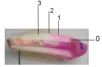
testers. For the assessment, the portion of the root with a highest degree of infiltration was selected. The penetration of the dye throughout the tooth-cement-dowel interface was measured by means of a scoring method (Table 1 and Fig. 1). The microleakage was analyzed in the cervical third, the middle third and the apical third of each root. After sectioning of all the roots, a picture of each root was taken with a digital camera, Canon Rebel XT, on a tripod, with a 3 X optical zoom, 100 macro lens, to decrease the distance between lens/object. All the images were taken under the same lighting conditions and with a ruler in mm close to the apical portion of the roots. These images were transferred to a digital archive and the leakages were measured using the Image Tool® program. This software enables magnifying an image by 2X, 4X, and 8X. After magnifying an image by 4 X, pixels (units or points that make up an image) become too obvious and thus jeopardize the quality of the image. Hence, magnifying images by 8X is limited to situations where there may be doubts regarding the dye markings and when gauging the program. The gauging of the program was undertaken to determine equivalence between a given amount of pixels and a given distance, thus enabling the conversion of a stroke seen on a screen into a definite metric unit. For this, images were zoomed in to the maximum level and marked with lines with the help of the software. One line was equivalent to the space between two lines in the ruler, that is, one millimeter. Thus, all measures that were taken were given in millimeters. For this procedure, the portion of the root with the highest degree of infiltration was selected.
Table 1. Description of scores
Score Description
0 Absence of microleakage 1 Microleakage not reaching the
middle third
2 Microleakage beyond the middle third
3 Microleakage of the dye throughout the root
3. RESULTS
The values of the scores that were obtained (Tables 2 and 3 and Fig. 2) were subjected to the Kruskal-Wallis statistical analysis test (p<0.05). It was possible to determine that there were no statistical differences between the cement placement techniques under analysis.
Fig. 1. Illustration of micro-infiltration scores
The values of scores were subjected to statistical analysis by Kruskal-Wallis test (p<0.05). Software
used was Bioestat 5.3
Table 2. Values of the scores that were obtained - Allcem / RelyX Luting
Allcem
Score 0 1 2 3 Total
G1 3 6 1 0 10
G2 1 5 1 3 10
G3 1 6 1 2 10
RelyX luting
G1 0 10 0 0 10
G2 0 8 2 0 10
G3 0 7 3 0 10
Marginal microleakage measures obtained via Image Tool® were statistically analyzed by ANOVA test (one-way), with a 95% degree of confidence (Table 2). Such values are detailed in Table 3. It was possible to find out that there had been no statistically significant differences among the manners of placement of the cementing agents.
4. DISCUSSION
Table 3. Microleakage means values (mm) and standard deviation
Lentulo L Centrix L
1 (± 0)a 1.3 (±0.46)a
Equal letters indicate that the values do not differ
Luting / 3M ESPE cement and the letter "
Fig. 2. Graphic illustration of microleakage scores (Letter L represents RelyX cement and letter A
drill allows a favorable distribution of the cement throughout the prepared space in the canal, facilitating the formation of a uniform continuous layer of cement. However, this technique should be avoided when using dual-cured resin cement, as it induces partial polymerization of the material before the proper seating of the pin. This can be explained by the fact that the heat generated by the lentulo drill can accelerate the polymerization reaction of the cementing a [26]. Yet some studies have reported the best performance of pins cemented with dual cementing agents inserted with letulo drill in comparison to other techniques [27,28
In this study there was no significant difference between the results of marginal microleakage of Allcem cement for the three application techniques that were used. These results can be explained by the fact that the dye leakage, initiated at the third cervical through restoration radicular dentin interface, is not influenced by the application technique of the cementing agent in this region. The cervical area enables easy access and presents a greater number of dentinal tubules, promoting the s
region, regardless of the cement insertion technique [29]. Fonseca et al. [27]
retention of pins cemented with dual polymerization cementing agent, by varying the application technique of cementing agent in the
0 2 4 6 8 10 12 LENTULO L PINO
Table 3. Microleakage means values (mm) and standard deviation
Manual L Lentulo A Centrix A
1.2 (±0.4)a 0.8(±0.6) 1.4(±0.92)
Equal letters indicate that the values do not differ statistically from one another. The letter "L" refers to the cement and the letter "A" refers to Allcem / FGM cement
Graphic illustration of microleakage scores (Letter L represents RelyX cement and letter A the Allcem cement)
drill allows a favorable distribution of the cement throughout the prepared space in the canal, facilitating the formation of a uniform continuous layer of cement. However, this technique should cured resin cement, as it induces partial polymerization of the material before the proper seating of the pin. This can be explained by the fact that the heat generated by the lentulo drill can accelerate the polymerization reaction of the cementing agent . Yet some studies have reported the best performance of pins cemented with dual cementing agents inserted with letulo drill in
27,28].
In this study there was no significant difference between the results of marginal microleakage of Allcem cement for the three application techniques that were used. These results can be explained by the fact that the dye leakage, he third cervical through restoration-radicular dentin interface, is not influenced by the application technique of the cementing agent in this region. The cervical area enables easy access and presents a greater number of dentinal tubules, promoting the sealing of the region, regardless of the cement insertion evaluated the retention of pins cemented with dual polymerization cementing agent, by varying the application technique of cementing agent in the
channel. The cement was inserted using only a lentulo drill, exclusively applying the agent at the pin surface or by combining both methods. The authors concluded that the best mechanical performance was obtained when the agent was taken to the radicular canal by means of drill and by the combination of both methods. In 2008, D'Arcangelo et al. [28] also found similar results when evaluating the adhesive strength through push-out test (compression). They used three types of fiber posts cemented with dual cured cement and self-adhesive system using three cement application techniques: using lentulo spiral, applying the cement at the pin surface and injecting the material with a specific syringe. They concluded that the best results with push-out test were obtained whe cementing agent was taken to channel with a lentulo drill or specific syringe. Only one system (ENA Post) showed similar bond strength values regardless of the application method of the cementing agent.
The glass ionomer cement has been used as an alternative to pin fixation. This indication takes place because of its biocompatibility
to release fluoride ions and because of its good thermal expansion coefficient [31
when comparing the cementing agents used, there were no statistically significant differences between them, for any of the application forms.
PINO-L CENTRIX
-L
LENTULO
A
PINO-A CENTRIX
-A ZERO UM DOIS TRES Manual A 1.6(±1.01) statistically from one another. The letter "L" refers to the RelyX
Graphic illustration of microleakage scores (Letter L represents RelyX luting
nt was inserted using only a lentulo drill, exclusively applying the agent at the pin surface or by combining both methods. The authors concluded that the best mechanical performance was obtained when the agent was taken to the radicular canal by means of lentulo drill and by the combination of both methods. In also found similar results when evaluating the adhesive strength test (compression). They used three types of fiber posts cemented with
dual-adhesive system using three cement application techniques: using lentulo spiral, applying the cement at the pin surface and injecting the material with a specific syringe. They concluded that the best results test were obtained when the cementing agent was taken to channel with a lentulo drill or specific syringe. Only one system (ENA Post) showed similar bond strength values regardless of the application method of the
The glass ionomer cement has been used as an lternative to pin fixation. This indication takes place because of its biocompatibility [30], ability to release fluoride ions and because of its good 31]. However, when comparing the cementing agents used, there were no statistically significant differences between them, for any of the application forms.
ZERO
UM
DOIS
Kunkel et al.; BJMMR, 21(2): 1-8, 2017; Article no.BJMMR.31531
Different results were found by Bonfante et al. [32], when the bond strength of glass ionomer and resin cementing agents were evaluated. The authors found the lower resistance values for glass ionomer cement because of its lower tensile strength. Perhaps the good performance of the glass ionomer cement found in this work is due to the frictional retention provided by the hygroscopic expansion that occurs after the cement maturation, which also helps the adhesive bonding at the cement-dentin interface [15].
The correlation between clinical success and in vitro study of the microleakage test was done by
Torabinejad et al. [33], when they reported that
in vitro tests experiments have produced
misleading results by simulating in vivo
conditions to a limited extent. The exact amount of infiltration is unknown, and is not even known how much infiltration is considered significant, being advisable the use materials and techniques that promote the most effective sealing. The major difficulty in in vitro studies, as
in this study, is the need for reproduction in the laboratory of clinical conditions to which these materials are submitted. One of these conditions is related to intraoral thermal changes which occur and produce changes in volume, allowing the ingress of bacteria and other elements in the tooth/restoration interface [34]. It can be observed in the specific literature that most studies seek to carry out thermal cycling, where the specimen alternately go through solutions containing dyes, saliva or water at different temperatures in an attempt to reproduce the variations that occur clinically, as conducted in this study [35,36]. Another factor of great importance when conducting a microleakage study is the penetration capacity of the dye used. To put leakage studies in context Oliver and Abbott [37] conducted a study with the aim to determine if there was a correlation between apical dye penetration and clinical performance of root fillings. They tested the length of apical dye penetration using a vacuum technique in vitro in 116 human roots that had been root-filled at least 6 months prior to extraction. Endodontic treatment was classified as clinically successful or unsuccessful and results for these groups were compared using an analysis of variance and the Student's t-test. Positive and negative controls were also used to test the experimental system. The dye penetrated significantly further in unsuccessful cases although the raw data suggested little difference. Overall, dye penetrated in 99.5% of the specimens, which
indicates that the presence of dye in a canal is a poor indicator as to whether a technique or material will succeed clinically. However, the
extent of dye penetration may be related to the clinical outcome. The authors concluded that clinically placed root canal fillings do not
provide an apical seal that prevents fluid penetration and therefore the outcome of treatment cannot be predicted based on the results of apical dye leakage studies. As early as 1993 Wu and Wesselink [38] already reviewed the shortcomings of various tests that had been reported in the literature. In 2016, Saffarpour et al. [39] evaluated the marginal microleakage of Class V restorations using chloramine chewing and thermal cycling as in the present study. They verified that this dye was effective for the evaluation of marginal microleakage.
According to the obtained results, the three insertion techniques were efficient in terms of marginal microleakage for both cementing agents used. However, some authors [27,33] note that the data from laboratory studies should be interpreted with caution, because it is impossible to reproduce in the laboratory the actual conditions of the mouth. More studies should be conducted with other variables to try to simulate the conditions of the oral environment.
5. CONCLUSIONS
Regarding the results obtained it was concluded that:
• All groups presented marginal microleakage regardless of the technique used;
• The three techniques had statistically similar performance relative to the degree of marginal microleakage found, regardless of the type of cementing agent used.
CONSENT
It is not applicable.
ETHICAL APPROVAL
It is not applicable.
COMPETING INTERESTS
REFERENCES
1. Soares CJ, Santana FR, Pereira JC, Araujo TS, Menezes MS. Influence of airborne particle abrasion on mechanical properties and bond strength of carbon/ epoxy and glass/bis-GMA fiber-reinforced resin posts. J Prosthet Dent. 2008;99:444-54.
2. Abduljawad M, Samran A, Kadour J, Al-Afandi M, Ghazal M, Kern M. Effect of fiber posts on the fracture resistance of endodontically treated anterior teeth with cervical cavities: An in vitro study. J
Prosthet Dent. 2016;116(1):80-4.
3. Manning KE, Yu DC, Yu HC, Kwan EW. Factors to consider for predictable post and core build-ups of endodontically treated teeth. Part I: Basic theoretical concepts. J Can Dent Assoc. 1995;61(8): 685-8, 690, 693-5.
4. Aggarwal V, Singla M, Miglani S, Kohli S. Comparative evaluation of fracture resistance of structurally compromised canals restored with different dowel methods. J Prosthodont. 2012;21(4):312-6. 5. Aggarwal V, Singla H, Yadav S, Yadav H, Sharma V, Bhasin SS. The effect of ferrule presence and type of dowel on fracture resistance of endodontically treated teeth restored with metal-ceramic crowns. J Conserv Dent. 2014;17(2):183-7.
6. dos Santos Alves Morgan LF, Peixoto RT, de Castro Albuquerque R, Santos Corrêa MF, de Abreu Poletto LT, Pinotti MB. Light transmission through a translucent fiber post. J Endod. 2008;34(3):299-302. 7. Rojas MS. et al. Novedades en relación a
la cementación de espigos muñones. Mundo Odonto. 2000;7(39):48-49.
8. Maroulakos G, Nagy WW, Kontogiorgos ED. Fracture resistance of compromised
endodontically treated teeth restored with bonded post and cores: An in vitro
study. J Prosthodont. 2015;114(3):390– 97.
9. Li XJ, Zhao SJ, Niu LN, Tay FR, Jiao K, Gao Y, Chen JH. Effect of luting cement and thermomechanical loading on retention of glass fibre posts in root canals. J Dent. 2014;42(1):75-83.
10. Carvalho RM, Carrilho MRO, Pereira LCG, Garcia FCP, Marquesini-Junior L, Silva SMA. Adhesive systems: Background information for understanding their clinical application and performance. Biodont. 2004;2(1):61-4.
11. Gomes GLS, Gomes RGC, Braz R. Cemento resinoso: ¿Todo cemento dual debe ser foto activado?. Acta Odontol Venez. 2009;47(4).
12. Alizadeh Oskoee P, Nooroloyouni A, Pornagui Azar F, Sajjadi Oskoee J, Pirzadeh Ashraf A. Effect of resin cement pre-heating on the push-out bond strength of fiber post to root canal dentin. J Dent Res Dent Clin Dent Prospects. 2015; 9(4):233-8.
13. Tay FR, Loushine RJ, Lambrechts P, Weller RN, Pashley DH. Geometric factors affecting dentin bonding in root canals: A theoretical modeling approach. J Endod. 2005;31(8):584-9.
14. Singh S, Thareja P. Fracture resistance of endodontically treated maxillary central incisors with varying ferrule heights and configurations: In vitro study. J Conserv
Dent. 2014;17(2):115-118.
15. Yiu CKY, Tay FR, King NM, Pashley DH, Sidhu SK, Neo JCL. Interaction of glass-ionomer cements with moist dentin. J Dent Res. 2004;83(4):283-9.
16. Almuhaiza M. Glass-ionomer cements in restorative dentistry: A critical appraisal. J Contemp Dent Pract. 2016;17(4):331-6. 17. Agha A, Parker S, Patel MP. Development
of experimental resin modified glassionomer cements (RMGICs) with reduced wateruptake and dimensional change. Dent Mater. 2016;32(6):713-22. 18. Raskin A, D'Hoore W, Gonthier S,
Degrange M, Déjou J. Reliability of in vitro
microleakage tests: A literature review. J Adhes Dent. 2001;3(4):295-308.
19. Aranha AC, Turbino ML, Powell GL, Eduardo Cde P. Assessing microleakage of class V resin composite restorations after Er: YAG laser and bur preparation. Lasers Surg Med. 2005;37(2):172-7. 20. Vichi A, Grandini S, Ferrari M. Comparison
between two clinical procedures for bonding fiber posts into a root canal: A microscopic investigation. J Endod. 2002; 28(5):355-60.
21. D’Arcangelo C, D’Amario M, Vadini M, De Angelis F, Caputi S. Influence of surface treatments on the flexural properties of fiber posts. J Endod. 2007;33(7):864-7. 22. Sfondrini MF, Scribante A, Cacciafesta V,
Gandini P. Shear bond strength of deciduous and permanent bovine enamel. J Adhes Dent. 2011;13(3):227-30.
Kunkel et al.; BJMMR, 21(2): 1-8, 2017; Article no.BJMMR.31531
Comparing the coronal seal of different thicknesses of MTA with gutta-percha after post space preparation. Scientific World Journal. 2015;2015:708639.
24. Subramaniam P, Pandey A. Assessment of Microleakage of a composite resin restoration in primary teeth following class III cavity preparation using Er, Cr: YSGG laser: An in vitro study. J Lasers Med Sci.
2016;7(3):172-176.
25. Garrido AD, Fonseca TS, Alfredo E, Silva-Sousa YT, Silva-Sousa-Neto MD. Influence of ultrasound, with and without water spray cooling, on removal of posts cemented with resin or zinc phosphate cements. J Endod. 2004;30(3):173-6.
26. Akgungor G, Akkayan B. Influence of dentin bonding agents and polymerization modes on the bond strength between translucent fiber posts and three dentin regions within a post space. J Prosthet Dent. 2006;95(5):368–78.
27. Fonseca TS, Alfredo E, Vansan LP, Silva RG, Sousa YT, Saquy PC. et al. Retention of radicular posts varying the application technique of the adhesive system and luting agent. Braz Oral Res. 2006; 20(4):347-52.
28. D’Arcangelo C, D’Amario M, Vadini M, De Angelis F, Caputi S. An evaluation of luting agent application technique effect on fibre post retention. J Dent. 2008;36(4):235-40. 29. Siviero M, Aivazoglou M, Camargo S,
Camargo C, Valera M. Análise topográfica, diametral e quantitativa de túbulos dentinários em canais radiculares de dentes humanos. Cien Odontol Bras. 2006;9(4):35-43.
30. Radke RA, Barkhordar RA, Podesta RE. Retention of cast endodontic posts: Comparison of cementing agents. J Prosthet Dent. 1988;59(3):318-20.
31. Badami V, Ahuja, B. Biosmart materials: Breaking new ground in dentistry. Scientific World Journal. 2014;2014:986912
DOI: 10.1155/2014/986912
32. Bonfante G, Kaizer OB, Pegoraro LF, do Valle AL. Tensile bond strength of glass fiber posts luted with different cements. Braz Oral Res. 2007;21(2):159-64.
33. Torabinejad M, Lee SJ, Hong CU. Apical marginal adaptation of orthograde and retrograde root end fillings: A dye leakage and scanning electron microscopic study. J Endod. 1994;20(8):402-407.
34. Camilotti V, Consalter AF, Dobrovolski M, Bosquiroli V, Busato PMR, Mendonça MJ. Microleakage of a self-adhesive resin cement after post cementation. Acta Odontol Latinoam. 2011;24(1):104-09. 35. Khoroushi M, Ehteshami A. Marginal
microleakage of cervical composite resin restorations bonded using etch-and-rinse and self-etch adhesives: two dimensional vs. three dimensional methods. Restor Dent Endod. 2016;41(2):83-90.
36. Rossomando KJ, Wendt SL Jr. Thermocycling and dwell times in microleakage evaluation for bonded restorations. Dent Mater. 1995;11(1):47-51.
37. Oliver CM, Abbott PV. Correlation between clinical success and apical dye penetration. Int Endod J. 2001;34:637-44. 38. Wu MK, Wesselink PR. Endodontic
leakage studies reconsidered. Part I. Methodology, application, and relevance. Int Endod J. 1993;26:37-43.
39. Saffarpour A, Ghavam M, Saffarpour A,
Sadighi K, Kharazifard MJ. Effect of Re-etching of oxalate-occluded dentin on Microleakage of composite resin restorations. J Dent (Tehran). 2016;13(5): 318-324.
_________________________________________________________________________________
© 2017 Kunkel et al.; This is an Open Access article distributed under the terms of the Creative Commons Attribution License (http://creativecommons.org/licenses/by/4.0), which permits unrestricted use, distribution, and reproduction in any medium, provided the original work is properly cited.
Peer-review history: