_____________________________________________________________________________________________________
*Corresponding author: E-mail: kirankumarrajyaguru@gmail.com;
www.sciencedomain.org
A Rare Presentation of Signet Ring Squamous Cells
in Ectocervical Squamous Epithelium Causing
Diagnostic Difficulty in Reporting Cervical
Intra-Epithelial Neoplasia
K. M. Rajyaguru
1*, Ahmad Zakuan
2and S. I. Shahruddin
31
Department of Pathology, Faculty of Medicine and Defence Health, National Defence University, Hospital Angkatan Tentera Tuanku Mizan, Kuala Lumpur, Malaysia.
2
Department of Pathology, Hospital Angkatan Tentera Tuanku Mizan, Kuala Lumpur, Malaysia.
3
Department of Obstetrics and Gynecology, Hospital Angkatan Tentera Tuanku Mizan, Kuala Lumpur, Malaysia.
Authors’ contributions This work was carried out in collaboration between all authors. Author KMR did histopathological study of tissue with special staining, laboratory diagnosis, references collection, literature review, case writing and submission. Author AZ did histopathology study with special stains and laboratory diagnosis. Author SIS examined the patient, did colposcopy examination, biopsy collection, treatment, follow up and obtained consent of patient. All authors read and approved the final manuscript.
Article Information
DOI: 10.9734/BJMMR/2016/29591 Editor(s): (1) Kate S. Collison, Department of Cell Biology, King Faisal Specialist Hospital & Research Centre, Saudi Arabia. Reviewers: (1) Gül Özcan, İstanbul University, Turkey. (2)Luca Valerio Messa, University of Siena, Italy. (3)Ajibola Idowu, Bowen University Teaching Hospital, Nigeria. Complete Peer review History:http://www.sciencedomain.org/review-history/16817
Received 19th September 2016 Accepted 24th October 2016 Published 7th November 2016
ABSTRACT
We present a case of cervical intraepithelial neoplasia Grade II having many areas full of signet ring squamous (SRS) cells in 6-7 parabasal layers, covered by the layers of koilocytotic changes in outermost layers, with a brief review of the literature. Such presentation is extremely rare. A cone biopsy of the cervix was sent to the laboratory after pap smear preparation and small biopsy from the aceto-white area during colposcopy examination, which revealed few malignant cells. We grossed the specimen and selected the tissue from 1-12 o’clock positions and processed for
paraffin blocks. H&E stained sections showed SRS cells at the majority of 1-12 positions. These SRS cells were negative for Alcian blue, Mucicarmine, Periodic acid–Schiff (PAS) and PAS-D stain and positive for cytokeratin. This presence of SRS cells has created difficulty in diagnosing the cervical intraepithelial neoplasia (CIN) case.
Keywords: Signet ring squamous cell; cervix; squamous epithelium; intraepithelial neoplasia.
1. INTRODUCTION
The ectocervical lining of the cervix is stratified squamous epithelium up to the squamocolumnar junctional area which is the prime site for cervical intraepithelial neoplasia (CIN) or Squamous cell carcinoma (SCC). The koilocytotic changes are commonly seen in cases of human papilloma virus (HPV) infection or in cervical intraepithelial neoplasia or in squamous cell carcinoma.
The signet ring cells are having a large round to oval vacuole in the cytoplasm, pushing and compressing the nucleus at the periphery. It is extremely rare to see the signet ring cells in non-neoplastic or non-neoplastic epithelial lining the ectocervical squamous epithelium[1].
A literature searches from Google, PubMed, and Science direct, revealed 19 cases of Primary signet cell adenocarcinoma of the endocervix [2,3]. However, signet ring squamous cells (SRS) in ectocervical squamous epithelium were found only in four cases reported by Kupryjaiqczyk et al. and in one case reported by Ohta et al. [1,4]. So total five cases are reported.
Because of the presence of such signet ring morphology in many squamous cells, it is very difficult to see the real dysplastic cells and so it required an extensive search to find the dysplasia. It is possible that diagnosis of CIN may be missed easily in such cases and later on the patient may develop invasive carcinoma which may have bad prognosis. Because of its rare occurrence and reported small number of cases, prognosis is difficult to predict.
In view of its rarity, we present a case of SRS cells in the ectocervical epithelium in a patient diagnosed as cervical intraepithelial neoplasia. To the best of our knowledge and web search, now this becomes the sixth reported case in English literature. A brief review of the literature on this topic is done.
2. CASE REPORT
A 52 -year- and 4 months old lady presented with foul-smelling vaginal discharge since one year.
She was a mother of 6 children. Last child birth was before 16 years. She was used to practice douching since young. Cervical cytology was done and Pap smear revealed High- Grade Squamous Intraepithelial Lesion, hence colposcopy was done.
Colposcopy was done by gynaecologist on 01-06-15 with the use of 3% acetic acid and it revealed thin acetowhite areas at 6 and 12 o’clock positions, with an impression of moderate dyskaryosis (Fig. 1). Patient was admitted to the hospital and a cone excision of the cervix was done in September 2015 and sent for histopathological examination (HPE). The cervical cone was sent in formalin and was found well fixed with preservative. There was no adnexal mass, the vulva and vagina were normal. Tests for HPV 16 and 18 and other high-risk HPV were negative. She was discharged in September 2015.
Fig. 1. Finding of colposcopy at the time of cone biopsy taken
Showing blood vessels seen under green filter (1). thin acetowhite lesion (2,3).
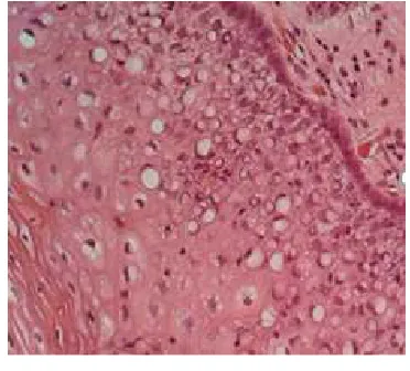
Histological study of the selected slide revealed plenty of typical SRS cells on H & E stain on 100 X and 400 X magnification, seen in 1/3 to 2/3 layers of squamous cells on majority area of ectocervical epithelium from 1-12 0’clock positions (Figs. 2 and 3).
Fig. 2. H & E 100 X: The slide of the ectocervical region showing plenty of signet
ring cells at different layers. The signet ring squamous cells are seen evenly distributed and limited to 2/3 thickness of squamous epithelium. Layers in superficial 1/3 thickness
of epithelium show koilocytotic changes
Fig. 3. H & E stain 400 X magnification showing clear signet ring cells up to 6-7 parabasal layers. The cells clearly show empty vacuoles within cytoplasm and nucleus is pushed to periphery giving signet
ring appearance to the squamous cells
The SRS cells were distributed evenly in lower one third to two third of squamous epithelium of ectocervix. The SRS cells are of variable sizes
and round to oval vacuoles were having clear to ground glass appearance pushing the nucleus to the periphery in semilunar shape. These SRS cells were quite different from koilocytotic changes in outermost layers of squamous cells.
We also did PAS (Fig. 4), PAS –D, Mucicarmine, Alcian blue, oil red O and Cytokeratin stains (Fig. 5) for one selected section from 12 o’clock positions slides. We also did Giemsa stain to rule out Chlamydia trachomatis infection. All these stains were negative in the vacuoles. Only the cytokeratin immunostain was positive for the squamous cells.
Fig. 4. PAS stained 100 X slide of ectocervical region showing plenty of signet ring cells at
different layers. Clear vacuoles in the cytoplasm of signet ring squamous cells is
negative for PAS stain
Fig. 5. Cytokeratin stained 100 X slide of ectocervical region showing positively stained squamous cell indicating these are
After an extensive search in different sections, there were few and small focal areas of squamous cells showing moderate dysplastic changes suggesting CIN. These cells were not looking like invasive cells or metastatic cells from other sites. The original squamous cells at this places have acquired changes of a signet ring. The cells were also not showing any obvious malignant changes. Because of such changes in morphology of squamous cells, it was difficult to decide the extent and presence of dysplastic changes due to the presence of such SRS cells.
The latest follow up in August 2016 revealed no gross abnormality or problem in the cervix.
3. DISCUSSION
Formation of signet ring cell changes in squamous cells is very rare. Many workers noted signet ring cells in adenocarcinoma of the endocervical region, but not in the stratified squamous epithelium of ectocervical region, except in our case and another four cases presented by Kupryjaiqczyk et al. and one case by Ohta et al. [1,4].
Kupryjaiqczyk et al have reported four rare cases of signet ring cells in the normal ectocervical squamous epithelium. The two were in normal ectocervical squamous epithelium and another two cases of squamous cell carcinoma of cervix having signet ring cells. In that four cases, the signet ring cells were negative for Mucicarmine, Alcian blue, and PAS stain. He also concluded that the cause of such cell is not clear and yet to establish [1].
Normally typical squamous cells of ectocervical epithelium are of polygonal shape having eosinophilic cytoplasm as well as intercellular bridges and are non-keratinising. The nucleus is larger, round and in the center of the cell. The cytoplasm of superficial and intermediate cells is rich in glycogen. The nucleus becomes smaller at superficial layers [5].
Usually, a signet ring cells acquire a characteristic appearance due to displacement and compression of the nucleus at one periphery of the cell and most common cause of this feature is the cytoplasmic accumulation of mucin.
Accumulation of mucin in the cytoplasm produce
a large clear vacuole which compresses and displaces the nucleus at one side in signet
ring cells in the stomach or prostatic
adenocarcinoma, and these mucins can be demonstrated by Alcian blue or Mucicarmine [6].
SRS cell carcinoma in skin is described by El Demellawy et al., Cramer [7,8]. Signet ring cells which are not positive for mucin usually are seen in non-mucin producing tumors and vacuoles are seen due to intermediate filaments [9,10], lipid droplets [9], or immunoglobulin [11], thyroglobulin [12], or by dilatation of cell organelles [12,13].
The presence of signet ring cells in the ectocervical epithelium is a rare occurrence.
Such cells are reported in Melanoma [10], T- cell lymphoma [13], carcinoma of stomach [14], Prostate [15], Gallbladder and Uterine cervix [16], Colon and Rectum [17], Testis [18], basal cell carcinoma [19], Breast [20], Ovary [21], Brain tumour–glioblastoma [22], eyelid skin sweat gland carcinoma [23], and other organs.
Sal et al. [3] reported a 19th case of primary signet ring cells in cervical carcinoma in a 48 years old lady. It was composed of 70% cells were signet ring cells and 30% endocervical adenocarcinoma cells.
Insabato et al. [24] described a case of primary signet ring cell adenocarcinoma purely arising from the endocervical region with an exceptional long-term survival. The signet ring cells, in this case, were neoplastic and on the head of an endocervical polyp.
Giordano et al. [25] reported a primary signet ring cells carcinoma of the cervix in which the signet ring cells were mucin positive so it was adenocarcinoma.
Yoon et al. [26] reported a signet ring cell adenocarcinoma of the uterine cervix case having 70% signet ring cells and 30% intestinal type of adenocarcinoma cells with aggressive behavior and the patient died in six months.
McKenna et al. [27] reported a case of signet ring cells in the uterine cervix which was found in the stroma of the cervix and he concluded that this was an artifact effect of the cauterization of the cervix. The cells in the stroma were negative for cytokeratin, EMA but positive for vimentin. Cracchiolo et al. [28] also noted one case primary signet ring cell carcinoma of the uterine cervix.
15 months after in situ cervical adenocarcinoma in a 40 years old female. In this case, focal signet ring cells were present in vaginal squamous epithelial in situ adenocarcinoma lesion.
Ohta et al. [4] reported a case of intraepithelial adenosquamous carcinoma of uterine cervix having p63 positive mucin-producing squamous cells having signet ring morphology in the uterine cervix.
Yiğit et al. [30] reported a case of signet ring cell morphology in squamous cell carcinoma of the lung in 85-year-old male.
Lugol’s Iodine can cause artifact effect in which the squamous cells become markedly retracted due to dehydration and intercellular bridges become very prominent [31]. In our case patient had colposcopy examination using acetone and not the Lugol’s Iodine.
4. CONCLUSION
Signet ring cells in the squamous epithelium of ectocervical epithelium is the rarest occurrence and the exact cause of this change is yet to be established. Only 5 such cases have been reported earlier. It is difficult to identify the dysplastic cells easily in the squamous epithelium due to presence of many signet ring cells. Hence, it requires an extensive search in many sections from various places in the cervix for the confirmation of dysplasia. In our case prognosis was found better since there was no gross abnormality of cervix after a follow up of 15 months.
CONSENT
All authors declare that written informed consent was obtained from the patient for publication of this paper and accompanying images.
ETHICAL APPROVAL
It is not applicable.
COMPETING INTERESTS
Authors have declared that no competing interests exist.
REFERENCES
1. Kupryjaiqczyk J, Kujawaf M. Signet-ring cells in squamous cell carcinoma of the
cervix and in non-neoplastic ectocervical epithelium. Int J Gynecol Cancer. 1992; 2:152-156.
2. Washimi K, Yokose T, Noguchi A, Ono K, Kawachi K, Maruyama Y, et al. Diagnosis of primary pure signet-ring cell carcinoma of the cervix. Pathol Int. 2015;65(7):393-5. 3. Sal V, Kahramanoglu I, Turan H,
Tokgozoglu N, Bese T, Aydin O, Demirkiran F, Arvas M. Primary signet ring cell carcinoma of the cervix: A case report and review of the literature. International Journal of Surgery Case Reports. 2016; 21.
4. Ohta Y, et al. Mixed mucin-producing and squamous differentiated tumor of the uterine cervix. Journal of Obstetrics and Gynecology Research. 2013;39(1):420– 423.
5. International agency for research on cancer, WHO.
Available:http://screening.iarc.fr/viavilichap 1.php?lang=1
(accessed on 8-07-16)
6. Remmele W, Weber A, Harding P. Primary signet-ring cell carcinoma of the prostate. Human pathol 1988;19:478-80. 7. EL Demellawy D, Onuma K, Alowami S.
SRS cell carcinoma–the forgotten variant: Case report and review of literature. J Cutan Pathol. 2011;38(3):306-8.
8. Cramer SF, Heggeness LM. SRS cell carcinoma. Am J Clin Pathol. 1989;91:488-91.
9. Mills SE, Fechner RE, Bruns DE, Bruns ME, O'Hara MF. Intermediate filaments in eosinophilic cells of epithelioid sarcoma. Am J Surg Pathol. 1981;5:195-202. 10. Sheibani K, Battifora H. Signet-ring cell
melanoma. Am J Surg Pathol. 1988;121: 28-34.
11. Henderson DW, Papadimitriou JM, Coleman M. Ultrastructural appearances of tumours, 2nd edn. Edinburgh London Melbourne and New York: Churchill Livingstone. 1986;88(97):290.
12. Mendelsohn G. Signet-cell-simulating micro-follicular adenoma of the thyroid. Am J Surg Pathol. 1984;9:705-8.
13. Grogan TM, Payne CM, Richter LC,
Rangel CS. Signet-ring cell lymphoma of T-cell origin an immunocytochemical
and ultrastructural study relating giant cell vacuole formation cytoplasmic
14. El-Zimaity HM, Itani K, Graham DY. Early diagnosis of signet ring cell carcinoma of the stomach: Role of the Genta stain. J Clin Pathol. 1997;50:867-8.
15. Roldán AM, Núñez NF, Grande E, García AA, Antón-Aparicio LM. A primary signet
ring cell carcinoma of the prostate with bone metastasis with impressive
response to folfox and cetuximab. Clinical Genitourinary Cancer. 2012;10(3):199-201. 16. Ragazzi M, Carbonara C, Rosai J.
Nonneoplastic signet-ring cells in the gallbladder and uterine cervix. A
potential source of overdiagnosis. Human Pathology. 2009;40(3):326-331.
17. Arifi S, Elmesbahi O, Riffi AA. Primary signet ring cell carcinoma of the colon and rectum, Bulletin du Cancer. 2015;102(10): 880-888.
18. Michal M, Hes O, Kazakov DV: Primary signet-ring stromal tumour of the testis. Virchows Arch. 2005;447:107-10.
19. Aroni K, Ch Lazaris A, Nikolaou I, Saetta A, Kavantzas N, Davaris PS. Signet ring basal cell carcinoma: A case study emphasizing the differential diagnosis of neoplasms with signet ring cell formation. Pathology - Research and Practice. 2001; 197(12):853-856.
20. Srinivas V, Subramanya H, Rajaram T, Vincent PJ, Rai R. Signet ring carcinoma of the breast: An uncommon type of breast carcinoma. Medical Journal Armed Forces India. 2005;61(1):84-85.
21. Roth LM, Ramzy I. Signet ring stromal cell tumour revisited and related signet ring cell lesions of the ovary, Human Pathology. 2014;45(3):636-642.
22. Martin SE, Bonnin J M, Hall DC, Hattab EM. Glioblastoma with signet-ring morphology: A case report and review of the literature. Human Pathology. 2010; 41(3):443-446.
23. Wollensak G, Witschel H, Böhm N, Signet ring cell carcinoma of the eccrine sweat glands in the eyelid. Ophthalmology. 1996; 103(11):1788-1793.
24. Insabato L, Simonetti S, De Cecio R, Di Tuoro S, Bifulco G, Di Spiezio Sardo A. Primary signet-ring cell carcinoma of the uterine cervix with long term follow-up: Case report. Eur J Gynaecol Oncol. 2007; 28(5):411-4. (PUB MED)
25. Giordano G, Pizzi S, Berretta R, D’Adda1 T. A new case of primary signet-ring cell carcinoma of the cervix with prominent endometrial and myometrial involvement: Immunohistochemical and molecular studies and review of the literature. World Journal of Surgical Oncology. 2012;10:7. 26. Yoon A, Kim SH, Kim HJ, Bae DS, Lee
JW, primary signet ring cell carcinoma of the Uterine cervix: A case report. Korean J Obstet Gynecol. 2011;54(9):570-573. 27. McKenna M, McCluggage WG. Signet ring
cells of stromal derivation in the uterine cervix secondary to cauterisation: Report of a previously undescribed phenomenon. J Clin Pathol. 2008;61(5): 648-51.
28. Cracchiolo B, Kuhn T, Heller D. Primary signet ring cell adenocarcinoma of the uterine cervix, A rare neoplasm that raises the question of metastasis to the cervix. Gynaecologic Oncology Reports. 2016;16: 9-10.
29. Clement PB, Benedet JL. Adenocarcinoma in situ of the vagina. Cancer. 1979;43(6): 2479-85.
30. Yiğit N, Çelik E, Yavan İ, Prominent signet ring cell morphology in a pulmonary squamous cell carcinoma, Turkish Journal Pathology. 2013;1:001-004.
31. Mehregan AH, Alberta E, Pinkus H. Artifacts in dermal histopathology. Arch Dermatol. 1966;94:218-225.
_________________________________________________________________________________ © 2016 Rajyaguru et al.; This is an Open Access article distributed under the terms of the Creative Commons Attribution License (http://creativecommons.org/licenses/by/4.0), which permits unrestricted use, distribution, and reproduction in any medium, provided the original work is properly cited.
Peer-review history: