Increased cellular turnover in response to fluoxetine
in neuronal precursors derived from
human embryonic stem cells
EUN-AH CHANG
1, ZEKI BEYHAN
1, MYUNG-SIK YOO
1, KANNIKA SIRIPATTARAPRAVAT
1, TAK KO
1,
KEITH J.
LOOKINGLAND
2, BURRA V.
MADHUKAR
3and JOSE B.
CIBELLI*
,4, 51Animal Science, 2Pharmacology & Toxicology, 3Pediatrics & Human Development, 4Animal Science and Physiology, Michigan State University, East Lansing, MI, USA and 5Programa Andaluz de Terapia Celular y Medicina Regenerativa, Andalucía, Spain
ABSTRACT Previous reports have shown that antidepressants increase neuronal cell prolifera-tion and enhance neuroplasticity both in vivo and in vitro. This study investigated the direct effects of one such antidepressant, fluoxetine , on cell proliferation and on the production of neurotrophic factors in neuronal precursors derived from human embryonic stem cells (hESCs; H9). Fluoxetine induced the differentiation of neuronal precursors, strongly enhancing neuronal characteristics. The rate of proliferation was higher in fluoxetine -treated cells than in control cells, as determined by MTT [3(4,5-dimethylthiazol-2-yl) 2,5-diphenyltetrazolium bromide] assay. The CPDL (cumula-tive population doubling level) of the fluoxetine-treated cells was significantly increased in comparison to that of control cells (p<.001). Bromodeoxyuridine incorporation and staurosporine-induced apoptosis assays were elevated in fluoxetine-treated cells. Quantitative RT-PCR analysis revealed no significant differences in the expression of neurotrophic factors, brain-derived neurotrophic factor (BDNF); glial-derived neurotrophic factor (GDNF) and cAMP-responsive element-binding protein (CREB) between cells treated with fluoxetine for two weeks and their untreated counterparts. These results may help elucidate the mechanism of action of fluoxetine as a therapeutic drug for the treatment of depression. Data presented herein provide more evidence that, in addition to having a direct chemical effect on serotonin levels, fluoxetine can influence hESC-derived neuronal cells by increasing cell proliferation, while allowing them to maintain their neuronal characteristics.
KEY WORDS:
human embryonic stem cell, neuronal precursor, fluoxetine, cellular turnover
Introduction
Human embryonic stem cells (hESCs), initially derived in 1998, show great potential as a source of cells for regenerative medicine and as a model for early human development (Thomson et al., 1998). However, hESCs could have a more immediate use in pharmacotoxicology. To date, such an approach has primarily been used with mouse ESCs (mESCs). For example, Wobus and colleagues (Wobus et al., 1991) generated differentiated cardi-omyocytes from mESCs. These cells were then evaluated for chronotropic responses to adrenergic and cholinergic agents and to Ca2+ channel blockers. While the utilization of hESCs for pharmacotoxicology research would be quite valuable, there exist
BIOLOGY
www.intjdevbiol.com*Address correspondence to: Dr. Jose B. Cibelli. B270 Anthony Hall, Michigan State University, East Lansing, Michigan 48824, USA. Fax: 517 432 8742. e-mail: [email protected]
Supplementary Material (four figures + two tables) for this paper is available at: http://dx.doi.org/10.1387/ijdb.092851ec
Accepted: 08 May 2009. Final author-corrected PDF published online: 10 July 2009.
ISSN: Online 1696-3547, Print 0214-6282
© 2009 UBC Press Printed in Spain
challenges associated with the standardization of culture condi-tions and methods (Wobus and Boheler, 2005). To this end, Dhara and colleagues (Dhara et al., 2008) have recently shown the advantages of using a neural progenitor cell line grown without a feeder layer, similar to the one we used in this study. The aforementioned techniques can facilitate further study of molecu-lar and biochemical mechanisms in early human neural differen-tiation and can potentially produce uniform neuronal cells for therapeutic use without concern about contamination from feeder
afflicts about 21% of the world population, and despite the multiple antidepressants available, not all patients respond to pharmaco-logical treatment (Schechter et al., 2005). While the use of postmortem human brain models in pharmacology and develop-mental toxicology has been developed, overall, human in vitro models to study the effects of drugs in treating mood disorders are scarce (Chen et al., 2001; Ongur et al., 1998; Rajkowska et al., 1999). Current models have inherent limitations, such as limited availability of tissues and anatomical differences that, in turn, can affect the interpretation and repeatability of the results. Most in vivo studies of mood disorders have used laboratory animals, and while useful, they also have shortcomings, i.e., (1) genetic causes of mood disorders are difficult to trace in animals, (2) major primary symptoms of mood disorders are difficult to evaluate, and (3) interspecies differences between animal models (Nestler et al., 2002).
Two mechanisms of depression have been hypothesized: chemical-based and network-based. Over the last few decades, most scientists and clinicians have accepted the premise that depression is caused by a chemical imbalance in the brain that can be corrected with medications. However, in the past decade, an alternative to simplistic chemical-based etiology has arisen; the network hypothesis suggests that depression reflects prob-lems in information processing within neural networks in the brain (Castren, 2005). While intriguing, more research is needed to categorically support the network hypothesis.
Fluoxetine (FLX), a widely used, potent antidepressant com-monly known as Prozac, was developed according to the chemi-cal hypothesis. FLX is a selective serotonin reuptake inhibitor (SSRI) with a high selectivity for the 5-hydroxytryptamine trans-porter (5-HTT). It modulates synaptic serotonin concentrations in the central nervous system (CNS) (Wong et al., 2005). Long-term administration of FLX can induce neural cell proliferation and neurogenesis in the hippocampus and prefrontal cortex of animal models both in vivo and in vitro (Chen et al., 2006; Encinas et al., 2006; Huang and Herbert, 2006; Kodama et al., 2004; Malberg and Duman, 2003; Santarelli et al., 2003). However, the mecha-nisms by which FLX promotes neural cell proliferation and neuroprotection in vivo are still unknown. Furthermore, the impact
human fetal development (Morrison et al., 2005).
To examine the effects of FLX and the mechanisms of depres-sion, hESCs with the potential to differentiate into“neuronal pre-cursors can offer a useful alternative to the current in vivo models to study the effect of FLX specifically on early human embryonic development. In the present study, hESC-derived neuronal pre-cursors were treated with FLX at a therapeutically relevant con-centration and for the period required for clinical efficacy. During the treatment period, we evaluated the effects of FLX on cell proliferation, cell differentiation patterns, and expression of neu-rotrophic factors. The results presented here provide new evi-dence that FLX can indeed influence neuronal cells in vitro, supporting to the notion that an SSRI not only can correct chemical imbalances in the CNS but can modify its cellular architecture as well.
Results
Characteristics and differentiation of hESC-derived NPs af-ter fluoxetine treatment
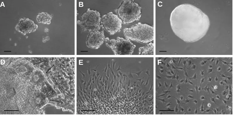
To avoid further potential contamination from feeder cells, which could alter accurate quantification of phenotypic markers during analysis, the differentiation of hESCs into neuronal precur-sors (NPs) was performed in the absence of any stromal cell line (Fig. 1). Generated hESC-derived NP cells showed a stable, normal karyotype (46, XX) (Suppl. Fig. 2).
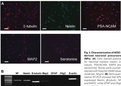
Immunofluorescence and RT-PCR analyses demonstrated that the hESC-derived NPs were strongly positive for nestin, PSA-NCAM, and β-tubulin. Neuronal differentiation was assessed by determining the number of cells expressing neuronal markers as a percentage of total cells counted in a selected area. The percentage of nestin-positive(+) cells was 88.2 ± 3.35%, while percentages for PSA-NCAM+, βIII-tubulin+, MAP2+, and seroto-nin+ were 67.9% ± 3.04%, 78.5% ± 6.45%, 50.6% ± 11.5%, and 29.3% ± 4.95%, respectively. A lower percentage of cells ex-pressed GFAP, CK-8, RIP, and TH, with percentages of 5.5% ± 3.2%, 3.1% ± 2.7%, 2.7% ± 1.5%, and 1.3% ± 0.8%, respectively (Fig. 3A). None of the cells expressed Oct4 (data not shown). Therefore, the hESC-derived NP cells expressed the
character-Fig. 1. Differentiation and isola-tion of hESC-derived neuronal pre-cursors (NPs). (A) hESC clumps floating on day 9. (B) Embryoid bod-ies (EBs) grown in DMEM/F12 with N2 supplementation for 8 days. (C) By day 18, EBs were clearly demar-cated with a smooth surface. (D) After attachment, EB formed neu-ronal rosettes (arrow). (E) NPs mi-grating from the periphery of neu-ronal rosettes. (F) Dissociated single NPs were propagated as attached monolayer cultures. Scale bars A,B,C, 50 μm; D,E,F, 1 mm.
B
C
D
A
istic markers of early neuronal cells. The fact that only a small percentage of cells were immunoreactive to RIP and CK-8 anti-bodies suggests that glial and epithelial cells were the least abundant types of cell in the cultures. The cells used in this study are fully capable of giving rise to astroglia, oligodendrocytes and neurons (data not shown) however, our goal in this differentiation protocol was to induce the NPs specifically into neuronal lineage. Semiquantitative RT-PCR analysis also showed results similar to the immunofluorescence data (Fig. 3B).
Following two weeks of culture with FLX, RT-PCR and immu-nofluorescence analyses showed that NPs expressed signifi-cantly more neuronal markers than control cells and continued to be negative for glial markers (Fig. 4A, 4B). No differences in the expression of MAP2 and serotonin were observed between control cells and those treated with FLX (Fig. 4A). However, the expressions of nestin detected by RT-PCR in control and FLX-treated cells are lower than Immunofluorescence analyses, This might be due to different transcriptional regulation and protein turn-over rates (Di Lieto et al., 2007).
treated cells were similar in their morphological characteristics with the appearance of large cell clumps, regardless of FLX treatment (Suppl Fig. 3-A1,A2). Due to the masking effect of FGF-2 on FLX treatment we observed, subsequent experiments were conducted without FGF-2 treatment.
Effects of fluoxetine on cell proliferation and death in neu-ronal precursors
The effects of FLX concentration on cell proliferation rates were determined using an MTT assay. When 10 μg/ml of FLX was used, 50% of NPs died within 24 hrs of exposure to the compound. When the dosage was reduced to 1 μg/ml of FLX, proliferation rates were similar to those of the control and of cells treated with 0.5 μg/ml. However, 50% of cells died after fourteen days of treatment with 1 μg/ml. On the other hand, NPs survived for up to fourteen days in less than 0.5 μg/ml of FLX, as did the control cells. An FLX concentration of less than 0.5 μg/ml is within the therapeu-tic range for FLX in adult human plasma, indicating that FLX has no cytotoxic effects as clinically used (Suppl Fig. 4). Briefly, the MTT assay showed that the proliferation rate of FLX-treated cells
Fig. 3. Characterization of hESC-derived neuronal precursors (NPs). (A) Cells stained positive for neuronal markers (nestin, b-tubulin, PSA-NCAM, MAP2 and serotonine). Nuclei were counter-stained with Hoechst 33342 (blue) (scale bar, 50 μm). (B) Semi-quan-titative RT-PCR showed that NPs expressed Nestin, β-tubulin, NF and MAP2, whileGFAP and Olig2 were absent.
Fig. 2. Schematic represen-tation shows culture condi-tions and protocol of neu-ronal differentiation. hESCs were plated on MEF feeders for eight days. hESC colonies were mechanically isolated and replated in feeder-free dishes as floating colonies (embryoid bodies; EBs) until day ten. EBs were attached on fibronectin-coated plates. Neural rosettes formed on days 21-27, were mechanically isolated and replated on poly-ornithine/laminin-coated plates. Neuronal precursors derived from neural rosettes were propagated in the presence of FGF-2 as attached monolayer cultures and passaged weekly.
Cell growth pattern of fluoxetine -treated NPs, with or without FGF-2
This study initially evaluated the influence of FGF-2 on FLX-treated cells.
NPs were cultured under four factorial combination of 4 ng/ml FGF-2 and 0.5 μg/ml FLX (FLX-/FGF-2+; Suppl Fig. 3-A1, FLX+/FGF-2+; Suppl Fig. 3-A2, FLX-/FGF-2-; Suppl Fig. 3B-, and FLX+/FGF-2-; Suppl Fig.3B-2). In the absence of FGF-2, NPs treated with FLX showed a clear difference in their morphology and growth pattern compared to those of non-FLX treated cells in two weeks. Numerous cell aggre-gates, similar to neurospheres were formed in FLX treated cells as opposed to scattered indi-vidual cells of control NPs (Suppl Fig. 3-B1,B2). When FGF-2 was present in the me-dia, FLX treated and non-FLX
B
increased more than those of control cells at less than 0.5 μg/ml of FLX during the first week, despite showing no statistically significant difference between different concentrations of FLX-treated NPs in overall proliferation rates.
CPDL was measured with and without FLX to determine the long-term proliferative potential of FLX-treated NPs. After 45 days of continuous passaging, totals of 6.81 and 5.03 CPDLs were found in FLX-treated and control cells, respectively. When the cells were passaged at regular intervals (every five to seven days), the CPDL of FLX-treated NPs was significantly increased at day 15 (p<.001; Fig. 5).
To examine the influence of FLX on DNA synthesis, the number of BrdU-positive NPs was counted by immunostaining at days 0, 3, and 7 after FLX treatment (0.5 μg/ml). The percentage of BrdU-positive cells at days three (54.9% ± 0.3%,) and seven (55.4% ± 0.4%) was significantly increased in FLX-treated cells compared to controls (Fig. 6, Table 1).
The rate of apoptosis induced by staurosporine (STS) in FLX-treated NPs was measured using DNA fluorochrome Hoechst staining. Upon exposure of the control cells to STS (200 nM) for 20 hrs, about 7.35% of the cells died at day 0. When NPs were treated with 0.5 μg/ml FLX, the percentage of apoptotic cells was
ing 1.0 μg/ml FLX, the percentage of apoptotic cells was 25.18% at day three, 60.59% at day seven, and 62.86% at day 14. These data indicate that apoptosis increases in NPs in the presence of FLX when the concentration and length of ex-posure increase compared to con-trols (p<.05 and p<.001, respec-tively; Fig. 7). It appears that FLX treatment itself does not promotes apoptosis but manifests the apop-tosis induced by STS. The role of FLX in apoptotic gene expression (Bcl-2, Bcl-xL, and Bax) in NPs was also investigated by real time RT-Fig. 4. Characteristics of hESC-derived neuronal precursors (NPs) changed after 2 weeks of
fluoxetine treatment. (A) Immunostaining showed that rates of Nestin, PSA-NCAM and β-tubulin positive staining significantly increased compared to that in control NPs (** = p<.01, * = p<.05). Rates of MAP2 and Serotonin positive NPs did not change. (B) RT-PCR showed that expression of Nestin, NF and β-tubulin were more increased in fluoxetine(FLX)-treated than in control cells. Fluoxetine-treated cells continued to be negative for glial markers (GFAP and Olig2). No difference in expression of MAP2 or serotonin was observed between control cells and those treated with fluoxetine.
Fig. 5 (Left). Cell proliferation assays for hESC-derived neuronal precursors (NPs) with and without fluoxetine. Cumulative population doubling level (CPDL) of NPs cultured with and without fluoxetine (FLX). CPDL of FLX-treated cells were significantly increased from day 15 until day 45 (* = p<.001).
Fig. 6 (Right). Immunofluorescent BrdU-incorporation of neuronal precursors (NPs). Percentage of BrdU-positive cells (red) was significantly increased in hESC-derived NPs cultured with fluoxetine (0.5 μg/ml) for 3 days (B) and 7 days (D) (p<.05 and p<.01, respectively), compared with control NPs cultured without fluoxetine for 3 and 7 days (A,C). Scale bar, 50 μm.
B
C
D
A
PCR analysis two weeks after the onset of FLX treatment. The expression of these genes showed no statistically significant differences between FLX-treated and control cells (data not shown).
Effects of fluoxetine on production of neurotrophic factors
weeks of FLX treatment. We found no statistically significant differences in gene and protein expression between the FLX-treated cells and controls (data not shown).
Discussion
Drug development presents numerous challenges beyond the demonstration of efficacy. Of great concern is the inability to anticipate toxicity in specific tissues, as well as potential teratoge-nicity. While numerous animal models can help predict the risks of drugs before the initiation of clinical trials, there is a tremendous need for human in vitro models that can be used to evaluate toxicity early in development.
Over the past ten years, ECVAM (the European Center for the Validation of Alternative Methods) has conducted several in vitro embryotoxicity tests — such as the whole-embryo culture (WEC) test, the rat limb bud micromass (MM) test, and the embryonic stem cell test (EST) — to evaluate chemicals known to have embryotoxic potential in vivo. These tests also very effectively reduce the number of in vivo animal experiments conducted (Genschow et al., 2002; Scholz et al., 1999). The most useful embryotoxicity test is the EST, which uses two well-established cell lines: a pluripotent mouse embryonic stem cell (mESC) line (D3) and differentiated 3T3 fibroblasts (Scholz et al., 1999). EST studies take advantage of the potential of mESCs to detect embryotoxicity in vitro. However, in vitro tests using mESCs have limitations due to species-specific developmental differences (Pouton and Haynes, 2007). Accordingly, hESCs can provide a more useful model to test new therapeutic compounds and also can be used as an in vitro model to test whether a specific drug promotes or interferes with lineage specific differentiation
pro-gram.
FLX, currently one of the most widely prescribed antidepres-sant medications. While it is known that FLX targets early progeni-tor cells in the adult brain (Encinas et al., 2006), its effect upon the developing brain has not been completely determined, in part due to the lack of a proper human model. In an effort to identify the therapeutic and toxic impacts of FLX in vivo, we describe an experimentally novel approach to examining various aspects of FLX’s effects on NPs derived from hESCs.
The results showed that FLX-treated NPs proliferated within one week after the addition of the drug and to a greater degree than untreated cells. This indicates that the mitogenic activity induced by FLX occurred during the early phases of these cultures. Our findings differ from previous studies, which argued that cell proliferation occurred after two or three weeks of FLX treatment in vivo or in vitro (Kodama et al., 2004; Malberg and Duman, 2003; Manev et al., 2001; Santarelli et al., 2003). The current study, however, produced results similar to those of Chen et al.,2007), who observed cell proliferation under in vitro condi-tions after seven days, though they used different cell type and culture conditions. It is highly likely that the discrepancies ob-served are due to differences in the experimental design — i.e., in vivo or in vitro tests.
The MTT assay and CPDL data demonstrated that there was a different proliferative effect between the two tests observed in the cells, depending primarily on the different cell culture methods in vitro. For the MTT assay, cells were not passaged. However, for the CPDL analysis, NPs were passaged at regular intervals, and they survived for up to 45 days. Control cells plateau their proliferation rate from Day 15 onward, while FLX-treated cells plateau on their proliferation on Day 23. We conclude that the effect on proliferation rate was higher in FLX-treated cell, as measured by CPDL. The results of the MTT assay showed that FLX has no cytotoxic effects at less than therapeutic concentra-tions (<0.5 μg/ml); however, this does not rule out the possibility of any teratogenic effect.
In the present study, the results of the STS-induced apoptosis assay were affected by the concentrations and time courses of the administration of FLX. Apoptosis increased as the concentrations and/or time increased, but no changes in apoptotic gene expres-sion (Bcl-2, Bcl-xl, and Bax). This indicates that although induced apoptosis is usually associated with changes in the amount of apoptotic gene transcripts, induced without changes in the tran-script level of these genes probably due to posttrantran-scriptional induction/regulation of the pathway. Previous studies examined the effects of FLX on apoptosis with different types of cell, such as adult neurons, hepatocytes, lymphoma cells, and neuroblastoma cells (Chen et al., 2007; Chiou et al., 2006; Koch et al., 2003; Lee et al., 2001; Levkovitz et al., 2005; Nahon et al., 2005; Serafeim et al., 2003). However, these studies used nonhuman CNS cells and animal models. Previous reports have shown conflicting evidence regarding the effect of FLX on apoptosis. Some studies showed an increase in apoptotic activity due to FLX in different cell lines using various methods (Koch et al., 2003; Levkovitz et al., 2005; Serafeim et al., 2003), while others found an increase in antiapoptotic activity (Chen et al., 2007; Chiou et al., 2006; Lee et al., 2001; Nahon et al., 2005). These conflicting findings may have resulted from differences in cell lines, FLX concentrations, or the duration of treatment. Our results, similar to those reported Time of drug treatment (% of BrdU-labeled cells)
Drug conc. (μg/ml) Day 0 Day 3 Day 7
0 41.4 ± 0.2 40.1 ± 0.1 33.0 ± 0.2 0.5 42.1 ± 0.4 54.9 ± 0.3* 55.4 ± 0.4**
TABLE 1
PERCENTAGE OF BrdU-IMMUNOPOSITIVE NEURONAL PRECURSORS, WITH AND WITHOUT FLUOXETINE, OVER TIME
(*; p<.05, **; p<.01)
of FLX treatment.
Interestingly,”FLX treatment increased in both cell proliferation and induced apoptotic activity in our experiments, consistent with the findings in the study by Sairanen et al. (2005) (Sairanen et al., 2005), which was conducted using an in vivo animal model after ten and 20 days of FLX treatment. We provide further evidence that FLX may activate cell proliferation and augment the effect of apoptotic agents in hESC-derived NPs.
The observed cellular characteristics and differentiation pat-terns demonstrate that FLX enhanced the neuronal rather than the glial properties. Markers increased by FLX were early neu-ronal markers, such as nestin, PSA-NCAM and β-tubulin. PSA-NCAM-induced expression by FLX is of interest since it has been described as a gene involved in neurogenesis (Kiss and Rougon, 1997; Varea et al., 2006). In addition, PSA-NCAM is closely associated with p-CREB involved in neuroplasticity and cell survival (Nakagawa et al., 2002). Therefore, the pattern of PSA-NCAM expression and cell proliferation observed in this study supports the hypothesis that one mechanism of action of FLX is via neurogenesis, providing further evidence that the mechanism of depression is associated with abnormal processing of the neural network.
Until recently, studies of depression and antidepressants have focused on intracellular pathways, which are known to be acti-vated by a number of extracellular signals, such as growth factors, stress, and neurotransmitters (Malberg, 2004; Malberg and Blendy, 2005). These signal pathways regulate various cellular processes associated with neuroplasticity and neurogenesis. The transcrip-tion factors CREB and BDNF are the targets of diverse types of antidepressants (Blendy, 2006; De Foubert et al., 2004; Josselyn and Nguyen, 2005). Chronic FLX administration promotes neurogenesis and synaptogenesis in the adult hippocampus (Grote et al., 2005; Malberg et al., 2000), as well as increased expression of the neurotrophins BDNF and GDNF (Hisaoka et al., 2007; Nibuya et al., 1995). These molecular and cellular events seem necessary to mediate the therapeutic effects of FLX. However, there are conflicting reports on the effect of FLX on CREB and BDNF. Some studies have observed increases in CREB and BDNF following in vivo FLX administration, and the authors attributed this increase to the type of anatomical brain lesions and to their experimental designs (Chen et al., 2001; Malberg and Blendy, 2005; Nibuya et al., 1995). Other studies were unable to detect increases in CREB or BDNF in response to FLX (Dias et al., 2003). The present study found no difference in the expression of neurotrophins in response to FLX in vitro. All of the studies conducted to assess the effect of FLX employed either brain-derived primary cultures or animal models, with generally using much higher concentrations of the drug. These differences may account for the discrepancy between the published data and ours. This may also suggest intrinsic differences between hES-derived NPs and primary cultures of the same type of cells directly isolated from animals or human. Considering the fact that the studies of FLX and tumor cell lines from lymphoma, neuroblas-toma, pheochromocytoma and glioblastoma can also increase neurotrophic factors and apoptotic activities (Bartholoma et al., 2002; Hisaoka et al., 2005; Koch et al., 2003; Levkovitz et al., 2005).
cell lines, are quite different from in vivo analogs (Gottlieb, 2002; Wobus et al., 1994).
Primary culture of human neural tissue is generally impractical, and while some neuronal cell lines can be induced to express markers of mature neurons under special culture conditions, they often fail to represent the properties of specialized neurons. This is often due, in part, to the origin of these cells in CNS-derived tumors, to a loss of viability, and to changes in phenotypic properties as a result of long-term culture.
Our study can help to elucidate the mechanism of action of FLX as a therapeutic drug for the treatment of depression by providing a valid alternative to current in vitro models. Their most attractive characteristic is their capacity to renew indefinitely while main-taining their differentiation potential. It is worth mentioning that standardization may be a challenge when working with hESCs. It has been shown that culture conditions will alter the gene and protein expression in these cells and, as a consequence, varia-tions may occur between replicates of the same study and among studies, even though the cell line might be the same. In addition, the process for generating large numbers of hESCs is labor intensive, and the possibility of irreversible genetic or epigenetic changes during experiments is always present (Pouton and Haynes, 2007). Regardless, hESCs can provide insights into a drug’s mechanisms of action and can reveal new targets for currently prescribed pharmaceuticals. Human ESCs may also help predict and avoid potential drug toxicity in humans. It is therefore essential that research and optimization of their use for these purposes continue.
Materials and Methods
All reagents were purchased from Sigma unless otherwise indicated.
Cell culture and differentiation
Human embryonic stem cell culture
Human H9 ESCs (passages 29 to 31) with a stable, normal (46, XX) karyotype were cultured as described previously (Zhang et al., 2001).
Briefly, cells were cultured on a mitotically inactive feeder layer of mouse embryonic fibroblasts. The culture medium consisted of Dulbecco’s Modified Eagle’s Medium (DMEM/F12; Invitrogen, Rockville, MD), 20% knockout serum replacement (Invitrogen), 0.1 mM β-mercaptoethanol, 4 ng/ml FGF-2 (Invitrogen), 1% nonessential amino acids (Invitrogen), and 2 mM L-glutamine (Invitrogen). The undifferentiated state of the hESCs was confirmed by characteristic morphology monitoring and immunostaining with anti-Oct4 antibody (Suppl. Fig. 1). All cultures were maintained in a humidified incubator with 5% CO2 at 37ºC.
Generation of hESC-derived neuronal precursors
A modification of the procedure described by Zhang was used to isolate hESC-derived neuronal precursors (Zhang et al., 2001). Human
ESCs colonies were mechanically isolated when they reached a diameter of 1 to 1.5 mm. Intermediate-sized clumps of cells were transferred to 35 mm dishes (BD Biosciences, Bedford, MA), and cultured in ESC medium at 37ºC for two days (Fig. 1A). Cell colonies that grew into nonattached embryoid bodies (EBs) were removed and transferred to new 35 mm dishes.
were cultured with 100% basal neural medium plus 3 μM retinoic acid (2+/ 2-; Fig. 1B and 1C). After ten days, well-demarcated EBs were attached to fibronectin-coated (Invitrogen) plates and cultured for nine days with basal neural medium plus 20 ng/ml FGF-2. A significant number of rosettes developed in the center of the differentiating EBs (Fig. 1D).
Rosette colonies were isolated, using a Pasteur pipette, and disso-ciated with a micropipette (Fig. 1E). Cells were replated on poly-L-ornithine/laminin-coated plates (50 μg/ml of poly-L-ornithine, 20 μg/ml of laminin) and cultured in basal neural medium containing 10 ng/ml FGF-2 for ten days (Fig. 1F). Subsequently, the cells, now called neuronal precursors (NPs), were cultured as a monolayer in basal neural medium containing 4 ng/ml FGF-2 (Fig. 2). NPs reached proliferative senescence at the tenth passage.
Treatment of hESC-derived neuronal precursors with fluoxetine
Stock solutions of FLX were prepared using DMSO and were diluted directly into the culture media at final FLX concentrations of 0.5
μg/ml. NPs were cultured in DMEM/F12-supplemented N2 culture medium in the presence of FLX. FGF-2 was excluded from the cell culture medium when the cells were treated with FLX. Culture medium was changed every two days and only DMSO was added to controls. NPs were cultured for two weeks in 0.5 μg/ml of FLX, at which point they were evaluated using immunostaining, RT-PCR, western blotting and real-time RT-PCR. For the MTT (3(4,5-dimethylthiazol-2-yl) 2,5-diphenyltetrazolium bromide) assay, NPs were cultured for various times (for 24, 48, and 72 hrs and for one and two weeks) in different concentrations of FLX (from 0 to 10 μg/ml). Cumulative population doubling level (CPDL) in continual subculture was calculated taking into account the total seeded and the total harvested cells from each culture dish. To determine the CPDL, NPs were cultured for 45 days in 0.5 μg/ml of FLX and passaged at five- to seven-day intervals. For the bromodeoxyuridine (BrdU) incorporation assay, NP cells were cultured for 72 hrs and one week in 0.5 μg/ml FLX. For the staurosporine-(STS-)induced apoptosis assay, NP cells were cultured for 72 hrs, one week, and two weeks in 0.5 μg/ml and 1.0 μg/ml of FLX.
Karyotype analysis
Karyotyping was performed on the NPs using standard methods (G-banding) by Cell Line Genetics (Madison, WI). At least 20 cells from each sample were examined.
Characterization of hESC-derived neuronal precursors
Immunofluorescence and quantification of immunopositive NPs
Protein expression in hESCs and NPs was analyzed by immunof-luorescence. Cells were fixed with 4% paraformaldehyde and permeabilized with 0.05% Triton X-100. After blocking with 3% bovine serum albumin, cells were incubated with primary antibodies. The following primary antibodies and dilutions were used: mouse anti-class III β-tubulin-Tuj1, 1:500; rabbit anti-glial fibrillary acidic protein (GFAP), 1:5000; rabbit nestin, 1:1000 (Abcam); mouse anti-cytokeratin-8 (CK-8), 1:200; mouse anti-microtubule-associated pro-tein 2 (MAP2), 1:500; goat anti-Oct4, 1:100 (Santa Cruz Biotechnol-ogy, Santa Cruz, CA); mouse anti-receptor interacting protein (RIP), 1:1000; mouse anti-polysialylated neuronal cell adhesion molecule (PSA-NCAM), 1:250 (Chemicon, Temecula, CA); and rabbit tyrosine hydroxylase (TH), 1:1000 (Pel-freez, Arkansas). The anti-gens were visualized under UV light using one of the following secondary antibodies: Alexa Fluor 594 or 488 (Molecular Probes, Eugene, OR). Cells were counterstained with 1 μg/ml of Hoechst 33342 for 10 min at room temperature. Semiquantitative measurement of immunopositive cells was determined using a 20X Zeiss LSC System objective lens mounted on a Nikon TE 2000-U microscope. More than 1,000 immunopositive cells for each primary antibody were photographed and counted. The number and the percentage of
immunopositive cells were calculated in relation to the total number of Hoechst-positive cells (>1,000 cells).
RT-PCR
Total RNA was extracted from hESC-derived NPs before and after two weeks with and without FLX, using the Picopure RNA Isolation Kit and following the manufacturer’s instructions (Arcturus, Sunnyvale, CA). After DNAse I digestion, cDNA was prepared from 1 μg total RNA, using the SuperScript III RT-PCR Kit (Invitrogen) as instructed by the manufacturer. Primer pair sequences are shown in Suppl. Table 1. The amplification procedure consisted of 30 cycles (denaturation at 94ºC for 30 sec, annealing at 58ºC for 40 sec, and extension at 72ºC for 45 sec). Amplification reactions were conducted in a final volume of 25 μl contain-ing 1.0 μl cDNA, 100 pmol each of forward and reverse primer and of PCR Master Mix (Promega, Madison, WI). RT-PCR products were separated by electrophoresis on 1% agarose gels and stained with ethidium bro-mide.
Assessment of proliferation, apoptosis, and production of neu-rotrophic factors in fluoxetine -treated neuronal precursors
MTT assay
MTT assays were performed at 0, 24, 48, and 96 hrs and at one and two weeks after FLX treatment. NPs growing without FLX were used as negative controls. The MTT assays were performed as previously de-scribed (Scholz et al., 1999). Briefly, 2 ß 104 NPs were seeded on 24-well
plates and grown in the presence of FLX at 0, 0.1, 0.5, 1, 10, and 100 μg/ ml. MTT (400 μg/ml) was added to each well for a 3 hr incubation period. HCl-isopropanol (0.04 M) was added to the cultures, and cells were incubated again for 15 min at 37ºC. The absorbance at 570 nm was measured using a spectrophotometer (Beckman Coulter, Inc., Fullerton, CA). Results were expressed as the percentage of blue formazan absorbance in treated cells relative to that in cells without FLX. Experi-ments were run in triplicate.
Cumulative population doubling level (CPDL)
FLX-treated (0.5 μg/ml) NPs were continuously passaged in neural media without FGF2 for 45 days, and each passage was five to seven day interval. The CPDL was calculated to determine their proliferation poten-tial. Nontreated NPs, cultured in same condition but without FLX, were used as controls. The CPDL at each passage was calculated from the cell count using the equation ln(Nf/Ni)/ln2, where Ni and Nf are the initial and
final cell numbers, respectively, and ln is the natural log(Lin et al., 2005).
5-Bromo-2-deoxyuridine (BrdU) incorporation assay
FLX-treated NP and control cells were cultured overnight with 30 μg/ ml of BrdU prior to immunostaining. Cells were fixed with 4% paraform-aldehyde and then incubated with blocking buffer (PBS containing 0.05% Triton X-100, 0.5% bovine serum albumin) for 1 hr at room temperature. Following incubation in 2.0 M HCl for 20 min at room temperature, cells were washed with 0.1 M sodium borate buffer. Immunostaining was done using primary mouse anti-BrdU antibody (1:100, Roche Applied Science, Indianapolis, IN), and the secondary antibody was Alexa Fluor 594 anti-mouse antibody (Molecular Probes). The nuclei were counterstained with Hoechst 33342 (1μg/ml) for 10 min at room temperature. The number and the percentage of BrdU-immunopositive cells were calculated in relation to the total number of Hoechst-positive cells (> 1,000 cells).
Staurosporine-induced apoptosis
Real time RT- PCR and quantification of transcripts
Isolated total RNA from NPs cultured with and without FLX was spiked with recombinant Venus-GFP (rVenusGFP) RNA (300 fg/μg total RNA) as an exogenous control. One microgram of total RNA was reverse tran-scribed for 60 min at 42ºC using SuperScript Reverse Transcriptase (Invitrogen) in a 50 μl reaction containing RT buffer (5X) with 500 μM random primers, 500 μM dNTPs, 10 mM DDT and 40 IU RNaseOUT. After heat inactivation of the reverse transcriptase for 15 min at 70ºC, samples were diluted to a concentration of 10 ng/μl of input RNA, using nuclease-free water. Quantities of transcripts were detected by real-time RT-PCR, using an ABI 7000 Sequence Detection System and SYBR green chemistry (Applied Biosystems, Foster, CA). The primer pairs were designed using Primer Express Software (Applied Biosystems) and human sequences available on the GenBank database (Suppl. Table 2). After optimizing the reaction conditions for‘all primer pairs, 25 μl PCR reactions were run in triplicate for each sample. Each reaction mixture consisted of SYBR Green PCR Master Mix (Applied Biosystems), 600 nM of sequence-specific primer pairs, 10 ng of input RNA equivalent template cDNA and water. For each primer pair, no-RT and no-template samples were run as controls. The thermal cycling profile of the reactions included one cycle at 60ºC for 2 min, one cycle at 95ºC for 10 min, and 40 cycles at 95ºC for 15 sec and at 60ºC for 1 min. Melting curve analyses were run at the end of each PCR reaction to monitor the PCR product sizes. The relative difference in the initial amount of each cDNA sample was determined by comparing the CT values. To quantify the mRNA concentrations, standard curves for each gene were generated using five 1/10 serial dilutions from a single stock of human genomic DNA and human reference cDNA mixture (10 pg to 100 ng). Before the statistical analyses, the amount of each transcript was normalized to the amount of exogenous control transcript (rVenusGFP) in the same sample.
Western blotting
NP pellets from cells incubated with and without FLX were collected and kept at 80ºC until used for western blotting. Briefly, cell lysates were prepared in TNE lysis buffer (50 mM Tris, 2 mM EDTA, 5 mM Na2P2O7, 100
μM Na3VO4, 5 mM NaF, 150 mM NaCl, 1% NP-40, 1μg/ml aprotinin, 1 μg/ ml leupeptin and 174 μg/ml PMSF), incubated on ice for 30 min, and centrifuged at 13,000 rpm for 30 min. Supernatants were evaluated to measure the amount of total protein using the Quick Start Bradford Protein Assay Kit (Bio-Rad, Inc., Hercules, CA). Cell lysates were then treated with SDS sample buffer(Sambrook and Russell, 2001). We loaded approxi-mately 13 μg of total protein into each lane of either 10% or 12% SDS-polyacrylamide gels. Proteins were separated by electrophoresis at 50 V for 4 to 6 hrs in a tris-glycine buffer and transferred onto PVDF membrane at 4ºC, 100 mA for 6 hrs in a transfer buffer. Blots were blocked in 5% skim milk in tris-buffered saline and 0.1% Tween 20 (TBST) with agitation for 90 min at room temperature. Primary antibodies were diluted in TBST with 3% skim milk, using the manufacturer’s recommended concentrations: mouse anti-CREB (1:1000, Chemicon), mouse anti-pCREB (Ser133) (1:1000, Cell Signaling Technology, Danvers, MA), mouse anti-5HTT (1:500, Advance Targeting Systems, San Diego, CA), mouse anti-BDNF (1:500; Abcam), and mouse anti-β actin (1:1000; Sigma). Blots were incubated overnight at 4ºC with each primary antibody and subsequently washed with TBST. Alkaline phosphatase-conjugated goat anti-mouse antibody (Bio-Rad) was diluted 1:3000 in TBST with 3% skim milk. The blots were incubated with the secondary antibody at room temperature for 90 min, and then exten-sively washed with TBST. Blots were immersed in NBT/BCIP substrate (Roche Applied Science) until the results were visualized. The images were generated by scanning and were analyzed using a densitometer (Bio-Rad).
Statistical analysis
One-way analysis of variance (ANOVA) and least significant difference
human GAPDH. The remaining data analysis employed two-tailed Student’s t-test. Each experiment was conducted at least three times (n ≥ 3) and a p value of less than 0.05 (p ≤ 0.05) was considered significantly different.
Acknowledgements
This project was supported by the Michigan State University Experi-ment Station, the office of the Vice President for Research and Graduate Studies, the MSU foundation and the Naylor Family foundation.
References
BARTHOLOMA, P., ERLANDSSON, N., KAUFMANN, K., ROSSLER, O.G., BAUMANN, B., WIRTH, T., GIEHL, K.M. and THIEL, G. (2002). Neuronal cell death induced by antidepressants: lack of correlation with Egr-1, NF-kappa B and extracellular signal-regulated protein kinase activation. Biochem Pharmacol
63: 1507-1516.
BENNINGHOFF, J., SCHMITT, A., MOSSNER, R. and LESCH, K.P. (2002). When cells become depressed: focus on neural stem cells in novel treatment strate-gies against depression. J Neural Transm 109: 947-962.
BLENDY, J.A. (2006). The role of CREB in depression and antidepressant treat-ment. Biol Psychiatry 59: 1144-1150.
CASTREN, E. (2005). Is mood chemistry? Nat Rev Neurosci 6: 241-246.
CASTREN, E., VOIKAR, V. and RANTAMAKI, T. (2007). Role of neurotrophic factors in depression. Curr Opin Pharmacol 7: 18-21.
CHEN, B., DOWLATSHAHI, D., MACQUEEN, G.M., WANG, J.F. and YOUNG, L.T. (2001). Increased hippocampal BDNF immunoreactivity in subjects treated with antidepressant medication. Biol Psychiatry 50: 260-265.
CHEN, H., PANDEY, G.N. and DWIVEDI, Y. (2006). Hippocampal cell proliferation regulation by repeated stress and antidepressants. Neuroreport 17: 863-867.
CHEN, S.J., KAO, C.L., CHANG, Y.L., YEN, C.J., SHUI, J.W., CHIEN, C.S., CHEN, I.L., TSAI, T.H., KU, H.H. and CHIOU, S.H. (2007). Antidepressant administra-tion modulates neural stem cell survival and serotoninergic differentiaadministra-tion through bcl-2. Curr Neurovasc Res 4: 19-29.
CHIOU, S.H., CHEN, S.J., PENG, C.H., CHANG, Y.L., KU, H.H., HSU, W.M., HO, L.L. and LEE, C.H. (2006). Fluoxetine up-regulates expression of cellular FLICE-inhibitory protein and inhibits LPS-induced apoptosis in hippocampus-derived neural stem cell. Biochem Biophys Res Commun 343: 391-400.
DE FOUBERT, G., CARNEY, S.L., ROBINSON, C.S., DESTEXHE, E.J., TOMLINSON, R., HICKS, C.A., MURRAY, T.K., GAILLARD, J.P., DEVILLE, C., XHENSEVAL, V. et al. (2004). Fluoxetine-induced change in rat brain
expres-sion of brain-derived neurotrophic factor varies depending on length of treat-ment. Neuroscience 128: 597-604.
DHARA, S.K., HASNEEN, K., MACHACEK, D.W., BOYD, N.L., RAO, R.R. and STICE, S.L. (2008). Human neural progenitor cells derived from embryonic stem cells in feeder-free cultures. Differentiation.
DI LIETO, A., LEO, D., VOLPICELLI, F., DI PORZIO, U. and COLUCCI-D’AMATO, L. (2007). FLUOXETINE modifies the expression of serotonergic markers in a differentiation-dependent fashion in the mesencephalic neural cell line A1 mes c-myc. Brain Res 1143: 1-10.
DIAS, B.G., BANERJEE, S.B., DUMAN, R.S. and VAIDYA, V.A. (2003). Differential regulation of brain derived neurotrophic factor transcripts by antidepressant treatments in the adult rat brain. Neuropharmacology 45: 553-563.
ENCINAS, J.M., VAAHTOKARI, A. and ENIKOLOPOV, G. (2006). Fluoxetine targets early progenitor cells in the adult brain. Proc Natl Acad Sci USA 103:
8233-8238.
GENSCHOW, E., SPIELMANN, H., SCHOLZ, G., SEILER, A., BROWN, N., PIERSMA, A., BRADY, M., CLEMANN, N., HUUSKONEN, H., PAILLARD, F. et al. (2002). The ECVAM international validation study on in vitro embryotoxicity
tests: results of the definitive phase and evaluation of prediction models. European Centre for the Validation of Alternative Methods. Altern Lab Anim 30:
151-176.
GROTE, H.E., BULL, N.D., HOWARD, M.L., VAN DELLEN, A., BLAKEMORE, C., BARTLETT, P.F. and HANNAN, A.J. (2005). Cognitive disorders and neurogenesis deficits in Huntington’s disease mice are rescued by fluoxetine.
Eur J Neurosci 22: 2081-2088.
HISAOKA, K., TAKEBAYASHI, M., NISHIDA, A., TSUCHIOKA, M., YAMAWAKI, S. and NAKATA, Y. (2005). [Mechanisms of antidepressants and serotonin (5-HT)-induced glial cell line-derived neurotrophic factor (GDNF) releases in rat C6 gliobrastoma cells]. Nihon Shinkei Seishin Yakurigaku Zasshi 25: 25-31.
HISAOKA, K., TAKEBAYASHI, M., TSUCHIOKA, M., MAEDA, N., NAKATA, Y. and YAMAWAKI, S. (2007). Antidepressants Increase Glial Cell Line-Derived Neu-rotrophic Factor Production through Monoamine-Independent Activation of Protein Tyrosine Kinase and Extracellular Signal-Regulated Kinase in Glial Cells. J Pharmacol Exp Ther 321: 148-157.
HUANG, G.J. and HERBERT, J. (2006). Stimulation of neurogenesis in the hippocampus of the adult rat by fluoxetine requires rhythmic change in corticos-terone. Biol Psychiatry 59: 619-624.
JOSSELYN, S.A. and NGUYEN, P.V. (2005). CREB, synapses and memory disorders: past progress and future challenges. Curr Drug Targets CNS Neurol Disord 4: 481-497.
KISS, J.Z. and ROUGON, G. (1997). Cell biology of polysialic acid. Curr Opin Neurobiol 7: 640-646.
KOCH, J.M., KELL, S. and ALDENHOFF, J.B. (2003). Differential effects of fluoxetine and imipramine on the phosphorylation of the transcription factor CREB and cell-viability. J Psychiatr Res 37: 53-59.
KODAMA, M., FUJIOKA, T. and DUMAN, R.S. (2004). Chronic olanzapine or fluoxetine administration increases cell proliferation in hippocampus and pre-frontal cortex of adult rat. Biol Psychiatry 56: 570-580.
LEE, H.J., KIM, J.W., YIM, S.V., KIM, M.J., KIM, S.A., KIM, Y.J., KIM, C.J. and CHUNG, J.H. (2001). Fluoxetine enhances cell proliferation and prevents apoptosis in dentate gyrus of maternally separated rats. Mol Psychiatry 6: 610,
725-618.
LEVKOVITZ, Y., GIL-AD, I., ZELDICH, E., DAYAG, M. and WEIZMAN, A. (2005). Differential induction of apoptosis by antidepressants in glioma and neuroblas-toma cell lines: evidence for p-c-Jun, cytochrome c, and caspase-3 involve-ment. J Mol Neurosci 27: 29-42.
LIN, T.M., TSAI, J.L., LIN, S.D., LAI, C.S. and CHANG, C.C. (2005). Accelerated growth and prolonged lifespan of adipose tissue-derived human mesenchymal stem cells in a medium using reduced calcium and antioxidants. Stem Cells Dev
14: 92-102.
MALBERG, J.E. (2004). Implications of adult hippocampal neurogenesis in antide-pressant action. J Psychiatry Neurosci 29: 196-205.
MALBERG, J.E. and BLENDY, J.A. (2005). Antidepressant action: to the nucleus and beyond. In Trends Pharmacol Sci, vol. 26 (ed., pp. 631-638.
MALBERG, J.E. and DUMAN, R.S. (2003). Cell proliferation in adult hippocampus is decreased by inescapable stress: reversal by fluoxetine treatment.
Neuropsychopharmacology 28: 1562-1571.
MALBERG, J.E., EISCH, A.J., NESTLER, E.J. and DUMAN, R.S. (2000). Chronic antidepressant treatment increases neurogenesis in adult rat hippocampus. J Neurosci 20: 9104-9110.
MANEV, H., UZ, T., SMALHEISER, N.R. and MANEV, R. (2001). Antidepressants alter cell proliferation in the adult brain in vivo and in neural cultures in vitro. Eur J Pharmacol 411: 67-70.
MORRISON, J.L., RIGGS, K.W. and RURAK, D.W. (2005). Fluoxetine during pregnancy: impact on fetal development. Reprod Fertil Dev 17: 641-650.
NAHON, E., ISRAELSON, A., ABU-HAMAD, S. and VARDA, S.B. (2005). Fluoxetine (Prozac) interaction with the mitochondrial voltage-dependent anion channel and protection against apoptotic cell death. FEBS Lett 579: 5105-5110.
NAKAGAWA, S., KIM, J.E., LEE, R., CHEN, J., FUJIOKA, T., MALBERG, J., TSUJI, S. and DUMAN, R.S. (2002). Localization of phosphorylated cAMP response element-binding protein in immature neurons of adult hippocampus. J Neurosci
22: 9868-9876.
NESTLER, E.J., BARROT, M., DILEONE, R.J., EISCH, A.J., GOLD, S.J. and MONTEGGIA, L.M. (2002). Neurobiology of depression. Neuron 34: 13-25.
NIBUYA, M., MORINOBU, S. and DUMAN, R.S. (1995). Regulation of BDNF and trkB mRNA in rat brain by chronic electroconvulsive seizure and antidepressant drug treatments. J Neurosci 15: 7539-7547.
NIBUYA, M., NESTLER, E.J. and DUMAN, R.S. (1996). Chronic antidepressant administration increases the expression of cAMP response element binding protein (CREB) in rat hippocampus. J Neurosci 16: 2365-2372.
ONGUR, D., DREVETS, W.C. and PRICE, J.L. (1998). Glial reduction in the subgenual prefrontal cortex in mood disorders. Proc Natl Acad Sci U S A 95:
13290-13295.
POUTON, C.W. and HAYNES, J.M. (2007). Embryonic stem cells as a source of models for drug discovery. Nat Rev Drug Discov 6: 605-616.
RAJKOWSKA, G., MIGUEL-HIDALGO, J.J., WEI, J., DILLEY, G., PITTMAN, S.D., MELTZER, H.Y., OVERHOLSER, J.C., ROTH, B.L. and STOCKMEIER, C.A. (1999). Morphometric evidence for neuronal and glial prefrontal cell pathology in major depression. Biol Psychiatry 45: 1085-1098.
SAIRANEN, M., LUCAS, G., ERNFORS, P., CASTREN, M. and CASTREN, E. (2005). Brain-derived neurotrophic factor and antidepressant drugs have differ-ent but coordinated effects on neuronal turnover, proliferation, and survival in the adult dentate gyrus. J Neurosci 25: 1089-1094.
SAMBROOK, J. and RUSSELL, D.W. (2001). Molecular cloning: a laboratory manual. Cold Spring Harbor Laboratory Press, Cold Spring Harbor, N.Y.
SANTARELLI, L., SAXE, M., GROSS, C., SURGET, A., BATTAGLIA, F., DULAWA, S., WEISSTAUB, N., LEE, J., DUMAN, R., ARANCIO, O. et al. (2003).
Require-ment of hippocampal neurogenesis for the behavioral effects of antidepres-sants. Science 301: 805-809.
SCHECHTER, L.E., RING, R.H., BEYER, C.E., HUGHES, Z.A., KHAWAJA, X., MALBERG, J.E. and ROSENZWEIG-LIPSON, S. (2005). Innovative approaches for the development of antidepressant drugs: current and future strategies.
NeuroRx 2: 590-611.
SCHOLZ, G., POHL, I., GENSCHOW, E., KLEMM, M. and SPIELMANN, H. (1999). Embryotoxicity screening using embryonic stem cells in vitro: correlation to in vivo teratogenicity. Cells Tissues Organs 165: 203-211.
SERAFEIM, A., HOLDER, M.J., GRAFTON, G., CHAMBA, A., DRAYSON, M.T., LUONG, Q.T., BUNCE, C.M., GREGORY, C.D., BARNES, N.M. and GORDON, J. (2003). Selective serotonin reuptake inhibitors directly signal for apoptosis in biopsy-like Burkitt lymphoma cells. Blood 101: 3212-3219.
THOMSON, J.A., ITSKOVITZ-ELDOR, J., SHAPIRO, S.S., WAKNITZ, M.A., SWIERGIEL, J.J., MARSHALL, V.S. and JONES, J.M. (1998). Embryonic stem cell lines derived from human blastocysts. Science 282: 1145-1147.
VAREA, E., BLASCO-IBANEZ, J.M., GOMEZ-CLIMENT, M.A., CASTILLO-GOMEZ, E., CRESPO, C., MARTINEZ-GUIJARRO, F.J. and NACHER, J. (2006). Chronic Fluoxetine Treatment Increases the Expression of PSA-NCAM in the Medial Prefrontal Cortex. Neuropsychopharmacology.
WOBUS, A.M. and BOHELER, K.R. (2005). Embryonic stem cells: prospects for developmental biology and cell therapy. Physiol Rev 85: 635-678.
WOBUS, A.M., KLEPPISCH, T., MALTSEV, V. and HESCHELER, J. (1994). Cardiomyocyte-like cells differentiated in vitro from embryonic carcinoma cells
P19 are characterized by functional expression of adrenoceptors and Ca2+ channels. In vitro Cell Dev Biol Anim 30A: 425-434.
WOBUS, A.M., WALLUKAT, G. and HESCHELER, J. (1991). Pluripotent mouse embryonic stem cells are able to differentiate into cardiomyocytes expressing chronotropic responses to adrenergic and cholinergic agents and Ca2+ channel blockers. Differentiation 48: 173-182.
WONG, D.T., PERRY, K.W. and BYMASTER, F.P. (2005). Case history: the discovery of fluoxetine hydrochloride (Prozac). Nat Rev Drug Discov 4:
764-774.
ZHANG, S.C., WERNIG, M., DUNCAN, I.D., BRUSTLE, O. and THOMSON, J.A. (2001). In vitro differentiation of transplantable neural precursors from human
Further Related Reading, published previously in the Int. J. Dev. Biol.
See Special Issue Pattern Formation edited by Michael K. Richardson and Cheng-Ming Chuong at: http://www.ijdb.ehu.es/web/contents.php?vol=53&issue=5-6
Mouse ES cells over-expressing the transcription factor NeuroD1 show increased differentiation towards endocrine lineages and insulin-expressing cells
Mlanie Marchand, Insa S. Schroeder, Suzy Markossian, Anouchka Skoudy, Didier Nègre, François-Loïc Cosset, Paco Real, Christian Kaiser, Anna M. Wobus and Pierre Savatier
Int. J. Dev. Biol. (2009) 53: 569-578
Mechanical control of tissue morphogenesis during embryological development
Donald E. Ingber
Int. J. Dev. Biol. (2006) 50: 255-266
Mouse induced pluripotent stem cells
Eamon Geoghegan and Lucy Byrnes Int. J. Dev. Biol. (2008) 52: 1015-1022
Neural differentiation from human embryonic stem cells in a defined adherent culture condition
Hossein Baharvand, Narges-Zare Mehrjardi, Maryam Hatami, Sahar Kiani, Mahendra Rao and Mahdi-Montazer Haghighi
Int. J. Dev. Biol. (2007) 51: 371-378
Transforming growth factor beta2 promotes the formation of the mouse cochleovestibular ganglion in organ culture
Junko Okano, Toshiya Takigawa, Kenji Seki, Shigehiko Suzuki, Kohei Shiota and Makoto Ishibashi
Int. J. Dev. Biol. (2005) 49: 23-31