Components, structure, biogenesis and function of the Hydra
extracellular matrix in regeneration, pattern formation and
cell differentiation
MICHAEL P. SARRAS (Jr).*
Department of Cell Biology and Anatomy, Rosalind Franklin University of Medicine and Science, North Chicago, IL., USA
ABSTRACT The body wall of Hydra is organized as an epithelial bilayer (ectoderm and endoderm) with an intervening extracellular matrix (ECM), termed mesoglea by early biologists. Morphologi-cal studies have determined that Hydra ECM is composed of two basal lamina layers positioned at the base of each epithelial layer with an intervening interstitial matrix. Molecular and biochemical analyses of Hydra ECM have established that it contains components similar to those seen in more complicated vertebrate species. These components include such macromolecules as laminin, type IV collagen, and various fibrillar collagens. These components are synthesized in a complicated manner involving cross-talk between the epithelial bilayer. Any perturbation to ECM biogenesis leads to a blockage in Hydra morphogenesis. Blockage in ECM/cell interactions in the adult polyp also leads to problems in epithelial transdifferentiation processes. In terms of biophysical param-eters, Hydra ECM is highly flexible; a property that facilitates continuous movements along the organism’s longitudinal and radial axis. This is in contrast to the more rigid matrices often found in vertebrates. The flexible nature of Hydra ECM can in part now be explained by the unique structure of the organism’s type IV collagen and fibrillar collagens. This review will focus on Hydra ECM in regard to: 1) its general structure, 2) its molecular composition, 3) the biophysical basis for the flex-ible nature of Hydra’s ECM, 4) the relationship of the biogenesis of Hydra ECM to regeneration of body form, and 5) the functional role of Hydra ECM during pattern formation and cell differentiation.
KEY WORDS:
Hydra, extracellular matrix, ECM biogenesis, laminin, collagen, morphogenesis, development
Introduction
The last comprehensive review of Hydra ECM was published over a decade ago (Sarras and Deutzmann, 2001). Because significant advances have been made since that publication, this review will update the reader on the advances made in this area of Hydra ECM biology and put these advances in context with our current understanding of matrix biology.
As a Cnidarian, Hydra arose early during metazoan evolution; approximately 580 million years ago before the divergence of pro-tosomes and deuterostomes. As a member of Cnidaria, Hydra is the first of existing groups with defined epithelial cell layers (seen as a bilayer) with junctional complexes present at the apical pole of the cells. Hydra therefore has true epithelial tissues as compared to the more primitive Porifera (sponges) that are thought to have diverged before Hydra. Hydra’s general body plan is organized as
www.intjdevbiol.com
*Address correspondence to: Michael P. Sarras (Jr.). Department of Cell Biology and Anatomy, Rosalind Franklin University of Medicine and Science, 3333 Green
Bay Road, North Chicago, IL., USA. e-mail: [email protected]
Final, author-corrected PDF published online: 5 June 2012
ISSN: Online 1696-3547, Print 0214-6282 © 2012 UBC Press
Printed in Spain
Abbreviations used in this paper: ECM, extracellular matrix.
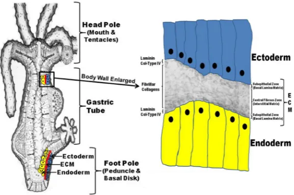
a gastric tube with a mouth and tentacle ring at the head pole and a peduncle and basal disk at the foot pole (Fig. 1). The entire body wall of Hydra (from the tip of its tentacles to the base of its foot process) is structured as an epithelial bilayer with an intervening extracellular matrix (ECM) (Fig. 1). In the basal-lateral compart-ment between the epithelial cells are the interstitial cells (i-cells) that comprise such phenotypes as nerve cells, nematocytes, and gametes for sexual reproduction (Fig. 1).
high regenerative capacity, Hydra has been used as a model for analysis of cell-ECM interactions during morphogenesis and cell differentiation. Over the last two decades, biochemical, and molecu-lar studies have established that the ECM of Hydra is composed of a broad spectrum of matrix components reflective of that seen in more complicated vertebrate systems. These matrix molecules have been shown to have an important role in morphogenetic and cell differentiation processes in Hydra. Molecular and biochemical studies have also established the structural basis for the biophysical properties of Hydra ECM. These studies in combination with early ultrastructural analyses indicate that the ECM of Hydra is a highly flexible matrix with elastic properties, mainly based on the macro-molecular structure of its collagens (type IV and fibrillar collagens).
This review will include a discussion of Hydra ECM in regard to 1) its general structure, 2) its molecular composition, 3) the biophysical basis for the flexible nature of Hydra’s ECM, 4) the relationship of the biogenesis of Hydra ECM to regeneration of body form, and 5) the functional role of Hydra ECM during pattern formation and cell differentiation in this simple invertebrate organ-ism that diverged early during metazoan evolution.
The general structure of the Hydra ECM based on early
and current studies
Early morphological and biochemical studies on the structure and composition of the Hydra ECM
Initial ultrastructural studies of Hydra ECM (mesoglea) identified a broad spectrum of structural components and described it as an amorphous matrix of low density containing fine fibrils ranging
Fig. 1. The Hydra body plan is formed of an epithelial bilayer wih an interven-ing extracellular matrix (ECM).Hydra exists as a gastric tube with a head process (mouth and tentacles) at the apical pole and a foot process (peduncle and basal disk) at the basal pole (left diagram). The entire body wall of Hydra (from the tip of the ten-tacles to the basal disk) is organized as an epithelial bilayer with an intervening ECM along the entire longitudinal axis of the organism. Hydra’s ECM is structured as two subepithelial zones (i.e. basal lamina matrix) with an intervening central fibrous zone (i.e. interstitial matrix). As shown in the composite diagram to the right that utilizes a transmission electron micrograph of Hydra ECM interposed between a drawing of two cell layers (ectoderm and endoderm), Hydra laminin and Type IV Collagen are localized to the two subepithelial zones of the matrix while Hydra fibrillar collagens (e.g. Hcol-I) are localized to the central fibrous zone or interstitial matrix.
junction with radiotracer precursor-labeling; they reported that Hydra ECM had biochemical characteristics similar to vertebrate basal lamina. They also concluded that the amino acid profiles suggested the presence of collagens in Hydra ECM. Experiments using lathyritic agents known to cause reduced cross-linking of collagens indicated a role for these macromolecules in Hydra morphogenesis (Barzansky and Lenhoff, 1974).
As a consequence of 1) the types of matrix components forming the supramolecular structure of Hydra ECM and 2) the unique structural features of these components, Hydra ECM exists as a highly flexible and elastic structure as compared to normal ECM of vertebrates. Also in contrast to vertebrate ECM, the collagens of Hydra ECM are more easily extracted due to a reduced amount of cross-linking as will be discussed later in the Hydra collagen sections (Deutzmann et al., 2000). The unique properties of Hydra ECM provide the necessary rigidity for maintenance of body shape while at the same time permitting sufficient plasticity to allow extensive but reversible shape changes along the longitudinal and radial axis. Studies by Shostak
et al., (1965) showed that when isolated Hydra ECM was
stretched to twice its original length, it would retract to its original length when released from tension. In this regard, it was noted in the ultrastructural studies of Davis and Haynes (1968) that in a contracted state, the fibrils of the central fibrous zone become irregular and fold upon themselves. This is unusual for vertebrate fibrillar collagens, which tend to be normally inelastic.
For a period of 16 years no further structural studies on from 5-50 nm in diameter (Hess, 1957; Wood, 1961; Gauthier, 1963). Cellular processes from the bilayer extend into the ECM and form cell-cell contacts between the endoderm and ectoderm (Hess, 1957; Wood, 1961; Haynes et al., 1968; Shimizu, 2008)
Davis and Haynes (1968) provided a detailed analysis of the ultrastructure of Hydra ECM under conditions in which the polyp was relaxed or contracted. In agreement with previous studies, they reported that Hydra ECM was approximately 0.5 to 2.0 mm in
diameter and noted it was thickest in the body column and thinnest in the tentacles. They also indicated that it had three structural components based on TEM analysis, namely, an amorphous ground substance, fibrils, and particulate material. The fibrils consisted of three types. The smallest fibrils were 5-9 nm in diameter and their density in the ECM varied depending on whether the polyps were in a relaxed or contracted state. During contraction, these fibrils were randomly arranged and more densely packed while in a relaxed state the fibrils were in a more orderly arrangement and less densely packed. A second type of fibril that was less frequent was a thicker banded fibril that ranged from 36-45 nm in diameter and had a periodicity of approximately 30 nm. The final class of fibrils consisted of short thin fibrils that formed bundles oriented perpendicular to the long axis of the polyp. These fibrils extend from the basal plasma membrane of the ectoderm cells into the ECM suggesting some type of linker function.
con-Hydra ECM were published. In 1991 the first of a series of papers
were published that utilized a combination of biochemical, molecu-lar, and cell biological approaches to clarify the macromolecular structure of Hydra ECM and to elucidate the role of cell-ECM interactions during cell differentiation and morphogenesis in this ancient metazoan. What follows, is an overview of what is currently known about the overall structure of Hydra ECM.
The Hydra ECM is a tri-laminar structure composed of two subepithelial basal lamina matrices and a central interstitial-like matrix
Beginning in the late 1980’s experiments were begun to utilize
Hydra as a developmental model to study the role of cell-ECM
interactions during pattern formation in epithelial systems. The specific questions being asked related to the role of Hydra ECM in cell differentiation and morphogenetic processes. As a first step, it was essential to clarify the molecular composition and structure of Hydra ECM. The strategy implemented involved: 1) isolation of Hydra ECM and use of biochemical and immunological ap-proaches to analyze the purified matrix preparation, 2) use of the purified Hydra ECM preparation to generate a battery of Hydra-specific polyclonal and monoclonal antibodies, 3) use of these
Hydra specific antibodies as reagents to screen expression cDNA
libraries and as probes to characterize the distribution of matrix components in Hydra ECM using morphological techniques, and 4) use of Hydra-specific antibodies and isolated matrix compo-nent domains as blocking reagents to study the role of cell-ECM interactions in Hydra using a number of regeneration bioassays. These functional studies were later complimented with antisense RNA studies to selectively knockdown Hydra ECM components during regenerative processes.
Initial biochemical and immunological studies using antibodies
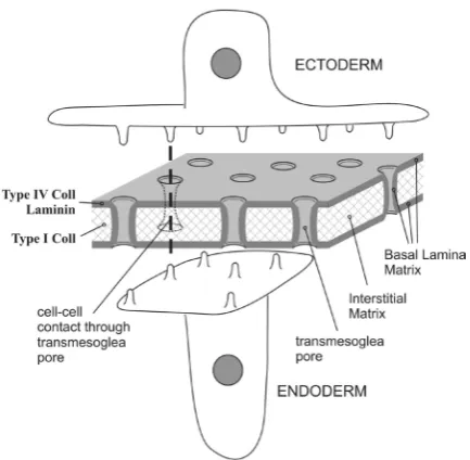
generated to vertebrate ECM components indicated that Hydra ECM had a spectrum of matrix components similar to those observed in more complicated invertebrates and in vertebrates. Specifically, evidence for the presence of collagen type IV, laminin, heparan sulfate proteoglycan and fibronectin-like molecules was presented (Sarras et al., 1991). Pulse-chase autoradiographic studies in conjunction with translational and post-translational processing inhibitor studies supported the presence of collagen and proteo-glycan components (Sarras et al., 1991). Use of Hydra-specific monoclonal antibodies (Sarras et al., 1993) in combination with special ultrastructural staining techniques (Sarras et al., 1994) clari-fied that Hydra ECM contained distinct structural regions. Adjacent to the basal plasma membrane border of each epithelial layer was a defined basal lamina-like region named the subepithelial zone and intervening between these two basal lamina–like regions was a central fibrous zone that appeared similar to interstitial matrix. Subsequent cloning studies resolved this issue by showing that laminin chains were confined to the subepithelial zones (i.e. basal lamina) (Sarras et al., 1994) and type I-like collagen was confined to the central fibrous zone (i.e. interstitial matrix) (Deutzmann et al., 2000). Along with molecular analysis (Fowler et al., 2000), mono-clonal antibody to the NC1 domain of collagen type IV has recently localized this macromolecule with laminin to the two basal lamina layers adjacent to the ectoderm and endoderm (Shimizu, 2008).
Recently, confocal microscopy used to further localize laminin,
Hydra collagen type IV (Hcol-IV), and Hydra fibrillar collagen type
I (Hcol-I) within the ECM, has determined that Hydra ECM is porous with multiple trans-ECM pores ranging from 0.5 to 1 mm
in diameter and about six pores per 100 mm2 in density. Cellular
processes from the ectoderm and endoderm, utilize these pores to form communications between the two layers within the ECM (Shimizu, 2008). The basal lamina components form a cylinder around these cellular processes as they pass through the ECM (Shimizu, 2008). The reader is referred to Fig. 2. for a diagram of this basal limina sheathed cell to cell communication system within
Hydra ECM. Based on these studies, an overall structure of Hydra
ECM is shown in Fig. 1. and a discussion of each of the major components (laminin, collagen type IV and fibrillar collagens) of
Hydra ECM follows. A list of the major components of Hydra ECM
with their general properties is shown in Table 1.
Molecular components of the Hydra ECM
LamininsThe laminin family of matrix proteins
Laminins represent a family of glycoproteins that are a major component of basement membranes (basal lamina). To date, at least sixteen different laminin heterotrimers have been identified in mammals and a number of laminins have been identified in invertebrates such as Drosophila, C. elegans, and sea urchin (Burgeson et al., 1994; Richards et al., 1994; Iivanainen et al., 1995; Martin et al., 1995; Miner et al., 1995; Champliaud et al., 1996; Benson et al., 1999; Hutter et al., 2000). Laminins are involved in basement membrane assembly and are an important component in the supramolecular architecture of matrix (Yurchenco and O’Rear, 1994; Yurchenco and Cheng, 1994; Timpl and Brown, 1996; Yurchenco, 2011). The heterotrimer isoforms are gener-ated from multiple subunits to include the a, b, and g chains. In
vertebrates, we find many different a, b and g chains and humans
have at least 16 heterotrimeric laminin species (Aumailley et al., 2005). At least some of these chains may exist as alternatively spliced forms (Kallunki et al., 1992; Galliano et al., 1995; Talts et al., 1999). ECM superstructure is based on assembly of a self-binding laminin mesh-like polymer, formation of a self-binding type IV col-lagen network polymer, and subsequent binding of various other ECM components to these networks (Colognato and Yurchenco, 2000). Interaction of laminin domains to cell surface ECM-binding proteins such as integrins and dystroglycan is involved with matrix assembly processes (Colognato et al., 1999) and with cell signaling events (Darribere et al., 2000; Wickstrom et al., 2011; Yurchenco, 2011; Alexi et al., 2011) associated with cell differentiation and morphogenesis.
Hydra laminin
Hydra laminin chains have been cloned and functionally analyzed
(Sarras et al., 1993; Sarras et al., 1994). As stated, Hydra laminin is localized to the two subepithelial zones (basal lamina) of Hydra ECM. A partial sequence for a Hydra b1-like chain was initially
reported (Sarras et al., 1994). Studies have since been completed for description of the entire ORF of the a1 chain and partial
descrip-tion of a a-5-like chain (Zhang et al., 2002). No gchain has thus
far been identified, although the typical trimeric form for isolated
Hydra laminin has been observed by rotary shadow TEM analysis
(Zhang et al., 2002). While Hydra laminin is localized to the two subepithelial zones (basal lamina) of Hydra ECM, it is synthesized exclusively by the endoderm, which means that the molecules have to diffuse through the mesoglea to reach the ectodermal layer (Sarras et al., 1993; Sarras et al., 1994). The location at the basal region of both cell layers suggests that laminin is required for proper cell function and differentiation. Consistent with this proposal, laminin secretion from the endoderm precedes secre-tion of Hydra collagen-I that arises from the ectoderm (Deutzmann
et al., 2000) and inhibition of laminin secretion through a RNA
antisense technique will block collagen secretion (Shimizu et al., 2002). Earlier studies have already established that antibodies to
Hydra laminin will block Hydra morphogenesis (Sarras et al., 1993)
and other ECM-related processes such as cell migration (Zhang and Sarras, 1994).
While the mechanism(s) of signal transduction in Hydra is not fully known, some published data (Agbas and Sarras, 1994) and unpublished studies (Sarras laboratory) suggest the involvement
of integrins, the primary class of ECM-receptors in higher animals (Darribere et al., 2000; Wickstrom et al., 2011). The Hydra ECM receptor data mainly pertains to a region in the short arm of the
Hydra b1-like chain. In this regard, sequence analysis of the b1-like
chain indicates the substitution of a FTGTQ sequence for the YIGSR receptor-binding sequence observed in vertebrates. Although the role of the YIGSR sequence in signal transduction-mediated processes has been questioned, published studies do indicate 1) its potential use as an inhibitor of human pre-B leukaemic cell growth and metastasis using SCID mice models (Yoshida et al., 1999) and 2) its role in the guidance of axon growth cones (Hopker
et al., 1999). Such studies and others support its involvement in
cell signaling processes.
More recent studies also validate the existence of the YIGSR ECM-receptor sequence (Saleh et al., 2008). The substituted FT-GTQ sequence in the Hydra b1-like chain has also been shown to
interact with the cell surface under both in-vitro and in-vivo conditions (Sarras et al., 1994) and affinity purification studies indicate that the FTGTQ sequence can interact with a Hydra integrin-like protein (Agbas and Sarras, 1994). Further analysis of 1) laminin-mediated cell signaling processes and 2) the role of laminin in the biogenesis and assembly of Hydra ECM is required to fully understand the relationship of pattern formation to ECM structure and assembly in this invertebrate organism. As an update, it is important to note that based on publication of the Hydra genome in 2010, it is now known that true integrin molecules are present in the genome of this organism (Chapman et al., 2010). Based on this sequence data, more extensive structural and functional studies regarding Cell/ECM interaction can now be conducted.
Collagens
Collagens are found in all animals and are the most abundant protein of the extracellular matrix. The basic structure of this large protein family consists of multiple Gly-X-Y repeats. Extensive struc-tural and functional diversity among collagens is accomplished by introducing interruptions in the triple helical domains and inclusion of various globular domains (Prockop and Kivirikko, 1995; Olsen and Niomiya, 1999; Ricard-Blum, 2011). Some collagens, like the basement membrane collagen type IV are found in all animals, whereas others are limited to particular groups. The fibrillar collagens for instance have previously only been found in vertebrates and have not been identified in invertebrates. In contrast, specialized
Basal Lamina ECM components
Laminin Contains α, β, and γ subunits in a trimeric cruciate (HLM) structure as viewed by rotary EM. Cloning studies indicate the alpha subunit is a vertebrate α5-like or Drosophila-like chain while the beta subunit is a vertebrate β1-like chain containing at least one defined cell binding domain (FTGTQ).
Collagen type IV
(Hcol-IV) A homotrimeric glycoprotein formed from three collagen IV a smaller non-collagenous domain (NC1) at its C-terminus. The collagen domain contains RGD cell binding motifs. TEM rotary shadow α1-like chains. Each subunit contains a collagen domain at its N-terminus and studies indicate that polymerization of Hcol-IV molecules within the ECM involves interaction of the NC1 domains. A typical 7S domain at the N-terminus appears not to be present in the mature ECM.
Interstitial ECM components (Fibrillar collagens)
1) Fibrillar Collagens type I
(Hcol1) A homotrimeric glycoprotein formed from three collagen type I terminal propeptide. During processing the C-terminal pro-peptide is removed, but the N-terminal propeptide is retained in the mature α1-like chains. Each chain contains an N-terminal propeptide and C-molecule. This results in the formation of flexible fibrils but not thickened banded fibrils as typically seen in vertebrate type I collagens. Hcol-1’s flexibility is enhanced by a reduction in its proline content and a loss of critical lysines involved in lysyl-cross bridging. 2) Hydra Collagen types 2, 3, 5, & 6
(Hcol 2; Hcol 3; Hcol 5 & Hcol 6) Hydra fibrillar collagens Hcol 2, 3, 5, and 6 were characterized only from cDNA sequence analysis and subsequent computer modeling, and so unlike Hcol1 where the protein was also purified and biochemically analyzed, there is some degree of speculation about the in vivo nature of these proteins.
TABLE 1
MAJOR COMPONENTS OF HYDRA EXTRACELLULAR MATRIX
collagens such as the cuticle collagens of C elegans (Kramer, 1994) and the mini-collagens of Hydra nematocyst capsules (Kurz et al., 1991) have only been found in invertebrates. Surprisingly, Kramer (1994) has reported that the cuticle collagens of C. elegans are encoded by more than a 100 genes. These observations indicate how diversity in collagen structure is utilized to meet the needs of a broad spectrum of specialized extracellular matrices. Accord-ingly, analysis of the structure of specialized collagens provides us with insight into the organization and function of ECM in both vertebrates and invertebrates.
While indirect evidence had suggested the existence of collagens in the ECM of Hydra (Hausman and Bernett, 1971; Barzansky and Lenhoff, 1974), more recent structural and functional analy-sis has provided a clear understanding of the types of collagens that exist in this invertebrate (Deutzmann et al., 2000; Fowler et
al., 2000). These collagens include a basement membrane-type
(Hydra collagen type IV, Hcol-IV) and an interstitial-type (Hydra fibrillar collagen, Hcol-1). Other Hydra fibrillar collagens have been more recently identified (Zhang et al., 2007). As will be discussed, Hcol-IV and Hcol-1 collagens have been characterized at both the cDNA and protein level.
Hydra collagen type IV (Hcol-IV)
Collagen type IV is the second most prominent constituent of basement membranes after laminin. These collagens are gly-coproteins composed of three subunits that form a polymerized network in conjunction with laminin (Hudson et al., 1994; Borza et
al., 2000; Colognato and Yurchenco, 2000). Currently, six different
types of collagen type IV subunits are known to exist in vertebrates (Yurchenco and O’Rear, 1994; Hopker et al., 1999; Borza et al., 2000; Vanacore R et al.., 2011). Invertebrate collagen type IV molecules have previously been identified in such organisms as
Drosophila (Blumberg et al., 1988), C. elegans (Guo et al., 1991)
and a number of other species (Kuehn, 1994).
As reported by Fowler et al. (2000) Hydra ECM also contains a collagen type IV. Analysis of the cDNA clone revealed a protein of 1723 amino acids, including an interrupted 1455 residue collag-enous domain and a 228 residue carboxyl-terminal non-collagcollag-enous domain. Hcol-IV is similar to all known a(IV) chains, but again,
most closely resembles vertebrate and invertebrate a1(IV) chains.
Like Hydra fibrillar collagen, Hcol-IV also forms homotrimeric molecules. Electron microscopy reveals an irregular network of rod-like structures interrupted by globular domains. This network can be depolymerized by reducing agents to dimeric collagen molecules, joined via their C-terminal non-collagenous domains. Under extensive denaturing conditions, depolymerization can only be taken to the dimeric but not monomeric stage. This suggests that the individual polypeptide chains are quantitatively held together by non-reducible cross-links in addition to disulfide bonds. This behavior is quite different from the vertebrate collagen type IV that needs pepsin digestion for solubilization. For vertebrate collagen type IV, a model has been proposed in which four molecules aggregate via their N-terminal domains to form a spider-like structure. The interactions are stabilized via disulfide bonds and lysine derived cross-links, resulting in a highly protease resistant 7S domain. In addition, the C-terminal globular domain, NC1, binds to itself, mainly via disulfide bridges, to form a linear dimer.
Both interactions at the N-terminal and C-terminal ends lead to the proposal of an open network structure that can further
polymer-ize via lateral aggregation of the triple helical domains (Yurchenco and O’Rear, 1994; Kuehn, 1994; Reinhart-King, 2011). In contrast, in Hydra while C-terminal interactions and lateral aggregation oc-curs, a stable 7S domain is not formed. A similar collagen type IV has also been reported in the worm, Ascaris suum (Noelken et al., 1986). This form of collagen type IV favors a more flexible ECM then that seen in typical vertebrate matrices.
Hydra fibrillar collagens
1) Hydra fibrillar collagen 1 (Hcol-1). Fibrillar collagens make up the majority of matrix components within the interstitial matrix of the animal kingdom. Likewise, Hydra fibrillar collagens are the major component of Hydra ECM (Deutzmann et al., 2000). The cDNA for the Hydra fibrillar collagen, Hcol-I, encodes a protein of 1412 amino acids. Hcol-I was the first fibrillar collagen to be identified in
Hydra. The polypeptide isolated from Hydra ECM has an apparent
molecular weight of 155 kDa. The subunit chains of Hcol-I form homotrimeric molecules that constitute the majority of the fibrils within the central fibrous zone (interstitial matrix). Sequence com-parisons clearly define Hcol-I as a fibrillar collagen. The highest similarity is to the a chains of vertebrate collagens type I and II. A
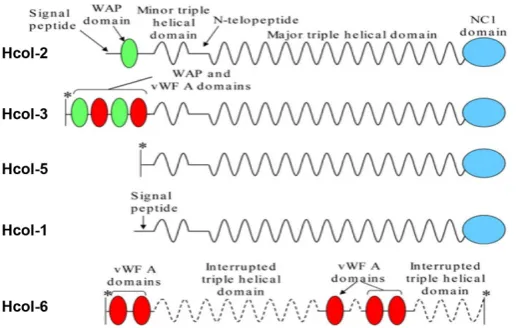
similar degree of similarity is found between Hcol-I and invertebrate sea urchin collagen (Exposito and Garrone, 1990) and a sponge fragment (Exposito et al., 1992). Corresponding to the similarity at the sequence level, Hcol-I also exhibits the characteristic domain structure of fibrillar collagens, consisting of a central triple helical domain flanked by an N-terminal propeptide-like domain and a C-terminal propeptide (Fig. 3). It is note worthy that the triple helical domain with 340 uninterrupted GLY-X-Y repeats has exactly the same length as the fibrillar collagens of vertebrates, suggesting similar fibril forming possibilities.
Despite marked similarities in the primary structure, there are distinct differences in the supramolecular organization of vertebrate fibrillar collagen networks as compared to that seen in Hydra ECM. Hcol-I forms a network of fine fibrils rather than thicker banded fibrils as seen by electron microscopy of vertebrate interstitial matrices. In contrast to vertebrate collagens that require pepsin digestion for solubilization, large polymeric structures of Hcol-I can be isolated
from the ECM of Hydra under native conditions.
Several factors are responsible for the special structure of Hydra Hcol-I. These factors include: 1) a low content of proline in the triple helical domain that is only about 40% that of vertebrate collagens: 2) a reduced degree of inter-chain cross-linking due to the lack of classical consensus sequences for lysine/lysine-aldehyde derived covalent bonds; and 3) most importantly, altered post-translational processing that results in retention of the N-terminal propeptide-like domain in the mature molecule. Combined, these factors result in a more flexible fibrillar collagen that can bend on itself as suggested by the early ultrastructural studies of Davis and Haynes (1968).
2) Hydra Fibrillar Collagens: Hcol2, Hcol3, Hcol5, Hcol6. Next to be identified through molecular cloning techniques were four fibrillar collagens named, Hcol2, Hcol3, Hcol5 and Hcol6 (Zhang et. al, 2007). Hcol6 was only a partial sequence of a collagen gene with a unique structural organization consisting of multiple von Willebrand factor-A domains interspersed with interrupted collagenous triple helices. Hcol2 and Hcol5 have major collagenous domains of clas-sical length (1020 amino acid residues), whereas the equivalent domain in Hcol3 is shorter (969 residues). The N-propeptide of Hcol2 contains a whey-acid-protein four-cysteine repeat (WAP) domain, and the equivalent domain of Hcol3 contains two WAP and two von Willebrand factor-A domains (Fig. 3).
Phylogenetic analyses revealed that the Hydra fibrillar collagen genes form a distinct clade that appeared related to the protostome/ deuterostome A-clade of fibrillar collagens. Data base searches revealed that Hcol2, Hcol5, and Hcol6 are highly conserved, which also provided preliminary evidence for the expression of a B-clade fibrillar collagen. In situ hybridization indicated an ectoderm expression pattern along the entire longitudinal axis of Hydra for the Hcol2 and Hcol3 fibrillar collagens as was previously reported for Hcol1 (Deutzmann,.et al., 2000). Hydra Hcol2 and Hcol3 also have high expression in the tentacles and forming buds. Fibrillar collagen Hcol5 has high expression in the tentacles, foot process, and forming buds of adult polyps while Hcol6 expression is high at the base of the tentacles and in forming buds. During head re-generation it is expressed in the ectoderm as observed with Hcol1. Hcol6 is expressed to a much lower degree than the other fibrillar collagens and its expression pattern is restricted to the base of the tentacles and to forming buds (Zhang et al., 2007).
3) Non-ECM collagens of Hydra. To be complete, it should be pointed out that Hydra has a variety of what are called “mini collagens”. These collagens are localized within the capsule of nematocysts and are not associated with Hydra’s ECM. They have a unique and interesting structure due to their role in maintaining high hydrostatic pressures within the cavity of the nematocyst capsule. Because these collagens are not associated with Hydra’s ECM, they will not be further discussed in this review, but the reader is referred to articles pertinent to their unique structure and function (Kurz et
al., 1991; Holstein et al., 1994), see also in this issue the review
by Beckmann and Ozbek (2012).
Evidence for other types of matrix components in Hydra ECM
During functional studies using a pharmacological approach, evidence was obtained by Sarras et al. (1991, 1993) and Zhang and Sarras (1994) indicating that proteoglycans also exist in the
Hydra ECM. These experiments involved analysis of the effect
of molecules that block proteoglycan biosynthesis during head regeneration (Sarras et al., 1991), Hydra cell aggregate formation, and during cell migration as studied in Hydra grafting experiments (Zhang and Sarras, 1994). The validity of these pharmacological studies was confirmed using pulse-chase autoradiographic tech-niques to demonstrate that proteoglycan-associated molecules such as SO4 were in fact blocked from appearing in the ECM following treatment with such agents as b-xyloside as compared
to its inactive isomer, a-xyloside. These studies indicated that
any blockage in proteoglycan biosynthesis resulted in a blockage in Hydra morphogenesis (head regeneration and cell aggregate morphogenesis) and a retardation in normal i-cell migration, sug-gesting that proteoglycans are components of the Hydra ECM. As of this date however, proteoglycans have not been isolated from Hydra ECM and therefore this evidence is indirect in nature.
The structural relationship of Hydra ECM molecular
components to its flexible biophysical properties
Hydra has enhanced the flexible nature of its fibrillar collagens
such as Hcol-I by reducing its proline content and by decreasing the degree of lysine/lysine inter-chain cross-links (Deutzmann, 2000; Zhang et al., 2007). In addition, alterations in the structure of Hydra collagen type IV are also observed and these structural alterations likely lead to a more flexible Hcol-IV network following polymerization of the ECM. In the case of Hcol-IV, C-terminal interactions and lateral aggregation occur too, but as indicated previously, a stable 7S domain is not formed. The reason might be that formation of a highly ordered 7S domain would impose steric constraints so that the four molecules leave at preferred angles. This might favor the formation of rigid basement membranes as needed by vertebrates to contribute to the shape of organs; but for formation of the flexible ECM of Hydra, as discussed above, a more relaxed cross-linking involving some cysteine residues in the N-terminal region might be more advantageous. In any case, the results for Hydra clearly show that the formation of a 7S domain is not essential for formation of a collagen type IV network. This is also supported by earlier findings for the worm Ascaris suum (Noelken et al., 1986). Taken together, the structural properties of Hydra Hcol-IV and the organism’s fibrillar collagens, such as Hcol-I, contribute to the flexible nature of Hydra ECM.
In summary, whereas vertebrates need a tight extracellular matrix that can withstand high tensile forces such as in the Achilles tendon, Hydra needs a highly flexible ECM, so that the animal can continuously contract and extend. The molecular and biochemical studies of Hydra Collagen Type IV and Hydra Fibrillar Collagens, such as Hcol-I, were required to understand the flexible nature of
Hydra ECM.
Biogenesis of Hydra ECM is intimately tied to
regenera-tion in this simple metazoan
As will be further discussed in the next section, Hydra ECM is required for general morphogenesis, pattern formation, and cell differentiation to occur in the adult polyp. This has been deter-mined through head and foot regeneration studies and through experiments utilizing Hydra cell aggregates. Development of
from non-enzymatically dissociated cells obtained from the adult polyp (Gierer et al.., 1972; Bode, 1974, Sarras et al., 1993). In all processes studied (head regeneration, foot regeneration, and development of Hydra cell aggregates), formation of a new ECM must occur for morphogenesis to proceed. If ECM biogenesis is blocked or perturbed in any way, morphogenesis is stopped. This has been shown through a broad array of approaches to include: 1) use of pharmacological agents (Sarras et al., 1991), 2) block-ing antibodies to Hydra ECM components (Sarras et al., 1993), 3) fragments of ECM components that are used to compete in the normal polymerization of the matrix components (Zhang et al., 1994), and 4) anti-sense RNA to ECM components introduced through electrophoresis techniques (Shimizu et al., 2002).
The sequence of events associated with Hydra ECM biogen-esis indicates that cross-talk occurs between the ectoderm and endoderm and through signals emanating from the ECM to both epithelial layers. A general description of Hydra ECM biogenesis is depicted in Fig. 4. The need for ECM biogenesis during exper-imentally-induced head or foot regeneration is explained by the unique elastic properties of Hydra’s ECM. Removal of the head or foot or a simple incision along the gastric tube wall of Hydra results in a retraction of the ECM from the cut edge, thereby creat-ing a newly sealed epithelial bilayer that has no ECM between the two cell layers (Shimizu et al., 2002). This ECM-deficient bilayer changes its morphology so that cells become flattened compared to the more cuboidal to columnar cellular phenotype normally seen along Hydra’s body wall (Fig. 4). Soon after sealing (within 1 hr post-decapitation), this bilayer immediately begins to synthesize a new ECM. The process involves initial up-regulation of ECM
component genes within 3 hrs of decapitation (in the case of head regeneration experiments as shown in Fig. 4). In this process, basal lamina mRNA expression (e.g. laminin and Hcol-IV) is specific to endodermal cells while interstitial matrix components expression (e.g. Hcol1, 2, and 3) is specific to the ectoderm layer of cells. Within 7 hrs of decapitation, basal lamina (BL) proteins are secreted from the endoderm and a newly formed BL is seen associated with the basal extracellular border of both the ectoderm and endoderm. Through ECM receptor systems (Agbas and Sarras, 1994) BL components trigger synthesis of fibrillar collagens, such as Hcol1, that are secreted from the ectoderm and polymerize in the interstitial matrix zone between to the two BL layers (Shimizu
et al., 2002). The linkage between BL formation and interstitial
matrix component translation and secretion is supported by RNA antisense experiments in which laminin translation is blocked and subsequent Hcol1 translation and secretion from the ectoderm is prevented (Shimizu et al., 2002), thereby blocking ECM formation and perturbing morphogenesis of the bilayer to a head or foot structure. Because Hydra cell aggregates form from dissociated tissues, the initial epithelial bilayer that forms lacks an ECM. If one uses pharmacological agents, blocking antibodies to Hydra ECM components, or fragments of ECM components to perturb ECM polymerization in Hydra cell aggregate; morphogenesis of the ag-gregates beyond the cystic stage will not occur (Zhang et al., 1994).
Taken in total, these studies indicate that 1) ECM biogenesis is essential for Hydra morphogenesis and 2) ECM biogenesis is a complicated process involving cross-talk between the endoderm and ectoderm as well as signaling from the ECM to the cells of the bilayer. This cross-talk involves sequential synthesis of basal
Fig. 4. Extracellular matrix (ECM) biogenesis following decapitation of Hydra. Experimentally-induced regeneration in Hydra is typified by surgical removal of the head pole in the adult Hydra polyp. This is a simple procedure involving excision of the head pole with a small scalpel. When decapitation is initiated, the open head pole (the gastric cavity is now open to the aqueous environment) seals within 1 hour by fusion of the two cut ends of the apical epithelial bilayer (Shimizu et al., 2002). The elastic properties of Hydra ECM cause the entire matrix to retract at the cut ends resulting in the sealed epithe-lial bilayer to now lack an intervening ECM. Because of the lack of an ECM, the apically sealed epithelial bilayer alters its morphology and becomes flattened as compared to the normal cuboidal-columnar phe-notype that body wall cells display. Within 3 hours, an up-regulation of ECM genes is observed in the ectoderm and endoderm (Shimizu et al., 2002). This up-regulation could be due to the abrupt lack of an ECM and subsequent ectoderm/endoderm cell to cell signaling. By 7 hours, basal lamina components such as laminin and collagen type IV (Hcol-IV) are
lamina (endoderm) and interstitial matrix (ectoderm) components that will in turn signal appropriately timed translation and secretion of ECM proteins resulting in the polymerization of a well ordered matrix. As with higher invertebrates, this also involves ECM receptor complexes that trigger appropriate signaling pathways associated with ECM biogenesis processes.
The relationship of ECM structure to cell-ECM
interac-tions in Hydra
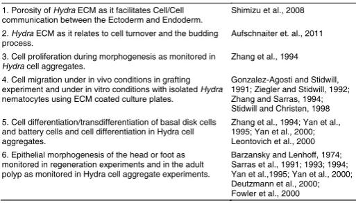
Cell-ECM interactions are central to normal tissue homeostasis, a broad spectrum of disease states, as well as developmental processes (Matejas et.al. 2010; Kim et al.., 2011; Watt and Fuji-wara, 2011; Wicksgtrom et al.., 2011; Yurchenco, 2011). While this review has focused on the relationship of Hydra ECM to the overall structural and cell behavior characteristics of this invertebrate, it should be noted that cell-ECM interactions have been determined to be important to a number of developmental processes in Hydra beyond general morphogenesis. These processes include, 1) cell proliferation, 2) cell migration, 3) cell differentiation, and as previ-ously discussed, 4) morphogenesis. These cell-ECM interactions in Hydra are listed in Table 2 with those references from which the studies were reported. A role for the ECM in the modulation of cell proliferation in Hydra was first reported by Zhang et al., (1974). These studies showed that introduction of exogenous matrix protein domains during ECM formation in Hydra resulted in alterations in cell proliferation rates. Likewise, alteration of ECM structure or ex-ogenous introduction of blocking antibodies to matrix components or matrix component domains would retard i-cell migration in Hydra grafting experiments (Zhang and Sarras, 1994). In vitro studies with isolated nematocytes have also indicated dependence of cell-ECM interactions for cell migratory processes in Hydra (Gonzalez et al., 1991; Ziegler and Stidwill, 1992; Stidwill and Christen, 1998). As discussed above, a number of studies involving head regeneration (Sarras et al., 1991) and morphogenesis of Hydra cell aggregates (Sarras et al., 1993) have clearly shown that any perturbation of ECM formation will result in blockage of Hydra morphogenesis. In addition, studies involving Hydra cell aggregates (Zhang et al., 1994) or Hydra foot regeneration (Leontovich et al., 2000) have shown that perturbation of ECM structure or turnover can affect cell
differentiation in the adult Hydra polyp. Signaling motifs in these cell/ECM interactions can be exposed or cryptic.
In this regard, the action of Hydra matrix metalloproteinases has been shown to be involved in exposing potentially cryptic signal-ing sites within ECM components (Leontovich et al., 2000). Hydra matrix metalloproteinase (HMMP) has been shown to specifically affect the maintenance of the phenotype of basal disk cells through mechanisms that remain unclear (Leontovich et al., 2000). A similar observation has been reported for Hydra metalloproteinase-1 that is localized to the tentacle ECM and has been shown to be involved in the maintenance of tentacle battery cell phenotypic markers (Yan et al., 1995, 2000). As reviewed by Schmid et al.,1999), the importance of cell-ECM interactions during morphogenesis and cell differentiation extends beyond Hydra to a number of classes within Cnidaria. Finally, and most recently, a new in vivo labelling technique for Hydra collagen 1 and laminin was used to track the fate of ECM in all body regions of the animal (Aufschnaiter et al., 2011). These studies revealed that Hydra “tissue movements” are largely displacements of epithelial cells together with associated ECM (Aufschnaiter et al., 2011). In contrast, during the evagination of buds and tentacles, extensive movement of epithelial cells relative to the matrix was observed together with local ECM remodelling (Aufschnaiter et al., 2011). Taken in total, these studies show a clear role for cell-ECM interactions in Hydra. These interactions are a dynamic process that is intimately associated with the continuous turnover of epithelial cells along the body axis of Hydra.
In conclusion, it is important to note, that Hydra Cell/ECM inter-actions represent highly conserved processes within the animal kingdom given the early divergence of Hydra during metazoan evolution. Accordingly, the functional role of Hydra ECM in a variety of its cellular and developmental processes is seen replicated in more complicated vertebrate systems (Kim et al.., 2011; Kruegel and Miosge, 2010; Watt and Fujiwara, 2011; Wicksgtrom et al.., 2011; Yurchenco, 2011). This serves to reinforce the fundamental nature of these cell-ECM processes and highlights their importance to the cell biology of all metazoans. This also reminds us that although Hydra is considered a relatively “simple” organism, this does not mean that its cellular and molecular biology is anything but simple to understand. In this regard, future studies with Hydra should be directed at 1) understanding how the epithelial bilayer of
Hydra coordinates the formation of its ECM through cell signaling
pathways and 2) the precise mechanisms underlying the role of ECM in Hydra morphogenesis and cell differentiation.
Acknowledgements
The author wishes to express his appreciation to the National Institutes of Health, USA (DK092721) for funds that supported preparation and writ-ing of this review.
References
AGBAS, A., SARRAS, M.P. JR. (1994). Evidence for cell surface extracellular matrix binding proteins in Hydra vulgaris. Cell Adhes Commun 2: 59-73.
ALEXI X, BERDITCHEVSKI F, ODINTSOVA E. (2011). The effect of cell-ECM adhe-sion on signaling via the ErbB family of growth factor receptors. Biochem Soc
Trans 39: 568-573.
AUFSCHNAITER, R., ZAMIR, E.A., LITTLE, C.D., OZBEK, S., MUNDER, S., DAVID, C.N., LI, L., SARRAS, M.P.J. (2011). In vivo imaging of basement membrane movement: ECM patterning shapes Hydra polyps. J Cell Sci 124: 4027-4038. AUMAILLEY, M., BRUCKNER-TUDERMAN, L., CARTER, W.G., DEUTZMANN, R.,
EDGAR, D., EKBLOM, P., ENGEL, J., ENGVALL, E., HOHENESTER, E., JONES,
1. Porosity of Hydra ECM as it facilitates Cell/Cell
communication between the Ectoderm and Endoderm. Shimizu et al., 2008 2. Hydra ECM as it relates to cell turnover and the budding
process. Aufschnaiter et. al., 2011
3. Cell proliferation during morphogenesis as monitored in
Hydra cell aggregates. Zhang et al., 1994
4. Cell migration under in vivo conditions in grafting experiment and under in vitro conditions with isolated Hydra
nematocytes using ECM coated culture plates.
Gonzalez-Agosti and Stidwill, 1991; Ziegler and Stidwill, 1992; Zhang and Sarras, 1994; Stidwill and Christen, 1998 5. Cell differentiation/transdifferentiation of basal disk cells
and battery cells and cell differentiation in Hydra cell aggregates.
Zhang et al., 1994; Yan et al., 1995; Yan et al., 2000; Leontovich et al., 2000 6. Epithelial morphogenesis of the head or foot as
monitored in regeneration experiments and in the adult polyp as monitored in Hydra cell aggregate experiments.
Barzansky and Lenhoff, 1974; Sarras et al., 1991; 1993; 1994; Yan et al.,1995; Yan et al., 2000; Deutzmann et al., 2000; Fowler et al., 2000
TABLE 2
J.C. et al. (2005). A simplified laminin nomenclature. Matrix Biol 24: 326-332. BARZANSKY, B., LENHOFF, H.M., BODE, H. (1975). Hydra mesoglea: similarity of
its amino acid and neutral sugar composition to that of vertebrate basal lamina.
Comp Biochem Physiol [B] 50: 419-424.
BARZANSKY, B., LENHOFF, H.M. (1974). On the chemical composition and devel-opmental role of the mesoglea of Hydra. Amer Zool 14: 575-581.
BECKMANN, A., OZBEK, S. (2012). The Nematocyst: A molecular map of the cnidar-ian stinging organelle. Int J Dev Biol 56: 577-582.
BENSON S, PAGE L, INGERSOLL E, ROSENTHAL E, DUNGCA K, SIGNOR D. (1999). Developmental characterization of the gene for laminin alpha-chain in sea urchin embryos. Mech Dev 81: 37-49.
BLUMBERG, B., MACKRELL, A.J., FESSLER, J.H. (1988). Drosophila basement membrane procollagen alpha 1(IV). II. Complete cDNA sequence, genomic structure, and general implications for supramolecular assemblies. J Biol Chem 263: 18328-18337.
BODE, H. (1974). Activity of Hydra cells in vitro and in regenerating cell reaggregates.
Am. Zool 14: 543-550.
BODE, H. (2011). Axis formation in Hydra. Annu Rev Genet 45: 105-117. BORZA, D.B., MINER, J.H., HUDSON, B.G. (2000). Basement Membrane and Cellular
Components of the Nephron. In: Massry SG, Glassock RJ. New York: Lippincott
Williams and Wilkins, p 37-42.
BURGESON, R.E., CHIQUET, M., DEUTZMANN, R., EKBLOM, P., ENGEL, J., KLEINMAN, H., MARTIN, G.R., MENEGUZZI, G., PAULSSON, M., SANES, J. et al., (1994). A new nomenclature for the laminins. Matrix Biol 14: 209-211. CHAMPLIAUD, M.F., LUNSTRUM, G.P., ROUSSELLE, P., NISHIYAMA, T., KEENE,
D.R. and BURGESON, R.E. (1996). Human amnion contains a novel laminin variant, laminin 7, which like laminin 6, covalently associates with laminin 5 to promote stable epithelial-stromal attachment. J Cell Biol 132: 1189-1198. CHAPMAN, J.A., KIRKNESS, E.F., SIMAKOV, O., HAMPSON, S.E., MITROS, T.,
WEINMAIER, T., RATTEI, T., BALASUBRAMANIAN, P.G., BORMAN, J., BUSAM, D. et al. (2010). The dynamic genome of Hydra. Nature 464: 592-596. COLOGNATO, H., WINKELMANN, D.A., YURCHENCO, P.D. (1999). Laminin
po-lymerization induces a receptor-cytoskeleton network. J Cell Biol 145: 619-631. COLOGNATO, H., YURCHENCO, P.D. (2000). Form and function: the laminin family
of heterotrimers. Dev Dyn 218: 213-234.
DARRIBERE T, SKALSKI M, COUSIN HL, GAULTIER A, MONTMORY C, ALFANDARI D. (2000). Integrins: regulators of embryogenesis. Biol Cell 92: 5-25.
DAVIS LE, HAYNES JF. An ultrastructural examination of the mesoglea of Hydra. (1968). Z Zellforsch Mikrosk Anat 92: 149-158.
DEUTZMANN, R., FOWLER, S., ZHANG, X., BOONE, K., DEXTER, S., BOOT-HANDFORD, R.P., RACHEL, R., SARRAS, M.P. (2000). Molecular, biochemical and functional analysis of a novel and developmentally important fibrillar collagen (Hcol-I) in hydra. Development 127: 4669-4680.
EXPOSITO JY, D’ALESSIO M, SOLURSH M, RAMIREZ F. (1992). Sea urchin col-lagen evolutionarily homologous to vertebrate pro-alpha 2(I) colcol-lagen. J Biol
Chem 267: 15559-15562.
EXPOSITO JY, GARRONE R. (1990). Characterization of a fibrillar collagen gene in sponges reveals the early evolutionary appearance of two collagen gene families.
Proc Natl Acad Sci, U S A 87: 6669-6673.
FOWLER, S.J., JOSE, S., ZHANG, X., DEUTZMANN, R., SARRAS, M.P., JR. and BOOT-HANDFORD, R.P. (2000). Characterization of hydra type IV collagen. Type IV collagen is essential for head regeneration and its expression is up-regulated upon exposure to glucose. J Biol Chem 275: 39589-39599.
GALLIANO MF, ABERDAM D, AGUZZI A, ORTONNE JP, MENEGUZZI G. (1995). Cloning and complete primary structure of the mouse laminin alpha 3 chain. Distinct expression pattern of the laminin alpha 3A and alpha 3B chain isoforms.
J Biol Chem 270: 21820-21826.
GAUTHIER, G.F. (1963). Cytological studies on the gastroderm of Hydra. J Exp
Zool 152: 13-40.
GIERER, A., BERKING, S., BODE, H., DAVID, C.N., FLICK, K., HANSMANN, G., SCHALLER, H. and TRENKNER, E. (1972). Regeneration of hydra from reag-gregated cells. Nature 239: 98-101.
GONZALEZ AGOSTI C, STIDWILL R. (1991). In vitro migration of Hydra nematocytes: the influence of the natural extracellular matrix (the mesoglea, of collagen type IV and type, laminin, and fibronectin) on cell attachment, migration parameters and
patterns of cytoskeletal proteins. Cell Motil Cytoskeleton 20: 215-227. GUO XD, JOHNSON JJ, KRAMER JM. (1991). Embryonic lethality caused by
muta-tions in basement membrane collagen of C. elegans. Nature 349(6311): 707-709. HAUSMAN RE, BURNETT AL. (1971). The mesoglea of Hydra: IV. A quantitative
radiographic study of the protein component. J Exp Zool 177: 435-446. HAYNES JF, BURNETT AL, DAVIS LE. (1968). Histological and ultrastructural study
of the muscular and nervous systems in Hydra. I. The muscular system and the mesoglea. J Exp Zool 167: 283-293.
HESS A. (1957). Observations on the structure of Hydra as seen with the electron and light microscope. Quart J. Micr Sci 98: 315-326.
HOLSTEIN TW, BENOIT M, HERDER GV, DAVID, CN, WANNER G, GAUB HE. (1994). Fibrous mini-collagens in Hydra nematocysts. Science 265: 402-404. HOPKER VH, SHEWAN D, TESSIER-LAVIGNE M, POO M, HOLT C. (1999).
Growth-cone attraction to netrin-1 is converted to repulsion by laminin- 1. Nature 401: 69-73. HUDSON BG, KALLURI R, GUNWAR S, NOELKEN ME. (1994). Structure and
or-ganization of type IV collagen of renal glomerular basement membrane. Contrib
Nephrol 107: 163-167.
HUTTER H, VOGEL BE, PLENEFISCH JD, NORRIS CR, PROENCA RB, SPIETH J, GUO C, MASTWAL S, ZHU X, SCHEEL J, HEDGECOCK EM. (2000). Con-servation and novelty in the evolution of cell adhesion and extracellular matrix genes. Science 287: 989-994.
IIVANAINEN A, SAINIO K, SARIOLA H, TRYGGVASON K. (1995). Primary structure and expression of a novel human laminin alpha 4 chain. FEBS Let 365: 183-188. KALLUNKI P, SAINIO K, EDDY R, BYERS M, KALLUNKI T, SARIOLA H, BECK K, HIRVONEN H, SHOWS TB, TRYGGVASON K. (1992). A truncated laminin chain homologous to the B2 chain: structure, spatial expression, and chromosomal assignment. J Cell Biol 119: 679-693.
KIM SH, TURNBULL J, GUIMOND S. (2011). Extracellular matrix and cell signaling: the dynamic cooperation of integrin, proteoglycan and growth factor receptor. J.
Endocrinol 209: 139-151.
KRAMER JM. (1994). Structures and functions of collagens in Caenorhabditis elegans.
FASEB J 8: 329-336.
KRUEGEL J, MIOSGE N. (2010). Basement membrane components are key players in specialized extracellular matrices. Cell Mol Life Sci 67: 992-1002.
KUEHN, K. (1994). Basement membrane (type IV) collagen. Matrix Biol 14: 439-445. KURZ, E.M., HOLSTEIN, T.W., PETRI, B.M., ENGEL, J., DAVID, C.N. (1991).
Mini-collagens in hydra nematocytes. J Cell Biol 115: 1159-1169.
LEONTOVICH, A.A., ZHANG, J., SHIMOKAWA, K., NAGASE, H., SARRAS, M.P. JR. (2000). A novel hydra matrix metalloproteinase (HMMP) functions in extracellular matrix degradation, morphogenesis and the maintenance of differentiated cells in the foot process. Development 127: 907-920.
MARTIN, P.T., ETTINGER, A.J., SANES, J.R. (1995). A synaptic localization domain in the synaptic cleft protein laminin beta 2 (s-laminin) [see comments]. Science; 269: 413-416.
MATEJAS V, HINKES B, ALKANDARI F, AL-GAZALI L, ANNEXSTAD E, AYTAC MB, BARROW M, BLÁHOVÁ K, BOCKENHAUER D, CHEONG HI et al. (2010). Muta-tions in the human laminin beta2 (LAMB2) gene and the associated phenotypic spectrum. Hum Mutat 31: 992-1002.
MINER JH, LEWIS RM, SANES JR. (1995). Molecular cloning of a novel laminin chain, alpha 5, and widespread expression in adult mouse tissues. J Biol Chem 270: 28523-28526.
NOELKEN ME, WISDOM BJ, JR., DEAN DC, HUNG CH, HUDSON BG. (1986). Intestinal basement membrane of Ascaris suum. Molecular organization and properties of the collagen molecules. J Biol Chem 261: 4706-4714.
OLSEN BR, NINOMIYA Y. (1999). Collagens. In: Kreis T, Vale R, editors. Guidebook to the Extracellular Matrix, Anchor, and Adhesion Proteins. Oxford: Oxford Uni-versity Press 380-408.
PROCKOP DJ, KIVIRIKKO KI. (1995). Collagens: molecular biology, diseases, and potentials for therapy. Annu Rev Biochem 64: 403-434.
REINHART-KING CA. (2011). How matrix properties control the self-assembly and maintenance of tissues. Ann Biomed Eng 39: 1849-56.
isolation of a partial cDNA encoding a variant laminin A chain. Genomics 22: 237-239. SALEH, A.F., AOJULA, H.S., PLUEN, A. (2008). Enhancement of gene transfer using
YIGSR analog of Tat-derived peptide. Biopolymers 89: 62-71.
SARRAS, M.P. JR., DEUTZMANN, R. (2001). Hydra and Niccolo Paganni (1782-1840) – Two peas in a Pod?: The Molecular Basis of Extracellular Matrix Structure in the Invertebrate, Hydra. BioEssays 23: 716-724.
SARRAS, M.P. JR., MADDEN, M.E., ZHANG, X.M., GUNWAR, S., HUFF, J.K., HUD-SON, B.G. (1991). Extracellular matrix (mesoglea) of Hydra vulgaris. I. Isolation and characterization. Dev Biol 148: 481-494.
SARRAS, M.P. JR., MEADOR, D., ZHANG, X.M. (1991). Extracellular matrix (meso-glea) of Hydra vulgaris. II. Influence of collagen and proteoglycan components on head regeneration. Dev Biol 148: 495-500.
SARRAS, M.P. JR., ZHANG, X., HUFF, J.K., ACCAVITTI, M.A., ST JOHN, P.L., ABRAHAMSON, D.R. (1993). Extracellular matrix (mesoglea) of Hydra vulgaris III. Formation and function during morphogenesis of hydra cell aggregates. Dev
Biol 157: 383-398.
SARRAS, M.P. JR., YAN, L., GRENS, A., ZHANG, X., AGBAS, A., HUFF, J.K., ST JOHN, P.L., ABRAHAMSON, D.R. (1994). Cloning and biological function of laminin in Hydra vulgaris. Dev Biol 164: 312-324.
SCHMID, V., ONO, S.I., MULLER, S.R. (1999). Cell-Substrate Interactions in Cnidaria.
Microsc Res Tech 44:254-268.
SHOSTAK, S., PATEL, N.G., BURNETT, A.L. (1965). The role of mesoglea in mass cell movement in Hydra. Dev Biol 12: 434-450.
SHIMIZU, H., AUFSCHNAITER, R., LI, L., SARRAS, M.P. JR., BORZA, D.B., ABRA-HAMSON, D.R., SADO, Y., ZHANG, X. (2008). The extracellular matrix of hydra is a porous sheet and contains type IV collagen. Zoology 111: 410-418. SHIMIZU, H., ZHANG, X., ZHANG, J., LEONTOVICH, A., FEI, K., YAN, L., SARRAS,
M.P. JR. (2002). Epithelial morphogenesis in hydra requires de novo expression of extracellular matrix components and matrix metalloproteinases. Development 129: 1521-1532.
STIDWILL, R.P., CHRISTEN, M. (1998). Alteration of fibronectin affinity during dif-ferentiation modulates the in vitro migration velocities of Hydra nematocytes. Cell
Motil Cytoskeleton 41: 68-73.
TALTS JF, ANDAC Z, GOHRING W, BRANCACCIO A, TIMPL R. (1999). Binding of the G domains of laminin alpha1 and alpha2 chains and perlecan to heparin, sulfatides, alpha-dystroglycan and several extracellular matrix proteins. EMBO
J 18: 863-870.
TIMPL R, BROWN JC. (1996). Supramolecular assembly of basement membranes.
Bioessays 18: 123-132.
VANACORE R, PEDCHENKO V, BHAVE G, HUDSON BG. (2011). Sulphilimine cross-links in Goodpasture’s disease. Clin Exp Immunol 164: Suppl 1: 4-6.
WATT, F.M., FUJIWARA, H. (2011). Cell-extracellular matrix interactions in normal and diseased skin. Cold Spring harb Perspect Biol 3: a005124.
WICKSTROM SA, RADOVANAC K, FASSLER R. (2011). Genetic analysis of integrins signaling. Cold Spring Harb Perspect Biol 3:a005116.
WOOD, RL. (1961). The fine structure of intracellular and mesoglea attachments of epithelial cells in Hydra. In: Lenhoff HLW, editor. The Biology of Hydra. Coral Gables, FL: Miami Press University
YAN, L., LEONTOVICH, A., FEI, K., and SARRAS, M.P. Jr. (2000). Hydra metallopro-teinase 1 (HMP1) is a pre/pro enzyme which has a restricted mRNA spatial expres-sion pattern during head regeneration in Hydra vulgaris. Dev.Biol 219: 115-128. YAN, L., POLLOCK, G., NAGASE, H., SARRAS, M.P. Jr. (1995). Hydra metallo-proteinase 1 (HMP1), a member of the astacin family, localizes to extracellular matrix in a head specific manner and has a functional role during development.
Development 121: 1591-1602.
YOSHIDA, N, ISHII E, NOMIZU M, YAMADA Y, MOHRI S, KINUKAWA N, MATSUZAKI A, OSHIMA K, HARA T, MIYAZAKI S. (1999). The laminin-derived peptide YIGSR (Tyr-Ile-Gly-Ser-Arg) inhibits human pre-B leukaemic cell growth and dissemina-tion to organs in SCID mice. Br J Cancer 80: 1898-1904.
YURCHENCO, P.D. (2011). Basement membranes: cell scaffoldings and signaling platforms. Cold Spring Harb Perspect Biol 3: a004911.
YURCHENCO, P.D., CHENG, Y.S. (1994). Laminin self-assembly: a three-arm interaction hypothesis for the formation of a network in basement membranes.
Contrib Nephrol 107: 47-56.
YURCHENCO, P.D., O’REAR JJ. (1994). Basal lamina assembly. Curr Opin Cell
Biol 6: 674-681.
ZHANG, X., FEI, K., AGBAS, A., YAN, L., ZHANG, J., O’REILLY, B., DEUTZMANN, R. and SARRAS, M.P. JR. (2002). Structure and function of an early divergent form of laminin in hydra: a structurally conserved ECM component that is essential for epithelial morphogenesis. Dev Genes Evol 212: 159-172.
ZHANG, X., HUDSON, B.G. and SARRAS, M.P. JR. (1994). Hydra cell aggregate development is blocked by selective fragments of fibronectin and type IV collagen.
Dev Biol 164: 10-23.
ZHANG, X. and SARRAS, M.P. JR. (1994). Cell-extracellular matrix interactions under
in vivo conditions during interstitial cell migration in Hydra vulgaris. Development
120: 425-432.
ZHANG, X., BOOT-HANDFORD, R.P., HUXLEY-JONES, J., FORSE, L.N., MOULD, A.P., ROBERTSON, D.L., LILI, ATHIYAL, M., SARRAS, M.P. JR. (2007). The collagens of hydra provide insight into the evolution of metazoan extracellular matrices. J Biol Chem 282: 6792-6802.
ZIEGLER, U., STIDWILL, R.P. (1992). The attachment of nematocytes from the primitive invertebrate Hydra to fibronectin is specific and RGD-dependent. Exp
Integrins contribute to the establishment and maintenance of cell polarity in the follicular epithelium of the Drosophila ovary
5 yr ISI Impact Factor (2010) = 2.961 Ana Fernández-Miñán, Laura Cobreros, Acaimo González-Reyes and María D. Martín-Bermudo Int. J. Dev. Biol. (2008) 52: 925-932
Soluble membrane-type 3 matrix metalloprioteinase causes changes in gene expression and increased gelatinase activity during Xenopus laevis development
Logan A. Walsh, Colin A. Cooper and Sashko Damjanovski Int. J. Dev. Biol. (2007) 51: 389-396
Mechanical control of tissue morphogenesis during embryological development Donald E. Ingber
Int. J. Dev. Biol. (2006) 50: 255-266
TGFbeta and TGFalpha, antagonistic effect in vitro on extracellular matrix accumulation by chick skin fibroblasts at two distinct embryonic stages.
P Locci, T Baroni, C Lilli, D Martinese, L Marinucci, S Bellocchio, M Calvitti and E Becchetti Int. J. Dev. Biol. (1999) 43: 157-165
Extracellular matrix and its receptors during development. D Gullberg and P Ekblom