Stemness in Hydra - a current perspective
BERT HOBMAYER*
,1, MARCELL JENEWEIN
1, DOMINIK EDER
1, MARIE-KRISTIN EDER
1,
STELLA GLASAUER
1, SABINE GUFLER
1,
MARKUS HARTL
2and WILLI SALVENMOSER
11Department for Evolutionary Developmental Biology, Institute of Zoology and Center for Molecular Biosciences, University of Innsbruck and 2Institute of Biochemistry and Center for Molecular Biosciences, University of Innsbruck, Austria
ABSTRACT Hydra is a classic and simple model for pattern formation and regeneration research. More recently, it has also been promoted as a model to study ancestral stem cell biology. Three independent cell lineages form the body of the polyp and exhibit characteristics of stem cell systems. In order to define differences in stemness between the ectodermal and endodermal epi-theliomuscular cell lineages and the interstitial cell lineage, we compare cellular properties and decision making. We argue that these three lineages are expected to show substantial variation in their stemness-related gene regulatory networks. Finally, we discuss Wnt signalling pathways and Myc oncoproteins, which are beginning to offer a perspective on how proliferation and differentia-tion might be regulated.
KEY WORDS:
Cnidaria, stem cell, self-renewal, Wnt, Myc
Introduction
The freshwater polyp Hydra is a classic model organism for pattern formation and the development of animal form. Its ex-tensive capacity for regeneration has been known for more than 250 years and remains a subject of intensive study (for review see Holstein et al., 2003; Bode, 2003; Galliot et al., 2006; Bosch, 2007). Furthermore, Hydra is regarded as virtually immortal. Since the 60s, Hydra lab strains have been cultivated by clonal propaga-tion, and ageing has so far not been observed (Martinez, 1998). Regeneration and immortality can be attributed to the cells of the gastric region of the polyp. These cells are in a state of continuous proliferation (Campbell, 1967a; Holstein et al., 1991) resulting in ongoing tissue renewal and tissue displacement along the major oral-aboral body axis towards the head (hypostome and tentacles) or the foot (basal disc) (Campbell, 1967b). At the terminal ends, differentiation takes place and the cells are finally lost. Due to this rapid turnover, individual cells can reach a maximum age of 3-4 weeks, before they divide to give rise to two daughter cells or be-fore they are replaced. Proliferation and tissue displacement also produce the material for lateral bud formation, the common asexual mode of reproduction under conditions of laboratory mass culture. Despite these extensive cellular dynamics, an adult polyp exhibits a constant spatial order of structures and body compartments (for review see Bode, 2009). Thus, in order to maintain this steady-state axial pattern, which is primarily controlled by a signalling center at the tip of the hypostome called the head organiser (Meinhardt,
www.intjdevbiol.com
*Address correspondence to: Bert Hobmayer. Department for Evolutionary Developmental Biology, Institute of Zoology and Center for Molecular Biosciences
University of Innsbruck, Technikerstr. 25, A-6020 Innsbruck, Austria. Tel: ++43-512-5076165. Fax: ++43-512-5072930. e-mail: [email protected]
Final, author-corrected PDF published online: 5 June 2012
ISSN: Online 1696-3547, Print 0214-6282 © 2012 UBC Press
Printed in Spain
1993, 2012), cells moving along the body axis must permanently sense changes in position and adapt their behaviour.
Three independent cell lineages arise during embryonic devel-opment and form the simple body plan of Hydra. Transition of a cell from one lineage to another has never been observed. Two lineages of epitheliomuscular cells build unicellular sheets which comprise the ecto- and endoderm of the polyp (Fig. 1). These cells are commonly termed epithelial cells in the Hydra literature. The two layers are separated by an extracellular matrix called the mesoglea and they are primarily responsible for the shape of the polyp and its morphogenetic capacities. A third lineage of inter-stitial stem cells and their differentiation products is embedded in the interstitial space between epithelial cells throughout the body column (Fig. 2). In some cases, differentiated interstitial cells can also be incorporated within ectodermal epithelial cells to perform specific functions.
Chera et al., 2009a).
With the increasing impact of stem cells in modern biology, Hydra researchers have started to emphasise that all three cell lineages of the polyp behave like stem cell systems in terms of permanent self-renewal plus differentiation (Bode, 1996). This culminated in several recently published reviews discussing Hydra as a model to investigate principles of stem cell biology, the role of stem cells to confer immortality, the search for stem cell-specific genes en-coded in Hydra’s genome, and the significance of a stem cell niche (Bosch, 2008, 2009; Watanabe et al., 2009; Bosch et al., 2010).
Here, we want to add to this discussion by addressing the term stemness in Hydra. Which features of stemness can be found in Hydra cells? Are there differences in stemness between the epi-thelial and interstitial cell lineages? What is known about stemness in Hydra at the molecular level?
How is stemness defined?
The commonly found textbook description of a stem cell is that it can self-renew and differentiate into one or more specialized cell type(s). Based on this simple definition, a large number of initial studies searched for a common transcriptional profile specific for different stem cells within one organism or even within a larger ani-mal taxon. However, based on the limited overlap in these studies, it has not been possible to define a core stem cell-specific genetic regulatory network up to now. Exceptions are the transcription factors Oct4, SoxB1, and Nanog, which were shown to induce pluripotency when activated in somatic cells in mammals, and the stem cell maintenance factors Myc and Klf4. It also has become clear that any stem cell-intrinsic molecular program is strongly influenced by external factors, leading to the concept of the stem cell “niche”. The niche provides the microenvironment around stem cells and coordinates the signals instructing their behaviour. More recently, stemness has been more broadly defined as the property of an entire cell lineage that regulates itself by feedback mechanisms and that is co-regulated by its environment. Thus, stemness is not
tion, i.e. during regeneration.
In the following sections, we will look in more detail at stemness and decision-making in the three cell lineages of the Hydra polyp.
Epitheliomuscular cell lineages
Epithelial cells of the gastric region of Hydra are multifunctional cells. They exhibit features of undifferentiated, stem cell-like cells with high developmental capacity. They give the polyp its shape and drive morphogenesis during asexual budding and regenera-tion. Polyps containing only epithelial cells have been obtained by chemical treatment or by heat shocking the mutant sf-1 strain, which contains a temperature-sensitive interstitial cell lineage. These animals produce buds and regenerate in a rather normal manner (Marcum and Campbell, 1978; Sugiyama and Fujisawa, 1978). In the most extreme case of regeneration - reaggregation - mechanically dissociated epithelial cells sort out into the original bilayered tissue structure and self-organise to develop completely normal polyps within a few days (Gierer et al., 1972; Technau and Holstein, 1992; Hobmayer et al., 2001).
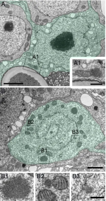
Cells of the gastric region show unlimited proliferation and renewal. The average cell cycle length is 3-4 days (David and Campbell, 1972). This corresponds to the doubling time of epithelial cells as measured in growth curves and also to the doubling time of polyps in asexual mass culture. With the exception of an initial characterisation of a cyclinB gene and its expression during the daily feeding rhythm and during head regeneration (Scheurlen et al., 1996), almost nothing is known about the molecular control of the cell cycle in these cells - an important gap of knowledge with respect to the basic role of proliferation and tissue turnover in this animal. By using electron microscopy, we recently detected mul-tiple nuages in epithelial cells mostly in the vicinity of the nucleus (Fig. 3A). Nuage was initially characterised in germ cell precur-sors including meiotic cells but is now more generally known as a structure specific for undifferentiated, proliferating cells (for review see Pek and Kai, 2011).
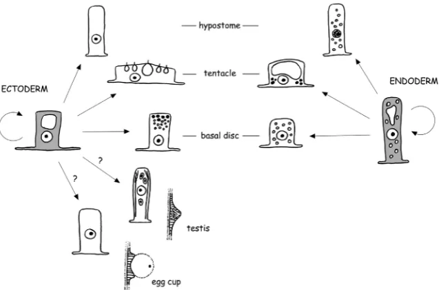
Fig. 1. Scheme of the proliferation and differentiation pathways of ectodermal and endodermal epitheliomuscular cells.
seen as a property of an individual cell, but of a cell lineage, which can generate and maintain a tissue during embryogenesis, regeneration or tis-sue turnover (for review see Lander, 2009, 2011; Wolkenhauer et al., 2011).
composi-Epithelial cells also exhibit features of a clearly differentiated and physiologically active cell type. They establish an outer epidermal tissue covered by a cuticle-like structure and an inner gastrodermal tissue responsible for digestion and uptake of food and the distribu-tion of nutrients. Ectodermal epithelial cells show all hallmarks of a true epithelium: apical junctional complexes (septate junctions), apical-basal polarity, and hemidesmosome-like junctions to attach to the mesoglea. Endodermal epithelial cells likewise show apical septate junctions and clear apical-basal polarisation, but defined junctional attachment sites to the mesoglea have so far not been found. Both epithelial cell types have basal muscle fibres attached to the mesoglea for coordinated movement under neuronal control. They also contain large vacuoles presumably involved in osmoregu-lation, and can act in a macrophage-like manner by phagocytising neighbouring cells under starvation conditions or when confronted with abnormal cells.
Ectodermal epithelial cells
Under conditions of asexual steady state growth, ectodermal epithelial cells give rise to three types of terminally differentiated cells: tentacle-specific battery cells, hypostome-specific cells, and basal disc-specific secretory cells (Fig. 1). The boundaries of the corresponding body regions, where terminal differentiation occurs, are defined by the expression of genes that act in position-dependent differentiation (see Böttger and Hassel, 2012). Regen-eration experiments and tracking of GFP-positive epithelial cells in transgenic polyps have demonstrated that every proliferating cell of
the body column has the potential to give rise to all three types of differentiated cells (Wittlieb et al., 2006). In all three cases, terminal differentiation and cell cycle arrest occurs in the G2 phase (Dübel, 1989; Dübel and Schaller, 1990). The arrest is irreversible, so that these cells can no longer participate in regeneration.
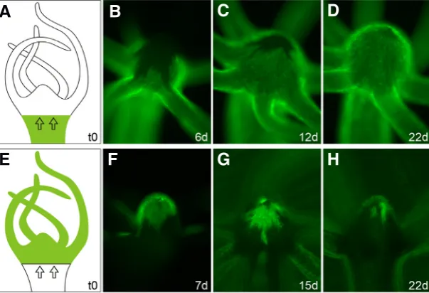
Previous data obtained by in vivo carbon labelling and tracking of ectodermal epithelial cells indicated that the lower hypostome may contain a separate and independent subpopulation of prolifer-ating cells (Dübel et al., 1987). It was argued that cells of the body column are displaced towards the tentacles, but do not contribute to the formation of the mouth. Instead, cells in the lower part of the hypostome self-renew and differentiate to tentacle cells and to cells of the upper hypostome and mouth. We reinvestigated this issue using Hydra expressing a stable lifeact-GFP transgene to visualise the actin cytoskeleton of ectodermal epithelial cells (Riedl et al., 2008; Aufschnaiter and Hobmayer, in preparation). We produced a number of transplants, where a wildtype head was grafted onto a transgenic body, and thereafter we followed the displacement of ectodermal epithelial cells into the head region under daily feeding (Fig. 4A). Immediately after healing, GFP-positive tissue of the upper body column started to move towards the tentacles and hypostome. After six days, the tentacles were almost entirely replaced by GFP-positive cells, and between the tentacle bases we could clearly observe the movement of transgenic cells towards the hypostome (Fig. 4B). After twelve days, only the upper part of the hypostome remained wildtype, and after three to four weeks, all the wildtype head tissue was replaced by transgenic cells from the gastric region, in agreement with Campbell (1967b) (Fig. 4 C,D). Equivalent results were obtained, when a transgenic head was transplanted to a wildtype body (Fig. 4 E-H). In summary, our data clearly argue against an independent sublineage of epithelial cells in the ectoderm of the lower hypostome.
During sexual reproduction, ectodermal epithelial cells contribute to the formation of the female egg cup, an egg-holding structure in the mid body column, and to the formation of testes localised closer to the head (Fig. 1). The egg cup is a support structure that holds the mature egg throughout fertilisation and cleavage stages. An egg finally detaches from this structure, when the egg shell forms in the blastula stage. The testis is responsible for matura-tion of sperm precursor cells and the controlled release of sperm. The differentiation pathways in these two sex organs are not well known. It is actually unclear, whether terminal differentiation occurs. A detailed morphological characterisation of the formation of egg cup and testis is ongoing in our lab.
Endodermal epithelial cells
At the base of tentacles and in the hypostome and basal disc, endodermal epithelial cells also undergo terminal differentiation. As in the ectodermal layer, the differentiated cells in the tentacles and basal disc are arrested in the G2 phase, while those in the upper hypostome are arrested in G1 (Dübel, 1989; Dübel and Schaller, 1990) suggesting that epithelial precursor cells can in principle go through a terminal mitosis. In contrast to the ectodermal lineage, endodermal epithelial cells do not seem to participate in sexual differentiation.
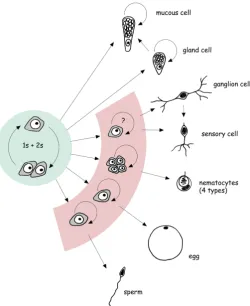
It should be noted that endodermal epithelial cells seem to have a greater capacity for changing their shape and for morphogenetic movement in comparison to ectodermal cells. During bud formation, it is a group of such endodermal cells that shows the first observ-Fig. 2. Scheme of the proliferation and differentiation pathways of the
able cell shape changes at the onset of evagination (Gelei, 1925; Philipp et al., 2009). Endodermal cells also show an unexpected mobility during reaggregation (Takaku et al., 2004) and during tis-sue recruitment into the newly forming bud (Wittlieb et al., 2006). In addition, endodermal cells are the first cells to change their shape and to close the wound, when regeneration starts (Znidaric, 1970; Murate et al., 1997; Chera et al., 2009b). The lack of cell-matrix contact sites in the endodermal tissue layer may contribute to a greater morphogenetic flexibility.
Interstitial stem cell lineage
The cells of this lineage are primarily located in the interstitial spaces between epithelial cells. But head-specific sensory nerve cells and mature nematocytes are also incorporated within
ecto-dermal epithelial cells. Interstitial cells comprise roughly 80% of the total cells of a Hydra polyp. Here, we just give a rough outline of the basic aspects of self-renewal and differentiation. In a related review in this issue, the cellular dynamics and the decision making within this lineage is described in more detail (see David, 2012).
Interstitial stem cells are small cells with an interphase nucleus and a ribosome-rich cytoplasm (David, 1973). Due to their occur-rence as single cells or pairs, they are commonly called 1s+2s in the literature. In transmission electron microscopy, multiple nuages are present at the nuclear membrane, associated with mitochondria and in the cytoplasm (Fig. 3B). The cell cycle of 1s+2s is much shorter (roughly 30 hrs) than that of epithelial cells (Campbell and David, 1974), but both cell types grow at the same rate since only about 60% of 1s+2s remain stem cells per cell cycle during steady state growth. Whether there are resting stem cells as known in bilaterian stem cell systems is unclear, but continuous labelling experiments have indicated that there is a small fraction of 1s+2s with a longer cell cycle (Campbell and David, 1974). Finally, clon-ing experiments revealed that interstitial stem cells can give rise to both somatic differentiation products and to gametes (Fig. 2) (Bosch and David, 1987).
There are three basic somatic differentiation pathways (Fig. 2). Nematocyte precursors go through several rounds of proliferation to produce nests of nematoblasts up to a size of 16 cells per nest (David and Gierer, 1974). These proliferating precursors may correspond to so-called “transit amplifying cells” as known from stem cell systems in higher metazoans. Transit amplifying cells are committed for a particular pathway and then undergo a limited number of rounds of proliferation, before terminal differentiation is initiated. In the case of a proliferating nematoblast nest, terminal mitosis produces a nest of differentiating nematocytes (David and Gierer, 1974). When differentiation is finished, the nest breaks up and individual nematocytes actively migrate toward the base of the tentacles, where they become incorporated into ectodermal battery cells (for review see Fujisawa et al., 1986).
Nerve cell differentiation starts with the formation of small inter-stitial cell precursors (David and Gierer, 1974; Holstein and David, 1990). The degree of proliferation in nerve cell precursors is under debate. While some studies emphasised a capacity to go through several rounds of mitosis (Heimfeld and Bode, 1984; Teragawa and Bode, 1995; Bode et al., 1990), Hager and David (1996) found, in most cases, just one terminal mitosis of the committed stem cell to produce two differentiating nerve cells. Two major types of nerve cells, ganglion cells and sensory cells, are formed. To a limited extent phenotype conversion has been observed, when mature ganglion cells are passively displaced from the body column into the head and thereby transform to head-specific sensory cells (Koizumi et al., 1988; Hobmayer et al., 1990).
Differentiation of secretory cells is the least intensively studied pathway. Interstitial precursor cells migrate through the mesoglea into the endodermal epithelium and there they differentiate in a position-dependent manner: gland cells in the gastric region and mucous cells in the hypostome (Smid and Tardent, 1984; Schmidt and David, 1986; Bode et al., 1987). It is unclear whether precursors of this pathway divide, but the differentiated gland and mucous cells still have a capacity to proliferate. New data with stable transgenic labelling have demonstrated phenotype conversion from gland cells to mucous cells during tissue displacement towards the head (Siebert et al., 2008).
Fig. 3. Electron microscopic visualisation of nuage in Hydra.(A) Part of an ectodermal epitheliomuscular cell (shown in green) including the nucleus and a mitochondrium-associated nuage (A1). (B) Interstitial stem cell (shown in green) with nuage in the cytoplasm (B1), associated to a mitochondrium (B2), and associated to the nuclear membrane (B3). Scale bars, 2.0 mm (A,B) and 500 nm (A1, B1-B3).
B
A1
B1
B2
B3
Both types of gametes are differentiation products of 1s+2s, and both gametogenic pathways exhibit transit amplifying cells, as the precursor cells undergo extensive proliferation to produce larger numbers of cells for final maturation (Fig. 2) (for more details see Munck and David, 1985; Littlefield et al., 1985; Nishimiya-Fujisawa and Sugiyama, 1993; Nishimiya-Fujisawa and Sugiyama, 1995; Miller et al., 2000; Alexandrova et al., 2005). The mature egg cell is mounted on an egg cup built by ectodermal epithelial cells. Likewise, huge numbers of sperm precursors terminate their final rounds of mitotic and meiotic divisions in testis formed by ectoder-mal epithelial cells. Hence, sex organ formation and maturation of gametes needs coordinated differentiation of ectodermal epithelial and interstitial stem cell lineages. We assume that processes of induction between the two lineages must occur to assure coordinated positional and temporal control of these processes. The molecular nature of these interactions is totally unexplored.
A stemness comparison
While there is no doubt that interstitial cells represent a prototypi-cal stem cell system, the situation for the two epithelial lineages is less clear. As outlined above, ectodermal and endodermal epithelial cells of the gastric region continuously self-renew and eventually give rise to several differentiation products, thus providing clear evidence for stemness in these two lineages. But can epithelial cells be regarded as bona fide stem cells? The answer depends on how stringently stem cells are defined. If stem cells are defined as cells just showing a capacity to self-renew and to produce dif-ferentiated cells, then epithelial cells are stem cells. If stem cells are defined more strictly as cells that self-renew and at the same time differentiate to other cell types, then epithelial cells are not
stem cells. In the paper by David (2012), evidence is presented for the first time that interstitial stem cells (1s+2s) show stochastic behaviour of self-renewal and pluripotency including asymmetric cell division. No such evidence exists for epithelial cells. In the gastric region, epithelial cells divide and the two daughter cells are again self-renewing cells; no differentiation occurs. When epithelial cells arrive at the base of the tentacles, the hypostome, or the basal disc, they dif-ferentiate, but they do so at sharp boundaries where all cells differentiate under tight positional control.
Additional differences between the epithelial and interstitial cell lines should be emphasized: (1) Interstitial stem cells respond to their own density (for review see David, 2012) and interact with the surrounding epithelial cells in a relatively well-defined niche. In the case of epithelial cells, it is not as easy to delineate a niche, neither in its dimensions and its composition, nor in its complexity of cellular interac-tions. (2) Epithelial and interstitial cell lineages arise differently during embryonic development. The ecto-dermal and endoecto-dermal epithelial lineages directly descend from the two ectodermal and endodermal germ layers established during gastrulation. In con-trast, the precise origin of the interstitial cell lineage is unknown in Hydra embryos, mostly because the critical embryonic stages are hidden under a thick, non-transparent, and rather inaccessible egg shell Fig. 4. Live tracking of ectodermal tissue movement into the hypostome. The head of a
wildtype AEP strain Hydra was transplanted onto the body of a transgenic polyp expressing a LifeactGFP fusion protein in ectodermal epitheliomuscular cells (A) and vice versa (E). Subsequent movement of ectodermal tissue into heads was visualised by fluorescence microscopy life imaging. (B-D) Transgenic tissue movement results in complete replace-ment of wildtype head cells after 3-4 weeks. Note the faster tissue movereplace-ment between the tentacles in (B). Wildtype tissue movement equivalently replaced transgenic cells as visualised by fluorescent microscopy (F) and phase contrast merge (G-H).
(Martin et al., 1997). In related hydrozoan species, however, where such data are available, interstitial cells develop after germ layer specification in the endoderm (Weiler-Stolt, 1960; reviewed in Tardent, 1978). (3) Epithelial and interstitial cell lineages also have a different evolutionary background. The ectodermal and endodermal cell lineages correspond to outer (epidermal) and in-ner (gastrodermal) cell layers, whose origins can be traced back to the most ancestral multicellular animals such as sponges. In contrast, hydrozoans are the only cnidarian class whose members evolved an interstitial stem cell lineage with variable degree of potency (Müller et al., 2004). In the other cnidarian classes in-cluding the anthozoans, which are thought to represent the most ancestral cnidarian group, there is no cell lineage in addition to the ectodermal and endodermal epithelium. Therefore, it is generally assumed that the interstitial stem cell system of Hydra represents an independent, hydrozoan-specific invention not directly related to bilaterian stem cell systems. (4) The Hydra genome was analysed for the presence of known stem cell-specific genes (see Table 1 in Watanabe et al., 2009). In all cases, where expression data are available, the genes are activated in the interstitial cell lineage and not in epithelial cells. Based on all these arguments, we expect significant differences in the gene regulatory networks mediating stemness in the three Hydra cell lineages. The data discussed below further support this view.
Molecular regulation of stemness in Hydra
Several recent and comprehensive reviews discuss the still rather limited current knowledge of molecular factors acting in stemness regulation in Hydra (Bosch, 2008; Watanabe et al., 2009; Bosch et al., 2010). These reviews discuss the interstitial stem cell niche,
G
B
C
D
E
F
H
the role of signalling and transcription factors in the interstitial stem cell lineage, and homologs of known stem cell-specific genes in the Hydra genome sequence. We will therefore focus here on some of the most recent and stemness-related findings.
Wnt signalling
Wnt signalling is among the best studied signalling systems due to its relevance in embryonic development and cancer. Secreted Wnt proteins are inducers of axis formation, organogenesis, and morphogenetic tissue movements, mechanisms invented during the evolution of the earliest multicellular animals. In Hydra, canonical Wnt/b-Catenin signalling is a central player in the head organiser
and in pattern formation along the oral-aboral body axis (Hobmayer et al., 2000; Broun et al., 2005; Gee et al., 2010; Nakamura et al., 2011). Noncanonical Wnt signalling, by comparison, acts in tissue movement and evagination during tentacle and bud formation, providing evidence for an ancestral complexity of the Wnt signal-ling network (Philipp et al., 2009).
More recently, it has become clear that the Wnt/b-Catenin
path-way plays an additional role in the maintenance and in cell fate decisions of stem cells; activated Wnt signalling is tightly linked to stemness (for review see Wend et al., 2010). For example, activated b-Catenin increases self-renewal of hematopoietic, skin,
and intestinal stem cells. In the case of bulge stem cells in hair fol-licles, Wnt/b-Catenin seems to control stem cell fate. On the other
hand, unbalanced Wnt/b-Catenin activity contributes to oncogenic
transformation and the maintenance of cancer stem cells. In these cases, one of the primary direct targets is the oncogenic transcrip-tion factor c-Myc (see below).
The Wnt pathway acts in many systems as an activator of cell proliferation (for review see Clevers, 2006). In Hydra, there is
ac-cumulating evidence that Wnt/b-Catenin signalling is also involved
in proliferation control. The head of the polyp exhibits a constant high level of S-phase labelling in epithelial cells and is insensitive to the fluctuations in epithelial cell division caused by the daily feeding rhythm. Furthermore, epithelial cells in evaginating buds generally exhibit higher proliferation rates (Holstein et al., 1991). In both regions, b-Catenin is in an activated state (Hobmayer et
al., 2000; Broun et al., 2005). During head regeneration, mitotic divisions have been associated with activated b-Catenin (Chera et
al., 2009b). There is also evidence that activation of b-Catenin by
treatment with GSK-3 inhibitors alters interstitial cell behaviour such that cells are prevented from initiating nematocyte differentiation (Khalturin et al., 2007). Finally, in a related hydrozoan, Hydractinia echinata, GSK-3 inhibition leads to an increased S-phase activity in interstitial stem cells resulting in an increase in the number of differentiated nematocytes and nerve cells (Teo et al., 2006).
Reprogramming factor Oct4
Mammalian somatic cells can be reprogrammed to pluripotency following forced expression of a small set of transcription factors (for review see Jaenisch and Young, 2008). Among those, Oct4, a class5 POU domain protein, is indispensable. New and exciting data suggest that this may be an evolutionarily old mechanism. In Hydractinia echinata, an oct4-related gene called polynem has been characterised (Millane et al., 2011). It is specifically expressed in embryonic cells and in interstitial stem cells of the adult polyp. Strikingly, ectopic expression of polynem in ectodermal epithelial cells of the polyp caused extensive neoplasia, and cells within the neoplasms started to express a set of stem cell marker genes.
In molecular phylogenies, polynem is either related to class5 or class3 POU proteins (Millane et al., 2011; data not shown). We
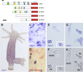
Fig. 5. Hydra Myc proteins and the expression patterns of Hydra myc1.(A) Schematic representa-tion of the Hydra Myc1 and Myc2 proteins and the predicted Myc-like proteins Myc3 and Myc4 in com-parison to c-Myc. The position of the conserved basic helix-loop-helix leucine Zipper (bHLH-Zip) domain and the putative Myc homology boxes (MBI-IV) are indicated. (B-H)myc1 is expressed in cells belong-ing to the interstitial stem cell system as visualised by in situ hybridisations using whole animals (B-D)
and single cell maceration preparations (E-H). (D)
Double in situ hybridisation using myc1 (blue) and the differentiation gene Nowa (red) demonstrates that myc1 is exclusively expressed in proliferating cells. 2s: interstitial stem cells; 4s, 8s: proliferating nematoblasts; ecto: ectodermal epitheliomuscular cell; endo: endodermal epitheliomuscular cell; nv: nerve cell. The insert in (E,F) shows a gland cell. (B-E,G)Bright field optics; (F,H)phase contrast optics (modified form Hartl et al., 2010, with permission from Proc. Natl. Acad. Sci. USA).
failed to identify members of these classes in the Hydra genome sequence. The most closely related POU domain-encoding genes detected in our analysis belonged to class4 and class6. Their functions and expression patterns are currently unknown.
G
B
C
D
E
F
H
Maintenance factors of the Myc family
Transcription factors of the Myc family control fundamental cellular processes including cell proliferation and ribosome biogen-esis. Deregulation of Myc leads to the development of cancer and recently it has become clear that Myc proteins play a decisive role in the control of proliferation and in the maintenance of stem cells in their undifferentiated state. Furthermore, Myc is among the core factors in reprogramming experiments with mammalian somatic cells (for review see Jaenisch and Young, 2008). We therefore started to investigate myc genes in Hydra.
The Hydra genome sequence contains four myc or myc-like genes (Fig. 5A). Two of them, myc1 and myc2, encode for pro-totypical Myc proteins displaying a highly conserved C-terminal bHLH-Zip DNA binding domain and most of the Myc-boxes in the larger N-terminal transactivation domain. The other two myc-like genes, myc3 and myc4, are more divergent. Their predicted C-terminal bHLH-Zip domains are Myc-related, but their predicted N-terminal domains almost completely lack Myc-boxes (Fig. 5A).
We have characterised Hydra Myc1 and Myc2 biochemically and in detailed expression studies (Hartl et al., 2010; Hartl et al., in preparation). The Hydra ortholog of Max has also been characterised (Hartl et al., 2010). Both Myc factors dimerise with their known interaction partner Max and bind to the E-box DNA recognition elements with high affinity. Furthermore, Myc1 and Myc2 contain structural elements capable of substituting for those present in the viral Myc protein to induce oncogenic transformation of avian fibroblasts. This suggests that the principle functions of this key factor were invented at the base of metazoan evolution.
The myc1 gene is specifically expressed in the proliferating somatic cells of the interstitial stem cell system including 1s+2s, proliferating nematoblasts, and gland cells (Fig. 5 B-H). Based on this pattern, we suggested that the primary function of Myc1 is in control of the comparably fast cell cycle in this lineage. Support-ing this view, knock-down of myc1 function by siRNA or the small molecule inhibitor 10058-F4 results in deregulated proliferation in 1s+2s (Ambrosone et al., 2012).
The myc2 gene shows expression in a broader set of cells and may therefore have a more basic function in the polyp. It is co-expressed with myc1 at a high level in proliferating interstitial cells, but it is also expressed at a lower level in ectodermal and endodermal epithelial cells throughout the gastric region. Terminally differentiated cells in the head and foot do not express myc1 or myc2. Interestingly, myc2 is also expressed during gametogenesis in proliferating egg and sperm precursor cells. Based on our molecular phylogenetic analysis, the myc1 and myc2 genes arose from a gene duplication within the cnidarian phylum, which obviously resulted in a functional separation of myc1 to the somatic compartment of the interstitial cell lineage and myc2 to gametogenesis. In summary, all these results suggest that the presence of Myc proteins keeps cells in a proliferative state.
We are just beginning to understand the molecular nature of stemness in Hydra. The availability of a well-annotated genome sequence (Chapman et al., 2010) and the establishment of new molecular and bioinformatic methods have opened up a new era of Hydra research. These innovations will provide the tools necessary to investigate the genetic signatures and molecular interactions that underlie the developmental capacity and stemness present in the three cell lineages of the Hydra polyp - a fascinating challenge.
Acknowledgements
We thank Charles David for comments and corrections. We also thank Brigitte Galliot, Monika Hassel, Hans Meinhardt, and Peter Ladurner for critical reading of the manuscript and Klaus Bister for ongoing support. Our work was supported by research grants of the Austrian Science Fund (FWF P16685, P19232, P20734).
References
ALEXANDROVA, O., SCHADE, M., BÖTTGER, A. and DAVID, C.N. (2005). Oo-genesis in Hydra: nurse cells transfer cytoplasm directly to the growing oocyte. Dev Biol 281: 91-101.
AMBROSONE, A., MARCHESANO, V., TINO, A., HOBMAYER, B. and TORTI-GLIONE, C. (2012). Hymyc1 downregulation promotes stem cell proliferation. PLoS ONE 7: e30660.
BODE, H.R. (1996). The interstitial cell lineage of hydra: a stem cell system that arose early in evolution. J Cell Sci 109: 1155-1164.
BODE, H.R. (2003). Head regeneration in Hydra. Dev Dyn 226: 225-236. BODE, H.R. (2009). Axial patterning in hydra. Cold Spring Harb Perspect Biol 1:
a000463.
BODE, H.R., BERKING, S., DAVID, C.N., GIERER, A., SCHALLER, H.C. and TRENKNER, E. (1973). Quantitative analysis of cell types during growth and morphogenesis in Hydra. Wilhelm Roux’s Arch Dev Biol 171: 269-285. BODE, H.R., FLICK, K.M. and SMITH, G.S. (1976). Regulation of interstitial cell
dif-ferentiation in Hydra attenuata. I. Homeostatic control of interstitial cell population size. J Cell Sci 20: 29-46.
BODE, H.R., HEIMFELD, S., CHOW, M.A. and HUANG, L.W. (1987). Gland cells arise by differentiation from interstitial cells in Hydra attenuata. Dev Biol 122: 577-585. BODE, H.R., GEE, L.W. and CHOW, M.A. (1990). Neuron differentiation in hydra
involves dividing intermediates. Dev Biol 139: 231-243.
BÖTTGER, A. and HASSEL, M. (2012). Hydra, a model system to trace the emergence of boundaries in eumetazoans. Int J Dev Biol 56: 583-591.
BOSCH, T.C.G. (2007). Why polyps regenerate and we don’t: Towards a cellular and molecular framework for Hydra regeneration. Dev Biol 303: 421-433.
BOSCH, T.C.G. (2008). Stem cells in immortal Hydra. In Stem cells: from Hydra to man (Ed. T.C.G. Bosch). Springer, New York, pp. 37-58.
BOSCH, T.C.G. (2009). Hydra and the evolution of stem cells. Bioessays 31: 478-486. BOSCH, T.C.G. and DAVID, C.N. (1984). Growth regulation in Hydra: relationship
between epithelial cell cycle length and growth rate. Dev Biol 104: 161-171. BOSCH, T.C.G. and DAVID, C.N. (1987). Stem cells of Hydra magnipapillata can
differentiate into somatic cells and germ line cells. Dev Biol 121: 182-191. BOSCH, T.C.G., ANTON-ERXLEBEN, F., HEMMRICH, G. and KHALTURIN, K.
(2010). The Hydra polyp: nothing but an active stem cell community. Dev Growth Differ 52: 15-25.
BROUN, M., GEE, L., REINHARDT, B. and BODE, H.R. (2005). Formation of the head organizer in hydra involves the canonical Wnt pathway. Development 132: 2907-2916.
CAMPBELL, R.D. (1967a). Tissue dynamics of steady state growth in Hydra littoralis. I. Patterns of cell division. Dev Biol 15: 487-502.
CAMPBELL, R.D. (1967b). Tissue dynamics of steady state growth in Hydra littoralis. II. Patterns of tissue movement. J Morphol 121: 19-28.
CAMPBELL, R.D. and DAVID, C.N. (1974). Cell cycle kinetics and development of Hydra attenuata. II. Interstitial cells. J Cell Sci 16: 349-358.
CHAPMAN, J.A., KIRKNESS, E.F., SIMAKOV, O. et al., (2010). The dynamic genome of Hydra. Nature 464: 592-596.
CHERA, S., BUZGARIU, W., GHILA, L. and GALLIOT, B. (2009a). Autophagy in Hydra: a response to starvation and stress in early animal evolution. Biochim Biophys Acta 1793: 1432-1443.
CHERA, S., GHILA, L., DOBRETZ, K. et al., (2009b). Apoptotic cells provide an unexpected source of Wnt3 signaling to drive hydra head regeneration. Dev Cell 17: 279-289.
127: 469-480.
DAVID, C.N. (2012). Interstitial stem cells in Hydra: multipotency and decision-making. Int J Dev Biol 56: 489-497.
DAVID, C.N. (1973). A quantitative method for maceration of hydra tissue. Wilhelm Roux’s Arch Dev Biol 171: 259-268.
DAVID, C.N. and CAMPBELL, R.D. (1972). Cell cycle kinetics and development of Hydra attenuata. I. Epithelial cells. J Cell Sci 11: 557-568.
DAVID, C.N. and GIERER, A. (1974). Cell cycle kinetics and development of Hydra attenuata. III. Nerve and nematocyte differentiation. J Cell Sci 16: 359-375. DAVID, C.N. and MACWILLIAMS, H. (1978). Regulation of the self-renewal probability
in Hydra stem cell clones. Proc Natl Acad Sci USA 75: 886-890.
DÜBEL, S. (1989). Cell differentiation in the head of Hydra. Differentiation 41: 99-109. DÜBEL, S., HOFFMEISTER, S.A. and SCHALLER, H.C. (1987). Differentiation
pathways of ectodermal epithelial cells in hydra. Differentiation 35: 181-189. DÜBEL, S. and SCHALLER, H.C. (1990). Terminal differentiation of ectodermal
epi-thelial stem cells of Hydra can occur in G2 without requiring mitosis or S phase. J Cell Biol 110: 939-945.
FUJISAWA, T. (1992). Homeostatic recovery of interstitial cell populations in Hydra. Dev Biol 150: 185-192.
FUJISAWA, T., NISHIMIYA, C. and SUGIYAMA, T. (1986). Nematocyte differentiation in hydra. Curr Top Dev Biol 20: 281-290.
GALLIOT, B., MILJKOVIC-LICINA, M., DE ROSA, R. and CHERA, S. (2006). Hydra, a niche for cell and developmental plasticity. Semin Cell Dev Biol 17: 492-502. GEE, L., HARTIG, J., LAW, L. et al., (2010). beta-Catenin plays a central role in
set-ting up the head organizer in hydra. Dev Biol 340: 116-124.
GELEI, J. (1925). Über die Sprossbildung bei Hydra grisea. Wilhelm Roux’s Arch Dev Biol 105: 633-654.
GIERER, A., BERKING, S., BODE, H.R. et al., (1972). Regeneration of Hydra from reaggregated cells. Nature New Biol 239: 98-101.
HAGER, G. and DAVID, C.N. (1997). Pattern of differentiated nerve cells in hydra is determined by precursor migration. Development 124: 569-576.
HARTL, M., MITTERSTILLER, A.M., VALOVKA, T., BREUKER, K., HOBMAYER, B. and BISTER, K. (2010). Stem cell-specific activation of an ancestral myc proto-oncogene with conserved basic functions in the early metazoan Hydra. Proc Natl Acad Sci USA 107: 4051-4056.
HEIMFELD, S. and BODE, H.R. (1984). Interstitial cell migration in Hydra attenuata. II. Selective migration of nerve cell precursors as the basis for position-dependent nerve cell differentiation. Dev Biol 105: 10-17.
HOBMAYER, E., HOLSTEIN, T.W. and DAVID, C.N. (1990). Tentacle morphogenesis in Hydra. II. Formation of a complex between a sensory nerve cell and a battery cell. Development 109: 897-904.
HOBMAYER, B., RENTZSCH, F., KUHN, K. et al., (2000). WNT signalling molecules act in axis formation in the diploblastic metazoan Hydra. Nature 407: 186-189. HOBMAYER, B., SNYDER, P., ALT, D., et al., (2001). Quantitative analysis of epithelial
cell aggregation in the simple metazoan Hydra reveals a switch from homotypic to heterotypic cell interactions. Cell Tissue Res 304: 147-157.
HOLSTEIN, T.W. and DAVID, C.N. (1990). Putative intermediates in the nerve cell differentiation pathway in hydra have properties of multipotent stem cells. Dev Biol 142: 401-405.
HOLSTEIN, T.W., HOBMAYER, E. and DAVID, C.N. (1991). Pattern of epithelial cell cycling in hydra. Dev Biol 148: 602-611.
HOLSTEIN, T.W., HOBMAYER, E. and TECHNAU, U. (2003). Cnidarians: an evo-lutionarily conserved model system for regeneration? Dev Dyn 226: 257-267. JAENISCH, R. and YOUNG, R. (2008). Stem cells, the molecular circuitry of
pluri-potency and nuclear reprogramming. Cell 132: 567-582.
KHALTURIN, K., ANTON-ERXLEBEN, F., MILDE, S., PLOTZ, C., WITTLIEB, J., HEMMRICH, G. and BOSCH, T.C. (2007). Transgenic stem cells in Hydra reveal an early evolutionary origin for key elements controlling self-renewal and differ-entiation. Dev Biol 309: 32-44.
KOBATAKE, E. and SUGIYAMA, T. (1986). Genetic analysis of developmental mecha-nisms in Hydra. XVII. Elimination of excess epithelial cells by phagocytosis in a mutant strain L4. Dev Biol 115: 249-255.
KOIZUMI, O., HEIMFELD, S. and BODE, H.R. (1988). Plasticity in the nervous system of adult hydra. II. Conversion of ganglion cells of the body column into epidermal
sensory cells of the hypostome. Dev Biol 129: 358-371.
LANDER, A.D. (2009). The ‘stem cell’ concept: is it holding us back? J Biol 8: 70. LANDER, A.D. (2011). The individuality of stem cells. BMC Biol 9: 40.
LITTLEFIELD, C.L., DUNNE, J.F. and BODE, H.R. (1985). Spermatogenesis in Hydra oligactis. I. Morphological description and characterization using a monoclonal antibody specific for cells of the spermatogenic pathway. Dev Biol 110: 308-320. MARCUM, B. and CAMPBELL, R.D. (1978). Development of Hydra lacking nerve
and interstitial cells. J Cell Sci 29: 17-33.
MARTIN, V.J., LITTLEFIELD, C.L., ARCHER, W.E. and BODE, H.R. (1997). Embryo-genesis in hydra. Biol Bull 192: 345-363.
MARTINEZ, D.E. (1998). Mortality patterns suggest lack of senescence in hydra. Exp Gerontol 33: 217-225.
MEINHARDT, H. (1993). A model for pattern formation of hypostome, tentacles, and foot in hydra: how to form structures close to each other, how to form them at a distance. Dev Biol 157: 321-333.
MEINHARDT, H. (2012) Modeling pattern formation in hydra: a route to understanding essential steps in development. Int. J. Dev. Biol. 56: 447-462.
MILLANE, R.C., KANSKA, J., DUFFY, D.J., SEOIGHE, C., CUNNINGHAM, S., PLICKERT, G. and FRANK, U. (2011). Induced stem cell neoplasia in a cnidar-ian by ectopic expression of a POU domain transcription factor. Development 138: 2429-2439.
MILLER, M.A., TECHNAU, U., SMITH, K.M. and STEELE, R.E. (2000). Oocyte development in Hydra involves selection from competent precursor cells. Dev Biol 224: 326-338.
MÜLLER, W.A., TEO, R. and FRANK, U. (2004). Totipotent migratory stem cells in a hydroid. Dev Biol 275: 215-224.
MUNCK, A. and DAVID, C.N. (1985). Cell proliferation and differentiation kinetics during spermatogenesis in Hydra carnea. Wilhelm Roux’s Arch Dev Biol 194: 247-256. MURATE, M., KISHIMOTO, Y., SUGIYAMA, T., FUJISAWA, T., TAKAHASHIIWANAGA, H. and IWANAGA, T. (1997). Hydra regeneration from recombined ectodermal and endodermal tissue. II. Differential stability in the ectodermal and endodermal epithelial organization. J Cell Sci 110: 1919-1934.
NAKAMURA, Y., TSIARIS, C.D., ÖZBEK, S. and HOLSTEIN, T.W. (2011). Autoregula-tory and repressive inputs localize Hydra Wnt3 to the head organizer. Proc Natl Acad Sci USA 108: 9137-9142.
NISHIMIYA-FUJISAWA, C. and SUGIYAMA, T. (1993). Genetic analysis of devel-opmental mechanisms in Hydra. XX. Cloning of interstitial stem cells restricted to the sperm differentiation pathway in Hydra magnipapillata. Dev Biol 157: 1-9. NISHIMIYA-FUJISAWA, C. and SUGIYAMA, T. (1995). Genetic analysis of devel-opmental mechanisms in Hydra. XXII. Two types of female germ stem cells are present in a male strain of Hydra magnipapillata. Dev Biol 172: 324-336. PEK, J.W. and KAI, T. (2011). Non-coding RNAs enter mitosis: functions, conservation
and implications. Cell Division 6.
PHILIPP, I., AUFSCHNAITER, R., OZBEK, S. et al., (2009). Wnt/beta-catenin and noncanonical Wnt signaling interact in tissue evagination in the simple eumetazoan Hydra. Proc Natl Acad Sci USA 106: 4290-4295.
RIEDL, J., CREVENNA, A.H., KESSENBROCK, K., et al., (2008). Lifeact: a versatile marker to visualize F-actin. Nature Methods 5: 605-607.
SCHEURLEN, I., HOFFMEISTER, S.A. and SCHALLER, H.C. (1996). Presence and expression of G2 cyclins in the coelenterate hydra. J Cell Sci 109: 1063-1069. SCHMIDT, T. and DAVID, C.N. (1986). Gland cells in Hydra: cell cycle kinetics and
development. J Cell Sci 85: 197-215.
SIEBERT, S., ANTON-ERXLEBEN, F. and BOSCH, T.C. (2008). Cell type complexity in the basal metazoan Hydra is maintained by both stem cell based mechanisms and transdifferentiation. Dev Biol 313: 13-24.
SMID, I. and TARDENT, P. (1984). Migration of I-cells from ectoderm to endoderm in Hydra attenuata Pall (Cnidaria, Hydrozoa) and their subsequent differentiation. Dev Biol 106: 469-477.
SPROULL, F. and DAVID, C.N. (1979). Stem cell growth and differentiation in Hydra attenuata. I. Regulation of the self-renewal probability in multiclone aggregates. J Cell Sci 38: 155-169.
TAKAKU, Y., HARIYAMA, T. and FUJISAWA, T. (2005). Motility of endodermal epi-thelial cells plays a major role in reorganizing the two epiepi-thelial layers in Hydra. Mech Dev 122: 109-122.
TARDENT, P. (1978). Coelenterata, Cnidaria. In Morphogenese der Tiere. Handbuch der ontogenetischen Morphologie und Physiologie in Einzeldarstellungen (Ed. F. Seidel). Gustav Fischer Verlag, Jena, pp. 73-415.
TECHNAU, U. and HOLSTEIN, T.W. (1992). Cell sorting during the regeneration of Hydra from reaggregated cells. Dev Biol 151: 117-127.
TEO, R., MOHRLEN, F., PLICKERT, G., MÜLLER, W.A. and FRANK, U. (2006). An evolutionary conserved role of Wnt signaling in stem cell fate decision. Dev Biol 289: 91-99.
TERAGAWA, C.K. and BODE, H.R. (1995). Migrating interstitial cells differentiate into neurons in hydra. Dev Biol 171: 286-293.
WATANABE, H., HOANG, V.T., MATTNER, R. and HOLSTEIN, T.W. (2009).
Immor-Further Related Reading, published previously in the Int. J. Dev. Biol.
An organizing region in metamorphosing hydrozoan planula larvae - stimulation of axis formation in both larval and in adult tissue
Melanie Stumpf, Britta Will, Karola Wittig, Jennifer Kasper, Benjamin Fischer, Jürgen Schmich, Stefanie Seipp and Thomas Leitz Int. J. Dev. Biol. (2010) 54: 795-802
5 yr ISI Impact Factor (2010) = 2.961
Wnt signaling in hydroid development: ectopic heads and giant buds induced by GSK-3beta inhibitors
Werner Müller, Uri Frank, Regina Teo, Ofer Mokady, Christina Guette and Günter Plickert Int. J. Dev. Biol. (2007) 51: 211-220
Patterning a multi-headed mutant in Hydractinia: enhancement of head formation and its phenotypic normalization
Werner A Müller, Regina Teo and Frank Möhrlen Int. J. Dev. Biol. (2004) 48: 9-15
Ca2+-ions and pattern control in Hydra
Stefanie Zeretzke, Fernando Pérez, Kirsten Velden and Stefan Berking Int. J. Dev. Biol. (2002) 46: 705-710
Pattern regulation properties of a Hydra strain which produces additional heads along the body axis
S Zeretzke and S Berking
Int. J. Dev. Biol. (2001) 45: 431-439
tality and the base of multicellular life: Lessons from cnidarian stem cells. Semin Cell Dev Biol 20: 1114-1125.
WEILER-STOLT, B. (1960). Über die Bedeutung der interstitiellen Zellen für die Entwicklung und Fortpflanzung mariner Hydroiden. Wilhelm Roux’s Arch Dev Biol 152: 398-455.
WEND, P., HOLLAND, J.D., ZIEBOLD, U. and BIRCHMEIER, W. (2010). Wnt signaling in stem and cancer stem cells. Semin Cell Dev Biol 21: 855-863.
WITTLIEB, J., KHALTURIN, K., LOHMANN, J.U., ANTON-ERXLEBEN, F. and BOSCH, T.C. (2006). Transgenic Hydra allow in vivo tracking of individual stem cells during morphogenesis. Proc Natl Acad Sci USA 103: 6208-6211.
WOLKENHAUER, O., SHIBATA, D.K. and MESAROVIC, M.D. (2011). A stem cell niche dominance theorem. BMC Systems Biol 5:4.