Cite as: Can Urol Assoc J 2018;12(5):E226-30. http://dx.doi.org/10.5489/cuaj.4831
Published online February 6, 2018
Abstract
Introduction: Once used primarily in the identification of renal metastasis and lymphomas, various urological bodies are now adopting an expanded role for the renal biopsy. We sought to evaluate the role of the renal biopsy in a Canadian context, focus-ing on associated adverse events, radiographic burden, and diag-nostic accuracy.
Methods: This retrospective review incorporated all patients under-going ultrasound (US)/computed tomography (CT)-guided biop-sies for T1 and T2 renal masses. There were no age or lesion size limitations. The primary outcome of interest was the correlation between initial biopsy and final surgical pathology. A binomial logistic regression analysis was conducted to determine any con-founding factors. Secondary outcomes included the accuracy of tumour cell typing, grading, the safety profile, and radiographic burden associated with these patients.
Results: A total of 148 patients satisfied inclusion criteria for this study. Mean age and lesions size at detection were 60.9 years (±12.4) and 3.6 cm (±2.0), respectively. Most renal masses were identified with US (52.7%) or CT (44.6%). Three patients (2.0%) experienced adverse events of note. Eighty-six patients (58.1%) proceeded to radical/partial nephrectomy. Our biopsies held a diag-nostic accuracy of 90.7% (sensitivity 96.2%, specificity 87.5%, positive predictive value 98.7%, negative predictive value 70.0%, kappa 0.752, p<0.0005). Binomial logistic regression revealed that age, lesion size, number of radiographic tests, time to biopsy, and modality of biopsy (US/CT) had no influence on the diagnostic accuracy of biopsies.
Conclusions: Renal biopsies are safe, feasible, and diagnostic. Their role should be expanded in the routine evaluation of T1 and T2 renal masses.
Introduction
Given the continued high use of cross-sectional imaging, the majority of renal cell carcinomas (RCCs) are now detected incidentally.1,2 Unlike most malignancies, intervention for suspected kidney cancer often proceeds based on radio-graphic findings, foregoing tissue diagnosis.3 Given the high proportion of clinical T1 and T2 renal lesions comprising this cohort, nephron-sparing approaches currently represent the gold standard of treatment for many suspected RCCs. Due to the associated surgical complications, there has been a recent drive to avoid surgery altogether through ablative tech-niques.4 When factoring in the relatively high frequency of benign pathology found on surgical resection and the desire for non-invasive treatment options, the urological community has been increasingly motivated to preoperatively risk-stratify and diagnose patients with small renal masses.5,6
Once used primarily in the identification of renal metas-tasis, lymphomas, and abscesses, various urological bodies are now adopting an expanded role for the renal biopsy.7-9 A recent meta-analysis published in European Urology high-lighted this increasing acceptance, noting a superb accuracy and a low rate of complications.10 We sought to evaluate the role of the renal biopsy in a Canadian academic con-text, focusing on associated adverse events, radiographic burden, and most importantly, the diagnostic accuracy of this modality.
Methods
This retrospective review incorporated all patients under-going biopsies for T1 and T2 renal masses. There were no age or lesion size limitations. Both computed tomography (CT)- and ultrasound (US)-guided biopsies were permitted. Patients were excluded if the primary indication for their biopsy was the investigation of medical renal disease or renal cyst aspiration.
Evaluating the role for renal biopsy in T1 and T2 renal masses:
A single-centre study
Dylan Hoare, MD
1; Howard Evans, MD
1; Heidi Richards
2; Rahim Samji, MD
2Our centre does not employ any standard biopsy request protocol. Prior to undergoing a biopsy, patients will be dis-cussed at length within our combined urology-radiology rounds. Biopsies are performed primarily by body-trained radiologists, and infrequently, by interventional radiology. US-guided biopsies employ 18-gauge core needle biopsies, without the use of a coaxial sheath. CT-guided biopsies use a 16-gauge coaxial sheath. Radiologists will take between two and four core samples at their own discretion using the Bard Mission Max-Core, the Cook Quick-Core, or the Argon Full Core devices.
Patients were identified from a billings database of renal biopsies maintained by our centre’s diagnostic imaging and interventional radiology department. Patient accrue-ment occurred from July 2013 through December 2016 at the Royal Alexandra Hospital in Edmonton, Alberta. Patient demographics were used to identify individuals within our provincial healthcare repository. Modality and date of initial detection was documented, as was the number of follo-wup images required. Lesion size and radiographically pre-sumed diagnosis were noted as well. Biopsy status included whether the lesion was malignant or benign, as well as its pathological subtype and Fuhrman grade. This data was paired with, when available, surgical date and pathology to elucidate our outcomes of interest. Surgical status was recorded up to May 2017.
The primary outcome of interest was the correlation between initial biopsy and final surgical pathology. This diagnostic accuracy was defined as the sum of true positives and true negatives divided by the total number of patients undergoing biopsy. Analysis of sensitivity was conducted with 95% confidence intervals (CI). A binomial logistic regression analysis was conducted to determine any con-founding factors affecting the binary success (diagnostic/ non-diagnostic) of renal biopsy.
Secondary outcomes included cell type and Fuhrman grade correlation with final pathology and the safety pro-file of the intervention as measured by the Clavien-Dindo classification system.11 In addition, the radiological burden of following patients leading up to their biopsy was evalu-ated. To do so, we quantified the number of surveillance tests (US, CT, magnetic resonance imaging [MRI], positron
emis-sion tomography [PET], renal scan) patients were exposed to between initial detection and the time of biopsy. All sta-tistical calculations were completed within SPSS.
Results
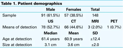
A total of 148 patients satisfied inclusion criteria for this study, with a higher proportion of males undergoing biop-sies (Table 1). Mean age at the time of initial detection was 60.9 years (±12.4). Lesion size at detection had a mean and median size of 3.6 cm and 3.1 cm (±2.0, range 1.0–15 cm), respectively. Most renal masses were identified with US (52.7%) or CT (44.6%). On average, patients under-went two additional scans prior to their biopsy, with CT representing the most common pre-biopsy modality (Table 2). Imaging tests were conducted for a variety of reasons, including improved resolution of the mass, evaluation of interval growth of the lesion, and investigation of potential metastatic disease. There was no defined imaging protocol and reasoning for tests was inconsistently reported. As such, we were unable to elucidate predictors of increased use of diagnostic imaging.
Initial biopsy was conducted within one year of detection for the majority of patients (Table 3). Most were conducted with US guidance (77.7%). A small number of patients (11) required repeat biopsy based on suspicious radiographical findings or non-diagnostic results. A greater proportion of patients (41.7%) required CT guidance for their repeat procedure.
Three patients (2.0%) experienced adverse events of note. Grade I Clavien-Dindo adverse events were not rou-tinely reported and could not be adequately assessed. One patient experienced a small, asymptomatic pneumothorax post-biopsy, and another developed a moderate perinephric hematoma associated with pain, both necessitating a short stay in hospital for observation. The final significant adverse event was a grade IVb post-biopsy bleed requiring emer-gent nephrectomy, inotropic support, and intensive care unit admission. This occurred in a patient with a history of both significant retroperitoneal bleeds and hypercoagulability.
Table 1. Patient demographics
Male Females Total
Sample 91 (61.5%) 57 (38.5%) 148
US CT MRI PET
Means of detection 78 (52.7%) 66 (44.6%) 3 (2.0%) 1 (0.7%)
Median Mean SD
Age at detection 61.4 years 60.9 years ±12.4 Size at detection 3.1 cm 3.6 cm ±2.0 CT: computed tomography; MRI: magnetic resonance imaging; PET: positron emission tomography; SD: standard deviation; US: ultrasound.
Table 2. Followup imaging
Modality Median Mean SD Patients
with only 1
Maintained on subcutaneous low-molecular weight hep-arin, this was held preoperatively, as is routine in our centre. Pre-biopsy markers of coagulation were normal. Although this was a CT-guided biopsy, it was noted to be exceedingly challenging, requiring traversal of the diaphragm to access the posterior mass, which was also found to be abutting the renal vein. No biopsy tract seeding was reported.
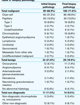
Initial biopsy reports found 32 benign (21.6%), 99 malig-nant (66.9%), and 17 non-diagnostic (11.5%) specimens (Table 4). Eleven patients underwent a second biopsy, one of whom proceeded to a third. Of these repeat biopsies, four patients were upgraded from a benign to malignant status, in addition to the three patients upgraded from non-diagnostic to malignant. The pathological subtypes of each biopsy are provided (Table 5).
Eighty-six patients (58.1%) had a combination of radio-graphical and/or biopsy results warranting radical/partial nephrectomy, and were suitable operative candidates. Sixty-six (76.7%) had final surgical pathology correspond directly with their most recent biopsy results (Table 6). Another nine patients (10.5%) were deemed malignant on both biopsy and surgical pathology, but had discordant cell types. No patients deemed benign were found to have malignant sur-gical resections. One patient with a chromophobe subtype on biopsy proceeded to be reclassified on surgical resec-tion as a benign oncocytoma. Of the three patients with both benign biopsy and surgical pathology, two patients proceeded to surgery due to ongoing concerns regarding followup and anxiety of their angiomyolipoma. The remain-ing patient had persistently concernremain-ing radiographical fea-tures. On final pathology, four (4.7%) and three (3.5%) non-diagnostic biopsies were returned benign and malignant, respectively. Our results culminated in a diagnostic accuracy of 90.7% of patients. A calculated sensitivity of 96.2% and a positive predictive value of 98.7% for biopsy detection of malignancy were generated (specificity 87.5%, negative
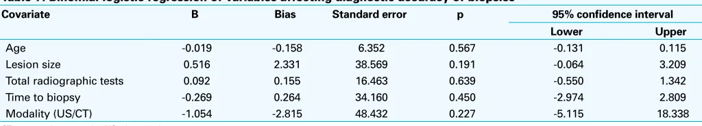
predictive value 70.0%, kappa 0.752, p<0.0005). Binomial logistic regression revealed that age, lesion size, number of radiographical tests, time to biopsy, and modality of biop-sy (US/CT) had no influence on the diagnostic accuracy of biopsies (Table 7). In patients who had Fuhrman grade reported on both biopsy and surgical pathology, 22 patients were adequately assessed, two underwent downgrading, and 17 were upgraded.
Discussion
Our single-centre, retrospective review fits into the growing body of evidence supporting the regular use, safety, and high diagnostic accuracy of renal biopsies. A recent meta-analysis from Lorenzo et al presented a diagnostic accuracy for
malig-Table 3. Biopsy time points
n Age Time from detection to biopsy (months) Modality
Median Mean SD Median Mean SD US-guided CT-guided
Biopsy 1 148 62.1 61.8 ±12.6 3.7 11.0 ±17.7 115 (77.7%) 33 (22.3%)
Biopsy 2 11 53.7 53.3 ±13.8 4.9 11.2 ±9.0 6 (54.5%) 5 (45.5%)
Biopsy 3 1 55.3 55.3 N/A 23.8 23.8 N/A 1 (100%) 0
CT: computed tomography; SD: standard deviation; US: ultrasound.
Table 5. Biopsy pathology
Initial biopsy pathology
Final biopsy pathology
Total malignant 99 (66.9%) 106 (71.6%)
Clear-cell 55 (37.2%) 60 (40.5%)
Papillary 20 (13.5%) 20 (13.5%)
Type 1 10 (6.8%) 10 (6.8%)
Type 2 4 (2.7%) 4 (2.7%)
Undefined/other 6 (4.1%) 6 (4.1%)
Chromophobe 9 (6.1%) 10 (6.8%)
Epithelioid angiomyolipoma 1 (0.7%) 1 (0.7%)
Lymphoma 5 (3.4%) 5 (3.4%)
Urothelial 3 (2.0%) 3 (2.0%)
Sarcomatoid 1 (0.7%) 1 (0.7%)
Metastatic from other site 1 (0.7%) 1 (0.7%) Undifferentiated malignant 4 (2.7%) 5 (3.4%)
Total benign 32 (21.6%) 28 (18.9%)
Oncocytoma 12 (8.1%) 11 (7.4%)
Angiomylipoma 6 (4.1%) 6 (4.1%) Arteriosclerosis/
No abnormal histology 8 (5.4%) 6 (4.1%)
Total non-diagnostic 17 (11.5%) 14 (9.5%)
Non-diagnostic chromophobe vs. oncocytoma
5 (3.4%) 5 (3.4%)
Other non-diagnostic 12 (8.1%) 9 (6.1%)
Table 4. Biopsy status
nancy of 92%.10 Our results compare favourably at 90.7%. Of note, our centre’s biopsies are performed primarily by body-trained radiologists under US guidance, and not by interven-tional radiology. This highlights a growing comfort with this sampling modality, necessary for its widespread adoption.
Despite the increasing acceptance from radiologists in our centre and across Canada, there remains some concern from the urological community regarding the regular use of renal biopsy. As such, routine use of biopsy has yet to become the standard of care in Canada, as per the most recent Canadian Urological Association guidelines for the management of the small renal mass.12 Similar stances are held by National Comprehensive Cancer Network and the European Association of Urology (EAU) in that renal biopsy remains a complementary, but unnecessary component of the small renal mass workup.13 Other, more contemporary opinions hold that biopsies should be used to define lesions of likely benign character, or to prepare for ablative/active surveillance strategies.14 The American Society of Clinical Oncology (ASCO) recently proclaimed that when accounting for competing mortality risks and tumour-specific findings, all small renal masses should undergo biopsy if management has the potential to be altered.15 With the advent of novel biomarkers and a greater appreciation of immunohistochem-istry, tissue diagnosis will be of even greater importance.14
Non-diagnostic results remain one of the most oft-cited concerns with kidney biopsies. Our series possessed an initial non-diagnostic rate of 11.5%. When enabling the use of repeat biopsies, this number decreased to 9.5%. A number of these samples were reported as chromophobe vs. oncocytoma, a well-documented diagnostic dilemma.16
This non-diagnostic rate ultimately compares well with other Canadian series, and highlights the importance of being open to repeat sampling.17 Importantly, a non-diagnostic status should not preclude surgery. In our series, 50% of non-diagnostic cases proceeded to nephrectomy and/or repeat biopsy. Given that our non-diagnostic rate represents an improvement over the literature reported rates of benign nephrectomy, this indicates a clinical advantage to the use of core needle sampling, despite the occasional diagnostic uncertainty. Identification of specific cell types remains a strong, albeit, imperfect feature of biopsies.18 This serves as an important feature, particularly in the comorbid patient, where prognosticating is a critical aspect of their care. Fuhrman grade characterization remains highly variable, however, both in our series and throughout the literature.19 This is believed to be, in large part, due to the grade hetero-geneity observed in renal masses.20
The concern regarding adverse events has been dampened with experience and evidence supporting low complication rates throughout the literature.21 Our review was comprised of only one event requiring operative management and two additional cases necessitating 24-hour monitoring. The Clavien IVb event we experienced highlights the importance of patient selection, as the patient had a known bleeding diathesis and may have benefitted from active surveillance. Based on current ASCO guidelines, this patient would have met the relative indications for active surveillance as well.15 It remains to be seen what role the renal biopsy will have in future active surveillance regimens.13 No needle tract seed-ing was observed in our review. Outside of rare reports, this remains consistent with the current body of evidence.22,23
In addressing the limitations of our study, we identify that this is, in fact, a retrospective series. The evaluation of renal biopsy will require prospectively randomized data before definitive guidelines can be established.10 In addition, our sample requires long-term followup to strengthen our out-comes of interest. The theoretical risk of needle tract seeding or deterioration in renal function may take years to develop. In addition, the assumption was made that benign biopsies not proceeding to surgery were definitively non-malignant. It is possible that in the years to come, these masses could begin demonstrating malignant character and require repeat biopsy or surgical resection.
Table 6. Biopsy and surgical pathology correspondence
Biopsy status Surgical status
Malignant Benign Total
Malignant correct cell type 66 (76.7%) 0 66 Malignant total 75 (87.2%) 1 (1.2%) 76
Benign 0 3 (3.5%) 3
Non-diagnostic 3 (3.5%) 4 (4.7%) 7
Total 78 8 86
True positives: total malignant biopsy pathology and malignant surgical pathology; false positives: total malignant biopsy pathology and benign surgical pathology; true negatives: benign + non-diagnostic biopsy pathology and benign surgical pathology; False negative = benign + non-diagnostic biopsy pathology and malignant surgical pathology.
Table 7. Binomial logistic regression of variables affecting diagnostic accuracy of biopsies
Covariate B Bias Standard error p 95% confidence interval
Lower Upper
Age -0.019 -0.158 6.352 0.567 -0.131 0.115
Lesion size 0.516 2.331 38.569 0.191 -0.064 3.209
Total radiographic tests 0.092 0.155 16.463 0.639 -0.550 1.342
Time to biopsy -0.269 0.264 34.160 0.450 -2.974 2.809
Modality (US/CT) -1.054 -2.815 48.432 0.227 -5.115 18.338
Moving forward, our centre would like to analyze the long-term followup of these patients. We plan to revisit our cohort in five years to assess rates of recurrence and malig-nant transformation. This will provide useful insight into the true negative rate or specificity of the renal biopsy. In addi-tion, we would like to perform a cost-benefit analysis. The goal is that a renal biopsy will help eliminate the unneces-sary cost of an operation planned for a benign lesion. The competing factors are the cost of the biopsy and the plethora of radiographical tests that are often ordered in surveillance regimens. Born out of a likely lack of trust in renal biopsy results, our study demonstrated a high radiographical burden attached to these patients. This undoubtedly factors into the cost analysis, but may improve with time, as urologists and radiologists alike grow more comfortable with this test.
Conclusion
Renal biopsies are safe, feasible, and diagnostic. Their role should be expanded in the routine evaluation of T1 and T2 renal masses.
Competing interests: The authors report no competing personal or financial interests related to this work.
This paper has been peer-reviewed.
References
1. Volpe A, Panzarella T, Rendon RA, et al. The natural history of incidentally detected small renal masses. Cancer 2004;100:738-45. https://doi.org/10.1002/cncr.20025
2. Rossi SH, Hsu R, Blick C, et al. Meta‐analysis of the prevalence of renal cancer detected by abdominal ultrasonography. Br J Surg 2017;104:658-9. https://doi.org/10.1002/bjs.10523
3. Kockelbergh R, Griffiths L. Renal tumour biopsy — a new standard of care? Eur Urol 2016;69:674-5. https://doi.org/10.1016/j.eururo.2015.09.017
4. Volpe A, Finelli A, Gill IS, et al. Rationale for percutaneous biopsy and histologic characterization of renal tumours. Eur Urol 2012;62:491-504. https://doi.org/10.1016/j.eururo.2012.05.009 5. Pahernik S, Zielger S, Roos F, et al. Small renal tumours: Correlation of clinical and pathological features
with tumour size. J Urol 2007;178:414-7. https://doi.org/10.1016/j.juro.2007.03.129
6. Remzi M, Ozsoy M, Klingler H, et al. Are small renal tumours harmless? Analysis of histo-pathological features according to tumours 4 cm or less in diameter. J Urol 2006;176:896-9. https://doi.org/10.1016/j.juro.2006.04.047
7. Novick AC, Campbell SC, Belldegrun A, et al. Guideline for management of the clinical stage 1 renal mass. J Urol 2009;182:1271-9. https://doi.org/10.1016/j.juro.2009.07.004
8. Richard PO, Jewett MA, Bhatt JR, et al. Renal tumour biopsy for small renal masses: A single-centre, 13-year experience. Eur Urol 2015;68:1007-13. https://doi.org/10.1016/j.eururo.2015.04.004 9. Halverson SJ, Kunju LP, Bhalla R, et al. Accuracy of determining small renal mass management with risk
stratified biopsies: Confirmation by final pathology. J Urol 2013;189:441-6. https://doi.org/10.1016/j. juro.2012.09.032
10. Lorenzo M, Dabestani S, Lam TB, et al. Systematic review and meta-analysis of diagnostic accuracy of percutaneous renal tumour biopsy. Eur Urol 2016;69:660-73. https://doi.org/10.1016/j.euru-ro.2015.07.072
11. Dindo D, Demartines N, Clavien P. Classification of surgical complications: A new proposal with evalua-tion in a cohort of 6336 patients and results of a survey. Ann Surg 2004;240:205-13. https://doi. org/10.1097/01.sla.0000133083.54934.ae
12. Jewett MA, Rendon R, Lacombe L, et al. Canadian guidelines for the management of small renal masses (SRM). Can Urol Assoc J 2015;9:160-3. https://doi.org/10.5489/cuaj.2969
13. Ljungberg B, Bensalah K, Canfield S, et al. EAU guidelines on renal cell carcinoma: 2014 update. Eur Urol 2015;67:913-24. https://doi.org/10.1016/j.eururo.2015.01.005
14. Burruni R, Lhermitte B, Cerantola Y, et al. The role of renal biopsy in small renal masses. Can Urol Assoc J 2016;10:E28-33. https://doi.org/10.5489/cuaj.3417
15. Finelli A, Ismaila N, Bro B, et al. Management of small renal masses: American Society of Clinical Oncology clinical practice guideline. J Clin Oncol 2017;35:668-80. https://doi.org/10.1200/JCO.2016.69.9645 16. Chang S, Loda M. Pathology and molecular pathology of renal cancer. Pathol Epidemiol Cancer
2017:335-46. https://doi.org/10.1007/978-3-319-35153-7_18
17. Leveridge MJ, Finelli A, Kachura J, et al. Outcomes of small renal mass needle core biopsy, non-diagnostic percutaneous biopsy, and the role of repeat biopsy. Eur Urol 2011;60:578-84. https://doi.org/10.1016/j.eururo.2011.06.021
18. Veltri A, Garetto I, Tosetti I, et al. Diagnostic accuracy and clinical impact of imaging-guided needle biopsy of renal masses. Retrospective analysis on 150 cases. Eur Radiol 2011;21:393-401. https:// doi.org/10.1007/s00330-010-1938-9
19. Abel EJ, Culp SH, Matin SF, et al. Percutaneous biopsy of primary tumour in metastatic renal cell car-cinoma to predict high-risk pathological features: Comparison with nephrectomy assessment. J Urol 2010;184:1877-81. https://doi.org/10.1016/j.juro.2010.06.105
20. Ball MW, Bezerra SM, Gorin MA, et al. Grade heterogeneity in small renal masses: Potential implications for renal mass biopsy. J Urol 2015;193:36-40. https://doi.org/10.1016/j.juro.2014.06.067 21. Volpe A, Kachura JR, Geddie WR, et al. Techniques, safety, and accuracy of sampling of renal tumours
by fine needle aspiration and core biopsy. J Urol 2007;178:379-86. https://doi.org/10.1016/j. juro.2007.03.131
22. Chang DTS, Sur H, Lozinskiy M, et al. Needle tract seeding following percutaneous biopsy of renal cell carcinoma. Korean J Urol 2015;56:666-9. https://doi.org/10.4111/kju.2015.56.9.666 23. Andersen, MFB, Norus TP. Tumour seeding with renal cell carcinoma after renal biopsy. Urol Case Rep
2016;9:43-4. https://doi.org/10.1016/j.eucr.2016.08.008