Role of Hox genes in regulating digit patterning
ROCÍO PÉREZ-GÓMEZ, ENDIKA HARO, MARC FERNÁNDEZ-GUERRERO,
MARÍA F. BASTIDA and MARÍA A. ROS*
Instituto de Biomedicina y Biotecnología de Cantabria, CSIC–SODERCAN Universidad de Cantabria, Santander, Spain
ABSTRACT The distal part of the tetrapod limb, the autopod, is characterized by the presence of digits. The digits display a wide diversity of shapes and number reflecting selection pressure for functional adaptation. Despite extensive study, the different aspects of digit patterning, as well as the factors and mechanisms involved are not completely understood. Here, we review the evidence implicating Hox proteins in digit patterning and the interaction between Hox genes and the Sonic hedgehog/Gli3 pathway, the other major regulator of digit number and identity. Currently, it is well accepted that a self-organizing Turing-type mechanism underlies digit patterning, this being understood as the establishment of an iterative arrangement of digit/interdigit in the hand plate. We also discuss the involvement of 5’ Hox genes in regulating digit spacing in the digital plate and therefore the number of digits formed in this self-organizing system.
KEY WORDS:
limb development, Hox gene, digit patterning, Shh, Gli3
Introduction
The basic plan of the tetrapod limb includes three distinct proximo-distal (PD) segments: the stylopod (arm), the zeugopod (forearm) and the autopod (hand/foot). The stylopod and the zeu-gopod contain one and two skeletal elements respectively and are highly conserved across species. In contrast, the distal segment or autopod, which contains the multiple skeletal elements of the hand or foot including the digits, shows considerable evolutionary variation reflecting the adaptation of the limb to such diverse uses as running, swimming or flying among others.
It is normally accepted that pentadactyly is the basal digit formula in tetrapods. Although the primitive tetrapods were polydactylous, the pentadactyl state was soon stabilized and it is considered that all extant tetrapods are descendants of an ancestor with a pentadactyl limb (Clack, 2002, Coates and Clack, 1990, Coates et al., 2002). The fact that the wide range of limb adaptations across species, when impacting digit number, always carries digit loss and that there is no species living today with more than 5 digits, led to the idea of the “pentadactyl constraint” meaning that the number of digits is limited to a maximum of five (Coates et al., 2002, Saxena et al., 2017). However, the recent reevaluation of the pre-hallux of amphibian hindlimbs as a sixth digit (Hayashi et al., 2015) to-gether with the reinterpretation of Ichthyostega anterior digits as deriving from anterior condensations (Mednikov, 2014), suggest that the pentadactylism may need to be reevaluated (Woltering
www.intjdevbiol.com
*Address correspondence to: Maria A. Ros. Instituto de Biomedicina y Biotecnología de Cantabria, c/ Albert Einstein, 22 (PCTCAN), E-39011 Santander, Spain.
Tel: + 34 942 201 933. E-mail: [email protected] - web: http://www.biodes.unican.es - https://orcid.org/0000-0002-1224-7671
Submitted: 2 July, 2018; Accepted: 13 September, 2018.
ISSN: Online 1696-3547, Print 0214-6282
© 2018 UPV/EHU Press Printed in Spain
Abbreviations used in this paper: AP, antero-posterior; PD, proximo-distal; ZPA, zone of polarizing activity.
and Meyer, 2015).
The digits are crucial elements for the function of the limb. They can be viewed as serial identical structures arranged along the antero-posterior (AP) axis of the autopod, thumb to little finger, or they can be viewed as distinct structures each one having its own independent and distinguishable identity. Therefore, the concept of “digit patterning” can be understood in different ways ranging from the basic iterative differentiation between digit and interdigit, to the specific digit formula (number of digits) and even to the specific digit morphologies (identities). The identity of the digits mainly relies on their phalangeal count. Based on the fossil record, the typical phalangeal count of the ancestral autopod is considered to be, from digit 1 to digit 5, 2-3-4-5-3 in the forelimb and 2-3-4-5-4 in the hindlimb (Smithson et al., 1993). In the synapsid lineages the phalangeal count evolved to the more familiar 2-3-3-3-3, as the mouse and human hand, with the most anterior digit, the thumb, having two phalanges and the rest of the digits having three.
discusses the factors and mechanisms controlling this process. Our goal is to present recent progress in this area with a special emphasis on the role played by Hox genes.
The formation of the autopod and of the digits
During limb development the three PD segments are specified progressively from the shoulder to digit tips under the influence of a specialized band of ectoderm riming the apex of the growing limb bud called the Apical Ectodermal Ridge (AER). The AER is a crucial signaling center for limb development that provides the signals needed for the survival, proliferation and specific gene expression of the limb progenitor cells (Fernandez-Teran and Ros, 2008). Fgf signaling is the main, if not the only one, AER signal (Mariani et al., 2017). AER cells secrete a battery of Fgfs, collectively called AER-Fgfs, of which the primary one is Fgf8 (Lewandoski et al., 2000, Mariani et al., 2008, Martin, 1998, Sun et al., 2002). The preponderant role of Fgf8 among other AER-Fgfs is due to its unique pattern and level of expression rather than to specific signaling properties (Boulet et al., 2004, Lewandoski et al., 2000, Lu et al., 2006, Mariani et al., 2008, Moon and Capecchi, 2000, Sun et al., 2002). Under the influence of Fgf signaling, the distal limb progenitor cells progressively transit through the specifica-tion states corresponding to the stylopod, the zeugopod and the autopod (Saiz-Lopez et al., 2015, Sato et al., 2007, Vargesson et al., 1997). There are good markers for each of the limb segments: Meis1 and Meis2 for the stylopod, Hoxa11 for the zeugopod and Hoxa13 for the autopod, but there is some discussion on whether they are necessary for the specification of the corresponding seg-ment and whether they can be considered as master genes for that segment (Tabin and Wolpert, 2007). It is currently accepted that signaling from the embryonic trunk and from the AER are conjointly necessary to specify the stylopod (Cooper et al., 2011, Rosello-Diez et al., 2011). In contrast, the specification of the two distal segments depends on an intrinsic program of the limb progenitors that is activated when, due to the growth of the bud, they become free of proximal influence (Rosello-Diez et al., 2014, Saiz-Lopez et al., 2015). This intrinsic program is controlled by an autonomous timer and includes, at least, the switch from a proximal to a distal mode of Hox gene expression, deceleration of proliferation rate and changes in cell adhesion properties (Andrey et al., 2013, Saiz-Lopez et al., 2015, Saiz-Lopez et al., 2017, Woltering et al., 2014). This results in the differentiation of an intermediate cylindrical-shaped segment, the zeugopod, and a distal paddle-shaped segment, the autopod. The initiation and dynamic expression of Hoxa13 marks the progressive specification of the autopod (Tamura et al., 2008).
Once the autopod forms, the first morphological evidence of a
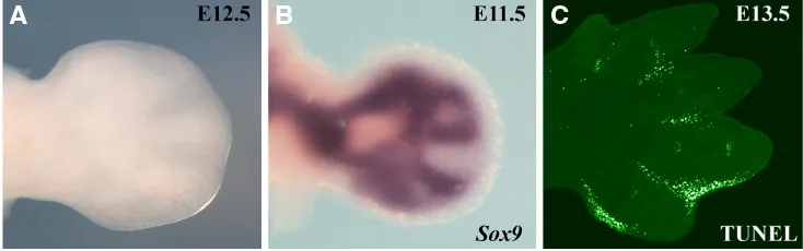
digit is an elongated longitudinal thickening, similar to a rod in a hand-fan (Fig. 1). The thickening corresponds to the chondrogenic condensation that contrasts sharply with the flattened flanking interdigital areas. The fate of the cells in these two regions is abso-lutely different, while the interdigital cells die by apoptosis, the cells in the condensations become chondrogenic, grow and segment to form the metacarpal/metatarsal and phalanges of each of the digital rays (Montero and Hurle, 2007). Before the digit thickening is evident, the pattern of expression of Sox9, a critical regulator of chondrogenesis, prefigures the pattern (Boehm et al., 2011).
The digit primordia emerge in sequence rather than simultane-ously and their number and distribution in the handplate seem to be fixed before their identity, which remains labile until much later stages of development (Dahn and Fallon, 2000, Sanz-Ezquerro and Tickle, 2003, Suzuki et al., 2008). The sequence of digit con-densation varies among species and in the mouse, a preferred animal model, it depends on the specific marker used (Boehm et al., 2011). Overall, it can be considered that a first and basic level in digit patterning is the generation of the digit-interdigit pattern upon which a second level of patterning, the generation of digit identities, is elaborated. It is clear that the number of digits corre-lates with the size of the handplate so that processes that lead to gain or loss of tissue in the handplate normally result in polydactyly or oligodactyly respectively. The thickness of the digits can also potentially impact the number of digits in a given-size handplate, but variability in digit thickness is not a common phenotype. It is also important to note that the final number of digits may not match the number of initial condensations as some of them may regress before forming the digit. For example, the apoptosis mediated elimination of the pre-chondrogenic condensations is a recently identified mode of evolutionary digit loss in mammals (Saxena et al., 2017; Cooper et al., 2014).
The two major factors involved in the control of digit patterning are the Hox genes and the Sonic hedgehog (Shh)/Gli3 pathway; their function and interactions will be considered next.
Hoxd and Hoxa gene expression in limb development
Hox genes are an important family of master genes codifying for transcription factors with functions in the control of the basic body plan of bilaterian animals (Lewis, 1978). Most vertebrates have 39 Hox genes that are organized in four clusters (HoxA, B, C, and D). In tetrapods, two of these clusters, the HoxA and the HoxD clusters, were coopted for the organization of limb morphology, a secondary axis in the embryo (Spitz et al., 2001, Zakany and Duboule, 2007). During limb development, similar to what occurs in the main embryonic axis, members of these two clusters are Fig. 1. Early morphological and molecularevidence of digit formation. (A) Dorsal view of a fresh E12.5 mouse embryo autopod in which the digits and interdigits are readily distinguishable. (B) The expression of Sox9, a key regulator of chondrocyte differentia-tion, envisages the pattern one day earlier. (C) Terminal deoxynucleotidyl transferase-mediated deoxyuridinetriphosphate nick-end labeling (TUNEL) assay in a 13.5 dpc mouse forelimb showing interdigital cell death.
B
C
activated sequentially in time and space, following their genomic position in the chromosome, a feature referred to as temporal and spatial collinearity (Tarchini and Duboule, 2006).
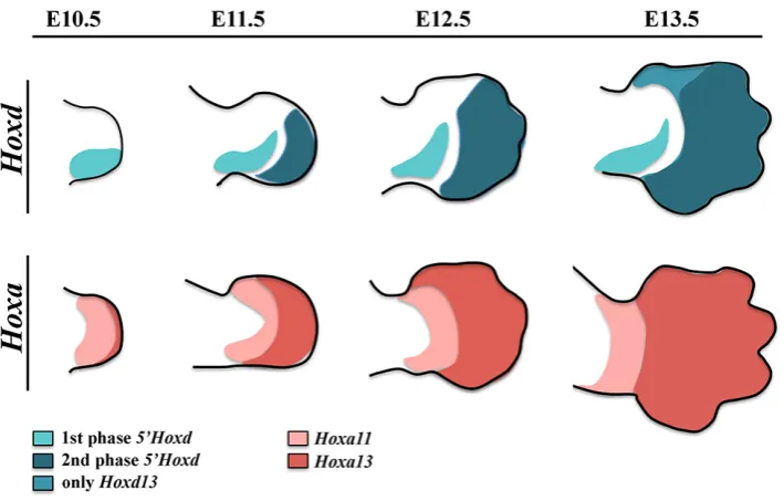
The expression of Hoxa and Hoxd genes in the developing limb is highly dynamic and specific for each cluster member. In the case of the HoxD cluster genes, their expression is best understood as occurring in two sequential and distinct waves that correlate with the PD anatomy of the tetrapod limb morphology (Zakany and Duboule, 2007) (Fig. 2). The correct morphology of the stylopod and zeugopod depends on the first phase of expression of Hoxd genes while the proper patterning of the digits, including their identity, depends on their second phase of expression.
The first or early phase of expression starts in the emerging limb bud and generates nested posterior-distal biased domains of expression with Hoxd9 to Hoxd11 showing the highest transcription level. The second or late phase of expression starts in the auto-pod and involves Hoxd10 to Hoxd13 with Hoxd13 displaying the higher level of expression (Montavon et al., 2008). It is important to note that, within each phase, the domains of the different Hoxd genes show a clear AP component. In the first phase, the domain of expression of more 5’genes is more posteriorly restricted and included within that of most 3’ genes (nested domains) while in the second phase the expression domains of Hoxd10, Hoxd11 and Hoxd12 show an anterior boundary coincident with the ante-rior border of digit 2 while Hoxd13 crosses this limit spreading all over the AP axis and therefore reversing the collinearity of the first phase (Kmita et al., 2002, Montavon et al., 2008).
The HoxA cluster is the other Hox cluster involved in limb development with 5’ located members exhibiting precise patterns of expression in correlation with the limb segments (Boulet and Capecchi, 2004, Davis et al., 1995, Fromental-Ramain et al., 1996, Kmita et al., 2005). Curiously, although Hoxa11 is initially expressed in the distal region of the early limb bud from the elbow/knee dis-tally, its distal expression fades concomitantly with the activation of Hoxa13 in the autopod progenitors. As a consequence, the expression of Hoxa11 becomes rapidly confined to the zeugopod
making Hoxa11 and Hoxa13 the best markers for the two distal segments of the limb, the zeugopod and autopod respectively (Tabin and Wolpert, 2007).
Extensive research over the past decade has exposed the complex regulatory landscapes that control Hox gene transcrip-tion in the developing limb. The HoxD cluster is located at the boundary between two topologically associating domains (TADs) that contain the limb regulatory regions (Rodríguez-Carballo et al., 2017). Transcriptional regulation during the early phase relies on enhancers located telomeric to the cluster (T-DOM) while en-hancers located centromeric to the cluster (C-DOM) govern the second phase of expression (Andrey et al., 2013, Montavon and Duboule, 2013, Montavon et al., 2011). The transition from the first to the second phase, which displaces the transcriptional efficiency towards genes located more 5’ in the cluster, requires the switch between these two regulatory regions (Andrey et al., 2013). One consequence of this switch is the emergence of a transverse band of tissue devoid of Hoxd expression that separates the two phases and that corresponds to the wrist/ankle.
Indeed, the analysis of several targeted mutations affecting genes in the HoxD cluster soon demonstrated that structural rearrangements of the genomic architecture had an impact in the expression patterns of remaining Hoxd genes (Kmita et al., 2002, Tarchini and Duboule, 2006). In most cases, the repositioning of the transcriptional units with respect to the cis-regulatory regions easily explained the alterations in expression patterns. However, other mutations that did not carry significant genomic change also had a strong impact in the expression of remaining Hox genes that was not explained by alterations in cis-regulation. Specifically, the removal of Hoxa13 or the joint removal of Hoxa13 and Hoxd13 resulted in the abolition of the biphasic expression of Hoxd genes (Sheth et al., 2014, Woltering et al., 2014). The investigation of the underlying molecular mechanisms showed that binding of Hoxa13 and Hoxd13 to the Hoxd regulatory domains was required to end the T-DOM regulation and activate the C-DOM regulation therefore governing the biphasic Hoxd expression (Beccari et al., 2016,
Fig. 2. Hoxd and Hoxa gene expression patterns during limb development.
Sheth et al., 2016). Therefore, Hoxa13 and Hoxd13, the terminal products of the HoxA and HoxD clusters, can influence in trans the transcriptional regulation of Hoxd genes.
considered that the proximal restriction of Hoxa11 is a requisite for the pentadactyl state (Kherdjemil et al., 2016).
The potential evolutionary implications of all these observa-tions have been widely discussed. It is easily conceivable that the progressive acquisition of Hoxa13 and Hoxd13 protein function in the regulatory landscapes together with the acquisition of the regulatory regions is at the root of the fin to limb transition. The transcriptional regulation of Hox genes is a fascinating topic that will be discussed in detail in other articles in this issue (Kherdjemil and Kmita, 2018, Woltering et al., 2014).
Hox genes regulate digit number and morphology
The influence of Hox genes in the control of the growth and morphological identities of the digits was already exposed with the generation of the first Hox targeted mutations. Alterations in digit number and morphology were a constant feature of the loss of function of Hoxd13 and Hoxa13, the genes at the 5’ edge of each cluster, and whose expression is characteristic of the autopod (Fromental-Ramain et al., 1996). The removal of Hoxd13 reduced the thickness and length of the digit condensations and caused polydactyly due to the presence of a postaxial slender extra digit (Dolle et al., 1993). The additional deletion of Hoxd11 and Hoxd12, the triple Hoxd11-13 deletion, produced a polydactyly phenotype similar to human synpolydactyly (Zakany and Duboule, 1996). In high contrast, the loss of function of Hoxa13, the only Hox gene whose removal is embryonic lethal, resulted in oligodactyly with the specific loss of digit 1, a phenotype that has not yet been fully explained. Interestingly, the reduction of Hoxa13 gene dose from the Hoxd13 or from the Hoxd11-13 null background exacerbated the polydactyly although the complete loss of Hoxa13 and Hoxd13 always led to digit agenesis possibly because of the interruption of the chondrogenic program (Fromental-Ramain et al., 1996). From these and other studies, it was inferred that 5’Hoxd genes tend to reduce the number of digits while Hoxa genes predisposed to increase the number of digits and that their progressive implementa-tion through evoluimplementa-tion could have contributed to the transiimplementa-tion from polydactyly to pentadactyly in basal tetrapods. The recruitment of the Hoxa genes first could have promoted polydactyly while the subsequent recruitment of Hoxd genes may have contributed to stabilize pentadactyl (Zakany et al., 1997).In addition, the analysis of multiple mutations with polydactyly or oligodactyly, such as Gli3 and Grem1, uncovered the strong correlation between the number of digits and the AP extension of the second phase domain of Hoxd13 expression (Buscher et al., 1997, te Welscher et al., 2002, Zuniga and Zeller, 1999). This observation established the notion that an increase in the number of digits required an expansion of the 5’Hoxd expression domains in the autopod.
The level of expression of Hoxd genes also correlates with the identity of the digits, in particular with the difference between the most anterior digit (two phalanges) and the rest of the digits (three phalanges). The precursors of digit 1 express a unique Hox code – this being Hoxd13 but not Hoxd12 or Hoxd11 – and a much lower dose of 5’Hoxd products than the rest of the digits (Montavon et al., 2008). Alterations of this Hox code leads to changes in digit 1 morphology (Morgan et al., 1992). This unique Hox code has been used to define the homology of the bird digits. The analysis of Hoxd12 and Hoxd13 expression in the chick wing showed that the
anterior most digit expresses Hoxd13 but not Hoxd12, consistent with a digit 1 identity disregarding its embryonical formation in position 2 (Towers et al., 2011, Vargas and Wagner, 2009, Wang et al., 2011).
The zone of polarizing activity and the Shh/Gli3 pathway
Besides Hox genes, the Shh/Gli3 pathway is the other major player in digit patterning. Indeed, the first clue on how digit forma-tion might be controlled came from the discovery of the Zone of Polarizing Activity (ZPA) (Saunders and Gasseling, 1968, Tickle, 2002). The ZPA is a group of mesodermal cells located at the posterior border of the limb bud that has the amazing ability to induce a mirror image duplication of the normal digital pattern when transplanted to the anterior border of a host limb bud. Since the ZPA lacks any morphological distinction, its spatial and temporal localization was precisely mapped through grafting assays in the chick embryo (Tickle and Towers, 2017, Zhu and Mackem, 2017). Two features were clearly salient in the limb phenotypes resulting from anterior ZPA grafts: (i) the extra growth that led to handplates much wider than normal and, (ii) the control of AP asymmetry, as indicated by the mirror image duplications. Therefore, the ZPA was clearly recognized as the primary organizer of the AP axis in the amniote limb bud regulating both growth and digit identity.The best interpretation of the function of the ZPA, particularly regarding patterning, came from the Positional Information (French Flag) model devised by Lewis Wolpert (Wolpert, 1969). This model posits that the ZPA produces a molecule, the morphogen, that spreads and creates a concentration gradient across the limb bud. The cells in the limb field are capable of reading the concentration of the morphogen and differentiate accordingly (Wolpert, 1969). After more than two decades of arduous search for the ZPA emanating morphogen, finally in 1993 it was identified as being Shh (Riddle et al., 1993). Shh, a potent signaling molecule with critical functions during development and homeostasis, was shown to elicit all ZPA properties indicating that it was the sole molecule responsible for ZPA function (Riddle et al., 1993) (Lopez-Martinez et al., 1995).
Although outside of the scope of this review, it is worth mention-ing that the production and secretion of Shh are highly regulated processes that involve the post-translational modification of the ligand including the proteolysis of the full-length molecule and the addition of cholesterol and palmitate residues to the C-terminal and N-terminal extremes respectively. The lipidic modifications raised doubts on whether such molecule could freely diffuse through the aqueous extracellular milieu but, there is compelling evidence supporting the diffusion of Shh protein away from the ZPA and establishing a gradient across the AP axis (Chen et al., 2004a, Gritli-Linde et al., 2001, Lewis et al., 2001, Li et al., 2006, Yang et al., 1997). It is currently accepted that Shh long-range signaling predominantly relies on specialized filopodia called cytonemes that connect distal cells (Kornberg, 2014).
2011). In the absence of Shh signaling, Gli3 protein is cleaved to produce a shorter N-terminal form that functions as a strong tran-scriptional repressor (Gli3R) (Wang et al., 2000). In the presence of Shh signaling, Gli3 processing is prevented and the full-length Gli3 functions as a weak transcriptional activator. The involvement of Shh signaling in the processing of Gli3 results in the genera-tion of an intracellular gradient of Gli3R opposite to that of Shh with maximum level in the anterior limb bud (Bastida et al., 2004, Litingtung et al., 2002, Wang et al., 2000).
In humans, mutations in the GLI3 gene are responsible for several syndromes, conjointly referred to as GLI3 morphopathies, all of them displaying malformations of the autopod including poly-dactyly and synpoly-dactyly. Thus, Gli3 is considered a major genetic cause of polydactyly (Biesecker, 2006, Demurger et al., 2015, Hui and Angers, 2011).
Multiple interactions between Hox genes and the Shh/
Gli3 pathway
Hox genes and the Shh/Gli3 pathway are highly interconnected. Actually, the initiation of Shh transcription requires the first phase of expression of the 5’Hoxd genes (Tarchini et al., 2006, Zakany et al., 2004). Hox proteins bind to the Shh limb specific enhancer
that was considered the cause of the polydactyly (Buscher et al., 1997, Chan et al., 1995, Masuya et al., 1997, Masuya et al., 1995). It was assumed that the ectopic source of Shh would cause the ectopic activation of 5’ Hoxd genes and therefore the polydactyly.
To test this assumption and revert the Gli3 polydactyly, Shh was removed from the Gli3 null background but, to the general surprise, double Shh;Gli3 mutant embryos were phenotypically indistinguish-able from Gli3 mutants, arguing that Shh was not necessary for the Gli3 polydactyly (Litingtung et al., 2002, te Welscher et al., 2002). Double mutant limb buds retained the anterior upregulation of Hox genes reinforcing their implication as the cause of the polydactyly, and consistent with Gli3R exerting a potent negative effect on their expression. The double Shh;Gli3 mutant clearly denoted that Shh’ major function was to control the production of Gli3R.
To directly determine the involvement of 5’Hoxd genes in the polydactyly, several experiments were conducted to remove or attenuate Hox function in the Gli3 null background (Sheth et al., 2007, Sheth et al., 2012, Zakany et al., 2007). Again contrary to the expectations, the removal of the complete HoxD cluster or the removal of the more 5’Hox genes (Hoxd11-13 or Hoxa13) neither restored pentadactyly nor even reduced the Gli3 polydactyly. Indeed, the compound genotypes either unchanged (Zakany et al., 2007) or exacerbated the Gli3 polydactyly (Sheth et al., 2007,
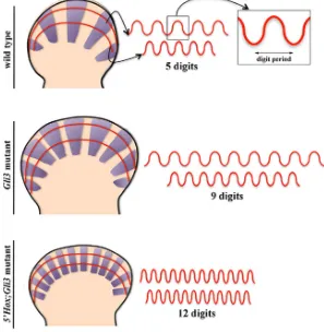
Fig. 3. Schemes representing the outline of E11.5 wild-type (WT, top), Gli3 mutant (middle) and 5’Hox;Gli3 mutant (bottom) autopods with the expression of Sox9 in the digit condensations depicted in purple. In each autopod, one proximal and one distal anterior posterior curved profile have been traced. The intensity of Sox9 ex-pression along each of these two profiles is represented on the right in which the digit period or wavelength can be appreciated. Note the effective proximo-distal increase of the digit wavelength in the WT and Gli3 mutant but not in the 5’Hox;Gli3 mutant. to synergistically activate Shh transcription (Capellini
et al., 2006, Kmita et al., 2005). Moreover, misexpres-sion of Hox genes at the anterior border also causes ectopic Shh activation (Charite et al., 1994, Knezevic et al., 1997).
Once Shh expression is on, it is in turn necessary for the establishment of the second phase of Hoxd expres-sion (Chiang et al., 2001, Kraus et al., 2001, Ros et al., 2003). The Shh-dependent activation of Hoxd genes is mediated by relieving the repression exerted by Gli3R (Vokes et al., 2008) (Lewandowski et al., 2015). In the absence of Gli3, as in the extratoes (Xt) spontaneous mutation in mice, the autopod is characterized by a prominent uniform AP expansion of Hoxd expression without a noticeable change in Shh expression. As in human, the mouse Gli3 mutant displays multiple abnor-malities including a severe polysyndactyly of 7-8 digits with no clear identity (Hui and Joyner, 1993, Schimmang et al., 1992). This is consistent with Gli3R restricting the expression of 5’Hoxd genes to the posterior mesoderm, a function that is modulated by its interaction with the 5’Hoxd products themselves. It has been shown that the physical interaction between Gli3R and Hoxd12 changes the repressor activity of Gli3R into an activator and that it is the balance between Gli3 and 5’Hoxd products that controls digit patterning (Chen et al., 2004b). This circumstance adds an additional level of complexity to the interaction between Hox genes and the Shh/Gli3 pathway and therefore to the control of digit patterning.
The Shh/Gli3 pathway and Hox genes as genetic
causes of polydactyly
Sheth et al., 2012). Remarkably, the removal of the three most 5’Hoxd genes plus one copy of Hoxa13 (Gli3-/-; Hoxd11-13-/-; Hoxa13+/-) produced autopods of 12-14 identical digits, the most severe polydactyly reported to date (Fig. 3). Thus, in the absence of Gli3, the progressive reduction in posterior Hox gene function led to a progressively increase in digit number supporting the conclusion that the dosage of 5’ Hox genes negatively correlated with the number of digits (Sheth et al., 2012). In addition, these results clearly showed that the gain of 5’ Hoxd expression was not required for the formation of extra-digits in the Gli3 mutant limb.
Importantly, the increase in digit number in the above mentioned mutants did not associate an increase in the size of the handplate but rather relied on thinner and more densely packed digits (Fig. 3). Thus, 5’Hox genes somehow modulate, in a dose dependent manner, the digit spacing or digit period and therefore the number of digits in a given space (Fig. 3). In addition, the phenotypes of the Gli3; Hoxd11-13; Hoxa13 allelic series consisting of regularly spaced digits the number of which within a similar space could change (according to the Hox gene dosage) pointed to a subja-cent self-organizing reaction-diffusion or Turing-type mechanism (Sheth et al., 2012).
A Turing-type model for digit-interdigit patterning
Turing or reaction-diffusion models are notorious for producing periodic patterns (Gierer and Meinhardt, 1972, Kondo and Miura, 2010, Maini and Solursh, 1991, Meinhardt and Gierer, 2000, New-man and Bhat, 2007, NewNew-man and Frisch, 1979, Turing, 1952). They are based on two molecules or chemicals, the activator and the inhibitor, that interact between them at the same time that they diffuse in a field of cells. The activator activates the production of the inhibitor and of itself, while the more rapidly diffusing inhibitor inhibits its production as well as that of the activator. With these conditions, the minimal oscillations inherent to biological processes are sufficient to generate periodic patterns, commonly stripes or spots, characterized by a particular spacing or wavelength (Econo-mou and Green, 2014, Marcon and Sharpe, 2012).Reaction-diffusion models had already been considered to explain digit patterning and the limb was one of the first develop-mental systems to be modeled using a Turing’s simple model (Ede et al., 1977, Newman and Frisch, 1979, Wilby and Ede, 1975). One indication that the digits may be generated via this mechanism was the ability of a cluster of randomized limb progenitor cells to self organize, in the absence of the ZPA, and generate symmetrical identical digits (Ros et al., 1994). Another indication in this direction was the periodic pattern of spots obtained in micromass cultures of the limb bud cells, a well-established in vitro model of chondro-genesis (Christley et al., 2007, Newman, 1996).
With the use of mathematical and computational approaches, a Turing-type model was devised that could account for normal and Gli3;Hox mutant digit patterning (Sheth et al., 2012). The Turing mechanism generates a periodic wave with a specific wavelength that defines the digit spacing or digit period. Given the fan shape of the handplate, during normal development, the wavelength needs to be scaled along the PD axis to maintain a constant number of digits and prevent digit bifurcations (Fig. 3). To fulfill this requirement the model considers the PD gradient of Fgf signaling and the level of 5’Hox product to both modulate the wavelength. Taking this into consideration, the simulations of the
reaction-diffusion model successfully reproduced the polydactylous patterns of Gli3;Hox double and triple mutants including the short-ening and distal displacement of the digit forming region (Sheth et al., 2012). Further studies designed to determine the molecules involved in the Turing system, identified Bmps, Sox9 and Wnt as the core network molecules of a substrate–depletion type model, termed the BSW model (Raspopovic et al., 2014). Alternative models have considered Galectin1a-Galectin8, Transformig growth factor beta2 and the Bmp-receptor interactions as the core mol-ecules in other Turing based models (Badugu et al., 2012, Glimm et al., 2014, Miura and Shiota, 2000, Newman et al., 2018). The different Turing-type models so far devised for digit development may coexist and overlap to a greater or lesser degree as well as coordinate with the signaling and other processes concomitantly occurring in the autopod.
Recently the Hox function in controlling the digit wavelength has been questioned (Hiscock et al., 2017). As explained above, due to the function of Hoxa13 and Hoxd13 in the transcriptional regulation of Hoxd genes, the reduction in their genetic dosage associates a delay in the onset of the second phase of Hoxd gene expression and, therefore, a distal displacement of the digit-forming region (Fig. 3) (Sheth et al., 2014). Because of the semicircular shape of the handplate, the distal displacement entails an increase in the AP extent of the digit-forming region that would explain an increase in the number of shorter digits without the need of a reduction in the wavelength. However, the quantification of the mutant phenotypes showed that this increase was not sufficient to accommodate the 12-14 digits observed in some of the Gli3;Hox mutants unless there is a concomitant reduction in the wavelength that actually occurs (Sheth et al., 2012). Further studies are required to better understand the spatial and temporal details of the Turing mecha-nism as well as the molecules involved.
Digit identity
Once the basic digit/interdigit pattern has been established, the digital rays continue elongation with the formation of the cor-responding phalanges and joints eventually attaining their identity. It was first shown in the chicken leg that digit identities were labile and susceptible of being modified until very late stages as their morphogenesis depends on continuous signaling, most probably BMP signaling, from the posterior interdigit (Dahn and Fallon, 2000, Sanz-Ezquerro and Tickle, 2003, Suzuki et al., 2008). The chick leg is an ideal system for the study of digit identity because it has four digits with readily identifiable identity as they maintain the ancestral phalangeal count (2-3-4-5 from digit 1 to 4). Lineage tracing analysis showed that the phalanges sequentially derived from the Phalange Forming Region (PFR), a crescent-shaped area at the tip of the digits where progenitors proliferate under AER influence. Subsequent studies showed that phalanges and interzones were coordinately specified in the PFR under signaling from the posterior interdigital space which in turn is controlled by the balance between Gli3 and 5’Hoxd products (Huang et al., 2016).
increased phospho-SMAD 1/5/8 staining at the tip of an already formed digit condensation and therefore a relatively late element (Suzuki et al., 2008). Whether the PFR can be equated with the initial digit condensation requires further studies to understand the sequence of involved events from the initial condensations to the final digit morphologies.
Summary
Digit patterning starts shortly after the specification of the auto-pod with the establishment of the periodic digit-interdigit pattern. Currently, this patterning is best understood as the result of a Turing-type self-organizing process that defines the spacing be-tween consecutive digits or wavelength. The details of the Turing type mechanism and its network core molecules are just beginning to be known. The features that give distinct identity to each digit, such as the number of phalanges, are elaborated upon this basic pattern although they may have been specified much earlier.
The Shh/Gli3 pathway together with the 5’Hox genes are the major regulators of digit patterning and they both are involved in early and late patterning events in an interconnected manner. The Shh/Gli3 pathway is highly involved in the control of progenitor cell proliferation controlling anterior-posterior expansion of the autopod and determining the size of the handplate and the domain of 5’Hoxd gene expression. It is clear that there is a link between early AP patterning in the limb bud and later digit morphogenesis but how this connection is propagated through development and integrated with other patterning events requires further investigation.
Acknowledgements
The M.A.R. laboratory is supported by the Spanish Ministerio de Economía, Industria y Competitividad (BFU2014-57216-P and BFU2017-88265-P).
References
ANDREY, G., MONTAVON, T., MASCREZ, B., GONZALEZ, F., NOORDERMEER, D., LELEU, M., TRONO, D., SPITZ, F. and DUBOULE, D. (2013). A switch between topological domains underlies HoxD genes collinearity in mouse limbs. Science 340: 1234167.
BADUGU, A., KRAEMER, C., GERMANN, P., MENSHYKAU, D. and IBER, D. (2012). Digit patterning during limb development as a result of the BMP-receptor interaction. Sci Rep 2: 991.
BASTIDA, M.F., DELGADO, M.D., WANG, B., FALLON, J.F., FERNANDEZ-TERAN, M. and ROS, M.A. (2004). Levels of Gli3 repressor correlate with Bmp4 expression and apoptosis during limb development. Dev Dyn 231: 148-160.
BECCARI, L., YAKUSHIJI-KAMINATSUI, N., WOLTERING, J.M., NECSULEA, A., LONFAT, N., RODRIGUEZ-CARBALLO, E., MASCREZ, B., YAMAMOTO, S., KUROIWA, A. and DUBOULE, D. (2016). A role for HOX13 proteins in the regulatory switch between TADs at the HoxD locus. Genes Dev 30: 1172-1186. BERLIVET, S., PAQUETTE, D., DUMOUCHEL, A., LANGLAIS, D., DOSTIE, J. and
KMITA, M. (2013). Clustering of tissue-specific sub-TADs accompanies the regula-tion of HoxA genes in developing limbs. PLoS Genet 9: e1004018.
BIESECKER, L.G. (2006). What you can learn from one gene: GLI3. J Med Genet 43: 465-469.
BOEHM, B., RAUTSCHKA, M., QUINTANA, L., RASPOPOVIC, J., JAN, Z. and SHARPE, J. (2011). A landmark-free morphometric staging system for the mouse limb bud. Development 138: 1227-1234.
BOULET, A.M. and CAPECCHI, M.R. (2004). Multiple roles of Hoxa11 and Hoxd11 in the formation of the mammalian forelimb zeugopod. Development 131: 299-309. BOULET, A.M., MOON, A.M., ARENKIEL, B.R. and CAPECCHI, M.R. (2004). The
roles of Fgf4 and Fgf8 in limb bud initiation and outgrowth. Dev Biol 273: 361-372.
BUSCHER, D., BOSSE, B., HEYMER, J. and RUTHER, U. (1997). Evidence for genetic control of Sonic hedgehog by Gli3 in mouse limb development. Mech Dev 62: 175-182.
CAPELLINI, T.D., DI GIACOMO, G., SALSI, V., BRENDOLAN, A., FERRETTI, E., SRIVASTAVA, D., ZAPPAVIGNA, V. and SELLERI, L. (2006). Pbx1/Pbx2 require-ment for distal limb patterning is mediated by the hierarchical control of Hox gene spatial distribution and Shh expression. Development 133: 2263-2273. CLACK, J.A. (2002). An early tetrapod from ‘Romer’s Gap’. Nature 418: 72-76. COATES, M.I. and CLACK, J.A. (1990). Polydactyly in the earliest known tetrapod
limbs. Nature 347: 66.
COATES, M.I., JEFFERY, J.E. and RUTA, M. (2002). Fins to limbs: what the fossils say1. Evo. Devo. 4: 390-401.
COOPER, K.L., HU, J.K., TEN BERGE, D., FERNANDEZ-TERAN, M., ROS, M.A. and TABIN, C.J. (2011). Initiation of proximal-distal patterning in the vertebrate limb by signals and growth. Science 332: 1083-1086.
CHAN, D.C., LAUFER, E., TABIN, C. and LEDER, P. (1995). Polydactylous limbs in Strong’s Luxoid mice result from ectopic polarizing activity. Development 121: 1971-1978.
CHARITE, J., DE GRAAFF, W., SHEN, S. and DESCHAMPS, J. (1994). Ectopic expression of Hoxb-8 causes duplication of the ZPA in the forelimb and homeotic transformation of axial structures. Cell 78: 589-601.
CHEN, M.H., LI, Y.J., KAWAKAMI, T., XU, S.M. and CHUANG, P.T. (2004a). Palmi-toylation is required for the production of a soluble multimeric Hedgehog protein complex and long-range signaling in vertebrates. Genes Dev 18: 641-659. CHEN, Y., KNEZEVIC, V., ERVIN, V., HUTSON, R., WARD, Y. and MACKEM, S.
(2004b). Direct interaction with Hoxd proteins reverses Gli3-repressor function to promote digit formation downstream of Shh. Development 131: 2339-2347. CHIANG, C., LITINGTUNG, Y., HARRIS, M.P., SIMANDL, B.K., LI, Y., BEACHY, P.A.
and FALLON, J.F. (2001). Manifestation of the limb prepattern: limb development in the absence of sonic hedgehog function. Dev Biol 236: 421-435.
CHRISTLEY, S., ALBER, M.S. and NEWMAN, S.A. (2007). Patterns of mesenchymal condensation in a multiscale, discrete stochastic model. PLoS Comput Biol 3: e76. DAHN, R.D. and FALLON, J.F. (2000). Interdigital regulation of digit identity and
homeotic transformation by modulated BMP signaling. Science 289: pp. 438-441. DAVIS, A.P., WITTE, D.P., HSIEH-LI, H.M., POTTER, S.S. and CAPECCHI, M.R.
(1995). Absence of radius and ulna in mice lacking hoxa-11 and hoxd-11. Nature 375: 791-795.
DEMURGER, F., ICHKOU, A., MOUGOU-ZERELLI, S., LE MERRER, M., GOUDE-FROYE, G., DELEZOIDE, A.L., QUELIN, C., MANOUVRIER, S., BAUJAT, G., FRADIN, M. et al., (2015). New insights into genotype-phenotype correlation for GLI3 mutations. Eur J Hum Genet 23: 92-102.
DOLLE, P., DIERICH, A., LEMEUR, M., SCHIMMANG, T., SCHUHBAUR, B., CHAM-BON, P. and DUBOULE, D. (1993). Disruption of the Hoxd-13 gene induces localized heterochrony leading to mice with neotenic limbs. Cell 75: 431-441. ECONOMOU, A.D. and GREEN, J.B. (2014). Modelling from the experimental
de-velopmental biologists viewpoint. Semin Cell Dev Biol 35: 58-65.
EDE, D.A., FLINT, O.P., WILBY, O.K. and COLQUHOUN, P. (1977). The develop-ment of precartilagecondensations in limb bud mesenchyme in vivo and in vitro.. In Vertebrate Limb and Somite Morphogenesis, (ed. EDE D.A., H. J. R., BALLS M.). Cambridge University Press, Cambridge pp.161-179.
FERNANDEZ-TERAN, M. and ROS, M.A. (2008). The Apical Ectodermal Ridge: morphological aspects and signaling pathways. Int J Dev Biol 52: 857-871. FROMENTAL-RAMAIN, C., WAROT, X., MESSADECQ, N., LEMEUR, M., DOLLE,
P. and CHAMBON, P. (1996). Hoxa-13 and Hoxd-13 play a crucial role in the patterning of the limb autopod. Development 122: 2997-3011.
GIERER, A. and MEINHARDT, H. (1972). A theory of biological pattern formation. Kybernetik 12: 30-39.
GLIMM, T., BHAT, R. and NEWMAN, S.A. (2014). Modeling the morphodynamic galectin patterning network of the developing avian limb skeleton. J Theor Biol 346: 86-108. GRITLI-LINDE, A., LEWIS, P., MCMAHON, A.P. and LINDE, A. (2001). The where-abouts of a morphogen: direct evidence for short- and graded long-range activity of hedgehog signaling peptides. Dev Biol 236: 364-386.
HISCOCK, T.W., TSCHOPP, P. and TABIN, C.J. (2017). On the Formation of Digits and Joints during Limb Development. Dev Cell 41: 459-465.
HUANG, B.L., TROFKA, A., FURUSAWA, A., NORRIE, J.L., RABINOWITZ, A.H., VOKES, S.A., MARK TAKETO, M., ZAKANY, J. and MACKEM, S. (2016). An interdigit signalling centre instructs coordinate phalanx-joint formation governed by 5’Hoxd-Gli3 antagonism. Nat Commun 7: 12903.
HUI, C.C. and ANGERS, S. (2011). Gli proteins in development and disease. Annu Rev Cell Dev Biol 27: 513-537.
HUI, C.C. and JOYNER, A.L. (1993). A mouse model of greig cephalopolysyndactyly syndrome: the extra-toesJ mutation contains an intragenic deletion of the Gli3 gene. Nat Genet 3: 241-246.
KHERDJEMIL, Y. and KMITA, M. (2018). Insights on the role of hox genes in the emergence of the pentadactyl ground state. Genesis e23046.
KHERDJEMIL, Y., LALONDE, R.L., SHETH, R., DUMOUCHEL, A., DE MARTINO, G., PINEAULT, K.M., WELLIK, D.M., STADLER, H.S., AKIMENKO, M.A. and KMITA, M. (2016). Evolution of Hoxa11 regulation in vertebrates is linked to the pentadactyl state. Nature 539: 89-92.
KMITA, M., FRAUDEAU, N., HERAULT, Y. and DUBOULE, D. (2002). Serial dele-tions and duplicadele-tions suggest a mechanism for the collinearity of Hoxd genes in limbs. Nature 420: 145-150.
KMITA, M., TARCHINI, B., ZAKANY, J., LOGAN, M., TABIN, C.J. and DUBOULE, D. (2005). Early developmental arrest of mammalian limbs lacking HoxA/HoxD gene function. Nature 435: 1113-1116.
KNEZEVIC, V., DE SANTO, R., SCHUGHART, K., HUFFSTADT, U., CHIANG, C., MAHON, K.A. and MACKEM, S. (1997). Hoxd-12 differentially affects preaxial and postaxial chondrogenic branches in the limb and regulates Sonic hedgehog in a positive feedback loop. Development 124: 4523-4536.
KONDO, S. and MIURA, T. (2010). Reaction-diffusion model as a framework for understanding biological pattern formation. Science 329: 1616-1620.
KORNBERG, T.B. (2014). The contrasting roles of primary cilia and cytonemes in Hh signaling. Dev Biol 394: 1-5.
KRAUS, P., FRAIDENRAICH, D. and LOOMIS, C.A. (2001). Some distal limb struc-tures develop in mice lacking Sonic hedgehog signaling. Mech Dev 100: 45-58. LEWANDOSKI, M., SUN, X. and MARTIN, G.R. (2000). Fgf8 signalling from the AER
is essential for normal limb development. Nat Genet 26: 460-463.
LEWANDOWSKI, J.P., DU, F., ZHANG, S., POWELL, M.B., FALKENSTEIN, K.N., JI, H. and VOKES, S.A. (2015). Spatiotemporal regulation of GLI target genes in the mammalian limb bud. Dev Biol 406: 92-103.
LEWIS, E.B. (1978). A gene complex controlling segmentation in Drosophila. Nature 276: 565-570.
LEWIS, P.M., DUNN, M.P., MCMAHON, J.A., LOGAN, M., MARTIN, J.F., ST-JACQUES, B. and MCMAHON, A.P. (2001). Cholesterol modification of sonic hedgehog is required for long-range signaling activity and effective modulation of signaling by Ptc1. Cell 105: 599-612.
LI, Y., ZHANG, H., LITINGTUNG, Y. and CHIANG, C. (2006). Cholesterol modifica-tion restricts the spread of Shh gradient in the limb bud. Proc Natl Acad Sci USA 103: 6548-6553.
LITINGTUNG, Y., DAHN, R.D., LI, Y., FALLON, J.F. and CHIANG, C. (2002). Shh and Gli3 are dispensable for limb skeleton formation but regulate digit number and identity. Nature 418: 979-983.
LOPEZ-RIOS, J. (2016). The many lives of SHH in limb development and evolution. Semin Cell Dev Biol 49: 116-124.
LU, P., MINOWADA, G. and MARTIN, G.R. (2006). Increasing Fgf4 expression in the mouse limb bud causes polysyndactyly and rescues the skeletal defects that result from loss of Fgf8 function. Development 133: 33-42.
MAINI, P.K. and SOLURSH, M. (1991). Cellular mechanisms of pattern formation in the developing limb. Int Rev Cytol 129: 91-133.
MARCON, L. and SHARPE, J. (2012). Turing patterns in development: what about the horse part? Curr Opin Genet Dev 22: 578-584.
MARIANI, F.V., AHN, C.P. and MARTIN, G.R. (2008). Genetic evidence that FGFs have an instructive role in limb proximal-distal patterning. Nature 453: 401-405. MARIANI, F.V., FERNANDEZ-TERAN, M. and ROS, M.A. (2017). Ectoderm-mesoderm crosstalk in the embryonic limb: The role of fibroblast growth factor signaling. Dev Dyn 246: 208-216.
MARTIN, G.R. (1998). The roles of FGFs in the early development of vertebrate
limbs. Genes Dev 12: 1571-1586.
MASUYA, H., SAGAI, T., MORIWAKI, K. and SHIROISHI, T. (1997). Multigenic control of the localization of the zone of polarizing activity in limb morphogenesis in the mouse. Dev Biol 182: 42-51.
MASUYA, H., SAGAI, T., WAKANA, S., MORIWAKI, K. and SHIROISHI, T. (1995). A duplicated zone of polarizing activity in polydactylous mouse mutants. Genes Dev 9: 1645-1653.
MEDNIKOV, D.N. (2014). Urodelans, Ichthyostega and the origin of the tetrapod limb. Paleontol. J. 48: 1092-1103.
MEINHARDT, H. and GIERER, A. (2000). Pattern formation by local self-activation and lateral inhibition. Bioessays 22: 753-760.
MIURA, T. and SHIOTA, K. (2000). TGFbeta2 acts as an “activator” molecule in reaction-diffusion model and is involved in cell sorting phenomenon in mouse limb micromass culture. Dev Dyn 217: 241-249.
MONTAVON, T. and DUBOULE, D. (2013). Chromatin organization and global regula-tion of Hox gene clusters. Philos Trans R Soc Lond B Biol Sci 368: 20120367. MONTAVON, T., LE GARREC, J.F., KERSZBERG, M. and DUBOULE, D. (2008).
Modeling Hox gene regulation in digits: reverse collinearity and the molecular origin of thumbness. Genes Dev 22: 346-359.
MONTAVON, T., SOSHNIKOVA, N., MASCREZ, B., JOYE, E., THEVENET, L., SPLINTER, E., DE LAAT, W., SPITZ, F. and DUBOULE, D. (2011). A regulatory archipelago controls Hox genes transcription in digits. Cell 147: 1132-1145. MONTERO, J.A. and HURLE, J.M. (2007). Deconstructing digit chondrogenesis.
Bioessays 29: 725-737.
MOON, A.M. and CAPECCHI, M.R. (2000). Fgf8 is required for outgrowth and pat-terning of the limbs. Nat Genet 26: 455-459.
MORGAN, B.A., IZPISUA-BELMONTE, J.C., DUBOULE, D. and TABIN, C.J. (1992). Targeted misexpression of Hox-4.6 in the avian limb bud causes apparent homeotic transformations. Nature 358: 236-239.
NEWMAN, S.A. (1996). Sticky fingers: Hox genes and cell adhesion in vertebrate limb development. Bioessays 18: 171-174.
NEWMAN, S.A. and BHAT, R. (2007). Activator-inhibitor dynamics of vertebrate limb pattern formation. Birth Defects Res C Embryo Today 81: 305-319.
NEWMAN, S.A. and FRISCH, H.L. (1979). Dynamics of skeletal pattern formation in developing chick limb. Science 205: 662-668.
NEWMAN, S.A., GLIMM, T. and BHAT, R. (2018). The vertebrate limb: An evolving complex of self-organizing systems. Prog Biophys Mol Biol 137: 12-24. RASPOPOVIC, J., MARCON, L., RUSSO, L. and SHARPE, J. (2014). Modeling digits.
Digit patterning is controlled by a Bmp-Sox9-Wnt Turing network modulated by morphogen gradients. Science 345: 566-570.
RIDDLE, R.D., JOHNSON, R.L., LAUFER, E. and TABIN, C. (1993). Sonic hedgehog mediates the polarizing activity of the ZPA. Cell 75: 1401-1416.
RODRÍGUEZ-CARBALLO, E., LOPEZ-DELISLE, L., ZHAN, Y., FABRE, P.J., BECCARI, L., EL-IDRISSI, I., HUYNH, T.H.N., OZADAM, H., DEKKER, J. and DUBOULE, D. (2017). The HoxD cluster is a dynamic and resilient TAD boundary controlling the segregation of antagonistic regulatory landscapes. Genes Dev. 31: 2264-2281. ROS, M.A., DAHN, R.D., FERNANDEZ-TERAN, M., RASHKA, K., CARUCCIO, N.C.,
HASSO, S.M., BITGOOD, J.J., LANCMAN, J.J. and FALLON, J.F. (2003). The chick oligozeugodactyly (ozd) mutant lacks sonic hedgehog function in the limb. Development 130: 527-537.
ROS, M.A., LYONS, G.E., MACKEM, S. and FALLON, J.F. (1994). Recombinant limbs as a model to study homeobox gene regulation during limb development. Dev Biol 166: 59-72.
ROSELLO-DIEZ, A., ARQUES, C.G., DELGADO, I., GIOVINAZZO, G. and TORRES, M. (2014). Diffusible signals and epigenetic timing cooperate in late proximo-distal limb patterning. Development 141: 1534-1543.
ROSELLO-DIEZ, A., ROS, M.A. and TORRES, M. (2011). Diffusible signals, not autonomous mechanisms, determine the main proximodistal limb subdivision. Science 332: 1086-1088.
SAIZ-LOPEZ, P., CHINNAIYA, K., CAMPA, V.M., DELGADO, I., ROS, M.A. and TOWERS, M. (2015). An intrinsic timer specifies distal structures of the vertebrate limb. Nat Commun 6: 8108.
SANZ-EZQUERRO, J.J. and TICKLE, C. (2003). Fgf signaling controls the number of phalanges and tip formation in developing digits. Curr Biol 13: 1830-1836. SATO, K., KOIZUMI, Y., TAKAHASHI, M., KUROIWA, A. and TAMURA, K. (2007).
Specification of cell fate along the proximal-distal axis in the developing chick limb bud. Development 134: 1397-1406.
SAUNDERS, J.W. and GASSELING, M.T. (1968). Ectodermal-mesenchymal interac-tions in the origin of limb symmetry. In Epitethelial-Mesenchimal interacinterac-tions, (ed. FLEISCHMAYER, R. and BILLINGHAM, R. E.). Williams & Wilkins, Baltimore, pp.78–97.
SAXENA, A., TOWERS, M. and COOPER, K.L. (2017). The origins, scaling and loss of tetrapod digits. Philos Trans R Soc Lond B Biol Sci 372.
SCHIMMANG, T., LEMAISTRE, M., VORTKAMP, A. and RUTHER, U. (1992). Expres-sion of the zinc finger gene Gli3 is affected in the morphogenetic mouse mutant extra-toes (Xt). Development 116: 799-804.
SHETH, R., BAROZZI, I., LANGLAIS, D., OSTERWALDER, M., NEMEC, S., CARL-SON, H.L., STADLER, H.S., VISEL, A., DROUIN, J. and KMITA, M. (2016). Distal Limb Patterning Requires Modulation of cis-Regulatory Activities by HOX13. Cell Rep 17: 2913-2926.
SHETH, R., BASTIDA, M.F., KMITA, M. and ROS, M. (2014). “Self-regulation,” a new facet of Hox genes’ function. Dev Dyn 243: 182-191.
SHETH, R., BASTIDA, M.F. and ROS, M. (2007). Hoxd and Gli3 interactions modulate digit number in the amniote limb. Dev Biol 310: 430-441.
SHETH, R., MARCON, L., BASTIDA, M.F., JUNCO, M., QUINTANA, L., DAHN, R., KMITA, M., SHARPE, J. and ROS, M.A. (2012). Hox genes regulate digit patterning by controlling the wavelength of a Turing-type mechanism. Science 338: 1476-1480. SMITHSON, T.R., CARROLL, R.L., PANCHEN, A.L. and ANDREWS, S.M. (1993).
Westlothiana lizziae from the Viséan of East Kirkton, West Lothian, Scotland, and the amniote stem. Earth and Environ. Sci. Trans. (Royal Soc. Edinburgh). 84: 383-412.
SPITZ, F., GONZALEZ, F., PEICHEL, C., VOGT, T.F., DUBOULE, D. and ZAKANY, J. (2001). Large scale transgenic and cluster deletion analysis of the HoxD complex separate an ancestral regulatory module from evolutionary innovations. Genes Dev 15: 2209-2214.
SUN, X., MARIANI, F.V. and MARTIN, G.R. (2002). Functions of FGF signalling from the apical ectodermal ridge in limb development. Nature 418: 501-508. SUZUKI, T., HASSO, S.M. and FALLON, J.F. (2008). Unique SMAD1/5/8 activity
at the phalanx-forming region determines digit identity. Proc Natl Acad Sci USA 105: 4185-4190.
TABIN, C. and WOLPERT, L. (2007). Rethinking the proximodistal axis of the verte-brate limb in the molecular era. Genes Dev 21: 1433-1442.
TAMURA, K., YONEI-TAMURA, S., YANO, T., YOKOYAMA, H. and IDE, H. (2008). The autopod: its formation during limb development. Dev Growth Differ 50 Suppl 1: S177-S187.
TARCHINI, B. and DUBOULE, D. (2006). Control of Hoxd genes’ collinearity during early limb development. Dev Cell 10: 93-103.
TARCHINI, B., DUBOULE, D. and KMITA, M. (2006). Regulatory constraints in the evolution of the tetrapod limb anterior-posterior polarity. Nature 443: 985-988. TE WELSCHER, P., ZUNIGA, A., KUIJPER, S., DRENTH, T., GOEDEMANS, H.J.,
MEIJLINK, F. and ZELLER, R. (2002). Progression of vertebrate limb development through SHH-mediated counteraction of GLI3. Science 298: 827-830. TICKLE, C. (2002). The early history of the polarizing region: from classical
embryol-ogy to molecular biolembryol-ogy. Int J Dev Biol 46: 847-852.
TICKLE, C. and TOWERS, M. (2017). Sonic Hedgehog Signaling in Limb
Develop-ment. Front Cell Dev Biol 5: 14.
TOWERS, M., SIGNOLET, J., SHERMAN, A., SANG, H. and TICKLE, C. (2011). Insights into bird wing evolution and digit specification from polarizing region fate maps. Nat Commun 2: 426.
TURING, A.M. (1952). The Chemical Basis of Morphogenesis Philosophical Transac-tions of the Royal Society of London. Series B, Biological Sciences 237: 37-72. VARGAS, A.O. and WAGNER, G.P. (2009). Frame-shifts of digit identity in bird
evolu-tion and Cyclopamine-treated wings. Evol Dev 11: 163-169.
VARGESSON, N., CLARKE, J.D., VINCENT, K., COLES, C., WOLPERT, L. and TICKLE, C. (1997). Cell fate in the chick limb bud and relationship to gene ex-pression. Development 124: 1909-1918.
VOKES, S.A., JI, H., WONG, W.H. and MCMAHON, A.P. (2008). A genome-scale analysis of the cis-regulatory circuitry underlying sonic hedgehog-mediated pat-terning of the mammalian limb. Genes Dev 22: 2651-2663.
WANG, B., FALLON, J.F. and BEACHY, P.A. (2000). Hedgehog-regulated process-ing of Gli3 produces an anterior/posterior repressor gradient in the developprocess-ing vertebrate limb. Cell 100: 423-434.
WANG, Z., YOUNG, R.L., XUE, H. and WAGNER, G.P. (2011). Transcriptomic analysis of avian digits reveals conserved and derived digit identities in birds. Nature 477: 583-586.
WILBY, O.K. and EDE, D.A. (1975). A model generating the pattern of cartilage skeletal elements in the embryonic chick limb. J Theor Biol 52: 199-217.
WOLPERT, L. (1969). Positional information and the spatial pattern of cellular dif-ferentiation. J Theor Biol 25: 1-47.
WOLTERING, J.M. and MEYER, A. (2015). The phantoms of a high-seven - or - why do our thumbs stick out? Front Zool 12: 23.
WOLTERING, J.M., NOORDERMEER, D., LELEU, M. and DUBOULE, D. (2014). Conservation and divergence of regulatory strategies at Hox Loci and the origin of tetrapod digits. PLoS Biol 12: e1001773.
YANG, Y., DROSSOPOULOU, G., CHUANG, P.T., DUPREZ, D., MARTI, E., BUM-CROT, D., VARGESSON, N., CLARKE, J., NISWANDER, L., MCMAHON, A. et al., (1997). Relationship between dose, distance and time in Sonic Hedgehog-mediated regulation of anteroposterior polarity in the chick limb. Development 124: 4393-4404.
ZAKANY, J. and DUBOULE, D. (1996). Synpolydactyly in mice with a targeted defi-ciency in the HoxD complex. Nature 384: 69-71.
ZAKANY, J. and DUBOULE, D. (2007). The role of Hox genes during vertebrate limb development. Curr Opin Genet Dev 17: 359-366.
ZAKANY, J., FROMENTAL-RAMAIN, C., WAROT, X. and DUBOULE, D. (1997). Regulation of number and size of digits by posterior Hox genes: a dose-dependent mechanism with potential evolutionary implications. Proc Natl Acad Sci USA 94: 13695-13700.
ZAKANY, J., KMITA, M. and DUBOULE, D. (2004). A dual role for Hox genes in limb anterior-posterior asymmetry. Science 304: 1669-1672.
ZAKANY, J., ZACCHETTI, G. and DUBOULE, D. (2007). Interactions between HOXD and Gli3 genes control the limb apical ectodermal ridge via Fgf10. Dev Biol 306: 883-893.
ZHU, J. and MACKEM, S. (2017). John Saunders’ ZPA, Sonic hedgehog and digit identity - How does it really all work? Dev Biol 429: 391-400.
An integrative framework for salamander and mouse limb regeneration Duygu Payzin-Dogru and Jessica L. Whited
Int. J. Dev. Biol. (2018) 62: 393-402 https://doi.org/10.1387/ijdb.180002jw
An integrative framework for salamander and mouse limb regeneration Duygu Payzin-Dogru and Jessica L. Whited
Int. J. Dev. Biol. (2018) 62: 393-402 https://doi.org/10.1387/ijdb.180002jw
The chick limb: embryology, genetics and teratology
Megan G. Davey, Matthew Towers, Neil Vargesson and Cheryll Tickle Int. J. Dev. Biol. (2018) 62: 85-95
https://doi.org/10.1387/ijdb.170315CT
Matrix remodeling associated 5 expression in trunk and limb during avian development
5 yr ISI Impact Factor (2016) = 2.421
J. Eli Robins and Anthony A. Capehart Int. J. Dev. Biol. (2018) 62: 335-340 https://doi.org/10.1387/ijdb.170225ac
Digit evolution in gymnophthalmid lizards
Juliana G. Roscito, Pedro M.S. Nunes and Miguel T. Rodrigues Int. J. Dev. Biol. (2014) 58: 895-908
https://doi.org/10.1387/ijdb.140255jg
The Hox Complex - an interview with Denis Duboule Michael K. Richardson
Int. J. Dev. Biol. (2009) 53: 717-723 https://doi.org/10.1387/ijdb.072558mr
Expression of Id2 in the developing limb is associated with zones of active BMP sig-naling and marks the regions of growth and differentiation of the developing digits Carlos I. Lorda-Diez, Nuria Torre-Pérez, Juan A. García-Porrero, Juan M. Hurle and Juan A. Montero