P17
Robotic-assisted Urachal Anomaly Excision: A Technically Feasible and Safe Option in Children
Anne G. Dudley¹, Jeremy N. Reese¹, Janelle A. Fox², Glenn M. Cannon³.
¹University of Pittsburgh Medical Center, Pittsburgh, PA, USA, ²Naval Medical Center, Portsmouth, VA, USA, ³Children’s Hospital of Pittsburgh, Pittsburgh, PA, USA.
Background: Excision of urachal anomalies is indicated for symptomatic
remnants or to prevent future malignant degeneration. Open and laparo-scopic approaches have been described as feasible and safe, but may be associated with a lower midline incision or technical difficulty respectively. Series of successful robotic urachal excision have been reported in adults but not in children. We hypothesized that robotic urachal excision with partial cystectomy of the bladder dome can be applied as a safe and efficient technique for these anomalies in pediatric patients.
Methods: A retrospective chart review was performed on all patients who
underwent a robotic assisted urachal remnant excision with partial cystec-tomy between 2010-2013. Patient demographics, surgical procedural data, surgical outcomes and complications were recorded.
Results: A total of 9 patients underwent urachal remnant excision with
partial cystectomy, with median age 10.9 years (range 2.6-18.5 years). 33% of the urachal anomalies were symptomatic, with drainage or history of infection. The majority of patients (56%) were found to have urachal cysts, and 22% of patients had a urachal diverticulum. One urachal sinus and one patent urachus were treated in the series. Average size of urachal rem-nant was 2.8 cm. Median operative time was 129 minutes, (range 96-238 min) with median estimated blood loss of 2cc (range 0-10cc). Urethral catheter removal and hospital discharge occurred on postoperative day 1 in all patients. Postoperative narcotic usage was low; median morphine equivalent=12mg during stay. No complications occurred in the series.
Conclusions: Robotic assisted laparoscopic urachal anomaly excision is
technically feasible and safe in pediatric patients. Complication rate is low and the approach is applicable for all encountered types of urachal remnants.
P18
Assessing the Impact of Recent Modifications in the Management of Urinary Tract Infection on the Severity of Disease in Children With Vesicoureteral Reflux Undergoing Ureteral Reimplantation
Marc Bienz, Tristan Martel, Diego Barrieras, Anne-Marie Houle, Julie
Franc-Guimond.
CHU Sainte-Justine, Universite de Montreal, Montreal, QC, Canada.
Background: The release of clinical practice guidelines in 1999 by the
American Academy of Pediatrics on the diagnosis, treatment, and evalu-ation of initial urinary tract infections (UTIs) in febrile infants and young children, further revised in 2011, led to significant practice changes in Pediatrics. Accordingly, our institution published in February 2012 an adapted plan for the management of UTIs. Such changes inevitably led to subsequent modifications in diagnosis and management of children with vesicoureteral reflux (VUR) associated UTIs. For those children especially, secondary renal damage remains of concern. Therefore, the aim of this study is to evaluate whether changes in local pediatric practices led to an increase in renal scarring among patients requiring surgical care.
Methods: Between 2005 and 2013, 300 surgeries were performed at our
institution for children with primary VUR (110 cases of ureteral reimplan-tations; 41 unilateral vs. 69 bilateral). Ureters operated prior to (group1;
n=137) and after (group 2; n=42) February 2012 were compared in terms of VUR grade, kidney bipolar size standardized for age, and renal scarring.
Results: The median age was 2.3 (1.7-4.4) years old, 58.2% patients
under-went bilateral surgery and 7.9% of ureters had a duplex collecting system. Groups 1 and 2 were similar for all three variables, and for both the median VUR grades (3.0 vs 4.0, p=0.447) and the kidney bipolar size (7.0 vs 7.4 cm, p=0.145). However, group 1 had a lower proportion of kidneys with >2 scars (27.3% vs 51.3%, p=0.007) and of diffuse renal damage (8.5% vs 21.6%, p=0.031). Additionally, group 2 was associated with a two-fold increased risk of kidney scaring (CI95%: 1.007-4.774, p=0.048).
Conclusions: Although cohorts are unpredictably similar, results show that
patients operated after the changes in management recommendations for UTI-associated VUR have increased renal scarring. Consequently, phy-sicians should take into account the risks associated with conservative management of UTI in children.
P19
Endoscopic Treatment With Dextranomer/Hyaluronic Acid Copolymer (Deflux) vs. Ureteral Reimplantation in the Management of Children With Vesicoureteral Reflux (VUR): An Overview of the Surgical Management Observed Overtime in a Teaching Institution
Tristan Martel, Marc Bienz, Anne-Marie Houle, Diego Barrieras, Julie
Franc-Guimond.
CHU Sainte-Justine, Universite de Montreal, Montreal, QC, Canada.
Background: Guidelines for the diagnosis and management of urinary tract
infections (UTIs) in infants and young children published in recent years did not necessarily help clarify how to care for patients later diagnosed with VUR. To prevent the complications associated with UTIs in such cases, the surgical management usually either consists of an endoscopic treatment or a ureteral reimplantation. A previous report from our institution showed comparable but disappointing success rates of Deflux injections during the 2005 to 2008 period. A part two analysis aimed to evaluate our change in practice during the 2009 to 2012 period, and showed a decrease in the number of endoscopic procedures performed and an increase in the volume injected, with no change in outcome. Thus, we now aim to assess how our practice changed accordingly.
Methods: This single-institution cohort is composed of patients diagnosed
with VUR whom underwent ureteral reimplantation or Deflux injections in 2005-2008 (group 1, n=153) and 2009-2012 (group 2, n=121). Patients with prior history of VUR surgery were excluded. A multivariate analysis was performed to evaluate which variables influenced the choice of surgery during both periods.
Results: Both groups were similar in terms of gender (males: 37.3% vs. 38%,
p=0.897), bilateral interventions (56.9% vs. 53.7%, p=0.687), and age (4.8 y.o. (2.1-7.5) vs. 3.7 y.o. (2.0-6.7, p=0.102). The proportion of reimplan-tations performed over endoscopic treatment increased from group 1 to group 2 (p<0.001). In fact, prior to 2009, 25.4% of refluxing ureters (62/244 ureters) were corrected with reimplantation, especially in younger patients (OR=0.835, p=0.021) with higher grade VUR (OR=1.705, p=0.007). After 2009, 50.5% (96/290 ureters) had ureteral reimplantations, the choice was most influenced by male gender (OR=3.395, p=0.001), a younger age (OR=0.711, p<0.001) and kidney scarring (OR: 2.248, p=0.044).
Conclusions: While both cohorts are unpredictably very similar, the
pro-portion of ureteral reimplantation performed over endoscopic treatment using Deflux injections has increased after 2009. Therefore, assessing the
Moderated Poster Session 2: Pediatrics
Thursday, November 13, 2014
disappointing success rate of Deflux injections in our teaching institution seems to have favored the choice for ureteral reimplantation
P20
Misuse of Ultrasound for Undescended Testis and the Diagnostic Discrepancies Between Primary Care Providers and Pediatric Urologists - A Prospective Study
Nathan C. Wong, David Kanters, Rahul Bansal, Luis H. Braga.
McMaster University, Hamilton, ON, Canada.
Background: Although previous evidence has shown that ultrasound (US)
is unreliable for the diagnosis of undescended testis (UDT), many primary care providers (PCP) continue to utilize it in their practice. We sought to assess the performance of US as a diagnostic tool for palpable UDT, as well as the agreement between PCP and pediatric urologists’ (PU) diagnosis of palpable UDT on physical examination.
Methods: A prospective database was created in 2011 for consecutive
boys referred with a diagnosis of UDT to our tertiary pediatric hospital. Patients referred without an US and those with non-palpable testes were excluded. Data on referring diagnosis, PU examination of testicular posi-tion and US reports were analyzed.
Results: Between March 2011 and October 2013, 339 boys referred with
UDT were entered into our database with 87 boys having bilateral UDT. Ultrasound was performed in 169 (50%) boys showing 256 UDT. The gestational age at time of referral was 45±3.3 mos. Only 21% of boys were referred within 12 mos of age. When US findings were compared to UDT diagnosis established by PU examination, agreement was only 34% (Kappa=-0.1). The performance of US as a diagnostic test for UDT was as followed: sensitivity = 100% (95%CI: 95-100%), specificity = 16% (95%CI: 11-22%), positive predictive value = 34% (95%CI: 28-41%), negative predictive value = 100% (95%CI: 88-100%), positive likelihood ratio = 1.2 (95%CI: 1.1-1.3), and negative likelihood ratio = 0. Diagnosis of UDT by PCP was confirmed by PU examination in only 30% of cases, with 70% re-diagnosed with normal or retractile testes (Kappa=-0.2).
Conclusions: Ultrasound performed poorly as a diagnostic tool to assess
for palpable UDT in boys. Positive US had a minimal increase in the probability of a boy having true UDT, changing the pre-test probability from 30% to 34%. As it does not alter further management, may delay time to optimal treatment and is a financial burden to the healthcare system, US should not be used for the diagnosis of palpable UDT in children. There is an increasing need for education and evidence-based guidelines directed towards PCP for the optimal management of UDT.
P21
The Role of Prophylactic Antibiotics after Laparoscopic Pyeloplasty with Ureteral Stent Placement in Children
Matthew C. Ferroni, Kevin J. Rycyna, Moira E. Dwyer, Francis X. Schneck,
Michael C. Ost, Steven G. Docimo, Heidi A. Stephany, Glenn M. Cannon, Jr. University of Pittsburgh Medical Center, Pittsburgh, PA, USA.
Background: Many pediatric urologic surgeons routinely prescribe
pro-phylactic antibiotics following laparoscopic pyeloplasty to reduce the risk of urinary tract infection while indwelling ureteral stents remain in place. We hypothesized that patients discharged with prophylactic antibiotics have a decreased incidence of postoperative urinary tract infections com-pared to those discharged without prophylactic antibiotics.
Methods: A retrospective review was performed of 143 consecutive
lapa-roscopic pyeloplasties (83 robotic and 60 pure lapalapa-roscopic) performed over a five-year period at a single institution. Patients less than 12 months old at the time of surgery were excluded. Based on surgeon preference, patients were discharged home either with or without daily prophylactic trimethoprim/sulfamathoxazole (TMP/SMX), continued until three days after ureteral stent removal. The primary outcome was incidence of cul-ture positive urinary tract infection between the two groups, both from the time of discharge to ureteral stent removal and for one week thereafter.
Results: 118 patients were discharged on daily TMP/SMX, while 25
patients were discharged without antibiotic prophylaxis. There were no statistically significant differences in median age (7.3 years vs. 11.8 years, p=0.11), percentage of male patients (64% vs. 68%, p=0.67),
his-tory of preoperative urinary tract infection (11.9% vs. 4.0%, p=0.22), or median days of postoperative indwelling urethral catheter (both equal to 1, p=0.64) between the two groups. The incidence of culture positive urinary tract infection after discharge and before stent removal was 3/118 (2.5%) in the antibiotic group and 0/25 in the group not on prophylaxis (p=1.00). At time of stent removal, perioperative urine culture was col-lected 35 times and was positive in 1/18 (5.6%) patients who received prophylactic antibiotics and in 1/17 (5.9%) patients who did not (p=0.74). There was not a single incidence of urinary tract infection occurring within one week of stent removal in either group.
Conclusions: The administration of extended prophylactic antibiotics
showed no significant impact on the rate of urinary tract infection fol-lowing laparoscopic pyeloplasty with ureteral stent placement. Further study with prospective, randomized analysis is necessary to determine if prophylactic antibiotics are beneficial in this setting.
P22
WITHDRAWN
P23
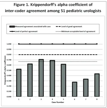
Significant Variability Exists in Coding Practices of American Pediatric Urologists Despite a Common Coding System Moira E. Dwyer, Steven G. Docimo, Heidi A. Stephany, Michael C. Ost,
Francis X. Schneck, Glenn M. Cannon, Jr.
Children’s Hospital of Pittsburgh of the University of Pittsburgh Medical Center, Pittsburgh, PA, USA.
Pittsburgh Medical Center, Pittsburgh, PA, USA.
Background: An increasing number of studies within the field of Pediatric
Urology are drawing conclusions based upon coding and billing data. Although the Current Procedural Terminology (CPT) codes used for surgi-cal billing are uniform throughout the nation, there is no mandatory or standardized training on the use of these codes. We hypothesized that variability exists in the coding practices of pediatric urologists.
Methods: The REDCap electronic data capture tool was used to send a
novel, anonymous survey to 108 pediatric urologists asking them to apply CPT codes to a variety of hypothetical pediatric urologic surgical cases according to their typical coding practice.
Results: A nationwide sample of 51 pediatric urologists (47%) responded.
The Krippendorff’s alpha coefficients for inter-coder agreement among the respondents are portrayed in Fig. 1. There was no association between the average or median number of codes that the pediatric urologists applied to a case and the level of inter-coder variability for that case (R -0.53, p=0.18 and R -0.57, p=0.13, respectively). Just over half of respondents (53%, 27) indicated that they do not think they have enough knowledge about billing and coding to code appropriately. Eighty-six percent (43) indicated that they had learned about coding on the job and were self-taught while 41% (21) reported having learned about coding in a formal fashion. Seventy-eight percent (40) felt it would be worthwhile to elimi-nate variability in coding practices among pediatric urologists.
Conclusions: Surgical coding practices among pediatric urologists have
P24
Assessment of the Unplanned Postoperative Morbidity in Children (UPMC) Scoring System for use in Pediatric Urology Moira E. Dwyer, Heidi A. Stephany, Glenn M. Cannon, Jr., Steven G.
Docimo, Francis X. Schneck, Michael C. Ost.
Children’s Hospital of Pittsburgh of the University of Pittsburgh Medical Center, Pittsburgh, PA, USA.
Background: Although use of the Clavien-Dindo Classification of Surgical
Complications is becoming more widespread within urology, this system was developed using a cohort of 6,336 adult general surgery patients. We proposed a novel Unplanned Postoperative Morbidity in Children (UPMC) scoring system to capture the global morbidity of unexpected post-surgical events on the child through surrogates for pain, traumatic experiences, and time away from home/school environments while accounting for the negative impacts of radiation exposure, general anesthesia, and short-term/lasting disabilities. We evaluated the acceptability and reproduc-ibility of this system and compared it to the Clavien-Dindo system for use in pediatric urology.
Methods: Replicating the methods of Dindo, Demartines, and Clavien,
we used the “Survey to Assess Acceptability and Reproducibility of the Classification” that had been sent to heads or senior surgeons of ten surgical departments with the request to distribute the survey to surgeons and trainees. We replaced the 14 adult surgical cases with 14 pediatric urology cases of the same Clavien-Dindo grade except for an additional disability suffix in two cases, and we added questions about
respon-dent demographics and the applicability of the classification systems to pediatrics. We used REDCap electronic data capture tool to send the survey to the 29 Pediatric Urology Fellowship Program Directors with a request to distribute to other pediatric urologists and trainees. Replies were anonymous.
Results: All respondents who completed the survey (20/20, 100%)
indi-cated that they think the UPMC scoring system is appropriate for pediat-rics and that it respects patient & hospital perspectives versus a respec-tive 50% (10/20; p=0.0004) and 63% (12/19; p=0.003) regarding the Clavien-Dindo system. The percentage of correct class assignment was significantly lower with the Clavien-Dindo system (79%, 229/291) than the UPMC scoring system (88%, 247/282; p<0.0001), where the percent-age did not significantly differ from that which was accepted in the origi-nal assessment of the Clavien-Dindo system (90%, 1816/2016; p=0.21). However, the inter-coder reliability was poor for the UPMC raw score (0.372), class (0.342), and death classification (0.120) (0.667=minimum acceptable level of agreement, 1.000=perfect agreement). Also, half of respondents (10/20) did not think the UPMC scoring system was simple.
Conclusions: While the proposed UPMC scoring system is uniformly
perceived as appropriate for pediatrics and is superior to the Clavien-Dindo system for accurate classification of morbidity in pediatric urol-ogy patients, it lacks inter-coder reliability. Efforts should be made to develop an appropriate system that is also user-friendly and has good inter-coder reliability.
P25
The Clavien-Dindo Classification of Surgical Complications is Not a Reproducible Way to Grade Morbidity in Pediatric Urology
Moira E. Dwyer, Heidi A. Stephany, Glenn M. Cannon, Jr., Steven G.
Docimo, Francis X. Schneck, Michael C. Ost.
Children’s Hospital of Pittsburgh of the University of Pittsburgh Medical Center, Pittsburgh, PA, USA.
Background: Although use of the Clavien-Dindo Classification of Surgical
Complications is becoming more widespread within urology, this system was developed using a cohort of 6,336 adult general surgery patients. We hypothesized that it is not well-suited to the pediatric urologic population.
Methods: Replicating the methods of Dindo, Demartines, and Clavien,
we used the “Survey to Assess Acceptability and Reproducibility of the Classification” that had been sent to heads or senior surgeons of ten surgical departments with the request to distribute the survey to surgeons and surgical trainees. We replaced the 14 adult surgical cases with 14 pediatric urology cases of the same Clavien-Dindo grade except for an added disability suffix in two cases, and we added questions about the applicability of the classification system to pediatrics and respondent demographics. We used REDCap electronic data capture tool to send the survey to the 29 Pediatric Urology Fellowship Program Directors in the United States and Canada with the same information about the Clavien-Dindo Classification that had accompanied the adult survey plus a request to distribute to other pediatric urologists and trainees. Replies were anonymous.
Results: We received 20 complete survey responses. Of complete
responders, 40% (8/20) had the position of Chairperson, Chief, or Program Director. Half (10/20) indicated that they think the Clavien-Dindo Classification is not appropriate for pediatrics. Also, 40% (8/20) indicated that they do not consider it useful for their patients versus only 10% (14/144) of the original adult surgical respondents (p=0.001). While 95% (19/20) of our respondents indicated that they think the Clavien-Dindo Classification is reproducible, the Kalpha coefficient for intercoder agreement was 0.416 for Clavien-Dindo grade and 0.086 for the disabil-ity suffix (0.667=minimum acceptable level of agreement, 0.800=good agreement, and 1.000=perfect agreement). The percentage of correctly graded pediatric urology cases was significantly lower than the percentage of correctly graded original adult surgical cases (Table 1).
Conclusions: The Clavien-Dindo Classification of Surgical Complications
is not a reproducible way to grade morbidity associated with pediatric urologic surgeries despite a perception that it is. Half of respondents to our survey think that this system is not appropriate for the pediatric population.
P26
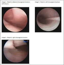
Previously Undescribed Intravaginal Structure Corresponds with Laterality of Dysgenetic Testis in Small Series of Patients with Gonadal Dysgenesis
Moira E. Dwyer¹, Selma F. Witchel¹, Svetlana A. Yatsenko², Francis X.
Schneck¹.
¹Children’s Hospital of Pittsburgh of the University of Pittsburgh Medical Center, Pittsburgh, PA, USA, ²University of Pittsburgh Medical Center, Pittsburgh, PA, USA.
Background: Endoscopic evaluation of the vagina at the time of
cysto-urethroscopy in pediatric patients with disorders of sex development
(DSD) is a safe and effective method to assess the lower genital tract. Vaginal anomalies have been recognized to exist as part of an non-linear spectrum. Findings have been categorized as congenital absence of the vagina, disorders of longitudinal fusion of the vagina, or disorders of transverse fusion of the vagina. The proposed formal classification is: (I) labial fusion, (II) distal urogenital sinus, (III) distal vaginal atresia & proxi-mal urethrovaginal fistula, and (IV) complete vaginal atresia. We report a series of three patients who had endoscopic identification of grossly similar intravaginal structures that have not been previously described and that do not fall under the proposed classification for vaginal anomalies.
Methods: All three patients underwent age-appropriate hormonal
evalua-tion, pelvic ultrasound, cystourethroscopy, endoscopic evaluation of the vagina, and laparoscopic abdominal exploration with bilateral gonadal biopsies or bilateral gonadectomies (Fig. 1).
Results: See Table 1.
Conclusions: We found grossly similar, heretofore undescribed vaginal
mucosal ridges in three patients with DSD who had common features: presence of a Y chromosome, cervical abnormalities, and, most interest-ingly, a dysgenetic testis corresponding to the side of the vaginal mucosal ridge. At present, the pathology of these structures and their developmen-tal etiologies are unknown to us. While it is possible that these structures resulted from a disorder of longitudinal fusion, the bilaterality in patient 1 makes this unlikely. Thus, the previous groupings used to describe vaginal anomalies do not readily accommodate this newly observed anomaly. We hypothesize that endoscopic identification of this ridge may be use-ful in predicting gonadal pathology prior to biopsy or gonadectomy in
Table 1. P25
Adult surgical cases correctly classified by adult surgeons & trainees
Pediatric urological cases correctly classified by pediatric urologists & trainees, grade
alone
Pediatric urological cases correctly classified by pediatric urologists & trainees, grade and
disability suffix 90%
(1816/2015)
79% (229/291)
p<0.0001
67% (174/259)
p<0.0001
the future. Evaluation of additional cases is necessary to determine if a significant correlation exists between the intravaginal structure and an ipsilateral dysgenetic gonad.
P27
Robotic Conversion of Cecostomy Tube to Malone Antegrade Continence Enema (ACE), Surgical Technique
Mourad Abouelleil, Jonathan Riddell.
Upstate, Syracuse, NY, USA.
Background: To our knowledge no previous report has described
conver-sion of cecostomy tube or a Mic-key button (usually in the cecum) to an antegrade continence enema (ACE). Several reports have described the procedure in both open and laparoscopic fashion using the appendix, but it has never been described as a conversion procedure. In general ACE is preferred over cecostomy in achieving continence, decreasing stool leakage and being free of appliances.
Methods: In this paper, we describe surgical techniques in converting
cecostomy to ACE laparoscopic robotic assisted.
Results: Outcomes have been satisfactory to this point in our small cohort
of three patients.
Conclusions: This approach appears to be simple and safe in achieving
the above listed goals. It carries minimal morbidity to patients and a quick recovery period.
P28
Comparison of Anesthesiologist-performed Transversus Abdominis Plane and Rectus Sheath Blocks to Surgeon-performed Local Site Injection for Postoperative Pain Management Following Minimally Invasive Pediatric Pyeloplasty
Kevin J. Rycyna, Matthew C. Ferroni, Moira E. Dwyer, Glenn M. Cannon,
Jr, Francis X. Schneck, Michael C. Ost, Steven G. Docimo, Heidi A. Stephany.
University of Pittsburgh Medical Center, Pittsburgh, PA, USA.
Background: Our institution has seen the adaptation of emerging
anes-thetic techniques for perioperative pain control, including the transversus abdominis plane (TAP) and rectus sheath (RS) blocks. The aim of our study was to compare the effectiveness of these techniques to traditional port site injection of local anesthetic in a population of patients undergoing minimally invasive pyeloplasty.
Methods: A retrospective chart review of all laparoscopic and
robotic-assisted laparoscopic pyeloplasties from January 1, 2009 to January 31, 2014 was performed. Inclusion criteria included a minimum age of 12 months at the time of surgery. Depending on the child’s age, either the Wong-Baker FACES pain rating scale or the Face Legs Arms Cry Consolability (FLACC) Scale data was collected to determine the maxi-mum pain score in the PACU and during the first 24 hours post-op, which were divided into the first and second 12 hour periods. All narcotics used in the first 24 hours were recorded and converted into parenteral morphine equivalents per kilogram. Pearson’s Chi-squared, Fisher’s Exact, and Kruskal-Wallis equality of populations rank tests were used for the analysis and a p-value of less than 0.05 was considered significant.
Results: There were a total of 143 cases and, of these, 31 had a TAP or
RS block and 92 had surgeon administered local site injection. There was
Table 1. P26. Characteristics of three patients with grossly similar previously undescribed intra-vaginal finding
Patient 1 2 3
Gender Assignment Female Female Female Karyotype 46,XY 46,XY 46,Y,dup(X)(p22.31p21.1) Disorder Testicular dysgenesis Testicular dysgenesis Mixed gonadal dysgenesis
Vaginal Finding
(Image 1)
Bilateral mucosal ridges with no visible os for cannulation Low confluence with urethra
into urogenital sinus
(Image 2)
Left mucosal ridge leading to cervix successfully cannulated with 0.035” wire
Low confluence with urethra into urogenital sinus
(Image 3)
Right mucosal ridge with distal suspected os that could not be cannulated with 0.035” wire
Cephalad tapering Gonadal Pathology Bilateral dysgenetic testes
Left dysgenetic testis Right focal mesothelial lining and
hydatid of Morgagni
Right dysgenetic testis with fallopian tube, epididymis, and vas
Left fibroadipose tissue Cervix Absent Duplicated Patulous, hypoplastic Uterus Atretic uterus Present Atretic uterus Phallus Normal female clitoris Prader III Prader II Perineal Opening(s) Prader IIISingle Prader IIISingle Prader IIDual
Hormone Profile
<1-month-old
Testosterone, total: <2.5 ng/dL Anti-Müllerian hormone: 2.60
ng/mL
Luteinizing hormone: 1.17 mIU/mL
Follicle-stimulating hormone: 7.47 mIU/mL
2-years-old
Testosterone, total: <2.5 ng/dL Anti-Müllerian hormone: 5.81 ng/mL
13-years-old
Testosterone, total: 21.0 ng/dL Luteinizing hormone: 11.18 mIU/mL*
Follicle-stimulating hormone: 16.17 mIU/mL*
*Normal ranges for age: Follicle-stimulating hormone:
0.04-10.80 mIU/mL
Luteinizing hormone: 0.87-9.16 mIU/ mL
no difference in gender ratios between the two groups, with 63/92 males in the local group vs 19/31 males in the TAP/RS block group (p=0.54). There was no difference in age between the local vs TAP/RS groups, with a median of 7.5 (95% CI 6.4, 9.2) years vs 10.4 (95% CI 5.6,14.1) years, respectively (p=0.285). There was no difference in maximum PACU pain score (2.2 vs 1.6, p=0.275) and maximum first (4.5 vs 4.9, p=0.457) or second (4.4 vs 3.7, p=0.173) twelve hour postoperative pain scores between the local and TAP/RS block groups, respectively. There was no difference in the total 24 hour parenteral morphine equivalent usage between the local and TAP/RS block groups, 25.97mg/kg vs 22.4 mg/kg (p=0.699), respectively.
Conclusions: There was no significant difference in subjective pain scores
or objective narcotic usage within the first 24 hours after minimally inva-sive pyeloplasty in our pediatric population. Ultimately, a prospective, randomized trial is needed to determine the utility of continuing to use the TAP or RS blocks over local site injection with pediatric minimally invasive pyeloplasty.
P29
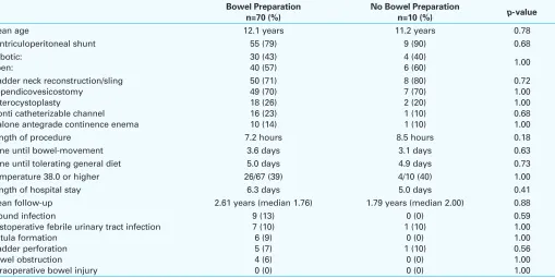
Preliminary Evaluation of the Impact of Bowel Preparation Prior to Reconstructive Urologic Surgery on Perioperative Measures & Postoperative Complications in Pediatric Myelomeningocele Patients with Neurogenic Bladder & Bowel
Moira E. Dwyer¹, Gwen M. Grimsby², Nicholas J. Farber³, Heidi A.
Stephany¹, Francis X. Schneck¹, Michael C. Ost¹, Patricio C. Gargollo², Micah A. Jacobs², Glenn M. Cannon, Jr.¹.
¹Children’s Hospital of Pittsburgh of the University of Pittsburgh Medical Center, Pittsburgh, PA, USA, ²Children’s Medical Center of Dallas, Dallas, TX, USA, ³University of Pittsburgh School of Medicine, Pittsburgh, PA, USA.
Background: The practice of bowel preparation had become a standard
for elective colorectal surgeries in the early 1970s with the intention of minimizing the risk of infections and complications through a reduction of the bacterial load associated with fecal matter. The actual benefits have been questioned in the last decade or so, and there have been limited reports in pediatric urology, where reconstructive procedures commonly involve the use of bowel. Investigations thus far have not isolated patients with neurogenic bowel as a select group, and the numbers of patients undergoing bladder neck reconstruction and the creation of catheterizable channels have been small. We sought to review our data to determine if bowel preparation for myelomeningocele patients prior to reconstruc-tive pediatric urologic surgery had impacted perioperareconstruc-tive measures or surgical complications.
Methods: We performed a multi-institutional retrospective chart review
on all patients ages 0 to 18 years with a history of myelomeningocele who underwent urologic surgery for management of neurogenic bladder from 2006 through 2013.
Results: See Table 1.
Conclusions: Although our cohort of pediatric patients with neurogenic
bowel due to myelomeningocele who had undergone reconstructive urologic surgery for management of their neurogenic bladder was small, we were able to compare a group of patients who had received bowel preparation with a group who had not. There was no significant increase in the rate of infection or bowel complications in the absence of a bowel preparation and, in fact, no wound infections or bowel events were observed in the group that had not received bowel preparation. This validates further study to better characterize the true risks and benefits of preoperative bowel preparation for these patients inclusive of study of additional factors such as patient morbidity and healthcare costs.
Table 1. P29. Comparison of perioperative measures and complications in the myelomeningocele population with and without bowel preparation at the time of reconstructive urologic surgery
Bowel Preparation n=70 (%)
No Bowel Preparation
n=10 (%) p-value
Mean age 12.1 years 11.2 years 0.78 Ventriculoperitoneal shunt 55 (79) 9 (90) 0.68 Robotic: Length of procedure 7.2 hours 8.5 hours 0.18 Time until bowel-movement 3.6 days 3.1 days 0.63 Time until tolerating general diet 5.0 days 4.9 days 0.73 Temperature 38.0 or higher 26/67 (39) 4/10 (40) 1.00 Length of hospital stay 6.3 days 5.0 days 0.41 Mean follow-up 2.61 years (median 1.76) 1.79 years (median 2.00) 0.88 Wound infection