Cite as: Can Urol Assoc J 2015;9(3-4):e114-21. http://dx.doi.org/10.5489/cuaj.2267 Published online March 11, 2015.
Abstract
Introduction: Partial bladder outlet obstruction (pBOO) is a
ubiqui-tous problem in urology. From posterior urethral valves to prostatic hypertrophy, pBOO results in significant morbidity and mortal-ity. However, the pathophysiology is not completely understood. Proteomics uses mass spectrometry to accurately quantify change in tissue protein concentration. Therefore, we have applied pro-teomic analysis to a rodent model to assess for protein changes after a surgically induced pBOO. We hypothesize that proteomic analy-sis after an acute obstruction will determine the most prevalent initial protein response and, potentially, novel molecular pathways.
Methods: Sprague Dawley rats underwent a surgically induced
pBOO (n = 3 per group) for 3, 7, or 14 days. Bladders were assessed for weight and urodynamic parameters. Proteomics used liquid-chromatography based mass spectrometry. Polymerase chain reac-tion (PCR) was performed on tissue samples to confirm increased mRNA transcription.
Results: Bladder weight and capacity increased over the
experi-mental period, but no changes were seen in bladder pressure. Statistically significant increases in protein quantities were seen in 3 proteins related to endoplasmic reticulum stress: GRP-78 (3.66-fold), RhoA (1.90-fold), and RhoA-GDP (1.95-fold), and 2 cytoskeleton molecules: actin (1.7-fold) and tubulin a/b (3.01-fold). Decorin and lumican, members of the small leucine rich proteoglycan (SLRP) family, were also elevated (0.35- and 0.34-fold, respectively). Real-time PCR data confirmed protein elevation.
Conclusion: Our experiment confirms that molecular changes
occur very soon after the initiation of pBOO, and implicates sev-eral molecular pathways. We believe these insights may provide insight into novel prevention and treatment strategies targeted at the pathophysiology of pBOO.
Introduction
Partial bladder outlet obstruction (pBOO) is a ubiquitous problem in urology, affecting the fetus with posterior urethral
valves, the child with spina bifida, the young adult with a urethral stricture, and the elderly male with prostatic hyper-trophy. However, despite significant medical and surgical advancements, pBOO still results in significant morbidity and mortality. Fundamentally, the deleterious consequences are not from the obstruction, as this is easily relieved, but from the secondary deterioration of the bladder. Multiple factors have been linked to this pathology, including inflammation, hypoxia, and remodeling of extracellular matrix (ECM) com-ponents.1,2 We have previously published results from our
animal model that demonstrates a programmed, progressive series of molecular and physical changes after pBOO. We described an initial period of inflammation, progressing to smooth muscle hypertrophy, and this eventually deteriorated into a poorly compliant, fibrotic bladder.3 We believe that
our model reasonably replicates the clinical scenarios seen with valve bladder syndrome and other obstructive states.
Mass spectrometry-based proteomic studies are used to identify and quantify the entire protein content of a cell, tis-sue, or organ.4 This is considered more sensitive and robust
than genomics, due to the dynamic state of protein expres-sion, depending on the physiological state of the biologic system. Because the amount of information can be over-whelming, most authors apply this technology to monitor changes in protein levels.
Herein, we explored the changes in protein levels of the urinary bladder over the first 14 days after pBOO, aiming to identify major changes in protein expression. This is an ear-lier time point than our previous experiments; therefore, we are examining the initial tissue response to the stress outlet obstruction. We hypothesize that proteomic analysis after an acute obstruction will determine the most prevalent protein response and, potentially, novel molecular pathways.
Methods
Approval from the University of Alberta Animal Care and Use Committee was obtained. Three un-obstructed Sprague Dawley rats were used as controls, and surgical induction of
Bader Alsaikhan, MD;
†Richard Fahlman, MD;
§Jie Ding, MD;
†Edward Tredget, MD;
¥Peter D. Metcalfe, MD, MSc, FRCSC
*†Division of Experimental Surgery, Department of Surgery, University of Alberta, Edmonton, AB; §Department of Biochemistry, University of Alberta, Edmonton, AB; ¥Division of Plastic Surgery, Department
of Surgery, University of Alberta, Edmonton, AB; *Division of Pediatric Surgery, Department of Surgery, University of Alberta, Edmonton, AB
pBOO was performed as previously described5 for 3, 7 and
14 days with 3 animals per time point. Main outcomes were bladder weight, urodynamic studies, and bladder proteomic analysis. Real-time polymerase chain reaction (RT-PCR) was performed to determine gene transcription.
Surgical induction of pBOO
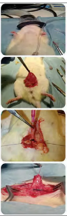
Isoflurane was used for anesthesia, and an 18-gauge angiocatheter was introduced into the bladder dome, and advanced into the urethra to serve as a stent for calibration. A 2-0 silk suture was tied outside the urethra, with the angio-catheter in place, to prevent complete obstruction. (Fig. 1).
Urodynamics and bladder weight
After the pre-determined time point, urodynamics were performed via cystotomy and the bladder filled with saline at 0.1 mL/minute. Bladder capacity and pressure (end-fill pressure) were recorded once urinary leakage was observed. Bladders were excised, dried, weighed, and fixed in 4% paraformaldehyde.
Protein extraction
In total, 200 mg of bladder was submerged in liquid nitro-gen, homogenized, and suspended in a cell lysis buffer. Supernatant was obtained and protein concentration was measured using the Bradford Bio-Rad protein assay (Bio-Rad, Hercules, CA) to yield a final concentration of 2 mg/mL for each sample. Bovine serum albumin was used as a standard.
Gel electrophoresis and staining
SDS-PAGE, 12% BisTris Criterion precast gels (Bio-Rad, Hercules, CA) were used. Molecular weight ladder was loaded into the first gel lane and 40 μL of each sample was loaded into the remaining lanes. Gels were run at 150V for 10 minutes, followed by 200V until the dye was seen at the bottom of the gel.
Liquid chromatography-mass spectrometry analysis
Each gel lane was cut into 10 regions, digested, and analyzed using liquid chromatography-mass spectrometry (LC-MS/MS) at the University of Alberta’s Institute of Biomolecular Design (IBD). The excised gels were de-stained twice in 100 mM ammonium bicarbonate (AmBic)/acetonitrile (ACN) (50:50). Samples were then reduced (10 mM β-mercaptoethanol 100 mM AmBic) and alkylated (55 mM iodoacetamide in 100 mM AmBic). After dehydration, trypsin (6 ng/uL) was added and digestion occurred overnight (about 16 hours) at room temperature. Tryptic peptides were extracted using
97% water/2% ACN/1% formic acid followed by a 1:1 mix-ture of extraction buffer and acetonitrile.
Fractions containing tryptic peptides were dissolved in aqueous 25% v/v ACN and 1% v/v formic acid and these were resolved and ionized by using nanoflow HPLC (Easy-nLC II, Thermo Scientific, Waltham, MA) coupled to the LTQ XL-Orbitrap hybrid mass spectrometer (Thermo Scientific). Peptide mixtures were injected onto the column at a flow rate of 3000 nL/min and resolved at 500 nL/min using 60-min linear ACN gradients from 0 to 45% v/v aqueous ACN in 0.2% v/v formic acid. The mass spectrometer was operated in data-dependent acquisition mode, recording high-accuracy and high-resolution survey Orbitrap spectra using external mass calibration, with a resolution of 60 000 and m/z range of 400 to 2000.
The 10 most intense multiply-charged ions were sequen-tially fragmented using collision-induced dissociation, and recorded in the linear ion trap. Data were processed using Proteome Discoverer 1.3 (Thermo Scientific) and the Rattus norvegicus protein database was searched using SEQUEST (Thermo Scientific). Search parameters included a precursor
mass tolerance of 10 ppm and a fragment mass tolerance of 0.8 Da, and spectral counting was used for quantification.6,7
Quantitative real-time polymerase chain reaction
RNA extraction was performed after homogenization and suspension in Trizol solution, using RNeasy spin columns (Qiagen, Mississauga, ON). DNA was digested for 60 min-utes, and random primers were used to synthesize first cDNA strand (Sigma, Oakville, ON). RT-PCR was then performed (Power SYBR Green PCR Master Mix, ABI, Foster City, CA). Hypoxanthine-guanine phosphoribosyltransferase (HPRT) gene was used as control. Amplifications were done using an ABI 7300 real-time system (Applied Biosystems, Foster City, CA). Table 1outlines primer sequences.
Statistical analysis
Only proteins that were present in all samples were used for statistical analysis. Data were reported as mean-fold change from normal control ± standard error, and analysis of variance (ANOVA) was performed (STATA Version 12.1). Statistical significance was set at p ≤ 0.05.
Results
All animals remained healthy, and there was no weight loss. pBOO resulted in an increase in bladder weight compared to unobstructed controls, with significant increases seen at 3 days (303.3 ± 24.0 mg vs. 200.7 ± 4.6 mg, p = 0.01), and 14 days (535.0 ± 82.5 mg vs. 200.7 ± 4.6 mg, p = 0.02).
Urodynamics
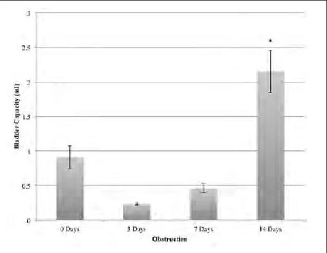
The bladder pressures recorded at the time of urethral leak-age (end-fill pressures) did not significantly change (Fig. 2). However, bladder capacity was significantly increased after 14 days (2.2 ± 0.3 vs. 0.9 ± 0.2 mL, p = 0.02) (Fig. 3).
Proteomic analysis
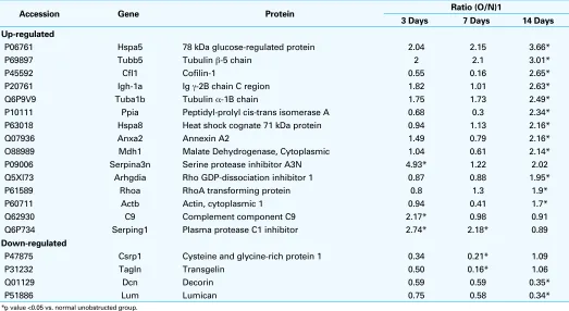
An average of 613 proteins per sample was identified ini-tially, and 140 proteins were detected in all samples. Table 2
outlines proteins with significant change from un-obstructed bladder tissue, and their fold-change from baseline in all groups. A total of 19 proteins had a statistically significant change from their baseline values. Several of these, includ-ing the cofilins, annexin, C9, serpinclud-ing1, Arhgdia, serpina3n, mdh1, Igh-1a, ppia, and csrp1, are of unknown metabolic significance.
We grouped the remaining molecules, which were felt to have relevant biologic roles, into markers of cellular stress, cytoskeleton molecules, and small leucine-rich proteogly-cans (SLRPs).
Markers of endoplasmic reticulum stress, GRP78 (3.66-fold increase), RhoA (1.90-(3.66-fold increase), and RhoA-GDP (1.95-fold increase) increased after 14 days of pBOO (all
p < 0.05). Cytoskeleton molecules, actin (1.7-fold) and tubulin-α/β (3.01-fold), also showed a statistically signifi-cant increase after 14 days. SLRPs decorin (0.35-fold) and lumican (0.34-fold) decreased after 14 days.
RT-PCR
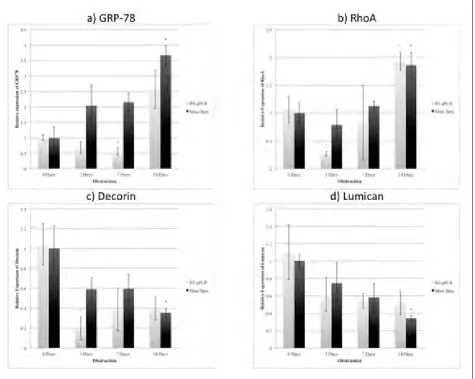
GRP78 mRNA was reduced after 7 days (0.56-fold), and then increased after 14 days (2.57-fold) (Fig. 4, part A). RhoA mRNA decreased in the 3-day group (0.27-fold) and then increased significantly after 14 days of pBOO (1.93-fold) (Fig. 4, part B) (all p < 0.05).
Down-regulation of decorin mRNA was significant in the 3-day group (0.20-fold) (Fig. 4, part C). Lumican mRNA changes, however, did not achieve statistical significance (Fig. 4, part D).
Discussion
The pathophysiologic progression from pBOO to bladder fibrosis remains a significant problem in urology, as these high-pressure, poorly compliant bladders result in inconti-nence, hydronephrosis, and eventually renal failure. Several causative factors have been identified, including mechanical strain, bladder wall hypoxia, and inflammation.1 Multiple
cytokines and pathways have been related to the aforemen-tioned stressors, including TGFβ, connecting tissue growth factor, HIF1α, and ECM proteoglycans.1,2 However, the exact
mechanisms and interplay have not been fully elucidated.
Table 1. List of PCR primer sequences
Gene Forward Sequence Reverse Sequence
GRP78 5’-CCAGCTTACTTCAATGATGCAC-3’ 5’-CTTCTCTCCCTCTCTCTTATCC-3’
RhoA 5’-AGGACCAGTTCCCAGAGGTT-3’ 5’-ACTATCAGGGCTGTCGATGG-3’
Decorin 5’-ACACCAACATAACTGCTATTCC-3’ 5’-AGACTGCCATTTTCCACAAC-3’
Lumican 5’-CACCAGAATGTAACTGTCCC-3’ 5’-TCAGCTTAGAGAAGACCTTTCC-3’
TGFβ1 5’-GAGGTGACCTGGGCACCAT-3’ 5’-GGCCATGAGGAGCAGGAA-3’
Fig. 2. Bladder pressure at end of filling, after 3,7, and 14 days of partial bladder outlet obstruction. There were no significant differences found between any of the bladder pressures.
Fig. 3. Bladder capacity for controls, and after 3,7, and 14 days of partial bladder outlet obstruction (pBOO). Rat baldders were significantly bigger after 14 days of pBOO. *p value <0.05 vs.
Mass spectrometry is an extremely powerful tool able to identify proteins and quantitate changes in concentra-tions. The proteins identified in this study can be grouped and related to cellular structure, inflammation, hypoxia and oxidative stress.
GRP78 and endoplasmic reticulum stress
GRP78 is a 70kDa heat shock protein, which is involved in protein folding in the endoplasmic reticulum (ER).1,2,8 During
states of stress, unfolded and misfolded proteins accumulate in the ER resulting in endoplasmic reticulum stress (ERS). This activates a quality control process known as the unfold-ed protein response (UPR),4,8 which is intended to restore the
proper folding of proteins, but may also trigger apoptosis.1,8
Our findings confirm the presence of ERS after 14 days of pBOO, evident by the increase in GRP78 protein levels.
ERS and UPR activation is seen in several fibrotic condi-tions, including heart failure, cystic fibrosis, renal and inter-stitial lung fibrosis.9-12 This has also been associated with
renal tubular apoptosis,13 and increased levels have been
used as a biomarker of cellular stress.5,14 In a rat model of
myocardial infarction, atorvastatin resulted in lower GRP78 and caspase-12 levels,15 and candesartan alleviated the ERS
response after ureteric obstruction.16 These findings suggest
an important role of ERS after pBOO. Further work
confirm-ing a causative role may lead to novel medical preventive strategies.
RhoA/RhoA kinase pathway
RhoA (Ras homolog transforming protein member A) increased significantly after 14 days of pBOO. Activation of RhoA results in increased contractility and cytoskeletal modulation.17 The RhoA/ROCK (RhoA Kinase) pathway is
involved in bladder smooth muscle contractility.18-20
Hypoxia and mechanical stress, two primary factors after pBOO, have been shown to activate RhoA/ROCK.21,22
Several studies have found that the increased contractility after pBOO23,24 can be improved with ROCK inhibition.23,25
Moreover, activation of RhoA/ROCK may further increase pathologic changes by reducing eNOS gene expression in vascular endothelium, reducing perfusion which will worsen hypoxia.26
Small leucine-rich proteoglycans
Small leucine-rich proteoglycans (SLRPs) are ECM proteogly-cans required for collagen fibrillogenesis.2,27 We detected a
significant reduction in decorin and lumican after 2 weeks of pBOO, consistent with a pro-fibrotic environment and deregulated collagen assembly.27
Table 2. Protein identification and quantification results, representing the ratio of obstructed to normal unobstructed groups and their fold change from normal unobstructed bladder tissue
Accession Gene Protein Ratio (O/N)1
3 Days 7 Days 14 Days
Up-regulated
P06761 Hspa5 78 kDa glucose-regulated protein 2.04 2.15 3.66*
P69897 Tubb5 Tubulin β-5 chain 2 2.1 3.01*
P45592 Cfl1 Cofilin-1 0.55 0.16 2.65*
P20761 Igh-1a Ig γ-2B chain C region 1.82 1.01 2.63*
Q6P9V9 Tuba1b Tubulin α-1B chain 1.75 1.73 2.49*
P10111 Ppia Peptidyl-prolyl cis-trans isomerase A 0.68 0.3 2.34*
P63018 Hspa8 Heat shock cognate 71 kDa protein 0.94 1.13 2.16*
Q07936 Anxa2 Annexin A2 1.49 0.79 2.16*
O88989 Mdh1 Malate Dehydrogenase, Cytoplasmic 1.04 0.61 2.14*
P09006 Serpina3n Serine protease inhibitor A3N 4.93* 1.22 2.02
Q5XI73 Arhgdia Rho GDP-dissociation inhibitor 1 0.87 0.88 1.95*
P61589 Rhoa RhoA transforming protein 0.8 1.3 1.9*
P60711 Actb Actin, cytoplasmic 1 0.94 0.41 1.7*
Q62930 C9 Complement component C9 2.17* 0.98 0.91
Q6P734 Serping1 Plasma protease C1 inhibitor 2.74* 2.18* 0.89
Down-regulated
P47875 Csrp1 Cysteine and glycine-rich protein 1 0.34 0.21* 1.09
P31232 Tagln Transgelin 0.50 0.16* 1.06
Q01129 Dcn Decorin 0.59 0.59 0.35*
P51886 Lum Lumican 0.75 0.58 0.34*
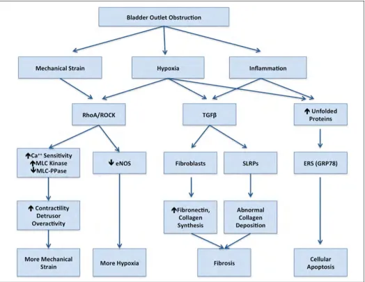
Figure 5 summarizes the changes in protein expression detected in this study, and their possible inducers, down-stream effects, and interactions after pBOO.
Although a very powerful tool, mass spectrometry-based proteomic analysis has its limitations, including extremes of molecular weight, protein length, peptide characteristics and the database selection. Furthermore, our technique relies upon several-fold increases in protein levels, and many mol-ecules exert their effect via post-translational changes and downstream mediators. Therefore, a small increase in an important upstream cytokine may not be detected, but the effects may be critical. Finally, the very acute time frame we studied in this manuscript may not reflect molecular pathways responsible for chronic changes.
Another fundamental concern is the question regarding the primary versus reactionary role of the proteins. We have
assumed that the physical stress of the acute pBOO creates a deliberate increase in the aforementioned protein levels, which results in downstream activation of the pathways. However, the increased concentrations may all be second-ary to an undetected mechanism.
Conclusion
The bladder damage secondary to pBOO is not completely understood. We have developed an animal model to further elucidate the primary factors involved, and this study uses proteomics to study the protein response very early after pBOO. Endoplasmic reticular stress (GRP-78), mechanical strain (Rho A/ ROCK), and inflammation (TGF-B) appear to be early, primary factors. We have confirmed that cellular and extracellular stress occurs very early after the initia-Fig. 4. A: Protein and mRNA expression of ER stress marker GRP78. B: Protein and mRNA expression of RhoA. C: Protein and mRNA expression of
tion of pBOO and that multiple interrelated pathways are involved. This work may lead to novel pharmacologic inter-ventions and reaffirms the need for early and aggressive care for our patients.
Acknowledgements: This work was supported by The Firefighters Fund, Edmonton Civic Employees, and an infrastructure grant from the Canadian Foundation for Innovation (CFI) to R.P.F.
Competing interests: Authors declare no competing financial or personal interests.
This paper has been peer-reviewed.
References
1. Metcalfe PD, Wang J, Jiao H, et al. Bladder outlet obstruction: Progression from inflammation to fibrosis.
BJU Int 2010;106:1686-94. http://dx.doi.org/10.1111/j.1464-410X.2010.09445.x 2. Maciejewski CC, Honardoust D, Tredget EE, et al. Differential expression of class 1 small leucine-rich
proteoglycans in an animal model of partial bladder outlet obstruction. J Urol 2012;188:1543-8. http:// dx.doi.org/10.1016/j.juro.2012.03.045
3. Metcalfe PD, Rink RC. Bladder augmentation: Complications in the pediatric population. Curr Urol Rep
2007;8:152-6. http://dx.doi.org/10.1007/s11934-007-0065-x
4. James P. Protein identification in the post-genome era: The rapid rise of proteomics. Q Rev Biophys
1997;30:279-331. http://dx.doi.org/10.1017/S0033583597003399
5. Sutherland RS, Baskin LS, Kogan BA, et al. Neuroanatomical changes in the rat bladder after bladder outlet obstruction. Br J Urol 1998;82:895-901. http://dx.doi.org/10.1046/j.1464-410X.1998.00873.x 6. Gao J, Opiteck GJ, Friedrichs MS, et al. Changes in the protein expression of yeast as a function of carbon
source. J Proteome Res 2003;2:643-9. http://dx.doi.org/10.1021/pr034038x
7. Liu H, Sadygov RG, Yates JR. A model for random sampling and estimation of relative protein abundance in shotgun proteomics. Anal Chem 2004;76:4193-201. http://dx.doi.org/10.1021/ac0498563
8. Brooks DA. Introduction: Molecular chaperones of the ER: Their role in protein folding and genetic disease.
Semin Cell Dev Biol 1999;10:441-2. http://dx.doi.org/10.1006/scdb.1999.0314
9. Dickhout JG, Carlisle RE, Austin RC. Interrelationship between cardiac hypertrophy, heart failure, and chronic kidney disease: Endoplasmic reticulum stress as a mediator of pathogenesis. Circ Res 2011;108:629-42. http://dx.doi.org/10.1161/CIRCRESAHA.110.226803
10. Dihazi H, Dihazi GH, Mueller C, et al. Proteomics characterization of cell model with renal fibrosis phenotype: Osmotic stress as fibrosis triggering factor. J Proteomics 2011;74:304-18. http://dx.doi. org/10.1016/j.jprot.2010.11.007
11. Baek HA, Kim DS, Park HS, et al. Involvement of endoplasmic reticulum stress in myofibroblastic dif-ferentiation of lung fibroblasts. Am J Respir Cell Mol Biol 2012;46:731-9. http://dx.doi.org/10.1165/ rcmb.2011-0121OC
12. Kerbiriou M, Le Drévo M-A, Férec C, et al. Coupling cystic fibrosis to endoplasmic reticulum stress: Differential role of Grp78 and ATF6. Biochim Biophys Acta 2007;1772:1236-49.
13. Zhang C, Gao J, Zhang L, et al. Effect of laminarin on the expression of GRP78 and GRP94 in rat after unilateral ureteral obstruction. Scand J Urol Nephrol 2012;46:267-72. http://dx.doi.org/10.3109/ 00365599.2012.663405
14. Liu T, Daniels CK, Cao S. Comprehensive review on the HSC70 functions, interactions with related molecules and involvement in clinical diseases and therapeutic potential. Pharmacol Ther 2012;136:354-74. http:// dx.doi.org/10.1016/j.pharmthera.2012.08.014
15. Song XJ, Yang CY, Liu B, et al. Atorvastatin inhibits myocardial cell apoptosis in a rat model with post-myocardial infarction heart failure by downregulating ER stress response. Int J Med Sci 2011;8:564-72. http://dx.doi.org/10.7150/ijms.8.564
16. Chiang C-K, Hsu S-P, Wu CT, et al. Endoplasmic reticulum stress implicated in the development of renal fibrosis. Mol Med 2011;17:1295-305. http://dx.doi.org/10.2119/molmed.2011.00131 17. Besson A, Assoian RK, Roberts JM, et al. Regulation of the cytoskeleton: an oncogenic function for CDK
inhibitors? Nat Rev Cancer 2004;4:948-55. http://dx.doi.org/10.1038/nrc1501
18. Wibberley A, Chen Z, Hu E, et al. Expression and functional role of Rho-kinase in rat urinary bladder smooth muscle. Br J Pharmacol 2003;138:757-66. http://dx.doi.org/10.1038/sj.bjp.0705109
19. Bing W, Chang S, Hypolite JA, et al. Obstruction-induced changes in urinary bladder smooth muscle contractility: A role for Rho kinase. Am J Physiol Renal Physiol 2003;285:F990-7.
20. Takahashi R, Nishimura J, Hirano K, et al. Ca2+ sensitization in contraction of human bladder smooth muscle. JURO 2004;172:748-52.
21. Wang Z, Jin N, Swartz DR, et al. Rho-kinase activation is involved in hypoxia-induced pulmonary vasocon-striction. Am J Respir Cell Mol Biol 2001;25:628-35. http://dx.doi.org/10.1165/ajrcmb.25.5.4461 22. Schiffrin EL, Touyz RM. From bedside to bench to bedside: Role of renin-angiotensin-aldosterone system
in remodeling of resistance arteries in hypertension. Am J Physiol Heart Circ Physiol 2004;287:H435-46. http://dx.doi.org/10.1152/ajpheart.00262.2004
23. Boberg L, Poljakovic M, Rahman A, et al. Role of Rho-kinase and protein kinase C during contraction of hypertrophic detrusor in mice with partial urinary bladder outlet obstruction. BJU Int 2012;109:132-40. http://dx.doi.org/10.1111/j.1464-410X.2011.10435.x
24. Burmeister D, AbouShwareb T, D’Agostino R Jr, et al. Impact of partial urethral obstruction on bladder function: Time-dependent changes and functional correlates of altered expression of Ca²+ signaling
regula-tors. AJP: Renal Physiology 2012;302:F1517-28.
25. Takahashi N, Shiomi H, Kushida N, et al. Obstruction alters muscarinic receptor-coupled RhoA/Rho-kinase pathway in the urinary bladder of the rat. Neurourol. Urodyn 2009;28:257-62. http://dx.doi. org/10.1002/nau.20625
26. Ming X-F, Viswambharan H, Barandier C, et al. Rho GTPase/Rho kinase negatively regulates endothelial nitric oxide synthase phosphorylation through the inhibition of protein kinase B/Akt in human endothelial cells. Mol Cell Biol 2002;22:8467-77. http://dx.doi.org/10.1128/MCB.22.24.8467-8477.2002 27. Iozzo RV. The biology of the small leucine-rich proteoglycans. J Biol Chem 1999;274:18843-6.