MP-8.01
Specific reduction of Fas-associated protein with death domain (FADD) in clear cell renal cell carcinoma
Kapoor A1,2, Tang D2, Khambati A1
1McMaster Institute of Urology, Hamilton, ON; 2Hamilton Centre for
Kidney Research (HCKR), Division of Nephrology, St. Joseph’s Healthcare, Hamilton, ON
Introduction and Objective:Most cancer therapeutic reagents induce apoptosis, and its attenuation thus contributes to treatment-resistance. Renal cell carcinoma (RCC) is known for its resistance to chemotherapy and radiotherapy. While apoptosis can be induced by a variety of stimuli, it is executed via 2 major pathways, death receptor (DR) or extrinsic path-way and mitochondrial or intrinsic pathpath-way. Fas-associated protein with death domain (FADD) plays a major role in the execution of apoptosis through the DR-dependent and independent pathways. One possibility for cancer to reduce DR mediated apoptosis is to compromise FADD’s pro-apoptotic function. To investigate this possibility, we compared FADD expression in normal and carcinoma kidney tissues of 85 RCC patients.
Materials and Methods:Immunohistochemistry was performed on 85 pairs of RCC and nontumour kidney tissues. Next, tissue lysate was prepared from these samples and Western blot was subsequently carried out to eval-uate the FADD expression. Using a densitometry software program (Scion Imaging for Windows), normalized FADD levels in RCCs were compared with the normalized FADD expressions in the respective nontumour kidney tissues to derive changes in FADD expression in RCC. Statistical analysis was performed using SPSS 10.0 for Windows
Results:We collected 85 pairs of RCCs, consisting of 62 clear cell RCCs (ccRCC), 17 papillary RCCs (pRCC) and 6 other RCCs, and their matched nontumour kidney tissues. Immunohistochemistry staining revealed that FADD was expressed largely in the cytosol of the proximal and distal tubu-lar epithelial cells with very faint staining occasionally seen in glomeruli. Western blot analysis suggested that in comparison to the normal kidney tissues, 65% (39/60) of ccRCC express reduced FADD, as their ratios of FADD (normal v. tumour) were larger than 1 fold, supporting that FADD is downregulated in ccRCC. Paired t test revealed that ccRCC expresses FADD at significantly lower levels than those in normal kidney tissues (p = 0.001).
Conclusion:Reduction of FADD in ccRCC might be an important mech-anism for ccRCC to evade apoptosis. This is consistent with our observa-tions showing for the first time that FADD is expressed in the epithelial cells of proximal and distal tubules, and that ccRCC expresses reduced FADD.
MP-8.02
Nonnarcotic analgesia in the treatment of renal colic: a systematic review of the literature and meta-analysis
Afshar K1,2, Marks A1, Jafari S1,2, Eftekhari A1, MacNeily A1
1Department of Urologic Sciences, University of British Columbia,
Vancouver, BC; 2Department of Health Care and Epidemiology, University
of British Columbia, Vancouver, BC
Introduction and Objective: This review aims to assess the benefits and harms of different nonnarcotic analgesics in patients with acute renal colic by using meta-analytic techniques on the available literature.
Materials and Methods: Our study protocol was designed according to the Cochrane Renal Group criteria for systematic reviews and published in the Cochrane database. Only randomized or quasi-randomized studies were included. Other inclusion criteria included adult patients with a diagno-sis of renal colic due to urolithiadiagno-sis; at least 1 treatment arm included a non-narcotic analgesic compared with placebo or another nonnon-narcotic drug, and a reporting of pain outcome or medication adverse effect. MEDLINE,
EMBASE, Cochrane Central Register of Controlled Trials and reference sec-tions of urology, nephrology and emergency medicine textbooks were sys-tematically searched. Abstracts were reviewed by at least 2 authors inde-pendently. Papers meeting the inclusion criteria were fully reviewed and relevant data were recorded in a standardized Cochrane Renal Group data collection form. For dichotomous outcomes relative risks and 95% con-fidence intervals were calculated. For continuous outcomes the weight-ed mean difference was estimatweight-ed. Both fixweight-ed and random models were used for meta-analysis. Heterogeneity was assessed by I2test.
Results:Eighty-one articles were fully reviewed with 39 meeting the inclu-sion criteria for analysis. Overall, 2294 patients receiving 21 different med-ications from 4 different classes of drugs were assessed in these studies. Nonsteroidal anti-inflammatory drugs (NSAIDs) were more effective than non-NSAIDs in relieving acute pain (RR 1.45, 95% CI 1.07–1.97). NSAIDS were more effective than placebo (RR 2.28, 95% CI 1.47–3.51). There was no significant difference between combinations of antispasmodics and NSAIDs versus NSAIDs alone (RR 0.98, 95% CI 0.81–1.19). Subgroup analysis did not show any difference in effectiveness of different types of NSAIDs. We identified a paucity of reports of adverse effects in the literature.
Conclusion: The effectiveness of NSAIDs in the treatment of renal colic is superior to non-NSAID, nonnarcotic analgesics. The addition of anti-spasmodics to NSAIDs does not improve the efficacy of pain relief. There is inadequate data on the adverse effects when these medications are used for renal colic.
MP-8.03
Modified surgical techniques minimize postoperative risk and improve male to female gender reassignment outcome Sievert K, Amend B, Kruck S, Stenzl A, Seibold J
University of Tuebingen, Department of Urology, Tuebingen, Germany
Introduction and Objective:Various techniques for neovaginal recon-struction in gender reassignment have been published in the past. The major problems after neovaginal reconstruction are vaginal stenosis and insufficient size resulting in patient dissatisfaction. In 2000 Perovic and colleagues described the incorporation of an excessive penile urethra into the neovaginal circumference. Our prospective patient follow-up inves-tigates the postoperative outcome of this technique. In addition, varying the surgical positions were analyzed to reduce the risk of compartment syndrome, thrombosis and peroneal paralysis caused by prolonged lithotomy position.
Materials and Methods: From October 2006 to October 2008, 9 patients underwent gender reassignment using the following technique: penile dis-assembly and orchiectomy in the supine position. The patient is then put in the lithotomy position and supplemented with pneumatic stockings. The corpora cavernosa is resected and neovaginal reconstruction is per-formed using the penile skin and urethral flap. Before the neovagina is inserted into the blund-disected rectoprostatic space and fixed to the sacrospinous ligament, the size-reduced vascularized and innervated glans is used to create the neoclitoris. The medial scrotal skin is used to form the labia majora and minora and the subcutaneous fat is moved to become the mons veneris. A unique approach to move the skin resulted in reduced skin incisions.
Results: In all 9 patients the cosmetic results in the external genitalia were excellent with only 2 almost invisible scars at the outer labia majora. In 2 patients, a minor correction of asymmetric skin of the labia majora was necessary. In all patients, a sensation in the neoclitoris was found. All patients had a satisfactory neovagina regarding depth and width and 8 patients of 9 reported normal sexual intercourse. One patient was
without partner. Mean operating time was 305 (270–405) minutes, of which the mean time in the lithotomy position was 185 minutes. Mean hospital-ization was 13 (9–18) days. In one patient, a small rectal lesion was closed during surgery that healed without problem. No blood transfusion was nec-essary. Using the 2-step positioning, no postoperative complications occurred, for example, compartment syndrome or peroneal paralysis.
Conclusion:The incorporation of vascularized distal urethra for the cre-ation of neovagina results in an optimal functional size regarding depth and width in addition to the glans use as the neoclitoris. The 2-step posi-tioning of the patients reduced operating time and postoperative com-plications were avoided. In most cases, gender reassignment from male to female could be achieved in one operation.
MP-8.04
Skeletal muscle induces human mesenchymal stem cells to differentiate into striated muscle in vivo
Baumann S1, Schäfer J1, Schäfer R2, Möhle R3, Feil G1, Stenzl A1, Sievert K1 1University of Tuebingen, Department of Urology, Tuebingen, Germany; 2Institute of Clinical and Experimental Transfusion Medicine, Tuebingen,
Germany; 3University of Tuebingen, Department of Medicine, Tuebingen,
Germany
Introduction and Objective:Since mesenchymal stem cells (MSCs) can be differentiated into smooth and skeletal muscle cells in vitro, autologous MSCs might be an option for a functional treatment of urinary inconti-nence. The aim was to study survival and myogenic differentiation of human MSCs in vivo in athymic rats.
Materials and Methods:Human MSCs were cultured from bone marrow aspirates. To induce myogenic differentiation in vitro, human MSCs were exposed to 5-azacytidine (AZA) in passage (P) 1. Native MSCs in P1 and P3 as well as AZA-exposed MSCs of subsequent P2 or P3 were directly injected into the rectus abdominis muscle of athymic nude rats. For in vivo tracking, MSCs were labelled with PKH26 red fluorescent cell linker. Integration and myogenic differentiation of MSCs in rat muscle tissue was histologically monitored from 4 days up to 16 weeks after cell injection. Immunohistochemistry was performed for the detection of the muscle mark-ers desmin and skeletal muscle myosin heavy chain with the specific antibodies clone D33 and clone NOQ7.54D, respectively.
Results:Native and AZA-exposed MSCs of all passages could be demon-strated in all animals investigated. Histology of animals in the short term experiments up to 8 days revealed well-defined clusters of transplanted MSCs (red PKH26 fluorescence) in the rectus abdominis muscle. After 4 and 8 weeks of cell injection, a continuous dissemination of trans-planted MSCs was detected. Histology of animals in the long-term study revealed PKH26-positive myofibres that were in parallel with the native skeletal muscle fibres. Immunohistochemistry for myogenic desmin and skeletal muscle myosin heavy chain demonstrated striated myofibres and skeletal muscle myosin in PKH26-positive myofibres.
Conclusion:This experimental athymic rat model demonstrates the poten-tial of bone marrow-derived human MSCs to differentiate into striated mus-cle. This is promising for a functional treatment of urinary incontinence based on autologous adult stem cells. Further in vivo studies to identify the functional innervation of newly developed striated muscle are required.
MP-8.05
Investigations on the biocompatibility of a new collagen-based matrix for urothelial implants
Maurer S1, Feil G1, Just L2, Krug J1, Kohler K3, Stenzl A1, Sievert K1 1University of Tuebingen, Department of Urology, Tuebingen, Germany; 2University of Tuebingen, Institute of Anatomy, Tuebingen, Germany; 3University of Tuebingen, Center For Regenerative Biology and Medicine,
Tuebingen, Germany
Introduction and Objective:Reconstructive surgery of the lower urinary tract often requires suitable grafts. Implantation of engineered tissue equiv-alents might be an option for urethral and ureteral reconstruction espe-cially in patients for whom autologous grafts are not available. Urothelial implants might require biomaterials as cell carriers. Matrices have to be biocompatible, induce tissue regeneration and must be subject to rapid
degradation in vivo. The aim of the study was to prove adherence, via-bility and growth pattern of human urothelial cells (HUCs) seeded on a new bovine collagen I–based matrix.
Materials and Methods:Ureter tissue specimens were obtained from adult patients undergoing open tumour surgery according to the ethics commit-tee approval. HUCs were isolated and labelled with the fluorescent cell linker PKH26, seeded onto the collagen matrix, and cultivated in serum-free medium. Cell adherence was indirectly ascertained by counting non-adherent cells in the culture supernatant. Growth behaviour was stud-ied by phase contrast microscopy and cryosections of the populated matrix. Viability of HUCs seeded onto the collagen matrix was analyzed with the WST-1 assay.
Results:HUCs grown on the collagen matrix were as homogeneously spread as HUCs seeded onto standard plastic surface. At day 1 after seeding the fraction of nonadherent HUCs was slightly increased (2.2%) compared with the controls (0.3%), whereas at day 3, both groups revealed similar rates (0.4% and 0.3%, respectively). Viability of HUCs growing on the matrix revealed 111% of the control group at day 3. The cell-matrix constructs could be easily detached from the culture dish and were manageable with surgical instruments.
Conclusion: The data demonstrate a good in vitro biocompatibility of the new bovine collagen I–based matrix. We conclude that the matrix might be well suitable for construction of urothelial cell-matrix implants for reconstructive ureteral and urethral surgery. Further experiments with urothelial multilayers grown on the matrix will be performed.
MP-8.06
Physician opinion of the privatization of health care services in Canada: a survey of Canadian urologists by the Canadian Urological Association Socioeconomic Committee
Mayson B1, Fleshner N2, So A1
1Prostate Centre, Department of Urologic Sciences, University of British
Columbia, Vancouver, BC; 2Divison of Urology, Department of Surgery,
University of Toronto, Toronto, ON
Introduction and Objective:Canadian health care policy faces unprece-dented pressures to reform. With new advances in health care technolo-gies and treatments, and increasing difficulties in obtaining timely access to necessary health care, the status quo is being challenged with the increased role of privately funded health care. In order to assess the opinions of Canadian urologists toward privatization of health care in Canada, the Socioeconomic Committee of the Canadian Urological Association surveyed all active members regarding their beliefs on the role and impact private health care should have in urology.
Materials and Methods:A short survey of 9 questions proposed by the Canadian Urological Association Socioeconomic Committee regarding private health care delivery was emailed to all active CUA members in April 2007. Responses were received by e-mail, fax or mail over a 1-month period.
Results:Of the 440 emails sent out, 90 were returned. The majority of respondents believed that a parallel private heath care system would short-en wait times and improve access to care (74%), improve outcomes for those with private health care (58.8%), not reduce the outcomes of those without private health care (74.2%), and not interfere with the accessi-bility of health care for most Canadians (73.3%). The majority of respon-dents (91.1%) felt that if a privately delivered health care was allowed, that urologists should spend a fixed amount of time providing services within the public health care system as well.
Conclusion: This survey of Canadian urologists regarding their beliefs on the role and impact private health care should have in urology indicated that the majority of respondents seem to anticipate a growing influence of private health care, and advocate for a regulated fixed proportion of service dedicated to the public system.
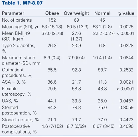
MP-8.07
Nunez R2, Bruhn A3, Altamar H4, Humphreys M2, Shah 03, Miller N4 1University of British Columbia, Vancouver, BC; 2Mayo Clinic, Scottsdale,
AZ, USA; 3New York University, New York, NY, USA; 4Vanderbilt
University, Nashville, TN, USA
Introduction and Objective: Surgical treatment of kidney stones remains a challenge in the obese population (BMI > 25 kg/m2) as shock wave
lithotripsy is not an option due to weight limitations. We sought to deter-mine the effectiveness of ureteroscopic lithotripsy in obese patients compared with nonobese controls from 4 centres with endourology-fellowship trained urologists.
Materials and Methods: Patients from 2004 to 2007 were retrospective-ly anaretrospective-lyzed providing a group of 266 patients who underwent uretero-scopic procedures for urolithiasis at 4 centres in the US and Canada.
Results:Of the 266 patients, 152 were obese (BMI ≥30 kg/m2), 69 were
overweight (BMI = 25–30), and 44 were normal controls (BMI < 25). There were significantly more patients in category ASA 3 in the obese and overweight groups than the normal group (p = 0.0021), and the only ASA 4 patients (n = 2) were in the obese group. The rate of diabetes was significantly higher in the obese and overweight groups (p = 0.0228). The percentage of obese patients requiring flexible ureteroscopy (79.6%) was higher than in the other groups (p < 0.0001). A slight significance was observed in the use of a ureteral access sheath between the groups (p = 0.0457). There was no difference in stone-free rates or number of patients who received postoperative stents. The complication rates did not differ between groups. In the obese group, postoperative complications included one episode each of MI and pneumonia, 2 strictures, 2 early stent insertions, 2 UTIs and 1 reintubation in the PACU (Table 1).
Conclusion: Ureteroscopic lithotripsy in the obese and overweight
pop-ulations produces the same stone-free rate as in the nonobese population. The complication rates are similar and postoperative stenting remain the same in all weight groups. Obese patients were more likely to require flexible ureteroscopy. Ureteroscopic laser lithotripsy is an effective and safe technique to treat urolithiasis in the overweight/obese patient.
MP-8.08
Anti-retropulsion devices increase stone fragmentation efficiency with holmium:YAG laser lithotripsy
Jonat L, Chew B, Paterson R, Teichman J
Stone Centre at Vancouver General Hospital, Department of Urologic Sciences, University of British Columbia, Vancouver, BC
Introduction and Objective:Stone fragmentation efficiency is proportion-al to the power applied with holmium:YAG laser lithotripsy; however, increased power leads to increased retropulsion that may result in high-er failure rates, prolonged procedures and necessitate lowhigh-er powhigh-er lithotrip-sy. We hypothesized that anti-retropulsion devices would increase stone fragmentation efficiency for a given amount of total energy by holding the stone in place during lithotripsy even at high power settings.
Materials and Methods: One hundred eighty UltraCal 30 stones of simi-lar size were divided into 6 groups, including a control group (no back-stop device), the 7-mm and 10-mm Stone Cone (Boston Scientific), the NTrap (Cook Urological), and the 7-mm and 10-mm Accordion (PercSys). Each group was further divided into 3 power setting groups: 0.5 J/10 Hz, 1 J/20 Hz, and 2 J/40 Hz to yield groups with sample sizes of 10. During each trial, a total of 500 J of energy was delivered. Stones were placed in a horizontal 8.5-mm diameter acrylic tube immersed in saline. The pri-mary end point was stone fragmentation efficiency as defined by stone mass change for a given unit of energy.
Results: At the lowest power setting, only the 7-mm Accordion and 10-mm Stone Cone increased stone fragmentation efficiency over control (p < 0.05). At the higher power settings (20 W and 80 W), all devices increased effi-ciency over controls (p < 0.001). Stone fragmentation effieffi-ciency was not improved with increasing power deployment in the control group. In all devices, however, stone fragmentation efficiency significantly increased as the power settings were raised. Stone retropulsion increased significantly with higher power settings in the absence of any devices (p < 0.01).
Conclusion:This trial demonstrates that anti-retropulsion devices increase stone fragmentation efficiency and facilitate high power lithotripsy com-pared with controls. No device was shown to be clearly superior to any other. Presumably, the improved efficiency was a result of preventing retropulsion and increasing the contact time between the laser fibre and stone. Migration of stone fragments was uncommon when using the devices and no fragments larger than 2 mm migrated past any device. These results may have application in the clinical setting in that it may decrease procedural times and increase stone-free rates.
MP-8.09
Discordance between ultrasound and computed tomography in the prediction of renal stone size
Ray A, Ghiculete D, Pace K, Honey R
St. Michael’s Hospital, University of Toronto, Toronto, ON
Introduction and Objective:Unenhanced helical computed tomogra-phy (CT) is the gold standard imaging modality for the assessment of uri-nary tract calculi. However, ultrasound (US) is often used as an adjunct, such as in the work-up of acute flank pain in the emergency setting, pregnant patients and the follow-up of urolithiasis. Despite a lack of evi-dence, it is commonly accepted in urological practice that US overesti-mates size in urolithiasis. Similarly, it is unknown whether patient obe-sity or stone location within the urinary tract contributes to measurement error.
Materials and Methods:We conducted a retrospective review of all patients at our institution undergoing both abdominal CT and renal US for soli-tary renal calculi since June 2004. Stones were identified on both CT and US in all cases. Imaging was conducted less than 1 month apart and patients could not have received treatment or passed their calculus in the interim period. Axial CT was considered the reference standard for determination of stone size.
Results: We identified a total of 71 calculi in 60 patients. The mean stone measurement on CT was 7.39 (standard deviation [SD] 4.4) mm. The mean interval between US and CT measurements was 9.59 (SD 9.5) days. For stones 5 mm or smaller, US measurements were a mean of 1.9 (SD 1.2) mm greater than CT (p < 0.001); for stones 5–10 mm, US meas-urements were 1.6 (SD 1.7) mm greater (p≤0.001); and for stones larg-er than 10 mm, US measurements wlarg-ere 2.1 (SD 4.0) mm greatlarg-er (p = 0.115). Compared with CT, US overestimated stone size, an effect that was progressively more pronounced with smaller calculi. The size Table 1. MP-8.07
Parameter Obese Overweight Normal p value No. of patients 152 69 45
Mean age (SD), yr 53 (15.18) 60.5 (13.3) 53.2 (2.8) 0.0025 Mean BMI 49
(SD), kg/m2
37.0 (2.78) 27.6 (1.27)
22.2 (0.27) < 0.0001
Type 2 diabetes, %
26.3 23.9 6.8 0.0228
Maximum stone diameter (SD), mm
8.9 (0.4) 7.9 (0.4) 10.4 (1.4) 0.0844
Outpatient procedures, %
85.5 92.8 88.7 0.2532
ASA = 3, % 36.6 21.7 11.3 0.0021 Flexible
uteroscopy, %
79.6 58.8 48.8 < 0.0001
UAS, % 44.1 33.3 25.0 0.0457 Stented
postoperation, %
84.2 78.3 75.0 0.8059
Stone-free rate, % 71.1 79.7 77.0 0.4423 Postoperative
complications, %
discrepancy was 49.7% for stones 5 mm or smaller, 23.7% for stones 5–10 mm and 14.9% for stones larger than 10 mm (p = 0.001). Discordance was associated with US measurement of skin-to-stone distance (p = 0.018), but not BMI (p = NS). Stone location in the urinary tract did not significantly affect stone measurement with US.
Conclusion: Ultrasound overestimates stone size in urolithiasis. The discordance in stone measurement varies with size and is greatest in stones 5 mm or smaller. SSD is an important determinant of error in US measurement of renal calculi.
MP-8.10
A randomized, double-blinded trial of ketorolac in addition to narcotic patient-controlled analgesia following laparoscopic donor nephrectomy
Pace K, Ghiculete D, Honey R St. Michael’s Hospital, Toronto, ON
Introduction and Objective:The ideal postoperative pain management regimen for laparoscopic donor nephrectomy (LapDN) has not been elu-cidated. The current standard for pain management after donor nephrec-tomy is patient-controlled analgesia (PCA). We sought to examine the impact of adding intravenous (IV) ketorolac to PCA following LapDN on postoperative recovery and quality of life (QOL).
Materials and Methods:Sixty-nine (of a preplanned sample size of 74) patients scheduled to undergo LapDN were enrolled and randomized. All patients underwent a transperitoneal, 3 trocar, LapDN performed by 2 sur-geons. All patients received intravenous narcotic PCA postoperatively, in addition to either 10 mg IV ketorolac (group 1) or saline q6h for 48 hours (group 2). Data were collected on analgesic usage, complications, bowel function, serum creatinine, length of stay, time to return to work, and QOL as assessed by the Postoperative Recovery Scale (PRS, a modification of the SF-36 that includes visual analog assessments of pain). Data were ana-lyzed with χ2, ANOVA, and repeated-measures ANCOVA where
appro-priate according to our preplanned blinded interim analysis.
Results:Sixty-nine patients: 42 were women (60.9%), with a mean age of 42.0 (SD 14.4) and mean BMI of 25.6 (SD 3.5) were enrolled between September 2003 and October 2008. There were no differences between the groups at baseline. Length of stay was similar (99.3 v. 101.2 hr, p = 0.75), but there was less PCA narcotic usage in group 1 (120.97 v. 149.52 mg mor-phine equivalent, p = 0.01). Quality of life scores were significantly high-er in group 1 throughout the immediate postophigh-erative phigh-eriod (p = 0.009). By 2 weeks and 3 months postoperatively, the difference in QOL scores between the 2 groups disappeared (p = 0.103). There were no differences in serum creatinine between the 2 groups (p = 0.215). Recipient outcomes were identical. Complications were rare: 1 patient in group 2 experienced an ileus; there were no conversions and no bleeding complications.
Conclusion:Ketorolac appears to improve QOL in the immediate postop-erative period, reduce morphine usage, improve pain control, and has no adverse impact on renal function in patients undergoing LapDN.
MP-8.11
The surgical personality: comparisons between urologists, nonurologists and nonsurgeons
MacNeily A1, Alden L2, Webber E3, Afshar K1,4
1Department of Urologic Sciences; 2Department of Psychology; 3Department of Surgery; 4Department of Clinical Epidemiology, University
of British Columbia, Vancouver, BC
Introduction and Objective: It is often perceived that the presence of a distinct surgical personality plays an important role in the selection process of surgical residents and may help predict successful completion of a surgical residency. We previously reported data demonstrating that urol-ogy residents possessed a distinct personality in comparison to the gen-eral population. The purpose of this study is to compare personality traits between urologists and nonurologist surgeons, as well as between surgeons and nonsurgeons.
Materials and Methods:Eighty-eight surgeons (57 faculty, 31 residents) completed the Revised NEO Personality Inventory (NEO-PI-R), a commer-cially available validated assessment tool of normal personality traits.
Participants agree or disagree to a compilation of 240 statements to gen-erate a score in each of 5 major character traits, according to the 5 fac-tor theory of personality: Extraversion (E), Openness (O), Conscientiousness (C), Agreeableness (A) and Neuroticism (N). Each factor is subdivided into 6 component facets. Scores for urology faculty and residents were pared with the other surgical groups. All surgeon scores were then com-bined into a single group for comparison to scores from 32 pediatricians (nonsurgeon group).
Results: Multivariate analysis of covariance controlling for age differences revealed no significant difference between urologists and other surgeons in any of the 5 personality factors. One-way ANOVA revealed that sur-geons scored significantly higher than nonsursur-geons on the Extraversion factor (p < 0.01). No significant differences emerged on Openness, Conscientiousness, Agreeableness or Neuroticism. Of the 6 extraversion facets, ANOVAs revealed significantly higher surgeon scores in Gregariousness (p < 0.05), Activity (p < 0.01), Excitement Seeking (p < 0.001), and Positive Emotions (p < 0.05). There were no significant differences in the facets of Warmth or Assertiveness.
Conclusion: Urologists do not appear to possess unique personality traits compared with other surgical specialists. The higher scores of surgeons compared with nonsurgeons in 4 facets of Extraversion reflects a prefer-ence for other people’s company, a rapid tempo and sense of energy with a need to keep busy while leading fast-paced lives and an enjoyment of excitement and stimulation, while maintaining an optimistic and high-spirited outlook. If these findings can be confirmed on a wider basis, the data may be helpful in resident selection, mentoring and evaluation.
MP-8.12
An interactive case-based learning tool in urological oncology for medical student education
Cox A1, MacLellan S2, Bell D 1, Rendon R1
1Department of Urology, Dalhousie University, Halifax, NS; 2Department
of Surgery, University of Toronto, Toronto, ON
Introduction and Objective:Oncology composes a large proportion of urological surgery. Medical learners are often present in the operating room for cases without being able to adequately view the anatomic struc-tures and surgical technique. This teaching module was created to pro-vide medical students with an approach to common urological oncolo-gy cases highlighting the anatomy, surgical approach and complications of common surgical procedures in urological oncology.
Materials and Methods:Adenocarcinoma of the prostate, urothelial cell carcinoma of the bladder and renal cell carcinoma were chosen as com-mon problems to be described in a case-based fashion. Patient consent was obtained. Photographs were taken intraoperatively during: an open radical retropubic prostatectomy, a transurethral resection of a bladder tumour, a cystectomy and ileal conduit, a neobladder diversion and a laparoscopic radical nephrectomy. Urological textbooks and journals were used to obtain relevant background information. Microsoft PowerPoint software was used to create the final interactive teaching module.
Results: Clinical scenarios are presented in an interactive manner for the viewer. Pertinent points on history and physical exam, as well as differential diagnoses, treatment options, and potential complications are presented for prostate cancer, urothelial cell carcinoma and renal cell carcinoma. The viewer is navigated through the relevant surgeries for each case as they are performed in the operating room, highlighting intraoperative anatomy.
Conclusion: This module will provide an educational aid for medical students and other observers of urological oncology surgery, including nursing staff. In addition, it will provide a reference for medical students throughout their clinical rotations. Lastly, this module will enhance the experience and exposure of medical students in the field of urology.
MP-8.13
Validation of real-time, intraoperative, surgical competence assessments linked to patient outcomes
Grober E1, Roberts M1, Mahdi M1, Jewett M2
of Toronto, Toronto, ON; University Health Network, Division of Urology, University of Toronto, Toronto, ON
Introduction and Objective: To determine if intraoperative evaluations of technical skill by expert surgeons using real-time intraoperative surgical competence (RISC) assessments, predicts clinical and operative outcomes in real patients.
Materials and Methods: Study design: a prospective analysis evaluating the predictive value of RISC assessments of technical skill in relation to clinically relevant patient outcomes. Study model: cystoscopic trans-urethral resection of bladder tumours (TURBT). Subjects include the fol-lowing: 1) Surgeons performing TURBT and 2) Patients identified at diag-nostic cystoscopy to have a bladder tumour requiring resection. RISC development: the RISC assessment tool was developed by study investi-gators following review of unedited surgical videos of 25 TURBT cases with tumour recurrence and 25 cases free of tumour recurrence following surgery. All surgical videos were analyzed by 3 expert surgeons with the goal to identify fundamental technical skill domains influencing a tumour recurrence/recurrence-free state. Twenty-five fundamental surgi-cal competency domains were identified and were used to create the RISC assessment instrument. The technical skill domains comprising the RISC assessment were then structured as a composite of previously validated global rating scales, surgical checklists and final product scores. Study intervention: live TURBTs (n = 160) will be recorded on video and prospec-tively evaluated in a blinded fashion by 3 expert surgeons as to the over-all technical quality of the tumour resection using the RISC assessment tool. Subjects will be followed for 18 months for signs of disease recur-rence or progression according to current standards of care. Bladder tumour recurrence (primary outcome), disease progression, operative complica-tions and need for further treatment will be correlated with RISC scores.
Results: The initial construct validity of the RISC assessment was tested on the first 50 TURBT cases. RISC scores discriminated between experienced and novice surgeons and correlated significantly with level of training and the number of previous surgical cases performed.
Conclusion: Lessons learned from this research will contribute significant-ly toward the objective assessment of technical skill in real operative settings, on real patients, based on clinically relevant patient outcomes. Similar methodology can be applied to develop RISC assessments for a variety of surgical procedures and disease states.
MP-8.14
The design and integration of a novel online problem-based learning resource to enhance undergraduate urology medical education
Wong W1, Butterfield M1, Koh K1, Chu K1, Clarke K1, Fraser T1, Moosaviasl S1,
Masterson J1,2, Goldenberg L1,2, Patterson L1,2
1University of British Columbia, Vancouver, BC; 2Urologic Sciences,
BC Children’s Hospital, Vancouver, BC
Introduction and Objective: To bridge the gap between preclinical and clinical education, the University of British Columbia (UBC) Department of Urologic Sciences created an online library of problem-based learning (PBL) modules to teach clinical approaches to common genitourinary con-ditions. The cases were published on the DiagnosisX server as a collabo-rative project between UBC, the Molson Medical Informatics Institute and McGill University with the aim of using this online resource as a new learning modality in medical education. At present the primary goal of this project is to investigate whether or not Web-based PBL cases can result in an immediate increase in the level of knowledge of the student.
Materials and Methods: The cases were written by medical students and reviewed by urology residents and faculty. DiagnosisX provides a tem-plate that allows for the creation of cases that simulate real-life patient encounters. Cases were developed in a pedagogical manner to provide a review of clinical presentation, patient assessment and treatment plans. Key features include the pre- and posttest questions. Both sets of questions are identical with the exception that the posttest questions are queried after case completion and the student is provided with the correct answers and an expert commentary.
Results: In April 2008, the FERGU (Fluids, Electrolytes, Renal, and Genitourinary) block of the UBC first-year medical curriculum was aug-mented with 4 online PBL cases as supplemental material. Data collected revealed that of the 35% of the class that accessed the cases, 40% found the cases useful as a self-learning tool to build on their understanding and for challenging their grasp of the concepts taught. The remaining 60% found the cases neutral as in they did not disagree with the benefits of the case.
Conclusion: Future plans include case enhancement and expansion and the promotion of DiagnosisX as a self-learning tool across all sites of the UBC distributed medical program. To demonstrate proof of knowledge acquisition upon case completion and thus lending support for the use of DiagnosisX as a new learning modality in medical education, a controlled study based on pre- and posttest scores is in consideration.
MP-8.15
Should peritoneal dialysis catheters be removed during kidney transplantation?
Warren J1, Drage M1, Taqi A1, Griffin S2, Watson C2, Luke P1
1Multi-Organ Transplant Unit, University Hospital, University of Western
Ontario, London, ON; 2Department of Transplantation, University of
Cambridge, Addenbrooke’s Hospital, Cambridge, UK
Introduction and Objective:Delayed graft function (DGF) following trans-plantation necessitates continued renal support. In patients undergoing peritoneal dialysis (PD) preoperatively, PD can be used for renal replace-ment therapy in the face of DGF. However, PD-related complication and failure rates are unknown. Herein we review our recent experience with PD catheter management in patients with DGF post–kidney transplantation.
Materials and Methods: All patients who were on PD at the time of kid-ney transplantation over a 3-year period at 2 separate institutions were ret-rospectively studied. Patients with retained PD catheters were compared with those in which it was removed at transplant.
Results: Of the 423 renal transplants performed during the study period, 137 received PD before transplantation. Of those 137 patients, only 19 had their PD catheters removed at the time of transplant. None of these patients required dialysis nor did they develop postoperative peritonitis. Of the remaining 118 patients, 103 had immediate graft function (89%). PD-related complications in this group included peritonitis (4), catheter related infections (2) and PD-related emergency laparotomy (1). Of the 15 patients requiring posttransplant PD, 5 developed peritonitis (33%) and 3 had fluid leaks (20%) necessitating initiation of hemodialysis. Overall, the practice of leaving a PD catheter in situ post–renal transplantation is associated with high rates of postoperative peritonitis (7.6%) compared with the rate of peritonitis if the PD is removed (0%, p = 0.09).