FORMULATION, OPTIMIZATION AND EVALUATION OF ENROFLOXACIN SOLID LIPID
NANOPARTICLES FOR SUSTAINED ORAL DELIVERY
SENTHIL KUMAR P
1*, ARIVUCHELVAN A
2, JAGADEESWARAN A
2, SUBRAMANIAN N
3, SENTHIL KUMAR C
3,
MEKALA P
21Department of Veterinary Pharmacology and Toxicology, Veterinary College and Research Institute, Orathanadu, Tamil Nadu, India. 2Department of Veterinary Pharmacology and Toxicology, Veterinary College and Research Institute, Namakkal, Tamil Nadu, India.
3Department of Pharmaceutical Technology, Anna University, Regional Centre, Tiruchirappalli, Tamil Nadu, India. Email: [email protected]
Received: 20 October 2014, Revised and Accepted: 10 November 2014
ABSTRACT
Objective: The objective of this study was to formulate and evaluate enrofloxacin solid lipid nanoparticles (SLNs) using a hot homogenization coupled with ultrasonication method for sustained oral delivery.
Methods: The SLNs were prepared using tripalmitin as lipid carrier, tween 80 and span 80 as surfactants and polyvinyl alcohol (PVA) as a stabilizer. The factors such as composition and concentration of lipid carrier and surfactant on the particle size were investigated to optimize the formulations. The optimized SLNs formulations were utilized to entrap enrofloxacin and characterized for particle size, polydispersity index (PDI), zeta potential (using dynamic light scattering), shape (using atomic force microscopy (AFM) and transmission electron microscopy [TEM]), drug encapsulation efficiency (EE) , loading capacity (LC) (using by dialysis and ultracentrifugation methods), and in vitro drug release (using by dialysis). The prepared SLNs were analyzed by Fourier transform infrared (FT-IR) spectroscopy to confirm the cross-linking reaction between drug, lipid and surfactants.
Results: The results demonstrated that the particle size, PDI, zeta potential, EE and LC of the enrofloxacin SLNs were 154.72±6.11 nm, 0.42±0.11,
−28.83±0.60 mV, 59.66±3.22% and 6.13±0.32%, respectively. TEM and AFM images showed spherical to circular particles with well-defined periphery.
In vitro drug release exhibited biphasic pattern with an initial burst release of 18% within 2 hrs, followed by sustained release over 96 hrs. FT-IR
study suggested that during the process of formulations, lipid and surfactants have not reacted with the drug to give rise to reactant products and it was only physical mixture.
Conclusion: The results indicated that SLNs might be a promising delivery system to prolong and enhance the pharmacological activity of enrofloxacin. Keywords: Enrofloxacin, Solid lipid nanoparticle, Hot homogenization, Tripalmitin, Atomic force microscopy.
INTRODUCTION
Enrofloxacin is a fluroquionolone antimicrobial agent developed solely for use in animals. It has potent bactericidal activity against a range of clinically relevant Gram-negative and Gram-positive pathogens as well as Mycoplasma and Chlamydiae. Enrofloxacin and its active metabolite ciprofloxacin possess high bactericidal activity, killing the bacteria in a concentration dependent manner. The relative safety of enrofloxacin, its low minimum inhibitory concentrations, broad spectrum of activity, long post-antibiotic effect and good tolerance has encouraged their use in veterinary medicine [1].
Despite the therapeutic potential of enrofloxacin, the very poor aqueous solubility of enrofloxacin leads to difficulty in designs of pharmaceutical formulation and variations in bioavailability [2]. In addition, all the oral enrofloxacin formulations are available as conventional, immediate-release form that necessitates administration twice daily for several days or weeks [3]. Numerous efforts have been made to develop alternative formulations of enrofloxacin to reduce frequency of administration.
Nanoparticle-based drug delivery systems have considerable potential in improving the bioavailability of the drug and as well reducing the dosing frequency. The solid lipid nanoparticles (SLNs)
introduced in 1991, are the forefront of the rapidly developing field
of nanotechnology, which is the most effective lipid based colloidal carriers system. They are submicron-sized (50-1000 nm) carriers composed of a lipid matrix stabilized by a surfactant. SLNs possess good tolerability, stability, scaling up feasibility and the ability to incorporate
hydrophilic/hydrophobic drugs [4]. The incorporation of poorly soluble drugs into SLNs can enhance gastrointestinal solubilization, absorption, and bioavailability of drugs [5]. Further, SLNs formulation has the ability to prolong, extend or sustain the release profile of the loaded molecules and hence reduce need for the repeated administration and increase the therapeutic value of the treatment [6].
Hence, the objective of this study is to formulate enrofloxacin SLNs with high loading capacity (LC) and sustained release profile using a hot homogenization and ultrasonication method.
METHODS
Drugs and chemicals
Enrofloxacin purchased from Himedia Laboratories Pvt. Ltd., India was used in this study. Tripalmitin (glyceryl palmitate), span 80 (polysorbate), tween 80 (sorbitate monooleate) and polyvinyl alcohol (PVA) procured from Sigma Aldrich Chemicals Pvt. Ltd., USA were utilized for the study. Dialysis membrane procured from Himedia Laboratories Pvt. Ltd., India was used. All other chemicals and solvents were analytical reagent grade and were used without further purification.
Formulation optimization
The preliminary studies were carried out by preparing various blank SLNs formulations (Table 1) with different variables in order to optimize the particle size fit into nanoscale.
• Hot homogenization, followed by ultrasonication technique was
selected to prepare blank SLNs
homogenization pressure, ultrasonication time and stirring speed
have been defined in previous study, the amount of lipid, and type
and concentration of surfactant was evaluated as variable
• The blank SLNs formulations were categorized into four sets,
three formulations in each category namely, formulations based on lipid concentration, secondly, formulations based on surfactant concentrations and third, formulations based on combination of surfactant
• The lipid tripalmitin was used in the concentration ranged from 0.5 to 1.5%
• The surfactants (span 80 and tween 80) were used in the concentration of 2-4%
• The combinations of surfactant (span 80 and tween 80) were used in the ratio of 1:1 (2%)
• Based on particle size, as an evaluation index (100-200 nm - Optimum
size for oral administration), the optimum blank SLNs formulations
were selected and utilized to entrap enrofloxacin.
Procedures for the preparation of enrofloxacin SLNs
Enrofloxacin SLNs were prepared by hot homogenization, followed
by ultrasonication method. Enrofloxacin (0.1% w/w), tripalmitin (0.5% w/w), span 80 (2% w/w) were added together to get organic phase
of preparation. The lipid content in the organic phase was melted by heating at 70°C using magnetic stirrer with hot plate. The contents in the organic phase were mixed properly by placing in the shaker (Spinix). An aqueous phase was prepared by dissolving hydrophilic surfactant tween
80 (2% w/w) and PVA (2% w/w) by heating to the same temperature as
the organic phase. The hot aqueous phase was added to the organic phase under magnetic stirring (Remi, Mumbai, India) at 1000 rpm to form pre-emulsion. The hot pre-emulsion was then homogenized at 10,000 psi
for 3 minutes using the high pressure homogenizer (Heidolph Electro,
Germany) kept in a water bath maintained at 70°C.
The hot emulsion so obtained was ultrasonicated (Sonics Vibra Cell, USA) using high-intensity (5/64’’ 2 mm tip diameter) microprobe
with amplitude 20% for 15 minutes to form nanoemulsion. Then, the
nanoemulsion was run under magnetic stirring at 1000 rpm for 4 hrs to obtain enrofloxacin loaded tripalmitin SLNs.
All the batches were prepared in triplicate and the average size was measured.
Characterization of enrofloxacin SLNs
Determination of particle size, polydispersity index (PDI) and zeta potential
Particle size and PDI of enrofloxacin SLNs were measured by photon correlation spectroscopy (PCS) using zetasizer nanoZS with the Malvern
PCS software version 6.20 (Malvern Instruments, UK). The aqueous SLNs dispersions were diluted with distilled water appropriately before analysis. Each value was the average of three measurements.
The zeta potential or the charge on the surface of colloidal particles in a liquid enrofloxacin nano suspension was measured by electrophoretic light scattering mode using zetasizer nanoZS. The particle charge of enrofloxacin SLNs were quantified at 25°C. The samples were diluted appropriately with the de-ionized water for the measurements of particle size. Each value was the average of three measurements.
Surface morphology
Transmission electron microscopy (TEM)
Surface morphology and shape of the enrofloxacin SLNs were examined individually using TEM (Philips, Tecrai10, Dutch). The enrofloxacin nanosuspension was dropped on copper grids, natively stained by phosphotungstic acid and tried at room temperature.
Atomic force microscopy (AFM)
The morphology of enrofloxacin SLNs was also analyzed using an AFM
(PARK XE-100). Briefly, 1 mL of enrofloxacin SLNs and acetone (1 mL)
were mixed. From the mixture, 10 µL was dispersed in a freshly cleaved mica substrate. After drying at room temperature, imaging of the samples were performed in non-contact mode with pyramidal silicon nitride tips.
Determination of LC and encapsulation efficiency
To determine the entrapment of enrofloxacin in the SLNs, 0.1 mL of
freshly prepared nanoemulsion was taken and diluted with 9.9 mL
chloroform. The obtained suspension was centrifuged for 45 minutes at 6000 rpm. The supernatant was separated and filtered through 0.2 µm filter. The filtrate was diluted using chloroform and analyzed
at 273.8 nm using ultraviolet (UV) spectrophotometer. The SLNs
formulated without enrofloxacin were treated similarly and used as
control for the measurements. The assay was repeated 3 times using
different preparations. LC and encapsulation efficiency were calculated as shown below:
Loading capacity Weight of enrofloxacin in SLNs Weight of S
=
L
LNs ×100%
Encapsulation efficiency Weight of enrofloxacin in SLNs Wei
= g
ght of enrofloxacin added ×100%
In vitro release studies
In vitro release of enrofloxacin SLNs and native enrofloxacin was performed by dialysis bag diffusion technique over a period of 120 hrs. Enrofloxacin nanosuspension equivalent to 5 mg of enrofloxacin was filled in dialysis bag (Himedia Laboratory Pvt. Ltd, India). The receiver solution containing 100 mL of phosphate buffer with
pH 6.7 was prepared and heated to 37°C under magnetic stirring
at a speed of 100 rpm. The drug containing dialysis bag (molecular weight 12-14 kDa, pore size 2.4 nm) was dialyzed against receiver compartment. To determine the enrofloxaxin diffused through the dialysis bag, 2 mL samples were withdrawn at regular intervals (0, 5,
10, 20, 30, 45, 60, 90 minutes, and 2, 4, 8, 12, 18, 24, 36, 72, 96 and
120 hrs) from the receiver solution and same amount of fresh receiver solution was added to maintain the volume constant. Enrofloxacin in
the samples was measured spectrophotometrically at 273.8 nm using a UV spectrophotometer (Systronics 2203 Smart, India). The control
nanoparticles without enrofloxacin were treated similarly and used as blanks for the measurements.
Compatibility studies using Fourier transform infrared (FT-IR) spectroscopic analysis
FT-IR spectral measurement for pure enrofloxacin, tripalmitin, span 80, tween 80, PVA and formulation were analyzed separately and then correlated for compatibility. In the present study, potassium bromide Table 1: Formulation design of blank SLNs by hot
homogenization and ultrasonication method
Formulation (%) Lipid (%) Tween 80 Span 80 PVA
F1 0.5 - 2 2
(KBr) pellet method was employed. A small drop of sample was placed on one of the KBr plates. The second KBr plate was placed on the top
and made a quarter turn to obtain an even film. Then, the plates were kept on the sample holder to run a spectrum.
Statistical analysis
The data obtained on particle size, PDI, zeta potential, LC and encapsulation efficiency were analyzed using a Statistical Package for Social Sciences (SPSS 11.00) [7]. All values are expressed as their mean±standard deviation (SD).
RESULTS
Formulation optimization
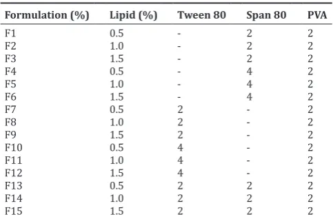
Various blank SLNs formulations were prepared on the basis of individual factors and the results are presented in Table 2. In all formulations, the mean±SD particle size was found within a range of
150.67±40.07-997.67±76.05 nm. Based on the comparison of visual clarity and particle size, the formulation F4, F10 and F13 having the mean±SD particle size from 150.67±40.07 to 229.67±57.57nm were
considered for enrofloxacin incorporation.
After encapsulation of enrofloxacin in the selected blank SLNs (F4, F10
and F13), mean±SD particle size, PDI, zeta potential, entrapment
efficiency (EE) and LC were evaluated and are presented in Table 3.
All the enrofloxacin SLNs formulations had shown nanosize range of
154.717-238.33 nm. From these studies, most efficient formulation of F13 with the particle size, PDI, zeta potential, EE and LC of 154.717±6.149 nm, 0.422±0.109-28.83±0.603 mV, 58.33±3.51 and 6.03±0.97, respectively were considered as final preparation.
Characterization of enrofloxacin SLNs
Particle size, PDI and zeta potential
The mean±SD particle size, PDI and zeta potential of the formulations
(F13) are given in Table 3.
Surface morphology
Transmission electron microscopic studies revealed that the enrofloxacin SLNs were spherical in shape (Fig. 1). In general, the particle size was with a diameter of <200 nm. These observations are consistent with PCS data of enrofloxacin SLNs recorded in this study.
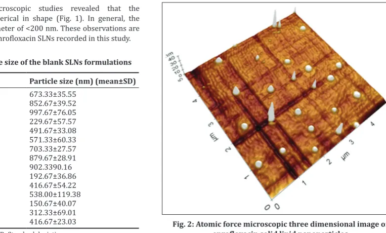
Atomic force microscopic analysis showed that the enrofloxacin SLNs were spherical and circular in shape (Fig. 2). The average sizes of SLNs were <200 nm. The particles were well dispersed with good particle size distribution. The surfaces of the nanoparticle were smooth.
Drug LC and encapsulation efficiency
The mean±SD encapsulation efficiency and LC of enrofloxacin SLNs
(F13) are presented in Table 3.
In vitro release studies
In vitro release of enrofloxacin from SLNs formulation and native enrofloxacin is illustrated in Fig. 3. The release curve of enrofloxacin
SLNs exhibited a biphasic pattern. There was an initial burst release
with about 39.23% drug released within the initial 24 hrs, followed by a
slow and sustained release. The amount of cumulated drug release over
96 hrs was 51.1%. In the native enrofloxacin, the release was 93.67% within 2 hrs and reached 100% by 24 hrs.
Table 2: Mean±SD particle size of the blank SLNs formulations
Formulation Particle size (nm) (mean±SD)
F1 673.33±35.55
F2 852.67±39.52
F3 997.67±76.05
F4 229.67±57.57
F5 491.67±33.08
F6 571.33±60.33
F7 703.33±27.57
F8 879.67±28.91
F9 902.3390.16
F10 192.67±36.86
F11 416.67±54.22
F12 538.00±119.38
F13 150.67±40.07
F14 312.33±69.01
F15 416.67±23.03
SLNs: Solid lipid nanoparticles, SD: Standard deviation
Table 3: Mean±SD particle size, PDI, zeta potential, EE and LC of selected enrofloxacin SLNs formulations
Formulations Particle size (nm) PDI Zeta potential (mV) EE (%) LC (%)
F4 238.33±30.98 0.59±0.072 −29.33±1.42 49.33±3.79 4.60±1.47 F10 195.67±6.81 0.55±0.05 −30.67±3.05 50.67±7.64 5.03±0.95 F13 154.717±6.149 0.422±0.109 −28.83±0.603 58.33±3.51 6.03±0.97
SD: Standard deviation, SLNs: Solid lipid nanoparticles, PDI: Polydispersity index, EE: Entrapment efficiency, LC: Loading capacity
Fig. 1: Transmission electron microscopic image of enrofloxacin solid lipid nanoparticles
Compatibility studies using FT-IR spectroscopic analysis
The FT-IR spectra of drug, tripalmitin, span 80, tween 80 PVA and formulation were exhibited the peaks of specific functional groups at their respective frequencies as presented in Fig. 4.
DISCUSSION
Formulation optimization
Hot homogenization followed by ultrasonicationwas reported to be an economic, simple, reproducible and most reliable method for the preparation of SLNs. In this method, the preparation of SLNs does not require any organic solvents, which could be difficult to remove after nanoparticle synthesis [8]. By this method, it is possible to
scale up to industrial level. Hence, hot homogenization coupled with ultrasonication method was employed in the present study to formulate SLNs.
Lipids must be selected based on their ability to solubulize the drug [9]. The lipid tripalmitin had high dissolution of enrofloxacin. The solubulizing potential along with already reported biocompatibility and
acceptability of lipid tripalmitin for oral route has favored its selection for the present study. In the present study, as the lipid concentration
increased from 0.5 to 1.5%, the mean particle sizes and particle size
distribution also increased, which is in agreement with Müller et al. [4] and Westesen et al. [9] higher concentration of lipid content increased the viscosity of the lipid dispersion which affected the homogenization efficiency and increased rate of particle agglomeration, hence, the lipid
content of the SLNs dispersion should not exceed 5% [4]. The inclusive
level of tripalmitin in this study was 0.5% which is well below the
recommended rate.
According to Souto and Müller [10], the right selection of surfactant with proper concentration was required to prepare physicochemically stable lipid nanoparticles. In the current study, the SLNs dispersion
stabilized by the combination of surfactant (2% span 80 and 2%
tween 80) had smaller particle size when compared with formulation stabilized by single surfactant. These findings are in accordance with Mehnert and Mäder [6] who had lower particle sizes and better stability when stabilized the formulation with surfactant mixtures compared with formulations with only one surfactant. Combination of surfactants prevented particle agglomeration more efficiently and also reduced the particle size of the SLNs [10]. In the present study, the mean particle size of SLNs tended to decrease with increase in the surfactant concentration. This observations are in agreement with the
findings of Bunjes et al. [11] and Lippacher et al. [12].
In the current study, 2% PVA was used to stabilize the SLNs formulation.
According to DeMerlis and Schoneker [13], PVA is the most commonly used emulsifier in the formulation of nanoparticles due to its excellent mechanical strength, biocompatibility and nontoxicity, and has been approved by the US FDA for medical and food applications.
From the optimization study, the suitable blank SLNs formulation
containing 0.1% tripalmitin, 2% span 80, 2% tween 80 and 2% PVA
were considered for further incorporation of drug.
Formulation of enrofloxacin SLNs
The blank SLNs selected in the optimization study were used to
entrap 0.1% enrofloxacin using a hot homogenization coupled with
ultrasonication method. According to Muller et al. [4], high temperature was performed in hot homogenization technique and thus, this method could not be used for temperature sensitive drugs. In this study, the Fig. 3: In vitro release of native enrofloxacin and enrofloxacin solid
lipid nanoparticles (mean±standard deviation, n=3)
temperature for the preparation of SLNs did not exceed the melting
point of enrofloxacin (219-233°C), hence the stability and antibacterial
activity will be maintained.
Homogenizationfollowed by ultrasonication technique applies high shear stress disrupting lipid particles down to the submicron range. According to Schwarz et al. [14], a sufficient high-energy input was necessary to break down the droplets into the nanometer range. A high energy such as high production temperature, high stirring rate, longer emulsification time and stronger ultrasound power were applied in this study to obtain a finer dispersion of formulation. In the present study,
the homogenization pressure 10,000 psi was applied for 3 minutes
and followed by ultrasonication resulted the mean±SD particle size of
154.717±6.149 nm with narrow size distribution. The result suggests
that the hot homogenization and ultrasonication method was a feasible and compatible method for preparing enrofloxacin loaded tripalmitin SLNs.
Characterization of enrofloxacin SLNs
The loading of drug with the blank SLNs in the present study resulted in a slight increase in the mean±standard error (SE) particle sizes from 150.67±40.07 to 154.72±6.15 nm. These findings are in consistent with Jensen et al. [15] who explained that the increase in size of SLNs after incorporation of drug reflected the dissolution of the drug in the lipid phase.
A narrow particle size distribution was an indication of nanoparticles stability and homogeneous dispersion [16]. PDI values ranging from 0 to 0.5 were considered to be monodisperse and homogenous, but those of more than 0.5 indicated non-homogenity and polydispersity [8,17]. In the present study, the particle size distribution was monodisperse and homogenous as formulation has less mean±SE PDI of 0.42±0.11.
According to Schwarz and Mehnert [18] and Zimmermann et al. [19], the negative charge of zeta potential was conferred by the lipids used in the SLNs. In agreement with this, the tripalmitin utilized in this study provided negative charge of zeta potential. Nanoparticle with zeta
potential values >+25 mV or <−25 mV typically have high degrees of
stability due to electric repulsion between particles. Dispersions with a low zeta potential value will aggregate due to Van Der Waal inter-particle attraction [4]. In this study, the mean±SD zeta potential of −24.90±1.00
mV was recorded and it could provide proper stability to the enrofloxacin SLNs. According to Srinivas and Sagar [20], the zeta potential with negative charge might not interfere in the absorption of the formulation.
TEM and AFM images revealed spherical and circular in shape with the presence of some particle aggregates. The presence of aggregates might be due to redistribution of particles after preparation. The images represented that the particles were ranging from 100 to 200 nm and well dispersed with smooth surfaces.
The enrofloxacin SLNs obtained in the present study had relatively
medium drug EE (59.67%). This could be attributed to the
physicochemical properties of the drug, most importantly, its lipophilic nature [21]. To get sufficient LC, the drug should have sufficiently high solubility in the lipid melt. The crystallization habits of tripalmitin nanoparticles also varied with the quantity of drug incorporated [22]. High temperature in production and high surfactant concentration might influence the drug loading and the shape of the loading profile [4]. The percentage encapsulation efficiency data obtained in this study are consistent with the findings of Xie et al. [23].
In the present study, enrofloxacin was having a higher melting point
(219-233°C) than the lipid base (67°C). Hence it was expected that
lipid phase solidify first upon cooling during the hot homogenization production process with the drug forming a core in the lipid phase [24]. Hence, the formulated SLNs in this study might bedrug enriched core model. Sadiq and Rassol [25] (2014) was formulated silibinin enriched core model using tripalmitin lipid.
In vitro releasedata obtained under sink conditions are consistent with drug release reported from different SLNs by Ji et al. [26] and Xie et al. [27]. The initial fast release (burst effect) could be attributed to the presence of a small fraction of unentrapped drug or drug embedded
near the SLNs surface. Other factors contributing to a fast release were
large surface area, high diffusion coefficient (small molecular size), low matrix viscosity and short diffusion distance of the drug. The slow release was mainly due to the low diffusion of drug molecules through the lipid matrix of the nanoparticles and hindering effects by surrounding solid lipid shell [4,11]. Slow drug release contributes to maintaining the effective therapeutic drug concentrations.
In the formulation spectrum, the peak at 1656.30/cm revealed the presence of C=O stretching of the carboxylic group of enrofloxacin, peak at 3433.21/cm referred to OH vibration bond, 2870/cm and 2954/cm were the stretching vibration of the secondary and tertiary
amine moiety of the drug. 1254.70/cm represented the C-N stretching which indicated presence of enrofloxacin without any change in the formulation [28].
From the IR spectra, it was clear that functionalities of drug have remained unchanged, including intensities of peak. This suggested that during the process of formulations, surfactants, lipid and stabilizer have not reacted with the drug to give rise to reactant products. Hence, it was only physical mixture and there was no interaction between them which is on favor to proceed for formulations.
CONCLUSION
Enrofloxacin was successfully incorporated into tripalmitin-SLNs by a hot homogenization coupled with ultrasonication method. The physico-chemical study of enrofloxacin loaded tripalmitin SLNs showed desired particle size, PDI, zeta potential, LC and encapsulation efficiency. The enrofloxacin SLNs had a sustained release effect in the in vitro release study. FT-IR study concluded that no interaction occurred between the drug excipients and polymer used in this study.
ACKNOWLEDGMENT
Tamil Nadu Veterinary and Animal Sciences University, Chennai is gratefully acknowledged. The authors wish to thank Dr. N. Punniamurthy, P.N. Richard Jagatheesan and Dr. Rukmani for their support of this work.
REFERENCES
1. Scheer M. Studies on the antibacterial activity of Baytril. Vet Med Rev 1987;2:104-8.
2. Martinez M, McDermott P, Walker R. Pharmacology of the fluoroquinolones: A perspective for the use in domestic animals. Vet J 2006;172(1):10-28.
3. Mitchell MA. Enrofloxacin. J Exotic Pet Med 2006;15:66-72. 4. Müller RH, Mäder K, Gohla S. Solid lipid nanoparticles (SLN) for
controlled drug delivery - A review of the state of the art. Eur J Pharm Biopharm 2000;50(1):161-77.
5. Müller RH, Runge S, Ravelli V, Mehnert W, Thünemann AF, Souto EB. Oral bioavailability of cyclosporine: Solid lipid nanoparticles (SLN) versus drug nanocrystals. Int J Pharm 2006;317(1):82-9.
6. Mehnert W, Mäder K. Solid lipid nanoparticles: Production, characterization and applications. Adv Drug Deliv Rev 2001;47(2-3):165-96.
7. Statistical Package for the Social Sciences. Computer Software 11.00 SPSS Inc., Chicago, Ilinois-60606, USA; 1999.
8. Zhang L, Pornpattananangkul D, Hu CM, Huang CM. Development of nanoparticles for antimicrobial drug delivery. Curr Med Chem 2010;17:585-90.
9. Westesen K, Bunjes H, Koch MH. Physicochemical characterization of lipid nanoparticles and evaluation of their drug loading capacity and sustained release potential. J Control Rel 1997;48:223-31.
10. Souto EB, Müller RH. Lipid nanoparticles: Effect on bioavailability and pharmacokinetic changes. Handb Exp Pharmacol 2010;115-41. 11. Bunjes H, Westesen K, Koch MH. Crystallization tendency and
12. Lippacher A, Müller RH, Mäder K. Semisolid SLN dispersions for topical application: Influence of formulation and production parameters on viscoelastic properties. Eur J Pharm Biopharm 2002;53(2):155-60. 13. DeMerlis CC, Schoneker DR. Review of the oral toxicity of polyvinyl
alcohol (PVA). Food Chem Toxicol 2003;41:319-24.
14. Schwarz C, Mehnert W, Muller R. Influence of production parameters of solid lipid nanoparticles (SLN) on the suitability for intravenous injection. Eur J Pharm Biopharm 1994;40:24-31.
15. Jensen LB, Magnussson E, Gunnarsson L, Vermehren C, Nielsen HM, Petersson K. Corticosteroid solubility and lipid polarity control release from solid lipid nanoparticles. Int J Pharm 2010;390(1):53-60. 16. Olbrich C, Kayser O, Müller RH. Lipase degradation of Dynasan 114
and 116 solid lipid nanoparticles (SLN) – Effect of surfactants, storage time and crystallinity. Int J Pharm 2002;237(1-2):119-28.
17. Anton N, Benoit JP, Saulnier P. Design and production of nanoparticles formulated from nano-emulsion templates-a review. J Control Release 2008;128(3):185-99.
18. Schwarz C, Mehnert W. Solid lipid nanoparticles (SLN) for controlled drug delivery. II. Drug incorporation and physicochemical characterization. J Microencapsul 1999;16(2):205-13.
19. Zimmermann E, Müller RH, Mäder K. Influence of different parameters on reconstitution of lyophilized SLN. Int J Pharm 2000;196(2):211-3. 20. Srinivas C, Sagar SV. Eenhancing the bioavailability of simvastatin
using microemulsion drug delivery system. Asian J Pharm Clin Res 2012;5:132-9.
21. Schäfer-Korting M, Mehnert W, Korting HC. Lipid nanoparticles for improved topical application of drugs for skin diseases. Adv Drug Deliv Rev 2007;59(6):427-43.
22. Bunjes H, Koch MH, Westesen K. Effect of surfactants on the crystallization and polymorphism of lipid nanoparticles. Prog Colloid Polym Sci 2002;121:7-12.
23. Xie S, Zhu L, Dong Z, Wang X, Wang Y, Li X, et al. Preparation, characterization and pharmacokinetics of enrofloxacin-loaded solid lipid nanoparticles: Influences of fatty acids. Colloids Surf B Biointerfaces 2011;83(2):382-7.
24. Jenning V, Gysler A, Schäfer-Korting M, Gohla SH. Vitamin A loaded solid lipid nanoparticles for topical use: Occlusive properties and drug targeting to the upper skin. Eur J Pharm Biopharm 2000;49:211-21.
25. Sadiq AA, Rassol AA. A formulation and evaluation of silibininloaded solid lipid nanoparticles for peroral use targeting lower part of gastrointestinal tract. Int J Pharm Pharm Sci 2014;1:55-67.
26. Ji J, Hao S, Wu D, Huang R, Xu Y. Preparation, characterization and
in vitro release of chitosan nanoparticles loaded with gentamicin and salicylic acid. Carbohydr Polym 2011;85:803-8.
27. Xie S, Zhu L, Dong Z, Wang Y, Wang X, Zhou W. Preparation and evaluation of ofloxacin-loaded palmitic acid solid lipid nanoparticles. Int J Nanomedicine 2011;6:547-55.
28. Pavia D, Lampman G, Kriz G. Introduction to Spectyroscopy. 6th ed.