Developmental Expression Pattern
Expression and regulation of Xenopus CRMP-4
in the developing nervous system
JACOB SOUOPGUI
1, TIEMO J. KLISCH, TOMAS PIELER and KRISTINE A. HENNINGFELD*
DFG-Center of Molecular Physiology of the Brain, Department of Developmental Biochemistry, University of Goettingen, Goettingen, Germany
ABSTRACT The collaspin response mediator proteins (CRMPs) are a family of cytosolic
phos-phoproteins which play a critical role in the establishment of neuronal polarity and growth cone
guidance. Here, we describe the temporal and spatial expression of CRMP-4 during early Xenopus
embryogenesis. CRMP-4 transcripts were first detected by whole mount in situ hybridization at
the end of gastrulation in the prospective neuroectoderm. During open neural plate stages,
CRMP-4 was expressed broadly throughout the anterior neural plate and in the three bilateral stripes of
the posterior neural plate where primary neurons arise. The expression in the territories of primary
neurogenesis prefigures that of the post-mitotic neuronal marker N-tubulin. At tadpole stages,
expression was maintained throughout the central nervous system and in the retina of the eye.
Consistent with the observed expression, CRMP-4 transcripts are positively regulated by
X-ngnr-1 and negatively by Notch signaling. The observed expression and regulation of CRMP-4 differ
from that of the CRMP-2, which is induced by the events of neural induction.
KEY WORDS:
Xenopus, primary neurogenesis, neurogenin, CRMP, Notch
The collapsin response mediator proteins (CRMPs) (also known
as TOAD (turned on after division), Ulip (unc-33 like protein) and
DRP (dihydropyrimidinase family) are a conserved family of
cytosolic phosphoproteins highly expressed in the nervous
sys-tem (Wang and Strittmatter, 1996). Even though the CRMPs
exhibit more than 60% amino acid identity to the amidohydrolase
family, they do not possess enzymatic activity (Wang and
Strittmatter, 1997).
The first CRMP was identified as an intracellular mediator of
semaphorin/collapsin growth cone collapse (Goshima
et al.,
1995). However, numerous studies have demonstrated that the
activities of CRMPs are not restricted to this repulsive guidance
cue and participate in a broad spectrum of additional activities,
with function being dependant on the specific interaction with
various protein partners (Arimura
et al., 2004). CRMP-2
partici-pates in LPA-induced growth cone collapse and regulates
axonogenesis through the binding of tubulin heterodimers (Inagaki
et al., 2001; Fukata et al., 2002). CRMP-2 also contributes to the
establishment of neuronal polarity through the association with
Numb and promoting Numb-mediated endocytosis of the
neu-ronal cell adhesion molecule L1 (Nishimura
et al., 2003).
0214-6282/2007/$30.00
© UBC PressPrinted in Spain www.intjdevbiol.com
Abbreviations used in this paper: CRMP, collapsin response mediator protein; DRP, dihydropyrimidinase; GSK3, glykogen synthase kinase 3; TOAD, turned on after division; Ulip, unc-33 like protein.
CRMP proteins are also targets of a variety of protein kinases.
CRMP-2 and CRMP-4 were identified as brain-specific
sub-strates for glycogen synthase kinase 3 (GSK3) and during growth
cone collapse, phosphorylation by Rho-associated kinase
inhib-its microtubule assembly and Numb-mediated endoyctosis
(Arimura
et al., 2005; Yoshimura et al., 2005; Cole et al., 2006).
Recently, CRMP-2 was identified as a negative regulator of p53
and it has been suggested to play a role in the regulation of
proliferation (Llanos
et al., 2006; Tahimic et al., 2006). Moreover,
the CRMPs may contribute to the pathogenesis of specific
neurodegenerative disorders (Charrier
et al., 2003).
Presently, we describe the expression analysis of
CRMP-4,
during early
Xenopus embryogenesis. CRMP-4 is expressed in
the differentiating primary neurons and later expression is
main-tained throughout the central nervous system and in the eye.
Correspondingly,
CRMP-4 is positively regulated by X-ngnr-1 and
negatively regulated by the Notch pathway.
*Address correspondence to: Kristine A. Henningfeld. DFG-Center of Molecular Physiology of the Brain, Department of Developmental Biochemistry, University of Goettingen, Justus-von-Liebig Weg 11, 37077 Goettingen, Germany. Fax: +49-551-391-4614. e-mail: [email protected]
Results and Discussion
Xenopus CRMP-4 was identified in a microarray screen aimed
at identifying genes induced in dissociated ectodermal explants
by the neuronal determination transcription factor, X-ngnr-1.
Comparison of the predicted amino acid sequence revealed
Xenopus CRMP-4 is 90% identical to the corresponding human
and mouse sequences. Lower identity was observed between
Xenopus CRMP-2 (75%) and other mammalian CRMP members
(71-74%) (Fig. 1). The Cdk5 priming and GSK3 phosphorylation
sites identified in the mammalian CRMP-2 are conserved (Uchida
et al., 2005; Yoshimura et al., 2005).
The expression of
CRMP-4 during early Xenopus
embryogen-esis was investigated by RT-PCR analysis with RNA isolated from
various stages of development (Fig. 2A). Zygotic transcripts of
CRMP-4 were detected at low levels by the end of gastrulation,
increased during neurula stages and were maintained at constant
levels throughout tailbud stages and later development.
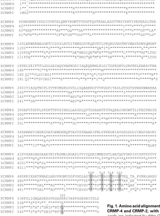
As shown in Figure 2B and 2C, transcripts are first detected at
stage 11.5 by whole mount
in situ hybridization, broadly
through-out the prospective neural ectoderm. At stage 12.5,
CRMP-4 is
enriched in the anterior neural plate and two longitudinal stripes
flanking the midline (Fig. 2D). As development proceeds, the
posterior expression becomes more prominent and
CRMP-4 is
strongly detected throughout the territories of primary neurogenesis
including the medial, intermediate and lateral stripes, where
motor-, inter- and sensory neurons will later differentiate,
respec-tively (Fig. 2E-G) (Chitnis
et al., 1995). This pattern of expression
is similar to that of proneural transcription factors and prefigures
that of the neural specific
β
-tubulin (N-tubulin), which marks
522
terminally differentiated neurons (Oschwald
et
al., 1991). Transcripts are still detected broadly
throughout the anterior neural plate and in a
lateral group of cells associated with the
trigemi-nal ganglia. Neurotrigemi-nal differentiation in the
ante-rior neural plate is delayed until tadpole stages,
thus in contrast to
CRMP-4, known proneural
genes are absent from this territory (Papalopulu
and Kintner, 1996). This restricted expression in
the posterior neural plate contrasts the panneural
expression of
Xenopus CRMP-2 (Kamata et al.,
1998).
During neurula and early tailbud stages,
CRMP-4 transcripts are found throughout the central
nervous system including the developing brain
and neural tube, as well as the eye (Fig 2I-K). As
shown by the transversal section, transcripts of
CRMP-4 are present primarily in the subventricular
and outer marginal layers of the neural tube (Fig
2L), where cells that initiate differentiation and
post-mitotic cells are localized, respectively.
Ad-ditionally, in the anterior ventrolateral region of the
embryo, a scattered ring of cells expresses
CMPR-4. A transversal section demonstrates that these
are isolated cells lying immediately below the
ectoderm (Fig. 2M). These
CRMP-4 expressing
cells are maintained through tailbud stages but
disappear at tadpole stages (compare Fig. 2K and
N). This punctuate pattern is similar to that of
Xphox2A and XHand2 and has been suggested to
be progenitors of smooth muscle cells or pericytes
of the forming vasculature (Smith
et al., 2000;
Talikka
et al., 2004). Interestingly, we have also
observed a similar punctuate expression by other
proneural genes such as
X-MyT1 (data not shown)
and
N-tubulin (Fig 2P) suggesting they are
neu-ronal cells. At tadpole stages (Fig. 2N),
CRMP-4
expression is maintained in the CNS, the eye, as
G
O
B
C
D
E
F
H
I
J
K
L
P
A
N
M
egg 7 9 11 13 15 20 25 27 36 -RT
H4 CRMP-4
--4
well as in the cranial ganglion IX (glossopharyngeal) and X
(vagus ganglion) cells. Consistent with the exclusion of
CRMP-4 from the proliferating cells of the neural tube, staining in the
eye is found in the central and marginal zones of the retina and
is absent from the ciliary marginal zone where proliferating
retinoblasts are found (Fig. 2O).
The regulation of
CRMP-4 was studied in ectodermal
plants (animal caps) from blastula stage embryos; these
ex-plants are normally fated to become epidermal tissue, but can
be converted to derivatives of all three germ layers. The animal
blastomeres of two-cell stage
Xenopus embryos were injected
bilaterally with mRNA encoding putative regulators and animal
caps were dissected at blastula stage. Total RNA was isolated
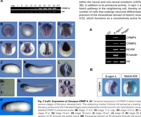
at stage 14 and analyzed by RT-PCR. As shown in Fig. 3A,
animal caps neuralized with the BMP inhibitor Noggin exhibited
strong induction of
NCAM as compared with uninjected animal
caps, but no influence on
CRMP-4 expression was observed.
This is in contrast to the regulation of the closely related
CRMP-2, which is activated by Noggin (Figure 3A and Kamata et al.,
1998). The neuronal determination factor X-ngnr-1 robustly
induced the activation of both
CRMP-4 and CRMP-2. The
positive regulation of
CRMP-2 by X-ngnr-1 is similar to the
activation of other panneural genes such as
Nrp-1 and NCAM
(Klisch
et al., 2006).
The regulation of
CRMP-4 was also investigated in whole
embryos. mRNAs encoding putative regulatory factors were
injected into one blastomere of two-cell stage embryos together
with
LacZ mRNA to localize the distribution of the injected
mRNA. Consistent with the results from the animal cap assay,
in whole embryos, X-ngnr-1 ectopically activated
CRMP-4
within the neural and non-neural ectoderm (100%, n=50; Fig.
3B). In addition to its proneural activity, X-ngnr-1 activates the
Notch pathway in the neighboring cell, thereby restricting the
number of cells that undergo neuronal differentiation.
Overex-pression of the intracellular domain of Notch1 receptor
(Notch-ICD), which functions as a constitutively active form of Notch
X-ngnr-1
Notch-ICD
CRMP4
X-B
A
Fig. 2 (Left).Expression of Xenopus CRMP-4. (A)Temporal expression of CRMP-4 determined by RT-PCR at various stages of Xenopus development. The ubiquitous marker Histone H4 served as a loading control. (B-O) Xenopus embryos at the indicated stages were analyzed by whole mount in situ hybridization using a digoxigenin-labelled CRMP-4 antisense probe. (B) Stage 11.5 d., (C) stage 11.5, veg., (D) stage 12.5 d., (E) stage 14 d., (F)
stage 15 d., (G)stage 15 ant., (H) stage 16 d-ant.,(I)stage 19 ant., (J)stage 24, (K) stage 28, (L) transversal section of (K) through the spinal chord, (M)transversal section of (K) showing through the punctuate anterior-lateral cells, (N)stage 37, lat., (O)transversal section of (N) through the midbrain region. (P) N-tubulin antisense probe, stage 27. anp, anterior neural plate; ant, anterior; cmz, cilliary marginal zone; d, dorsal; i, intermediate; l, lateral; mz, marginal zone; m, medial; r, retina; svz, subventricular zone; tg, trigeminal placode; veg, vegetal; vz, ventricular zone; IX, glossopharngeal ganglion; X, vagus ganglion. Red arrowheads indicate scattered cells.
(Coffman
et al., 1993), inhibits CRMP-4 expression on the
injected side (97%, n=57; Fig. 3B).
Taken together, the temporal expression pattern observed in
the territories of primary neurogenesis, as well as the regulation
of
CRMP-4 by Notch signaling and X-ngnr-1, support an early
role for CRMP-4 in neuronal precursor cells as they initiate
differentiation during primary neurogenesis. Both expression
and regulation of
CRMP-4 differ from that of CRMP-2, which is
induced by the events of neural induction. It will be of interest
to elucidate the function of CRMP-4 during the differentiation of
primary neurons in
Xenopus, as the CRMP family has primarily
been characterized during neuronal maturation in mammalian
systems.
Experimental Procedures
Xenopus CRMP-4
CRMP-4 was identified in a Xenopus cDNA library obtained from the
German Resource Center for Genome Research (IMAGE ID: 4408246, Accession number BC082618). The Xenopus CRMP-4 cDNA clone
contained 1909 bp insert comprising 146 bp of 5’-UTR, 1713 bp of coding sequence and 50 bp of 3’-UTR in pCMVSport6.
Xenopus embryo collection and whole mount in situ hybridization
Xenopus laevis embryos were obtained by HCG induced
egg-laying, dejellied in 2% cysteine pH 8.0, washed and cultured in 0.1X MBS. Embryos were fixed in MEMFA at the desired stage according to Nieuwkoop and Faber (1967). The spatial expression patterns were determined by whole mount in situ hybridization (Harland, 1991) using
a DIG labelled antisense probe. XCRMP-4pCMVSport6 was linearized with EcoRI and transcribed with T7 polymerase. Embryos were embed-ded in gelatine and 30 µm sections were prepared using a vibratome.
Microinjection of embryos
Capped mRNA for microinjections were prepared by in vitro
tran-scription (mMessage-mMachine™ Ambion) and purified over an RNeasy column (Qiagen). Embryos were injected in one or both blastomere of the two-cell stage with the indicated amount of RNA: 50 pg Noggin
(Smith et al., 1993), 50 pg Notch-ICD (Coffman et al., 1993), 25 pg X-ngnr-1 (Ma et al., 1996). As a lineage tracer, 50 pg nuclear lacZ mRNA
was coinjected (Chitnis et al., 1995).
Animal cap assay and RT-PCR
Two-cell stage embryos were injected bilaterally, animal caps dis-sected from stage 8-9 embryos and cultured until sibling controls reached stage 14. Total RNA was extracted from the various embryonic stages or animal caps (Qiagen RNeasy Kit) and cDNA prepared using random hexamer primers and MuLV reverse transcriptase (Perkin– Elmer). PCR was performed with Taq polymerase using the following gene specific oligonucleotide primer pairs:
Histone H4 (26 cycles)
forward: 5´-CGGGATAACATTCAGGGTATCACT-3´ reverse: 5´-ATCCATGGCGGTAACTGTCTTCCT-3´
NCAM (32 cycles)
forward: 5´-CACAGTTCCACCAAATGC-3´
reverse: 5´-GGAATCAAGCGGT5ACAGA-3´ (Hemmati-Brivanlou and Melton, 1994).
N-tubulin (28 cycles)
forward: 5´-ACACGGCATTGATCCTACAG-3´
reverse: 5´-AGCTCCTTCGGTGTAATGAC-3´ (Good et al., 1989). CRMP-2 (30 cycles)
forward: 5´-GGAGAACATGGTTCACACTA-3´
reverse: 5´-TGCAGCATTTGTACTGGTGAC-3’ (Kamata et al., 1998).
CRMP-4 (28 cycles)
forward: 5´-GGAACATTGGCGAGAGAGAAC-3´ reverse: 5´-GTTGTCTCCAATCTGCTTGAT-3´.
Acknowledgements
We thank K. Ditter for excellent technical assistance. This work was supported by a grant from the DFG (CMPB) to T.P. and T.K. was supported by a Lichtenberg stipend in the International Ph.D. Molecu-lar Biology Program, International Max Planck Research School Uni-versity of Goettingen.
References
ARIMURA, N., INAGAKI, N., CHIHARA, K., MENAGER, C., NAKAMURA, N., AMANO, M., IWAMATSU, A., GOSHIMA, Y. and KAIBUCHI, K. (2000). Phos-phorylation of collapsin response mediator protein-2 by Rho-kinase. Evidence for two separate signaling pathways for growth cone collapse. J. Biol. Chem.
275: 23973-80.
ARIMURA, N., MENAGER, C., FUKATA, Y. and KAIBUCHI, K. (2004). Role of CRMP-2 in neuronal polarity. J. Neurobiol. 58: 34-47.
ARIMURA, N., MENAGER, C., KAWANO, Y., YOSHIMURA, T., KAWABATA, S., HATTORI, A., FUKATA, Y., AMANO, M., GOSHIMA, Y., INAGAKI, M., MORONE, N., USUKURA, J. and KAIBUCHI, K. (2005). Phosphorylation by Rho kinase regulates CRMP-2 activity in growth cones. Mol. Cell. Biol. 25: 9973-84.
CHARRIER, E., REIBEL, S., ROGEMOND, V., AGUERA, M., THOMASSET, N. and HONNORAT, J. (2003). Collapsin response mediator proteins (CRMPs): in-volvement in nervous system development and adult neurodegenerative disor-ders. Mol. Neurobiol. 28: 51-64.
CHITNIS, A., HENRIQUE, D., LEWIS J., ISH-HOROWICZ, D. and KINTNER, C. (1995). Primary neurogenesis in Xenopus embryos regulated by a homologue
of the Drosophila neurogenic gene Delta. Nature 375: 761-6.
COFFMAN, C.R., SKOGLUND, P., HARRIS, W.A. AND KINTNER, C.R. (1993). Expression of an extracellular deletion of Xotch diverts cell fate in Xenopus
embryos. Cell 73: 659-71.
COLE, A.R., CAUSERET, F., YADIRGI, G., HASTIE, C.J., MCLAUCHLAN, H., MCMANUS, E.J., HERNANDEZ, F., EICKHOLT, B.J., NIKOLIC, M. and SUTHERLAND, C. (2006). Distinct priming kinases contribute to differential regulation of collapsin response mediator proteins by glycogen synthase kinase-3 in vivo. J. Biol. Chem. 281: 16591-8.
FUKATA, Y., ITOH, T. J., KIMURA, T., MENAGER, C., NISHIMURA, T., SHIROMIZU, T., WATANABE, H., INAGAKI, N., IWAMATSU, A., HOTANI, H. and KAIBUCHI, K. (2002). CRMP-2 binds to tubulin heterodimers to promote microtubule assembly. Nat. Cell Biol. 4: 583-91.
GOOD, P.J., RICHTER, K. and DAWID, I.B. (1989). The sequence of a nervous system-specific, class II beta-tubulin gene from Xenopus laevis. Nucleic Acids Res. 17: 8000.
GOSHIMA, Y., NAKAMURA, F., STRITTMATTER, P. and STRITTMATTER, S.M. (1995). Collapsin-induced growth cone collapse mediated by an intracellular protein related to UNC-33. Nature 376: 509-14.
HARLAND, R.M. (1991). In situ hybridization: an improved whole mount method for”Xenopus embryos. Methods Cell Biol. 36: 685-95.
HEMMATI-BRIVANLOU, A. and MELTON, D.A. (1994). Inhibition of activin recep-tor signaling promotes neuralization in Xenopus. Cell 77: 273-81.
INAGAKI, N., CHIHARA, K., ARIMURA, N., MENAGER, C., KAWANO, Y., MATSUO, N., NISHIMURA, T., AMANO, M. and KAIBUCHI, K. (2001). CRMP-2 induces axons in cultured hippocampal neurons. Nat. Neurosci. 4: 781-2.
KAMATA, T., DAAR, I.O., SUBLESKI, M., COPELAND, T., KUNG, H.F. and XU, R.H. (1998). Xenopus CRMP-2 is an early response gene to neural induction. Brain Res. Mol. Brain Res. 57: 201-10.
KLISCH, T.J., SOUOPGUI, J., JUERGENS, K., RUST, B., PIELER, T. and HENNINGFELD, K.A. (2006). Dev. Biol. 292: 470-85.
LLANOS, S., EFEYAN, A., MONSECH, J., DOMINGUEZ, O. and SERRANO, M. (2006). A High-Throughput Loss-of-Function Screening Identifies Novel p53 Regulators. Cell Cycle 5: 1880-5.
ANDERSON, D.J. (1998). Neurogenin1 is essential for the determination of neuronal precursors for proximal cranial sensory ganglia. Neuron 20: 469-82.
NIEUWKOOP, P.D. and FABER, J. (1967). Normal Table of Xenopus laevis
(Daudin), 2nd ed. Elsevier/North Holland, Amsterdam.
NISHIMURA, T., FUKATA, Y., KATO, K., YAMAGUCHI, T., MATSUURA, Y., KAMIGUCHI, H. and KAIBUCHI, K. (2003). CRMP-2 regulates polarized Numb-mediated endocytosis for axon growth. Nat. Cell Biol. 5: 819-26.
OSCHWALD, R., RICHTER, K. and GRUNZ, H. (1991). Localization of a nervous system-specific class II beta-tubulin gene in Xenopus laevis embryos by whole
mount in situ hybridization. Int. J. Dev. Biol. 35: 399-405.
PAPALOPULU, N. and KINTNER, C. (1996). A posteriorising factor, retinoic acid, reveals that anteroposterior patterning controls the timing of neuronal differen-tiation in Xenopus neuroectoderm. Development 122: 3409-18.
SMITH, W.C., KNECHT, A.K., WU M. and HARLAND, R.M. (1993). Secreted noggin protein mimics the Spemann organizer in dorsalizing Xenopus
meso-derm. Nature 361: 547-9.
SMITH, S.J., KOTECHA, S., TOWERS, N. and MOHUN, T. J. (2000). Xenopus
Hand2 expression marks anterior vascular progenitors but not the developing heart. Dev. Dyn. 219: 575-81.
TAHIMIC, C.G., TOMIMATSU, N., NISHIGAKI, R., FUKUHARA, A., TODA, T., KAIBUCHI, K., SHIOTA, G., OSHIMURA, M. and KURIMASA, A. (2006). Evidence for a role of Collapsin response mediator protein-2 in signaling pathways that regulate the proliferation of non-neuronal cells. Biochem.
Bio-phys. Res. Commun. 340: 1244-50.
TALIKKA, M., STEFANI, G., BRIVANLOU, A.H. and ZIMMERMAN, K. (2004). Characterization of Xenopus Phox2a and Phox2b defines expression domains
within the embryonic nervous system and early heart field. Gene Expr. Patterns
4: 601-7.
UCHIDA, Y., OHSHIMA, T., SASAKI, Y., SUZUKI, H., YANAI, S., YAMASHITA, N., NAKAMURA, F., TAKEI, K., IHARA, Y., MIKOSHIBA, K., KOLATTUKUDY, P., HONNORAT, J. and GOSHIMA, Y. (2005). Semaphorin3A signalling is medi-ated via sequential Cdk5 and GSK3beta phosphorylation of CRMP2: implication of common phosphorylating mechanism underlying axon guidance and Alzheimer’s disease. Genes Cells 10: 165-79.
WANG, L.H. and STRITTMATTER, S.M. (1996). A family of rat CRMP genes is differentially expressed in the nervous system. J. Neurosci. 16: 6197-207.
WANG, L.H. and STRITTMATTER, S.M. (1997). Brain CRMP forms heterotetramers similar to liver dihydropyrimidinase. J. Neurochem. 69: 2261-9.
YOSHIMURA. T., KAWANO. Y., ARIMURA. N., KAWABATA. S., KIKUCHI. A. and KAIBUCHI. K. (2005). GSK-3beta regulates phosphorylation of CRMP-2 and neuronal polarity. Cell 120: 137-49.
The International Journal of Developmental Biology
UBC Press - Editorial Service University of the Basque Country E-48940 Leioa, (Vizcaya) SPAIN
ORDER BY
Web:
http://www.intjdevbiol.com
E-mail: [email protected]
(include the information indicated above)FAX:
+34-94-601-3266
Preface
by E.M. De Robertis and J. Aréchaga
HERITAGE OF THE 1924 ARTICLE BY HANS SPEMANN AND HILDE MANGOLD Introducing the Spemann-Mangold organizer: experiments and insights that generated a
key concept in developmental biology
by K. Sander and P. Faessler
Induction of embryonic primordia by implantation of organizers from a different species
Reprint of the original 1924 article by Hans Spemann and Hilde Mangold, translated into English by Viktor Hamburger
Developmental biology of amphibians after Hans Spemann in Germany
by H. Grunz
Spemann´s heritage in Finnish developmental biology
by L. Saxén
Spemann´s influence on Japanese developmental biology
by M. Asashima and T. Okada
Contribution of the Belgian school of embryology to the concept of neural induction by the organizer
by H. Alexandre
Contrasting influences of the organizer and induction concepts on the scientific activity of French embryologists
by J.-C. Beetschen and A.-M. Duprat
Consequences of the Spemann-Mangold organizer concept for embryological research in Russia: personal impressions
by A.T. Mikhailov and N.A. Gorgolyuk
The organizer concept and modern embryology: Anglo-American perspectives
by T. Horder
THE ORGANIZER CONCEPT: OVERVIEWS AND THEORETICAL APPROACHES Evolution of the organizer and the chordate body plan
by J. Gerhart
Continuity and change: paradigm shifts in neural induction
by S. Gilbert
Formation and maintenance of the organizer among vertebrates
by K. Joubin and C.D. Stern
Organizer and axes formation as a self-organizing process
by H. Meinhardt
ORGANIZER RESEARCH TODAY
Molecular mechanisms of cell-cell signalling by the Spemann-Mangold organizer
by E.M. De Robertis, O. Wessely, M. Oelgeschläger, B. Brizuela, E. Pera, J. Larraín, J. Abreu and D. Bachiller
Formation of a functional morphogen gradient by a passive process in tissue from the early Xenopus embryo
by N. McDowell, J.B. Gurdon and D.J. Grainger
A study of Xlim1 function in the Spemann-Mangold organizer
by L. Kodjabachian, A.A. Karavanov, H. Hikasa, N.A. Hukriede, T. Aoki, M. Taira and I.B. Dawid
Making mesoderm - upstream and downstream of Xbra
by J. Smith
Regulation of convergent extension in Xenopus by Wnt5a and Frizzled-8 is independent of the canonical Wnt pathway
by J.B. Wallingford, K.M. Vogeli and R.M. Harland
Generation of the germ layers along the animal-vegetal axis in Xenopus laevis
by H. Yasuo and P. Lemaire
Dickkopf1 and the Spemann-Mangold head organizer
by C. Niehrs, O. Kazansakaya, W.Wu, and A. Glinka
Siamois cooperates with TGFβββββ signals to induce the complete function of the Spemann-Mangold organizer
by M.J. Engleka and D. S. Kessler
ORDER FORM
I would like to order ____ cop(y/ies) of the Int. J. Dev. Biol. Special Issue “The Spemann-Mangold Organizer” (Vol. 45, Nº 1)
at US$ 70 or Euro 70 per copy (including post and packaging). Total to be charged: __________US$ / Euro (please specify currency)
Organizer
Edited by Eddy M. De Robertis and Juan Aréchaga
The Spemann-Mangold organizer: the control of fate specification and morphogenetic rearrangements during gastrulation in Xenopus
by T. Bouwmeester
Functional analysis of the Xenopus frizzled 7 protein domains using chimeric receptors
by R.K. Swain, A. Medina and H. Steinbeisser
Fox (forkhead) genes are involved in the dorso-ventral patterning of the Xenopus mesoderm
by H. El-Hodiri, N. Bhatia-Dey, K. Kenyon, K. Ault, M. Dirksen and M. Jamrich
In vitro induction systems for analyses of amphibian organogenesis and body patterning
by T. Ariizumi and M. Asashima
The avian organizer
by T. Boettger, H. Knoetgen, L. Wittler and M. Kessel
Nodal signaling and the zebrafish organizer
by A.F. Schier and W.S. Talbot
The role of the homeodomain protein Bozozok in Zebrafish axis formation
by L. Solnica-Krezel and W. Driever
Role of the anterior visceral endoderm in restricting posterior signals in the mouse embryo
by A. Perea-Gomez, M. Rhinn and S.-L. Ang
Roles of Sox factors in neural determination: conserved signaling in evolution?
by Y. Sasai
Getting your head around Hex and Hesx1 : forebrain formation in mouse
by J.P. Martínez Barbera, R.S.P. Beddington
The role of Otx2 in organizing the anterior patterning in mouse
by A. Simeone and D. Acampora
Defects of the body plan of mutant embryos lacking Lim1 , Otx2 or Hnf3βββββ activity
by S.J. Kinder, T.E. Tsang, S.-L. Ang, R.R. Behringer and P.P.L. Tam
Otx2 and Hnf3βββββ genetically interact in anterior patterning
by O. Jin, K. Harpal, S.L. Ang and J. Rossant
The isthmic organizer and brain regionalization
by S. Martínez
Early neurogenesis in amniote vertebrates