EFFECTS OF SURFACTANT SYSTEMS ON
METFORMIN TRANSPORT
A thesis presented by
Dejana Dimitrijevic
in partial fulfilment of the requirements for the degree of Doctor of Philosophy
of the
University of London
February 1998 The School of Pharmacy
ProQuest Number: 10104149
All rights reserved
INFORMATION TO ALL USERS
The quality of this reproduction is dependent upon the quality of the copy submitted. In the unlikely event that the author did not send a complete manuscript and there are missing pages, these will be noted. Also, if material had to be removed,
a note will indicate the deletion.
uest.
ProQuest 10104149
Published by ProQuest LLC(2016). Copyright of the Dissertation is held by the Author. All rights reserved.
This work is protected against unauthorized copying under Title 17, United States Code. Microform Edition © ProQuest LLC.
ProQuest LLC
789 East Eisenhower Parkway P.O. Box 1346
They did not know that a new hfe is not given for nothing; that it has to he paid dearly for, and only acquired by much patience and suffering, and great future efforts...”
To my parents, Katarina and Nikola
CONTENTS
TITLE PAGE NUMBER
Acknowledgements 6
Abstract 7
Abbreviations 8
List of Figures 9
List of Tables 16
Chapter 1 - Introduction 17
1.1 Outline of the work 18
1.2 Absorption of drugs from the gastrointestinal tract 19
1.2.1 Anatomy and physiology of the gastrointestinal tract 19
1.2.2 The con^onents and structure of cell membrane 26
1.2.3 Tight junctions 29
1.2.4 Absorptive mechanisms of drugs across intestinal
mucosa 29
1.2.4.1 Passive diffusion 30
1.2.4.2 Carrier mediated transport 30
1.2.4.3 Bulk transport 31
1.2.5 Pathways of drug absorption 31
1.3 Methodology to study intestinal transport, metabolism and
absorption enhancement 32
1.3.1 Cell cultures as models for dmg absorption studies 34
1.3.1.1 Caco-2 cell culture system 36
1.4 Factors affecting drug absorption 38
1.5 Enhancement of drug absorption from the gastrointestinal tract 3 9
1.5.1 Absorption enhancers 39 %
TITLE PAGE NUMBER Chapter 1 - Introduction (cont.)
1.5.1.2 Bile salts and mixed micelles 46
1.5.1.3 Sodium tauro-24,25-dihydrofusidate (STDHF) 47
1.5.1.4 Fatty acids and medium-chain glycerides 47
1.5.1.5 Non-steroidal anti-inflammatory drugs
(NSAIDs) and derivatives as
absorption enhancers 50
1.5.1.6 Chelating agents (Calcium binding agents) 51
1.5.2 Other possibilities of increasing drug absorption 52
1.5.2.1 Protease inhibitors 52
1.5.2.2 Prodrugs and analogues 52
1.5.2.3 Particulate carriers 54
1.5.2.4 Emulsions 55
1.5.2.5 Niosomes and lÿosomes 57
1.5.2.6 The Macrosol™ and Bridgelock™technology 58
1.6 Efl&cacy and potential toxicity of permeability enhancers for
oral drug delivery 59
1.7 Metformin 60
1.7.1 Non-insulin dependent diabetes melhtus 62
Chapter 2 - Influence of emulsion formulations on metformin
absorption in the rat 64
2.1 Introduction 65
2.2 Materials 66
2.3 Methods 66
2.3.1 Preparation of emulsions 66
2.3.2 Characterisation of emulsions 71
2.3.3 In vitro release measurements 71
TITLE PAGE NUMBER Chapter 2 - Influence of emulsion formulations on metformin
absorption in the rat (cont.)
2.3.5 Recovery experiments 72
2.3.6 Histological investigation 72
2.3.7 Scanning Electron Microscopy (SEM) 73
2.4 Results and Discussion 74
Chapter 3 - Effects of some non-ionic surfactants on transepitheUal permeability and metformin transport in Caco-2 cells: microscopic and
in vivo studies 92
3.1 Introduction 93
3.2 Materials 95
3.3 Methods 96
3.3.1 Caco-2 cells 96
3.3.2 Trypan blue method 98
3.3.3 Determination of Critical Micellar Concentration (CMC) 98
3.3.4 Osmolality measurements 99
3.3.5 Absorption studies in Caco-2 cell monolayers 99
3.3.6 Integrity of the Caco-2 cell monolayers 100
3.3.7 Cell viability (MTT test) 100
3.3.8 Transmission Electron Microscopy (TEM) 101
3.3.9 Fluorescence microscopy 101
3.3.10 Scanning Electron Microscopy (SEM) 102
3.3.11 Absorption, recovery and histology study in rats 102
3.3.12 Matrix-assisted laser desorption mass spectroscopy 103
TITLE PAGE NUMBER Chapter 4 - The effects of monomers and vesicular forms
of Solulan C24 and Solulan 16 on Caco-2 cell
monolayers 142
4.1 Introduction 143
4.2 Materials 144
4.3 Methods 144
4.3.1 Caco-2 cells 144
4.3.2 Preparation of niosomes 144
4.3.3 Determination of Critical Micellar Concentration (CMC) 145
4.3.4 Osmolality measurements 145
4.3.5 Absorption studies in Caco-2 cell monolayers 145
4.3.6 Transepithelial electrical resistance (TEER) 146
4.3.7 Cell viability (MTT test) 146
4.4 Results and Discussion 146
Chapter 5 - Effects of fluorinated and hydrogenated surfactants on transepithelial permeabUity in Caco-2 cell
monolayers 164
5.1 Introduction 165
5.2 Materials 166
5.3 Methods 166
5.3.1 Caco-2 cells 166
5.3.2 Transepithelial electrical resistance (TEER) 166
5.3.3 Osmolality measurements 166
5.3.4 Absorption studies in Caco-2 cell monolayers 166
5.3.5 Cell viability (MTT test) 167
5.3.6 Transmission Electron Microscopy (TEM) 167
TITLE PAGE NUMBER Chapter 5 - Effects of fluorinated and hydrogenated surfactants
on transepithelial permeability in Caco-2 cell monolayers (cont.)
5.4 Results and Discussion 167
Final conclusion 190
Acknowledgements
I am greatly indebted to my supervisor, Professor Alexander Florence, for the
guidance, freedom and advice I received and for the interest he has always shown for my
work, and especially for the support he gave to me at the beginning and the end of my
thesis. I will have eternal gratitude to him for offering me assistance in dire personal
circumstances.
I extend my gratitude to Dr. Andy Shaw who opened the door leading to the
wonderftd world of cell cultures for me. Much appreciation to my friend Dr. Stefrn
Dojcinov for his great help and e^gert contribution of microscopy work. Thanks to Mr
Dave McCarthy and Ms Wendy Tynan for heÿfril discussions and for developing the
photographs which were used in this thesis. Many thanks to Ms Charlotte Lamandin for
her contribution to this thesis, and for the pleasure of supervising. I would also like to
thank Mr Mark Domin for his mass spectroscopy work and Mr Colin James for molecular
modelling.
The work was carried out under the Lçha Pharmaceuticals grant.
Much love and gratitude to all my colleagues from Centre for Drug Delivery
Research, Toxicology Department and Centre for Polymer Therapeutics. I wish also to
thank Miss Barbara Grant for her assistance and kindness at the beginning of my thesis.
Many thanks to all nty friends (the IvovioHohs, Ljiljana, Sandra, Agata, Antonela,
Tanja, Olivera, Bohan, Misica, Branko, Zoran, Lidija, Biba, Snezana, Beba, Maca, Misa)
for love and frimdsh^ without which I could not work or live. I deeply appreciate the
unwavering support which I received from members of my family.
Finally, to my Sasha, for making a dream possible, and for the encouragement to
Abstract
This work has focused on the investigations of carrier and absorption enhancing systems for the hydrophihc drug metformin when delivered orally.
The influence of both water-in-oil (W/0) and oil-in-water (OAV) emulsions containing metformin in the aqueous phase on oral metformin bioavailabihty was studied in the rat. A water-in-isopropyl myristate emulsion stabilised with sorbhan monooleate (Span 80) and bovine serum albumin (BSA), an OAV emulsion stabilised with cholesteryl poly (24) oxyethylene ether (Solulan C24) and Span 80 prepared with soybean oil and an OAV emulsion stabilised with sorbitan monostearate (Span 60) with sesame oil as the continuous phase were formulated, characterised and tested in vivo. The oral administration of emulsions modifies the absorption of metformin in rat. Histological investigations provided some clues as to the cause of increase in metformin absorption. Most likely are changes to the intestinal membrane and, in some cases to the junctional conçlexes caused by the sur&ce active compounds and fetty adds present in the formulations. Knowledge of the reversibility of these effects was crucial Allowing the intestinal cells to recover after dosing for three days, suggests that all changes that were detected were, indeed, reversible.
The effects of the non-ionic sur&ctants polysorbate 20, 60, 85, Solulan C24 and a lanolin-based poly(16)oxyethylene ether (Solulan 16) on epithelial integrity of the Caco-2 cells monolayers were studied. The effects of the surfactants on cell permeability were assessed by measurements of the transport of metformm, transepithelial electrical resistance (TEER) and tests for cell viability (determined by the dphenyhetrazolium bromide test). The concentration-dependent effects on TEER correlated with cell viability, Le. increased TEER and increased cell-monolayer permeability for metformm corresponded to decreased cell viability. The results indicate that the Solulans and polysorbates were active as absorption enhancer; in quite different concentration ranges, the former being more effective than the latter. The effects of the sur&ctants were also assessed by transmission and scanning electron microscopy. The results prove a correlation between the increase of metformin transport caused by the surjetants and toxic effects on cell monolayers. The effects of the most potent surjetants (polysorbate 20, Solulan C24 and 16) were tested in vivo to establish a correlation with in vitro data. It was concluded that the effects of the surjetants used at certain concentrations on the rat intestinal membrane were reversible.
Several niosome formulations were prepared in order to assess potential toxic effect of these systems on Caco-2 cell monolayers. It may be concluded that the toxic effect of niosomes is due to the amount of free surjetant present in the niosome suspension.
Abbreviations
BLMV - Basolateral membrane vesicles
BBMV - Bmsh border membrane vesicles
CMC - Critical micellar concentration
DMEM - Dulbecco’s modified Eagle medium
DMSO - Dimethylsulfoxide
EDTA - Ethylenediaminetetraacetic acid
PCS - Foetal calf serum
HBSS - Hank’s balanced salt solution
IPM - Isopropyl myristate
LM - Light microscopy
LNA - Linolenic add
MTT - 3-(4,5-dimethylthiazol-2-yl)-2,5-diphenyltetrazolium bromide
NIDDM - Non-insulin dependent diabetes mellitus
NEAA - Non-essential amino-acids
PEG - Polyethyleneglycol
PI - Propidhun iodide
SA - Saturated fetty acid
SEM - Scanning Electron Microscopy
TEM - Transmission Electron Microscopy
List of Figures
Figure 1.1 A Schematic diagram of intestinal epithelial cells.
Figure 1.1 B Cell types of intestinal epithelium
Figure 1.1 C Schematic diagram of intestinal absorptive cells.
Figure 1.1 D Electron micrograph of the rat intestinal absorptive cells.
Figure 1.2 Shematic diagram of intercellular junctions.
Figure 1.3 A ‘Thud mosaic” model of membrane structure.
Figure 1.3 B Structure of a phospholçid.
Figure 1.3 C How membranes become more fluid or more sohd.
Figure 1.3 D Possible relationship between unsaturated fetty acids,
LNA (linolenic) add and suÿhur-containing proteins and
SA (saturated 6tty adds).
Figure 1.4 Schematic drawing of the cell culture model
Figure 1.5 Three possible sites of action for enhancers.
Figure 1.6 Solubilizing membrane proteins with a detergent.
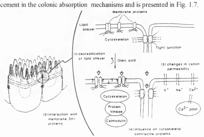
Figure 1.7 Proposed model for oldc add-induced enhancement
in the colonic absorption.
Figure 1.8 Schematic diagram of the princÿal stages in %id digestion.
Figure 1.9 Chemical structure of metformin.
Figure 2.1 Photomicrograph of aW/IPM emulsion
stabilised with Span 80 and BSA.
F ^u re 2.2 Photomicrograph of an OAV emulsion stabilised with Solulan C24
and Span 80, prepared with soybean oil
Figure 2.3 Photomicrograph of an OAV emulsion stabilised with Span 60,
prepared with sesame oil
Figure 2.4 Scanning Electron Micrograph of rat intesine.
List of Figures (cont.)
Figure 2.6 A Plasma concentration of metformin as a fimction of time
after oral administration of a W/IPM emulsion
stabilised with Span 80 and BSA.
Figure 2.6 B Plasma concentration of metformin as a fimction of time
after oral administration of an OAV emulsion stabilised
with Solulan C24 and Span 80, prepared with soybean oil
Figure 2.6 C Plasma concentration of metformin as a function of time
after oral administration of an OAV emulsion stabilised
with Span 60, prepared with sesame oil
F ^u re 2.7 A Rat small bowel mucosa, control sangle
Figure 2.7 B Rat small bowel mucosa, control sangle
F ^ure 2.8 A Rat small bowel mucosa, after the treatment for 2 h with
a W/IPM emulsion stabilised with Span 80 and BSA.
Figure 2.8 B Rat small bowel mucosa, after the treatment for 3 h with
a W/IPM emulsion stabilised with Span 80 and BSA.
F%ure 2.9 A Rat small bowel mucosa, after the treatment for 2 h with
an 0/W emulsion stabilised with Solulan C24 and Span 80,
prepared with soybean oü.
Figure 2.9 B Rat small bowel mucosa, after the treatment for 3 h with
an O/W emulsion stabilised with Solulan C24 and Span 80,
prepared with soybean oil
Figure 2.10 A Rat small bowel mucosa, after the treatment for 2 h with
an O/W emulsion stabilised with Span 60, prepared with sesame oil
F%ure 2.10 B Rat small bowel mucosa, after the treatment for 3 h with
an O/W emulsion stabilised with Span 60, prepared with sesame oil
Figure 2.11 A Rat small bowel musosa after 2 h of e?q)osure to
a W/IPM emulsion stabilised with Span 80 and BSA
List of Figures (cont.)
Figure 2.11 B Rat small bowel musosa after 2 h of exposure to
an OAV emulsion stabilised with Solulan C24 and Span 80,
prepared with soybean oil and than a recovery phase for 3 days.
Figure 2.11 C Rat small bowel musosa after 2 h of exposure to
OAV emulsion stabilised with Span 60, prepared with sesame oil
and than a recovery phase for 3 days.
Figure 3.1 Caco-2 cells in tissue culture flask.
Figure 3.2 A Percentage of PEG transport over filters with
and without Caco-2 cells.
Figure 3.2 B Percentage of metformin transported across filters with
and without Caco-2 cells.
Figure 3.3 A Transport of metformin as a fimction of the concentration
of polysorbate 20.
Figure 3.3 B Transport of metformin as a function of the concentration
of polysorbate 60.
Figure 3.3 C Transport of metformin as a fimction of the concentration
of polysorbate 85.
Figure 3.4 A Transport of metformin as a fimction of the concentration
of Solulan C24.
Figure 3.4 B Transport of metformin as a fimction of the concentration
of Solulan 16.
Figure 3.5 A The surface tension-log concentration relationshç of Solulan C24.
Figure 3.5 B The surface tension-log concentration relationship of Solulan 16.
Figure 3.6 A Effect of polysorbate 20 on the transepithelial electrical resistance
of Caco-2 cell monolayers.
Figure 3.6 B Effect of polysorbate 60 on the transepithelial electrical resistance
List of Figures (cont.)
F ^ure 3.6 C Effect of polysorbate 85 on the transepithelial electrical resistance
of Caco-2 cell monolayers.
Figure 3.6 D Effect of Solulan C24 on the transepithelial electrical resistance
of Caco-2 cell monolayers.
Figure 3.6 E Effect of Solulan 16 on the transepithelial electrical resistance
of Caco-2 cell monolayers.
Figure 3.7 Correlation between TEER and % metformin transported at t=3 h.
Figure 3.8 A Cytotoxicity of polysorbate 20, 60 and 85.
Figure 3.8 B Cytotoxicity of Solulan C24 and Solulan 16.
Figure 3.9 Senh-representational views of Solulan C24,
polysorbate 20, 60 and 85.
Figure 3.10 A TEM of Caco-2 cells. Control
Figure 3.10 B TEM. Caco-2 cells e?q)osed for 2 h to 0.5% polysorbate 60.
Figure 3.10 C TEM. Caco-2 cells exposed for 2 h to 0.5% polysorbate 85.
F ^ure 3.10 D TEM. Caco-2 cells exposed for 2 h to 0.01% Solulan 16.
F ^ure 3.10 E TEM. Caco-2 cells e>q)osed for 2 h to 0.01% Solulan C24.
Figure 3.10 F TEM. Caco-2 cells e?q)osed for 2 h to 0.5% polysorbate 20.
Figure 3.10 G TEM. Caco-2 cells e>q)osed for 2 h to 0.1% polysorbate 20.
Figure 3.10 H TEM. Caco-2 cells e?q)osed for 2 h to 0.01% Solulan C24.
Figure 3.101 TEM. Caco-2 cells e?q)osed for 2 h to 0.1% Solulan C24.
Figure 3.11 A Scanninig electron micrograph of control Caco-2 cells.
Figure 3.11 B Scanninig electron micrograph of Caco-2 cells e?qposed to
0.01% Solulan C24.
Figure 3.11 C Scanninig electron micrograph of Caco-2 cells e?q)Osedto
0.5% Solulan C24.
List of Figures (cont.)
Figure 3.12 B Cells stained with propidium iodide,
after e?q)osure to 0.5% Solulan C24.
Figure 3.13 Plasma concentration of metformin as a fimction of time
after oral administration of control sarrq)le,
Solulan C24, Solulan 16 and polysorbate 20.
F ^ure 3.14 A L.M. Photomicrograph of histological section of rat
intestinal mucosa, control sangle.
F%ure 3.14 B L.M. Photomicrograph of histological section of rat
intestinal mucosa after treatment for 2 h with 0.1% Solulan C24.
Figure 3.14 C L.M. Photomicrograph of histological section of rat
intestinal mucosa after treatment for 2 h with 0.1% Solulan 16.
Figure 3.14 D L.M. Photomicrograph of histological section of rat
intestinal mucosa after treatment for 2 h with 3% polysorbate 20.
F ^ure 4.1 A Transport of metformin across Caco-2 cells as a fimction
of the concentration of Solulan C24.
Figure 4.1 B Transport of metformin across Caco-2 cells as a fimction
of the concentration of Solulan 16.
Figure 4.2 A Cytotoxicity of Solulan C24, fi'ee and in niosome formulations.
Figure 4.2 B Cytotoxicity of Solulan 16, free and in niosome formulations.
Figure 4.3 Stmcture of the vesicle forming confounds.
Figure 4.4 A Effect of (0.43%) Solulan C24, free and in niosomes on
TEER of Caco-2 cells as a fimction of time.
Figure 4.4 B Effect of (0.8%) Solulan C24, free and in niosomes on
TEER of Caco-2 cells as a fimction of time.
Figure 4.4 C Effect of (1.7%) Solulan C24, free and in niosomes on
TEER of Caco-2 cells as a fimction of time.
Figure 4.4 D Effect of (2.6%) Solulan C24, free and in niosomes on
List of Figures (cont.)
Figure 4.4 E Eflfect of (3.46%) Solulan C24, free and in niosomes on
TEER of Caco-2 cells as a fimction of time.
F ^ure 4.5 Cytotoxidty in the presence of Solulan C24 in niosomes
(sanq)les 1-5).
Figure 4.6 Effect of pellet, supernatant and niosomes.
Figure 4.7 The apparent free concentration of Solulan C24 as fimction
of the total concentration of Solulan in niosomes.
Figure 5.1 Structure of surfrctants used.
Figure 5.2 A Transport studies of metformin as a fimction
of the concentration of AF-87.
Figure 5.2 B Transport studies of metformin as a fimction
of the concentration of A-87.
Figure 5.2 C Transport studies of metformm as a fimction
of the concentration of AF-66.
F ^ure 5.2 D Transport studies of metformin as a fimction
of the concentration of A-109.
Figure 5.2 E Transport studies of metformin as a fimction
of the concentration of AF-107.
Figure 5.3 A Effect of AF-87 on the TEER of Caco-2 cell monolayers
as a fimction of time and surfr ctant concentration.
Figure 5.3 B Effect of A-87 on the TEER of Caco-2 cell monolayers
as a fimction of time and surfrctant concentration.
Figure 5.3 C Effect of AF-66 on the TEER of Caco-2 cell monolayers
as a fimction of time and surfactant concentration.
Figure 5.3 D Effect of A-109 on the TEER of Caco-2 cell monolayers
as a fimction of time and sur6ctant concentration.
Figure 5.3 E Effect of AF-107 on the TEER of Caco-2 cell monolayers
List of Figures (cont.)
Figure 5.4 A Cytotoxicity of AF-87, A-87, AF-66.
Figure 5.4 A Cytotoxicity of AF-107, A-109.
F%ure 5.5 A TEM of Caco-2 cells after the treatment with 5%AF-87.
F%ure 5.5 B TEM of Caco-2 cells after the treatment with 1% A-87.
Figure 5.5 C TEM of Caco-2 cells after the treatment with 5% A-87.
Figure 5.5 D TEM of Caco-2 cells after the treatment with 1% A-109.
F ^ure 5.5 E TEM of Caco-2 cells after the treatment with 5% A-109.
Figure 5.5 F TEM of Caco-2 cells after the treatment with 5% AF-107.
Figure 5.6 Correlation between % metformin transported across
Caco-2 cell monolayers after the treatment with A-87, AF-87,
List of Tables
Table 1.1 Summary data for in vitro models.
Table 1.2 Classes of enhancers of intestinal drug absorption
and some of their representatives.
Table 1.3 Various kind of surjetants used for absorption enhancement
of proteins and peptides enq)loying intestinal, rectal and nasal route.
Table 1.4 The use of prodrugs and analogues for in^roving absorption.
Table 3.1 The osmolality measurements for polysorbates 20, 60, 85
and Solulan C24 anl6.
Table 3.2 Properties of the surjetant used.
Table 4.1 The osmolality of niosomes containing Solulans C24 and 16.
Table 4.2 The osmolality measurements of niosomes and Solulan C24 in HBSS.
Table 4.3 The osmolality measurements of pellet, supernatant and niosomes
with no Solulan C24.
Table 4.4 Effect of pellet, supematant and niosomes with no Solulan C24
on cell vmbility.
Table 5.1 Molecular weights and CMC of surjetant used.
Table 5.2 Osmolality measurements for Af-87, A-87, AF-66, A-109
Chapter 1
1.1 Outline of work
The objective of the work described in this thesis was to explore the possibilities of
increasing the oral absorption of the highly hydrophilic drug, metformin. Metformin
hydrochloride is a biguanide hypoglycaemic agent used in the treatment of non-insulin
dependent diabetes mellitus (NIDDM). Metformin has been on market for more than 20
years and so fer all attenqits to increase its bioavailabihty (of 30-50%) have been
unsuccessfiiL The work was designed to investigate the effects of non-ionic surfactants
and surfectant-based systems such as emulsions on metformin absorption. Their potential
to act as absorption enhancing systems for metformin has been evaluated.
The rationale of using emulsions was to investigate the influence of both
sur&ctants and oils present in the formulations. It is known that sur&ctants may affect the
rate of gastrointestinal absorption through different mechanisms, involving alteration of the
normal state of biological membranes, in general increasing their permeability. The
influence of oils on the retention time in the small intestine and of fetty acids present in the
oil phases of emulsions has also been considered. The work has demonstrated the difi&culty
of using metformin because of three &ctors: its high water solubility, its high dosage and
its electrolyte character. AU Simulations were tested in vivo. The question of formulation intact on intestinal morphology and recovery afl;er dosing was demonstrated.
The potential toxic effects of surfectants on isolated and less coirplex system such
as Caco-2 ceU monolayers was studied. The Caco-2 ceU line is the most commonly used in
studies of drug absorption and absorption enhancement, due to its ease of cultivation and
the spontaneous differentiation to enterocyte-like cells under standard ceU culture
conditions. Different techniques were used to assess the interaction of surfactants with the
ceU membranes and junctional corqplexes. Metformin transport across the Caco-2 ceU line
aUowed the characterisation of the narrow range of surfectant concentrations capable of
increasing metformin transport with minimum toxicity to the ceU monolayer. The study
was extended to investigate the effect of nonionic surfectant vesicles containing some of
determine the influence of novel fluorocarbon and hydrocarbon surjetants on metformin
transport.
A relationship between the increase in transport caused by surjetants and their
toxicity has been established.
1.2 Absorption of drugs from the gastrointestinal tract
The oral route is the most common and convenient method of drug administration.
However, on oral administration some drugs exhibit rektively low bioavaflability. Low
bioavailability may be caused by poor penetration of the intestinal mucosa, precÿitation or
binding of the drug in the gastro-intestinal tract or by extensive first-pass metabolism. Oral
administration of drugs often leads to degradation, due to the highly acidic gastric
environment, the enzymes of the mucosa, or by liver before the drugs enter the systemic
circulation.
This work examined reported methods for enhancement of the oral absorption of
highly polar drugs, including polar peptides and proteins. In order to improve the intestinal
absorption of these poorly absorbed drugs, many have investigated the effects of
absorption enhancers, protease inhibitors, prodrugs and analogues, and the use of a variety
of dosage forms.
The anatomical and physiological aspects of the gastrointestinal tract and some
approaches to enhance the intestinal absorption of poorly absorbed drugs will be discussed
first.
1.2.1 Anatomy and physiology of the gastrointestinal tract
The gastrointestinal tract is a highly specialised region of the body v\hose ftmctions
may be described by the processes of secretion, digestion, and absorption (Mayersohn,
the drug molecules, it is inçortant that the anatomy and physiology of the gastrointestinal
tract is thoroughly understood, along with the different uptake mechanisms in the intestine.
The absorption of drug molecules from the stomach is generalty not high when
conq)ared with absorption from the intestine, but gastric emptying is an important
physiological event which significantly influences the uptake of drug substances from the
intestine (Kararli, 1989).
The small intestine is conq)osed of the duodenum, jejunum and ileum In humans,
the duodenum is 20cm long, the jejunum, 275cm long and the ileum about 425cm in
length (Rhodin, 1977). The wall of the small intestine is made up of four concentric layers:
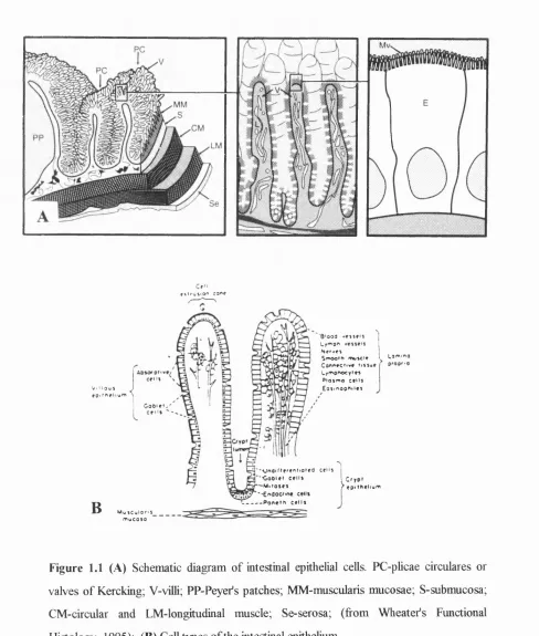
the mucosal layer, submucosa, musculaiis externa and serosa (Smith, 1986) (Fig. 1.1 A).
Three structural specializations increase the surface area of the intestinal mucosa
to enhance the absorptive process: the plicae circulares (valves of Kercking), intestinal villi and microvilh (Fig. I.IB and C). The plicae circulares are large, permanent folds which consist of the intestinal mucosa with the central core of the submucosa. The more
effective means of augmenting the surfrce area of the mucosa is the presence of enormous
number of intestinal villi They cover the entire suffice of mucosa. A villus averages 0.5 to
1.5 mm in length and 0.1mm in diameter (Krause and Cutts, 1981). It has been estimated
that there are some 10-40 villi/mm^ of mucosal suffice (Bloom and Fawcett, 1975).
Pfojecting from surface of the vilh are fine stmctures, the microvilli The surfice area of
the small intestine is increased approximately 30-fold by the presence of microvilh (Krause
and Cutts, 1981). The microvilh are about 1pm in length, about 0.1pm in width, and there
are about 2000 per ceh (Rhodin, 1977). The microvilh constitute the striated border of the
hght microscopy. The area immediately beneath the microvilli, cahed the terminal web, is
relatively free of organeUes except for filaments that extend into the cores of the microvilh
(Moon, 1983). Filaments of the microvilh and terminal web contribute to the cytoskeleton
and give stability to the microvillus border (Fig. 1.1 C and D).
The mucosa is most important to the digestive and absorptive function of the
intestine. The mucosa consists of villi, intestinal glands, lamina propria, and musculaiis
absorptive epithelial cells. The nucleus of the absorptive cells is oval and usually located in
the basal one-third of the cell Mitochondria are scattered throughout the absorptive cells.
Mucufr-secreting goblet cells are between the cells and they have the shape of a wine glass
(goblet). Emptying between the bases of the villi are tubular glands called intestinal glands
(Crypts of Lieberkuhn). The intestinal glands represent sites for continual renewal of the
intestinal epithelial ceUs. Mucous cells and absorptive cells move to the tq) of the villus
Wiere they are continually shed (Rhodin, 1977; Krause and Cutts, 1981). The lamina
propria is a loose connective tissue v\hich forms the core of the intestinal vilh and surounds
the intestinal glands. Fibroblasts, reticular and collagenous fibrils, lyrqphocytes,
macrophages, plasma cells and eosinophils make up its cells firamework. The lamina
propria of the smah intestine contains a great number of lynçhatic nodules and groups of
nodules called Peyer s patches. The muscularis mucosae consists of an inner circular and
an outer longitudinal layer of smooth muscle, and provide the means by which vilh
contract.
The submucosa forms the core of the permanent plicae circulares. In the ileum it may contain large aggregates of lyn^hoid tissue, derived from Peyers patches and in
duodeum, duodenal glands (Brunner’s glands) which secrete a mucus. The muscularis
externa is composed of inner circular and outer logitudiaal layers. A serosa surrounds ah
parts of the smah intestine and consists of loose conective tissue.
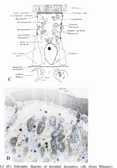
The absorptive cells are separeted from the core of the villus by a thin basal lamina.
The base of the cells rests on this basal lamina and apical parts of neighbouring cehs are
conected by junctional complexes. Ceh junctions can be classified into three fiinctional
groups: 1) occluding junctions, 2) anchoring junctions, 3) communicating junctions
(Alberts et al, 1994) (Fig. 1.2). Occluding junctions, also known as tight junctions or
zonula occludens., are discussed below (section 1.2.3). Anchoring junctions occur in three stmcturally and fimctionally different forms: adherens junctions ( zonula adherens ), are connection sites for actin filaments, while desmosomes {macula adherens ) are connection sites for intermediate filaments between the cells and hemidesmosomes are connection
occludens^ zonula adherens and macula adherens is known as a junctional conçlex (Wheater, 1995).
The epithelial cells of the entire intestine are covered by a mucous layer, consisting
of water, glycoproteins (mucins), electrolytes, proteins, and nucleic acids. The layer is
bound to the apical cell sur&ce by the glycocalyx, a 500nm thick glycoprotein structure
vsèich is covalently linked to %ids and proteins of the brush border membrane. This
glycoprotein layer Wiich has been termed the surfece coat, is resistant both to mucolytic
and proteolytic agents and has a protective fimction as well as being involved in the
digestive process (Krause and Cutts, 1981). The mucous layer is a part of the unstirred
water layer and it is supposed that the minimal thickness of the unstirred water layer is
about 500pm (Thomson and Dietschy, 1984). Because of its aqueous nature, the unstirred
water layer is considered to form an absorption limiting barrier for the conq)ounds with a
high lipid-water partition coefl&dent, e.g. aromatic hydrocarbons (Rahman et al, 1986)
and long-chain fetty acids (Thomson and Dietschy, 1981).
The anatomy and physiology of gastrointestinal tract of the humans described so
&r is different from that of commonly used laboratory animals. In addition to metabolic
differences, the anatomical, physiological and biochemical differences in the
gastrointestinal tract of the human and laboratory animals can cause significant variation in
drug absorption from oral route (Kararh, 1995). The diameter of the small intestine in the
human is 5cm and in the rats (in our in vivo e?q)eriments we have used rats) it is 0.3- 0.5cm Villi are finger shaped in the mouse, pig and human and tongue shaped in the rats
V . I I O u J j
f o> m ei i wm )
B
f t I f VI M O " JO " »
C f l l i
G o O l » ! , '
8 ' o o a . » > i » n
t y i " 0 " N» f »»»
Smooim fnufci*
Co""»cii»* ii»»u»
Lyi Ti onocy»»» P l O j m o C»l l * £ g s i " 0 0 " i l « t
L a m . n o
propr.O
' - \ j " O i l t f f * " i i a t » d c e i l »
- • C o 6 i » i c e l l * I C f y p i - M i t o s e * y e p i t h e l i u m ' • E n d o c r i n e cel l*
P o n e t h c e l l *
M v i i C u l o n * m u c o s a
Figure 1.1 (A) Schematic diagram of intestinal epithelial cells. PC-phcae circulares or
valves of Kercking; V-vilh; PP-Peyer’s patches; MM-muscularis mucosae; S-submucosa;
CM-circular and LM-longjtudinal muscle; Se-serosa; (from Wheatefs Functional
Histology, 1995); (B) Cell types of the intestinal epithelium
[ n t f f C f l l u l o f j p a c t
N u c l e u s
• M I C R O VI L L I
Figure 1.1 (C) Schematic diagiam of intestinal absoiptive cells (from Wheatefs
Functional Histology, 1995); (D) Electron micrograph of the rat intestinal absoiptive
cells, TJ; Tight Junction, M: Mitochondria, X 32,000.
___________&___________
A ftcV Tight junction
r ¥ - ' :
^ ^ r ZonulcTadherens
eren s (d esm osom e)
Figure 1.2 Shematic diagram of intercellular junctions. Between the cells, a junctional
complex prevents access of luminal contents to the intercellular spaces and strengthening
the luminal sur&ce against cellular degradation (from Wheatefs Functional Histology,
1995).
1.2.2 The components and structure of cell membrane
The major structural confound of the cell membrane is the %id bilayer (Fig. 1.3
A). The major %id corcponents are phosphatidylcholine, glycolçids, cholesterol,
phosphatidylethanolamine, phosphatidylglycerol, phosphatidylserine, ^hingomyelin
(Alberts et al, 1994). The composition of the acyl chains of the phospho%ids and
glycolipids exhibits heterogeneity, with respect to both chain length and degree of
unsaturation. The polar heads are mainly derived j&om glycerol conjugated to a
nitrogenous conq)ound such as choline, ethanolamine or serine via a phosphate bridge.
The phosphate group is negatively charged whereas the nitrogenous group is positively
charged. The non-polar tail of the phospho%id molecules consists of two long-chain 6tty
acids each covalently linked to the glycerol conçonent of the polar head. In most
mammalian cell membranes, one of the fetty acids is a straight-chain saturated fetty acid
\Ahilst the other is an unsaturated fetty acid which is "kinked" at the position of the double
bond (Wheater, 1995) (Fig. 1.3 B, C, D).
The fluidity of a %)id bilayer depends on both its con^oshion and temperature
(Alberts et aL, 1994). The fluidity of the membrane in increased by the presence of the
unsaturated fatty acids Wdch prevents close packing of the hydrophobic chain. Natural
6tty acids influence membrane order due to their c/j-double bonds, disrupting
phospho%id arrangement (Stubbs and Smith, 1984). Carrying shght negative charges,
unsaturated 6tty acids repel each another, do not easily aggregate and maintain the
membrane in its fluid state. Their negative charge makes them weakly basic and able to
form weak hydrogen bonds with weak acid groups such as the su^hydryl groups found in
proteins.
Proteins are embedded in the lipid bilayer by their hydrophobic segments. A
variety of protein molecules make up almost half of the total mass of the membrane.
Because optimal activities of membrane-bound enzymes require fluid-state membranes,
transition fio m the gel phase to the fluid liquid-crystalline phase occurs) below
environmental temperature.
Cholesterol, amphipathic and having a "kinked" conformation, intercalates
between phospholipid molecules with the non-polar ting embedded in the hydrocarbon region and exerts a regulating action on membrane structure, increasing fluidity of gel-
state membranes and decreasing fluidity of liquid-crystalline membranes (Wheater, 1995).
Cholesterol prevents too close packing of the phospholipid fatty acids tails whilst filling
the gaps between the "kinks" of the unsaturated fatty acids tails. By decreasing the
mobility of the first few CH] groups of the hydrocarbon chains of the phospholipid
molecules, cholestrol makes the lipid bilayer more rigid in this region and therfore
decreases the peimeability of the bilayer to the small water-soluble molecules (Albeits et
al., 1994). Sphingomyelin has been suggested to enhance the ordering effect of cholesterol
(Van Blitterswijk et al., 1987). For these reasons, the fluidity of fluid-state membranes may
increase with decreasing cholesterol/phospholipid molar ratio or increasing total-
lipid/protein ratio. Saturated fatty acids also provide membrane rigidity.
Many of the membrane proteins and some of the membrane lipids are conjugated
with short chains of polysaccharide; these glycoproteins and glycolipids respectively project fi om the surface of the bilayer forming an outer coating which may be analogous
to tlie cell walls of plants, bacteria and fungi. This polysacchaiide layer has been termed
the glycocalyx. The apical membrane, which faces the intestinal lumen is rich in
glycolipids, and serves to rigidify this membrane at body temperature due to the high Tm of these lipids.
Figure 1.3 (A) “fluid mosaic” model of membrane structure (fi'om Wheatefs Functional Histology, 1995).
B
where ‘R’ = choline inositol ethanolamine serine
glycerol backbone'
18;2w6 bonds
more solid more fluid
18:2w6 18:3w3 18:1w9 18:0'
(saturated)
cMestBfol
18:1w9 18:3w3
phospholipid protein
D
1.2.3 Tight junctions
The tight junction is a narrow beh that wraps apical ends of epithelial cells regions,
separates the intestinal lumen from the paracellular space, and forms a barrier to the
diffusion of the molecules and ions across the epithelial cell layer through the paracellular
pathway (Gumbiner, 1987; Madara, 1988). In freeze-fracture electron microscopy, the
tight junction appears as a set of continuous, anastomosing intramembrane strands This
seal restricts diffusion of small molecules in a charge specific manner. Tight junctions are
cation selective and they have been suggested to be mpermeable to cations with a
diameter exceeding 1. Inm or with molecular weight higher than 350 (Madara, 1989; Van
Hoogdalem, 1989). Four tight junction proteins have been identified and include: ZO-1,
Z 0 2 , cmgulin and 7H6 antigen (Stevenson et al, 1988, 1989; Madara, 1989; Hochman
and Artursson, 1994). The tight junction is also important to the organization of the
transcellular pathway, because it participates in the polarization of the epithelial plasma
membrane into the conposhionally distinct apical and basolateral domains (Gumbiner,
1987). Only four tight junction proteins have been identified and include: ZO-1, ZO-2,
cmguhn and 7H6 antigen (Stevenson et al, 1988, 1989; Madara, 1989; Hochman and
Artursson, 1994). Biochemical structure and regulation of tight junction need more
investigations in order to design more specific way to ‘loose” tight junctions.
1.2.4 Absorptive mechanisms of drugs across intestinal mucosa
Drugs are transported across the intestinal mucosa by two major mechanisms:
passive dififiision or a carrier-mediated transport system, either by fidlitated diffusion or an
1.2.4.1 Passive diffusion
Passive diflUsion is entirely dependent on the presence of a concentration gradient
across the plasma membrane, from the region of high relative concentration to a region of
lower relative concentration. The transfer of substances across the membranes can be
explained on the basis of Picks law in the form:
dQ /d t= -D A K (C o-C i)/h
Wiere Q = the amount of drug at the absorption she, D - difSision coefi&dent, A = area
of absorption surfrce, Co = concentration of drug in the outer phase of membrane, Cj =
concentration of drug in the inner phase of membrane and h = thickness of the membrane.
Lÿids and lipid-soluble compounds such as ethanol pass freely through plasma
membranes which also offer little barrier to the dififiision of the gases such as oxygen and
carbon dioxide. The plasma membrane is, in general, impermeable to hydrophilic
molecules. Nevertheless some small molecules, including water and urea, and inorganic
ions such as bicarbonate, are able to pass down osmotic and electrochemical gradients
through the membrane via hydrophilic regions.
1.2.4.2 Carrier mediated transport
Facilitated dififiision is also concentration-dependent and involves the transport of
larger hydrophilic metabolites such as glucose and amino acids. This process is passive,
but requires the presence of "carriers" to which the metabolites bind reversibly. In the
small intestine, drugs with structural similarities to nutrients may be taken up by frdlitated
dififiision or by active transport. Active transport is not only independent of concentration
gradients but also operates against extreme concentration gradients. The classic example
of this form of transport is the continuous transport of sodium out of cell by the "sodium
Active transport is mediated by "dynamic pores" consisting of transmembrane
protein systems. Many nutrients such as sugars, bile salts, and vitamins are transported
across the intestinal membrane by this way (Kararli, 1989). The anticancer drug 5-FU
(Schanker and Jefrey, 1961), a-methyldopa (Hu et al, 1989), cephadroxil (Kimura et al,
1983) are also transported by this mechanism
1.2.4.3 Bulk transport
Bulk transport involves large molecules or small particles being absorbed by the
plasma membrane forming membrane-bound vacuoles (vesicles) within the cytoplasm
When the process involves the creation of small vacuoles it is known as pinocytosis, and Wien large vacuoles are formed it is called phagocytosis.
1.2.5 Pathways of drug absorption
Drugs may be absorbed across the epithelial cell (transcellular) through the lipoidal
membrane or between the cells via the tight junctions and intracellular space (paracellular)
(Jackson, 1987; Gumbinger, 1987; Madara, 1989). Drugs lAiiich are presented in the
lumen can enter the blood stream through three processes: active or fecihtated transport,
passive transcellular transport, passive paracellular transport. Lipid-soluble confounds
with low molecular size are believed to pass mostly across the lÿoidal barrier of the
epithelium by a passive or carrier-mediated transport system Ionized or polar organic
solutes are absorbed with difficulty, probably via both permeation routes. The paracellular
route is considered to be most inçortant for transport of ions and small molecules carrying
a positive charge. There are reports of different classes of confounds Wiich could
increase paracellular route flux, such as calcium chelators, sodium dodecyl suphate,
palmitoylcamitine, sodium salts of fatty adds and a pentapepide (Pz^peptide) (Hochman
and Artursson, 1994; Yen and Lee, 1994, 1995). Polar macromolecules cannot be
1.3 Methodology to study intestinal transport,
metabolism and absorption enhancement
The methods used to investigate intestinal transport and metabolism can be
classified into three categories: 1) in vitro, 2) in situ and 3) in vivo methods.
In vitro systems include the everted intestinal segment, everted intestinal sac, Ussing chambers, isolated cells (discussed in following part, 1.3.1), brush border
membrane vesicles (BBMV) and basolateral membrane vesicles (BLMV) often from small
rodents such as mice, rats and guinea pigs (Wood and Lawrence, 1991) (Table 1.1). Due
to their sinpbcity and controllability, these systems have been widely used and have
provided information on transport of drugs and nutrients (Baker et al, 1991). The everted
intestinal segment (also known as the intestinal ring) and everted intestinal sac are the most
common in vitro techniques and in both cases the v^ole small intestine (colon) is removed fiom starved rat and everted onto a glass rod, ensuring that the mucosal 6ce is in contact
with bathing solutions. Alternatively, small sections of isolated intestine (colon) may be
mounted in Ussing chambers. Ussing chambers may be used to study transfer of
macromolecules across Peyefs patches. The major limitation of in vitro methods is the presence of urmatural absorption barriers (comiective and muscle tissue) which is the rate
limiting step to in vitro absorption. In vivo, the drug will be removed by the vasculature immediately after crossing the enterocytes (Wood and Lawrence, 1991). Therefore, Wolfe
at al (1973) proposed a method of stuping the musculature Wiich caused no change in
the integrity of the epithelium BBMV and BLMV allow the processes by Wiich a
molecule is transferred across each individual membrane barrier to be studied in isolation.
They model only transcellular processes. They are very stable and can be made from fresh
or frozen tissue (usually rat and rabbit), but the techniques can be adapted to allow vesicle
preparation from the small tissue sangles obtained with human biopsy and post-mortem
Isolated intestinal cells are thought to retain some active transport systems (study
of active and passive absorption), but not via paracellular pathway. The isolated cells lose
their polarity and are viable for only 30-60 minutes and therefore they are rarely used for
absorption studies.
Table 1.1 Summary data for in vitro models (from Wood and Lawrence, 1991) Model Everted Segment Everted Sac Ussing chamber
BBMV BLMV Isolated Cells
Cell Mono layers
diffixsion Y Y Y Y Y Y Y
active transport
Y Y Y Y Y Y Y
trans cellular
Y Y Y Y Y Y Y
para cellular
Y Y Y N N N Y
inte stinal
Y Y Y Y Y Y Y
colonic Y Y Y N Y ? Y
muscle barrier
Y* Y* Y* N N N N
viability H H H H H H W
^ +1tissue can be stripped; Y=yes; N=no; ?=unknown; H=hours; W=weeks
Lawrence, 1991). In these methods, after the animal is anaesthetized, a midline incision is
made and the small intestine e?q)osed. The rate of drug disappearance from the lumen or
its appearance in the portal venous (blood) is a measure of drug transport (Baker et al,
1991). Solutions do not require buffering, as intestinal tissue retains its natural buffering
capacity throughout the e?q)eriment. In in situ methods, although the animal has been surgically manÿulated and anesthesized, the mesenteric blood flow is intact.
In vivo systems include animal and human models and they are, of course, the most ircq)ortant ones. There are certain situations that allow absorption to be studied in
man such as scintigraphic methods using radiolabelled drug etc. Human absorption
e?qperiments tend to rely on studies involving serial blood, urine and frecal sanqrling
following oral administration of the tested compound. The understanding of physiological,
anatomical and biochemical differences between the G.I. tracts of different animal species
can lead to the selection of the correct animal model to mimic the bioavailability of
compounds in the human. In general, current data indicate that no single animal can mimic
the G.I. characteristics of humans (Kararh, 1995). Therefore, human studies cannot be
substituted by animals, but for a given purpose, selection of the right animal model is
possible.
More than one method has to be adopted to study mechanisms of uptake and to
be able to evaluate the hqportance of the mechanism in the wfrole animal. Each method
reported so frr has its relative advantages and drawbacks \\ftich must be understood if the
results are to give accurate correlations between e^qrerimental and in vivo absorption proiSles.
1.3.1 Cell cultures as models for drug absorption studies
Three different strategies have been used to obtain intestinal epithehal cell
cultures: 1) cultivation of normal intestinal cells, 2) cultivation of intestinal explants,
and 3) cultivation of neoplastic epithehal cells derived from human adenocarcinomas
way to obtain a source of intestinal epithelial cells, because primary dififerentiated cell
lines are difficult to maintam. Small pieces of small intestinal mucosa have been
maintained in cell culture for up to 24h, while explants from rat intestine have
survived for longer periods (Artursson, 1991). Isolated intestinal tissues are composed
of the normal complement of epithelial cell types found in that region of the gut, vdiile cell
cultures are typically composed of only one cell type.
Some advantages of cell culture drug absorption model Advantages of cell culture techniques include the abihty to
1) facihtate characterization of drug delivery mechanisms at the molecular/ceUular
level
2) aUow rapid assessment of potential drug permeabihty, metabohsm and transport
3) allow rapid evaluation of strategies for achievmg drug targeting and enhancement
4) achieve absolute control of physicochemical environment, temperature, osmotic
pressure, concentration of solutes, pH
5) achieve good reproducibihty
6) make assessments of drug absorption with low drug concentration,
and
7) minimize animal use, but can not completely replace them
Legal, moral and ethical questions of animal experiments are avoided
Disadvantages of cell culture Disadvantages include:
1) the need to maintain strict aseptic conditions
2) the need to take care in interpretation of results
3) the difficulty in predicting therapeutic benefit from the data
Most studies on drug transport and absorption enhancement have been performed
in a few epithelial cell lines originating either from the canine kidney (MDCK) or human
1.3.1.1 C aco-2 cell c u ltu re system
The Caco-2 cell line is the most commonly used in studies of drug absorption and
absorption enhancement, due to its ease of cultivation and spontaneous differentiation to
enterocyte-like cells under standard cell culture conditions (Hidalgo et al., 1989;
Artursson, 1990; Hilgers et al., 1990; Anderberg and Artursson, 1994; Hillgren et al.,
1995; Borchardt, 1995).
In contrast to other in vitro systems, the formation of an intact epithelial layer on filter support, allows the undirectional and quantitative transport of molecules across the
cell layer to be measured (Wilson et al., 1990) (Fig. 1.4). Polycarbonate filters allow
access of drugs to both the apical and basolateral sides of the epithelium, and Caco-2 cells
form tight and well differentiated monolayers on these filters without the need for
extracellular matrix proteins such as collagen (Artursson et al., 1996).
A P I C A L
c h a m b e r
m o n o l a y e r
F I L T E R B A S O L A T E R A C H A M B E R
M I C R O V I L L I
T I G H T J U N C T I O N
N U C L E U S
M E M B R A N E f i l t e r
Figure 1.4 Schematic drawing of the cell culture model (from Anderberg and Artursson,
1994)
Characterization of the morphological appearance with transmission and scanning
electron microscopy is usually followed by the investigation of the permeability properties.
The permeability characteristics are obtained by measurement of the transepithelial
electrical resistance (TEER) and by transport experiments with paracellular markers such as
the potential diflference between the apical and basolateral sides of the cell monolayer and
is inversely related to the mainly sodium and chloride ions across the monolayer
(Anderberg and Artursson, 1994). The passive ion flow through the tight junction and the
“active” ion tranport in the cell membrane both contribute to the measured TEER The
absence of well-developed tight junctions will result in leaky monolayers, detected by
decrease in TEER Hydrophilic marker molecules permeate the normal epithelial cells
through the paracellular spaces and as a results the permeability of these markers across
intact epithelia is very low. The permeability of the paracellular pathway in Caco-2
monolayers is relatively low, probably due to colonic origin of the cells.
The bmsh border enzymes are of great irtq)ortance in mucosal drug metabolism
and include: peptide-metabolizing enzymes such as d^eptidylpeptidase IV and
aminopeptidase N, drug-metabolizing enzyme phenol sulfotransferase, enzymes involved
in the detoxication of anticancer dmgs glutathione-S-transferase.
Caco-2 cells form confluent monolayers of polarized epithelial cells joined by tight
junctions and e?gress a bush border. The subepithelial barriers such as the basement
membrane, lamina propria, and the capillary wall are not generally considered to be rate
limiting for the absorption of small drug molecules. With the exception of mucin, the
Caco-2 cell model seems to contain the three major permeability barriers to the
asbsorption of the drugs: a) the “unstirred” water layer, b) the junctional con^lex, and c)
cell membrane (Artursson, 1990).
Caco-2 cell monolayers have been used extensively to predict intestinal absorption
of drugs such as atenolol, dexamethasone and proteins in human large intestinal tissue,
calcium transport, various vasopressin analogues, safety of P-cyclodextrins, (Rubas et al,
1996; Surendran et al, 1995, 1996; Artursson and Magnusson, 1990; Brondsted et al,
1995; Totterman et al, 1997). Starch microspheres induced a pulsed delivery of insulin
across the monolayers of human epithelial (Caco-2) cells in vitro. The pulsed delivery
correlated with a reversible appearance of focal dilatations in the tight junctions between
the epithelial cells, indicating enhanced delivery of insulin by the paracellular route
and markers through Caco-2 cell monolayers will be mentioned in part 1.5.1.1 of
Introduction and in Chapter 3.
1.4 Factors affecting drug absorption
The Actors i\hich influence drug absorption are: a) physicochemical Actors:
lÿophilicity, ionization of drugs, so Ability of drugs, stability m the gastromtestinal tract,
food and additives such as surActants, oils, sugars, alcohol and b) biological Actors:
structure of the gastromtestinal tract, gastromtestinal pH, mtestinal motility, blood and
lynph flow, gastromtestinal mucin and bile, gastric ençtying rate, disease state, drug
metabolism (Mayersohn, 1979).
The primary physicochemical properties of the drug infhiencmg its passive
absorption into and across biological membranes are its oil/water partition coefficient, the
extent of ionization m biological fluids which is determined by its pKa value and the pH of
the fluid. The most inq)ortant piincÿle of drug absorption is the pH-pardtion theory,
which treats the mtestine membrane as a lÿoidal barrier and proposes that the unionized
form of the drug molecule is preferentially absorbed (Shore et al, 1957).
In general, it is well known that a drug molecule, which has a molecular weight of
more than 1000, would not be readily absorbed in an intact form from the gastromtestinal
tract. Polysaccharide (starch), molecular weight around 50000 and inulin, molecular
weight around 5000, have absorption rates pmoklOOmg wet wt/h of zero, whereas
pentose,molecular weight 150, has an absorption rate 2.2 pmol/lOOmg wet wt/b
1.5 Enhancement of drug absorption from the
gastrointestinal tract
Many current therapeutic drugs such as antibiotics, peptides and proteins, are
poorly absorbed because of their hydrophilic properties, ionic charge, high molecular
weight and extensive hydrolysis. The question is vsiiat kind of methodologies should be
adopted to inqirove the intestinal absorption of various confounds? Methods for
in^rovement include: 1) the use of absorption enhancers such as surfece active
confounds and protease inhibitors; 2) analogue synthesis-change in the physicochemical
properties of drugs such as %ophihcity, charge, solubility, configuration, chemical
stabihty, enzymic liability and 3) different dosage forms such as lÿosomes, niosomes,
emulsions, particulate carriers and novel systems (Hayashi, 1993).
1.5.1 Absorption enhancers
The use of enhancer systems which are directed to change the membrane
permeability is a complex topic and involves a variety of mechanisms. Absorption
enhancers belong to different chemical groups (Table 1.2). Figure 1.5 illustrates three
A.
a o i c a l s i a e
m
iuiM
t ’QOt junction
o . o , o.
b a so la tera l stoe
Figure 1.5 Surface active absorption enhancers are believed to increase the permeabihty of the cell membrane (A) calcium chelators are beheved to act mainly at the tight junctions (B) but there are many indications that most enhancers affect both the cell membranes and tight junctions (C) (from Hochman and Artursson, 1994).
Mechanisms for improvement of intestinal membrane permeability by absorption
enhancers can be summarized as follows:
Transcellular pathways
1. Membrane perturbation by interaction between the enhancer and membrane lipid or
protein, eg. surfactants, medum chain fatty acids, carnitine, sahcylic acid, mixed micelles.
2. Disorder of membrane status by decrease in membrane nonprotein thiol, eg. diethyl
maleate, sahcyhc acid.
Paracellular pathways
1. Chelation between enhancer and calcium or magnesium around tight junction, eg.
EDTA, bile sahs, medium chain fatty acids.
2. Stimulation of junctional actomyosin contraction, eg. glucose, aminoacids, mixed
Table 1.2 Classes of enhancers of intestinal drug absorption and some of their
representatives
Surfactants
non-ionic: polyoxyethylene ethers anionic: sodium laurylsulfate
cationic: quaternary ammonium conq)ounds
Bile salts
dihydroxy bile salts: deoxycholate, taurodeoxycholate, chenodeoxycholate, glycochenodeoxycholate, ursodeoxycholate
trihydroxy bile salts: sodium cholate, taurocholate, glycocholate
Mixed micelles
glyceryl monooleate, fatty acids+sodium taurocholate
Sodium tauro-24,25-dihydrofusidate (STDBDF)
Fatty acids
capryhc, capric, lauric, oleic, linoleic, linolenic acid
Medium-chain glycerides
glyceryl- 1-monooctanoate, mono/di-glycerides
Non-steroidal anti-inflammatory drugs and derivatives
sodium sahcylate
sodium 5-methoxysahcylate indomethacin
diclofenac
Chelating agents (Calcium binding agents)
1.5.1.1 Non-ionic, anionic and cationic surfactants
The eflFect of surfectants on drug absorption have been already reported (Florence,
1981; Gibaldi and Feldman, 1970; Attwood and Florence, 1983). Amphiphilic
(anq)hÿathic) coropounds are those which possess both polar and non-polar groups and
most amphiphilic confounds are surjetants. Non-ionic surjetants possess a polar
“headgroup”, often a polyoxyethylene chain and a non-polar hydrocarbon chain. Most
commercial polyoxyethyjted non-ionic detergents are polydisperse in the length of
polyoxyethylene chain, and some in relation to their hydrophobic chain length. It has been
generally accepted that surfactants vsftich are too hydrophobic to be water soluble are poor
enhancers, Wiile surjetants which are very hydrophilic cannot partition effectively into the
hydrophobic environment of the lipid bdayer (Attwood and Florence, 1983).
Surfactants winch increase cellular permeability generally do so by disturbing the
membrane (Fig. 1.6). When a small amount of detergent is present, the molecules of
detergent are incorporated into the membrane without breaking, but changing the physical
properties of the cell membrane. At high surjetant concentrations in the plasma
membrane surjctant-surjctant contacts occur, and the membrane can be dissolved into
surjctant-membrane mixed micelles. Mixed protein-lq)id-detergent micelles are in
equilibrium with detergent micelles and free detergent molecules. Pure protein-detergent
micelles may he obtained in equilibrium with detergent-lÿid and detergent micelles (Gulik-
Krzywicld, 1975; Helenuis and Simons, 1975; Lichtenberg, 1985).
The size and shape of both the alkyl chain and the polar group influence absorption
enhancing capability. Florence et al (1978) and Walters et al (1981) studied the influence
of a range of polyoxyethyjted non-ionic surjetant upon the transport of paraquat
dichloride across rabbit iso jte d gastric mucosa. By varying both the alkyl chain length and
polyoxyethyjted chain length, the most effective absorption promoter was found to be
Brij 36T (C12E10, wMe Eio = a polyoxyethylene chain with 10 ethylene oxide units) and
rank order for other surjetants in promoting paraquat absorption was CisiEio > CigEio >
solubility between water and oil and ability to penetrate the lipid bilayer easily. Lauryl
surfactants may penetrate the lipid bilayer readily and because of their aqueous solubility
possess a greater monomer concentration and higher CMC than the longer ones. From
C12 to C l8, the thermodynamic tendency to partition into non-aqueous environments
increases, and the decreasing concentration of the surjetants monomers may produce
only lower response. However, Guarini and Ferrari (1984) demonstrated that C16 and
C18 ethers were more effective than C12 ethers in studies on absorption of orally dosed
heparin in rats.
m e m b ra n e pro tein in lipid b ilay er
w a te rs o lu b le p ro te in -lip id -d e te rg e n t c o m p le x
d e te rg e n t m ic elles
d e te rg e n t m o n o m e rs
s o lu b le m ix ed lip id -d e te rg e n t
m ic elles
Figure 1.6 Solubilizing membrane proteins with a detergent. The detergent disrupts the lipid bilayer and brings the proteins into solution as protein-lipid-detergent complexes. The phospholipids in the membrane are also solubilized by detergent (from Alberts et al, Molecular Biology of the Cell, 1994).
The effects of sur&ctants on drug absorption are often correlated with toxic
effects on mucosal integrity, involving epithelial cell loss, release of proteins and %ids, and
unspecified musocal damage (Van Hoogdalem et al, 1989; Anderberg et al, 1993;
Anderberg and Artursson, 1992, 1993; Sugimura, 1974 a, b; Swenson and Curatolo,
1992). Ishizawa et al (1987) noted that a 1% polyoxyethylene lauryl ether was effective in
increasing fosfomycin absorption. Polyoxyethylene-23-lauryl ether (C12E20) significantly
enhanced the absorption of cefoxitin from the rat rectum, but released protein from the
rectal mucosa (Nishihata et al, 1985).The intestinal absorption of ergot peptide alkaloids
was enhanced by micellar solutions of polyoxyethylene-24-cholesteryl ether, equivalent to
Sohdan C24 discussed later in the thesis (Franz and Vonderscher, 1981).
Polysorbates 20, 21, 40, 60, 80 and 85, (the Tweens^) were investigated for their
effect on the permeability of the rat rectal membrane by Nakanishi et al (1983),
polysorbate 20 being the most potent. When added in aqueous solutions, they did not
significantly affect permeability, wMe they affected the histological nature of the rectal
tissue and enhanced the permeability of sut&nilic add wfien they were added in oils such
as Miglyol and olive oil Azmin et al (1982 and 1985a) demonstrated that polysorbate 80
could increase the absorption of methotrexate from the mouse gastrointestinal tract and
enhance the drug uptake into the brain without a damaging effect on the gastrointestinal
mucosa.
The effect of the anionic surfectant, sodium lauryl suÿhate on absorption of
ce&droxil in the rat colon has been studied by Sancho et al (1995). The well-known
ability of sodium dodecyl suphate to disrupt membranes and denaturate proteins suggests
its possible role as an absorption enhancer. Heparin absorption has been shown to be
increased by incorporation of sodium dodecyl suphate in an enteric coated capsule dosage
form in dogs (Engel and Riggi, 1969).
The effects of different surfectants have been tested on Caco-2 cell monolayers
such as: sodium caprate, sodium dodecyl sulfrte, sodium dioctyl sulfosucdnate,
polysorbate 80, polyoxyl 40 hydrogenated castor oil, bile salts (Anderberg et al, 1993;