International Journal of Nanomedicine
Dovepress
open access to scientific and medical researchOpen Access Full Text Article O R I g I N A L R e s e A R c h
Binding fullerenol c
60
(Oh)
24
to dsDNA
Mariana Pinteala Andrei Dascalu cezar Ungurenasu
Petru Poni Institute of Macromolecular chemistry, Aleea grigore ghica, Iasi, Romania
correspondence: cezar Ungurenasu Petru Poni Institute of Macromolecular chemistry, Aleea grigore ghica Voda 41 A, 700487 Iasi, Romania
email [email protected]
Abstract: The first C60(OH)24-DNA complex and its fluorescence enhancement is reported. The enhanced fluorescence intensity of fullerenol C60(OH)24 is in proportion to the concentration of DNA in the range of 1 × 10⁻⁹ to 8 × 10⁻⁵ molL-1 and the detection limit was 1.3 ng mL-1.
Fullerenol C60(OH)24 binds significantly to the phosphate backbone of native dsDNA and to base-pairs within the major groove of sodium salt of dsDNA.
Keywords: nanomedicine, fullerenol, DNA complexation, fluorescent probe
Introduction
Nanoscale materials seems to offer great opportunities for biomedical applications such as therapeutic and diagnostic tools.1–9 Biomedical applications under
develop-ment include drug delivery systems targeted to the brain and cancer tissues, gene transfection, and intravascular nanosensor and nanorobotic devices for imaging and diagnosis.3,9
In this context, the biological activities of fullerene derivatives have attracted much attention in the past 25 years.10–17 As potent free-radical scavengers and antioxidants18–23
the water-soluble polyhydroxylated [C60]fullerenes, fullerenols, exhibit an exciting range of biological activities as glutamate receptor antagonists,24 antiproliferative,25–27
neuroprotective,28–31 and anticancer agents.32–37
Knowing the ways fullerenols interact with proteins and nucleotides is a prerequisite for understanding their biological effects at membrane penetration and the intracellular level, only two studies deal with their binding to proteins38,39 and their interaction with
DNA has not been reported to date.
On the other hand, the solution-based assays and quantitative analysis of nucleic acids are critical in current biochemistry and biomedical science. Throughout the years, a number of fluorimetric methods for the determination of nucleic acids have been developed with ethidium bromide,40–42 lanthanide cations,43–45 ruthenium complexes,46–48
and asymmetric cyanine dyes as fluorescence probes.49–51
Despite the prominence of fullerenes in bionanotechnology, the exploration of their fluorescent properties in solution remains still at a very early age. Several studies have been devoted to dsDNA/single-walled carbon nanotube hybrid systems,52,53 but
only very few deal with their fluorescent proprieties54,55 when dispersed in aqueous
solution.
Herein, we are happy to report the first complexation of dsDNA with C60(OH)24 in aqueous media in the absence of a buffer in physiological pH-range.
International Journal of Nanomedicine downloaded from https://www.dovepress.com/ by 118.70.13.36 on 23-Aug-2020
For personal use only.
Number of times this article has been viewed
This article was published in the following Dove Press journal: International Journal of Nanomedicine
Pinteala et al Dovepress
Materials and methods
C60 (99.5+%) was purchased from MER Corp (Tuscon, AZ, USA). KOH (99.99%, semiconductor grade) was purchased from Sigma-Aldrich (St. Louis, MO, USA). DNA (low molecular weight, salmon sperm) was purchased from Fluka (St. Louis, MO, USA). All other reagents were purchased from Sigma-Aldrich.
Fluorescence spectroscopy was performed with a Perkin Elmer LS55 spectrometer Perkin Elmer, Wellesley, MA, USA). To prepare fluorescence samples, the only opera-tion was the mixing of two soluopera-tions before fluorescence measurements. X-ray photoelectron spectroscopy (XPS) measurements were carried out using a Leybold LHS 10 spectrometer (Leybold, Cologne, Germany).
To the best of our knowledge, most of these fullerenols are not pure C60(OH)n, but a complex mixture of products. For instance, those synthesized through sulfuric/nitric acid,56
hydroboration,57 or nitronium chemistry58 afforded products
with average composition of C60Ox(OH)y. The so-called fullerenols prepared by alkaline polyhydroxylation of C60 under phase transfer conditions59 are not simply C
60(OH)x,
but stable radical anions with the molecular formula, Na+
n[C60Ox(OH)y]n
-,60 and the fullerenol obtained by
alkaline hydrolysis of C60Br24 is not C60(OH)24 as claimed by Bogdanovi and Dvordjevic,37,61 but C
60(ONa)8(OH)16.62 In the
light of this, many biomedical studies involving fullerenols species in the literature may need to be reconsidered. The pure fullerenol C60(OH)24 used in this study was prepared by a modified method of alkaline hydrolysis of C60Br24,62
followed by demetallation of the obtained C60(OK)8(OH)16 with a cation exchange resin and exhaustive purification by dialysis.
Representative procedure for synthesis
of c
60(Oh)
24All experiments were performed with Schlenk techniques under argon and protected from light. According to the literature, before the synthesis of the polyhydroxylated fullerene, bromofullerene C60Br24 was synthesized first.62
In the synthesis of the C60(OH)24, to a sonicated (40 W, 15 min) suspension of C60Br24 (200 mg, 0.075 mmol) in de-aerated water (100 mL), fresh KOH (200 mg, 3.57 mmol) was added under argon protection and stirred for 10 days at room temperature. After the reaction was completed, the resulting dark-brown solution was passed to a centrifuge at 4000 rpm for 30 min and the supernatant was brought to dryness in a rotavapor apparatus at 40 °C. The dark-brown residue was dissolved in 50 mL of deionized water, stirred
with ion exchange resin AMBERJET™ 1200[H] (Rohm and Haas Company, Philadelphia, PA, USA) (20 mL) for 8 h and subjected to dialysis (Spectra/Por® 1000 D; Spectrum
Laboratories, Rancho Dominguez, CA, USA) for four days. Finally, the dialyzed solution was brought to dryness in a rotavapor apparatus at 60 °C and dried at 80 °C and 10-4 Torr for 24 h. The fullerenol thus obtained contained
24 hydroxyl groups as characterized by elemental analysis, Fourier transform infrared (FT-IR) spectroscopy, and XPS spectroscopic measurements.
elemental analysis
Calculated for C60H24O24: C, 63.82; H, 2.12. Found: C, 63.66; H, 1.98. FTIR (KBr): ν max, 3436 (-OH), 1605 (C = C), 1430 (δ –OH), 1095, 1046 (ν C-OH), 1018, 994, 825, 877, 570, 530 cm-1.
XPs analysis
C1s components: % C = C (284.6 eV) 59.77 (clcd. 60); % C-OH: (285.8 eV) 39,76 (clcd. 40); O/C = 0.42 (clcd. 0.40).
Results and discussion
The fullerenol water solution, exhibited different maxima depending on the concentration (Figure 1a). In the range of 1.6 × 10⁻⁵ to 4.4 × 10⁻⁵ molL-1 one fluorescent maximum
was observed at 469 nm, while two fluorescence maxima where found for lower concentrations located at 469 nm and 492 nm at λex= 420 nm.
These emission profiles of fullerenol at different concen-trations provided the baseline for understanding perturbation upon interaction with dsDNA. As shown in Figure 1a, the most appropriate concentrations of fullerenol in water for fluorescence measurements at λex= 420 nm are within the range of 1.6 × 10⁻⁵ to 4.4 × 10⁻⁵ molL-1 (λ
em= 469 nm).
As can be seen from Figure 1b, the fluorescence intensity of fullerenol alone is dependent on its concentration in the range of 4.4 × 10⁻⁵ to 1.6 × 10⁻⁵ molL-1 according to a very
significant linear relationship.
Figure 2 shows the fullerenol and DNA emission spectra recorded at different excitation wavelengths. Inspection of how fluorescence emission spectra of fullerenol and DNA change as a function of excitation wavelength yield additional supporting information on the appropriate fullerenol excitation wavelength suitable for the fluorescence investigation of C60(OH)24 – DNA complex in aqueous media. One can observe that for a concentration higher than 1.6 × 10⁻⁵ molL-1, the
emission spectra of fullerenol do not overlap with emission spectra of DNA when excited at 340, 360, 380, and
International Journal of Nanomedicine downloaded from https://www.dovepress.com/ by 118.70.13.36 on 23-Aug-2020
Binding fullerenol c60(Oh)24 to dsDNA
Dovepress
420 nm, respectively. Apparently, all these fluorescence excitations should be suitable for a fluorescence study of DNA-fullerenol interaction. However, the emission maxima of fullerenol at concentrations 1.6 × 10⁻⁵ molL-1 (Figure 1a)
and DNA (Figure 2a) overlap at 492 nm when recorded at
λex= 420 nm. This is the reason why, to cover a large concen-tration range (1-9-4.5-5 molL-1) of fullerenol, the fluorescence
excitation at 420 nm and emission at 469 nm were used for fluorescence intensity measurements in this work.
In Figure 3a, the emission spectra of DNA-fullerenol complexes, with constant DNA concentration and increasing fullerenol content are shown. It can been seen that increasing the concentration of the fullerenol results in a strong increase in fluorescence intensity of fullerenol from 50 to 500 nm, without causing any perceptible shifts of the fluorescence maximum at λ= 469 nm. In order to establish the DNA binding affinity of fullerenol, these fluorescent-enhancing
data were plotted (Figure 3b) according to the equation (6) derived from the equilibrium equation (1):
n[C (OH) ] + [DNA]60 24 K [C (OH)60 24-DNA] (1) K = [C60(OH)24- DNA]/[C60(OH)24]n [DNA] (2)
[C60(OH)24- DNA] = K[C60(OH)24]n [DNA] (3)
log [C60(OH)24- DNA] = log K + nlog [C60(OH)24] + log [DNA] (4) log [C60(OH)24- DNA]/[DNA] = log K + nlog [C60(OH)24]
(5) log F-F1/F0= log K + nlog [C60(OH)24] (6)
where F1 is the fluorescence intensity from the fullerenol in the absence of DNA (Figure 1b), F0 is the fluorescence intensity from the DNA in the absence of fullerenol at 467 nm for λex= 420 ( Figure 2a), F is the fluorescence intensity from the DNA-fullerenol complex in the presence of different concentrations of the fullerenol (Figure 3b) and n is the number of associated molecules of fullerenol with one base pair of DNA. From the linear plot for (log(F-F1)/F0) vs (log[C60(OH)24]) (Figure 5), according to equation (1), the values of K and n were estimated to be 6 × 10⁵ M-1 and
0.8 ± 0.2, respectively.
In order to evaluate the range of [DNA] determina-tion, the binding of fullerenol to DNA was characterized [C60(OH)24] rnol/L
[C60(OH)24]−5 rnol/L
400
350
300
250
200
150
100
50
0
400
B A
350
300
250
200
150
100
50
0
0 1 2 3 4 5
440
4.4 × 10⁻⁵
2.3 × 10⁻⁵
1.6 × 10⁻⁵
8.8 × 10⁻⁶
1.0 × 10⁻⁹
460 480 500 520 540 560 580 600
Wavelength (nm)
Fluorescence intensity
Fluorescence intensity
Figure 1 Fluorescence data of c60(Oh)24 in aqueous media. A) Fluorescence emission
spectra of c60(Oh)24 after 5 min incubation in water, with excitation at 420 nm.
B) Plot of fluorescence intensity versus [C60(Oh)24], with excitation at 420 nm;
average standard error, 3.76%.
Wavelength (nm) Wavelength (nm) Wavelength (nm)
Wavelength (nm)
Fluorescence intensity
400 600 800
200 0
450 500 550 600
400 600 800
200 0
400 600 800
200 0
400 600 800
200 0
450 500 550 600
450
400 500 550 600
450
400 500 550 600
B
C
D A
Figure 2 Fluorescence emission spectra of fullerenol ( - - - ) and dsDNA (_____)
for different excitation wavelength. A) λ= 420 nm. B) λ= 380 nm. C) λ= 360 nm. D) λ= 340 nm. [C60(Oh)24] = 1 × 10⁻⁴ molL-1; [DNA] = 1 × 10⁻⁴ molL-1.
International Journal of Nanomedicine downloaded from https://www.dovepress.com/ by 118.70.13.36 on 23-Aug-2020
Pinteala et al Dovepress
through fluorescence emission titration of fullerenol. The enhancement of the fluorescence intensity of fullerenol with DNA at increasing concentrations is shown in Figure 4. One can observe that even for nanoscale concentration of DNA the fluorescence intensity of fullerenol increases from 25 to 100 (Figure 4a). The plot in Figure 4b is bro-ken down into two regimes corresponding to ranges from 1.3 × 10⁻⁹ to 3.1 × 10⁻⁶ gL-1 and 2.5 × 10⁻⁵ to 5 × 10⁻⁵ gL-1.
The low [DNA] range in the plots of Figure 4b (detec-tion limit = 1.3 ng/mL) are close to what can be accom-plished with current available fluorescence probes, ie, Hoechst 33258 (20 ng/mL) and YO-PRO-1/YOYO-1 (0.5–2.5 ng/mL). In addition, from the shape and intensity of emission spectrum recorded for [DNA] = 2.1 × 10⁻⁹ molL-1
(Figure 4a), it is useful to point out that the sensitivity can be extended into lower regions.
As regards the chemical interactions between fullerenol and the DNA target, the electrostatic and intercalative binding are ruled out and hydrogen-bonding interaction can only be taken under consideration. Earlier studies38,39 have pointed out that
hydrogen-bonding plays the main role in the interaction between fullerenols and proteins. Thus, the major groove binding of fullere-nol through the hydrogen-bonding between its hydroxyl groups and free or bridged –NH2 in base-pairs of DNA is predictable.
Taking into account that phenols can interact with phosphates,63 the hydrogen bonding between fullerenol and
phosphate backbone of DNA can be also suggested as a possibility for the binding of fullerenols with DNA.
From the linear plot in Figure 3b according to equation (1) the value of n was estimated to be 0.8 ± 0.2. This value 600
400
300
100
0 200 500
540 560 580 600 520
500 480 460 440
Wavelength (nm)
Fluorescence intensity
log[C60(OH)24](mol/L)
−5.5 −5.0 −4.5 −4.0 −6.0
−6.5 −7.0 2.0
1.5
1.0
0.5
0.0
−0.5
logF−F1
A
B
C60 (OH)24 M
4.50 × 10⁻⁵
2.70 × 10⁻⁵
0.94 × 10⁻⁵
4.50 × 10⁻⁶
0.94 × 10⁻⁶
4.50 × 10⁻⁷
0.94 × 10⁻⁷
F0
Figure 3 Fluorescence data of c60(Oh)14 in the presence od dsDNA. A) Fluorescence
emission spectra of fullerenol (_____) in the presence of dsDNA ( - - -) with excitation at 420 nm, as a function of fullerenol concentration; [dsDNA] = 6.31 × 10⁻⁵ molL-1. B) Plot for DNA-c60(Oh)24 system as a function of fullerenol concentration in the range of 0.94 × 10⁻⁷ to 4.5 × 10⁻⁵ molL-1; [dsDNA] = 6.31 × 10⁻⁵ molL-1; average standard error, 0.17%.
0 50 100 150 200 250 300 350 400 450 500 0
440 460 480 500 520 540 560 580 600
50 100 150 200 250 300 350 400 450 500
1.3 E-9 1.3 E-8 1.3 E-7 3.1 E-6 2.5 E-5 3.75 E-5 5 E-5 8.0 × 10⁵ 4.0 × 10⁵ 2.1 × 10⁵ 5.0 × 10⁻⁶ 2.1 × 10⁻⁷
2.1 × 10⁻⁹ 2.1 × 10⁻⁸ [DNA] mol/L
[DNA] (g/mL)
Fluorescence intensity
Fluorescence intensity
Wavelength (nm) A
B
Figure 4 Dependence of fluorescence intensity of C60(Oh)24 on dsDNA concentration.
A) Fluorescence spectra of fullerenol (- ⋅ - ) with increasing concentration of DNA
(_____) with excitation at 420 nm ; B) Plot of fluorescence intensity of fullerenol as a function of [dsDNA] in the concentration range of 1.3 × 10⁻⁹ to 4.4 × 10⁻⁶ molL-1; [C60(Oh)24] = 4.5 × 10⁻⁵ molL-1; average standard errors: 2.69% for 2.5e-5-5e-5 region and 10.80% for 1.3e-9-3.1e-6 region.
International Journal of Nanomedicine downloaded from https://www.dovepress.com/ by 118.70.13.36 on 23-Aug-2020
Binding fullerenol c60(Oh)24 to dsDNA
Dovepress
groove of DNA (6 Å). The width of the major groove (12 Å) is larger than 9.8 Å and thus the fullerenol molecule can fit snugly according to a nonintercalation model as shown in Scheme 1a.
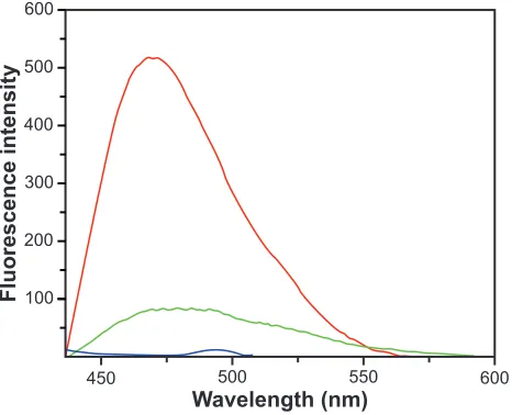
Indeed, upon binding to sodium salt of DNA (Figure 5), the fluorescence intensity of fullerenol does not increase, but decreases, which suggests that only in the absence of hydrogen bond-forming P-OH moieties of DNA, fullerenol binds to base-pairs into the major groove of DNA, according to a nonintercalative model, which strongly change its average local environment. The perceptible shift of the emission maximum of fullerenol also support a strong change of its average local environment. We can thus conjecture that, under the present experimental condi-tions with native dsDNA, hydrogen binding to phosphate backbone to the outside of dsDNA helix is the main bind-ing mode of fullerenol C60(OH)24 to DNA, as shown in Scheme 1b.
Conclusions
Fullerenol C60(OH)24 binds to phosphate backbone to the outside of native dsNNA and to base-pairs within major groove of sodium salt of dsDNA. The fluorescence of fullerenol C60(OH)24 is highly enhanced by dsDNA due to the binding of the probe to DNA in a nonintercalative way. Because of its high binding affinity (K = 105 M-1) and
sensitivity (1.2 × 10⁻⁹ g/mL) towards DNA, there are good
prospects that C60(OH)24 will be used as versatile fluorescent probe for DNA quantification. In addition to its high sensitivity, other advantages of this fullerenol-based method include its simplicity, nontoxicity, and rapidity.
Acknowledgments
We are grateful to the Romanian Ministry of Education and Research for funding through the CNCSIS-GRANT Nr.306. The authors report no conflicts of interest in this work.
References
1. Wilkinson JM. Nanotechnology applications in medicine. Med Device
Technol. 2003;14:29–31.
2. Roco MC. Nanotechnology: convergence with modern biology and medicine. Curr Opin Biotechnol. 2003;14:337–346.
3. Tong R, Cheng J. Anticancer polymeric nanomedicines. Polymer
Review. 2007;47:345–381.
4. Moghimi SM, Theme editor and T. Kissel T. Particulate nanomedicines.
Adv Drug Deliv Rev. 2006;58:1451–1455.
5. Torchilin VP. Targeted pharmaceutical nanocarriers for cancer therapy and imaging. AAPS J. 2007;9:E128–E147.
6. Chan WCW. Bio-applications of Nanoparticles. New York, NY: Springer Science; 2007.
7. Lammers T, Hennink WE, Storm G. Tumor-targeted nanomedicines: principles and practice. Br J Cancer. 2008;99:392–397.
450 500 550 600
Wavelength (nm)
Fluorescence intensity
600
500
400
300
200
100
Figure 5 emission spectra of c60(Oh)24 (red line), DNA-sodium salt (blue line) and
c60(Oh)24- DNA-sodium salt complex (green) line.
accounts for a single fullerenol molecule associated with a base pair of DNA. An important question is whether the fullerenol binds significantly to the phosphate backbone of DNA in addition to nonintercalative groove binding to base-pairs of DNA.
Because of its diameter (9.8 Å) the globular three-dimensional C60(OH)24 molecule does not fit the minor
Scheme 1 Binding fullerenol c60(Oh)24 to dsDNA. A) Binding fullerenol to the major
groove of sodium salt of dsDNA. B) Binding fullerenol to the outside of the dsDNA.
A
DNA DNA
B
C60(OH)24
International Journal of Nanomedicine downloaded from https://www.dovepress.com/ by 118.70.13.36 on 23-Aug-2020
Pinteala et al Dovepress
8. Muthu MS, Singh S. Targeted nanomedicines: effective treatment modalities for cancer, AIDS and brain disorders. Nanomed. 2009;4: 105–118.
9. Kreuter J. Nanoparticulate systems for brain delivery of drugs. Adv
Drug Deliv Rev. 2001;47:65–81.
10. Da Ros T. Twenty years of promises: Fullerenes in medicinal chemistry In: Cataldo F, Da Ros T, editors. Medicinal Chemistry and
Pharma-cological Potential of Fullerenes and Carbon Nanotubes. New York,
NY: Springer-Verlag; 2008.
11. Injac R, Radic N, Govedarica B, Dvordjevic A, Borut S. Bioapplication and activity of fullerenol C60(OH)24. Afr J Biotech. 2008;7:4940–4950. 12. Yamawaki H, Iwai N. Cytotoicity of water-soluble fullerene in vascular
endothelial cells. Am J Physiol Cell Physiol. 2006;290:1495–1502. 13. Cui D. Medicinal chemistry and pharmacological potential
of fullerenes and carbon nanotubes. In: Cataldo F, Da Ros T, editors. Functionalized Carbon Nanotubes and their Applications. New York, NY: Springer-Verlag; 2008.
14. Beuerle F, Lebovutz R, Hirsch A. In: Antioxidant properties of water-soluble fullerene derivatives. Cataldo F, Da Ros T, editors. Medicinal Chemistry and Pharmacological Potential of Fullerenes and Carbon
Nanotubes. New York, NY: Springer-Verlag; 2008.
15. Bosi S, Da Ros T, Spalluto G, Balzarini J, Prato M. Synthesis and ant-HIV propreties of new water-soluble bis-functionalized [C60]fullerene derivatives. Bioorg Med Chem Lett. 2003;13:4437–4440.
16. Nakamura E, Isobe M. Functionalized fullerenes in water. The first 10 years of their chemistry, biology, and nanoscience. ACC Chem Res. 2003;36:807–815.
17. Bosi S, Da Ros T, Spalluto G, Prato M. Fullerene derivatives: an attractive tool for biological applications. Eur J Med Chem. 2003;38:913–923. 18. Satoh M, Takayanagi I. Pharmacological studies on fullerene C60,
a novel carbon allotrope, and its derivatives. J Pharmacol Sci. 2006;100:513–518.
19. Lai HS, Chen Y, Chen WJ, et al. Free radical scavenging activity of fullerenol on grafts after small bowel transplantation in dogs. Transplant Proc. 2000;32(6):1272–1274.
20. Lai HS, Chen WJ, Chiang LY. Free radical scavenging activity of fullerenol on the ischemia-reperfusion intestine in dogs. World J Surg. 2000;24(4):450–454.
21. Wang H, Joseph JA. Quantifying cellular oxidative stress by dichlo-rofluorescein assay using microplate reader. Free Radic Biol Med. 1999;27:612–616.
22. Tsai MC, Chen YH, Chang LY. Polyhydroxylated C60, Fullerenol, a novel free-radical trapper, prevented hydrogen peroxide – and cumene hydroperoxyde – elicited changes in rat hippocampus in-vitro. J Pharm
Pharmacol. 1997;49:438–454.
23. Chiang LY, Lu FJ, Lin JW. Free radical scavenging activity of water- soluble fullerenols. J Chem Soc Chem Commun. 1995;12:1283–1286. 24. Jin H, Chen WQ, Tanh XW, et al. Polyhydroxylated C60, fullerenols, as
glutamate receptor antagonists and neuroprotective agents. J Neurosci Res. 2000;62:600–607.
25. Gelderman MP, Simakova O, Clogston JD, et al. Adverse effects of fullerenes on endothelial cells: Fullerenol C60(OH)24 induced tissue factor and ICAM-1 membrane expression and apoptosis in vitro. Int J
Nanomedicine. 2008;3:59–68.
26. Huang HC, Lu LH, Chiang LY. Antiprofilative effect of polyhydrox-ylated C60 on vascular smooth muscle cells. Proc Electrochem Soc. 1996;403:96–100.
27. Zhao QF, Zhu Y, Ran TC, et al. Cytotoxicity of fullerenols on Tetrahymena pyriformis. Nucl Sci Techniq. 2006;17:280–284. 28. Silva GA. Neuroscience nanotechnology: progress, opportunities and
challenges. Nat Rev Neurosci. 2006;7:65–74.
29. Silva GA. Nanotechnology approaches for the regeneration and neuroprotection of the central nervous system. Surg Neurol. 2005;63: 301–306.
30. Dugan LL, Lovett EG, Quick KL, Latharius J, Lin TT, O’Malley KL. Fullerene-based antioxidants and neurodegenerative disorders.
Parkinsonism Relat Disord. 2001;7:243–246.
31. Dugan LL, Gabrielsen JK, Yu SP, et al. Buckminsterfullerenol free radical scavengers reduce excitotoxic and apoptotic death of cultured cortical neurons. Neurobiol Dis, 1996;3:129–135.
32. Injac R, Radic N, Govedarica B, et al. Acute doxorubicin pulmotoxicity in rats with malignant neoplasm is effectively treated with fullerenol C60(OH)24 through inhibition of oxidative stress. Pharmacol Rep. 2009;61:335–342.
33. Injac R, Perse M, Cerne M, et al. Protective effects of fullerenol C60(OH)24 against doxorubicin-induced cardiotoxicity and hepatotoxicity in rats with colorectal cancer. Biomaterials. 2009;30: 1184–1196.
34. Injac R, Boskovic M, Perse M, et al. Acute doxorubicin nephrotoxicity in rats with malignant neoplasm can be successfully treated with fullerenol C60(OH)24 via suppression of oxidative stress. Pharmacol Rep. 2008;60(5):742–749.
35. Yin JJ, Lao F, Meng J, et al. Inhibition of tumor growth by endohedral metallofullerenol nanoparticles optimized as reactive oxygen species scavenger. Mol Pharmacol. 2008;74:1132–1140.
36. ZhuJ, Ji Z, Wang J, et al. Tumor-inhibitory effect and immunomodula-tory activity of fullerol C60(OH)x. Small. 2008;4(8):1168–1175. 37. Bogdanovi G, Kojic V, Dvordjevic A, Vojinovic M, Baltiv VV.
Modulating activity of fullerol C60(OH)24 on doxorubicin-induced cytotoxicity. Toxicol in Vitro. 2004;18:629–637.
38. Bingshe X, Xuguang L, Xiaoqin Y, Jinli Q, Weijun J. Studies on the interaction of water-soluble fullerols with BSA and effects of metallic ions. Mat Res Soc Symp Proc. 2001;675:W7.4.1–W7.4.5.
39. Zhao GC, Zhang P, Wei XW, Yang ZS. Determination of proteins with fullerol by a resonance light scattering technique. Anal Biochem. 2004;334:297–302.
40. Nafisi S, Saboury AA, Keramat N, Neault J-F, Tajmir-Riahi HA. Stability and structural features of DNA intercalation with ethidium bromide, acridine orange and methyleneblue. J Molec Struct. 2007;827:35–43.
41. Heller DP, Greenstock CL. Fluorescence lifetime analysis of DNA intercalated ethidium bromide and quenching by free dye. Biophys Chem. 1994;50:305–312.
42. Le Pecq JB, Paoletti C. A new fluorometric method for RNA and DNA determination. Anal Biochem. 1966;17:100–107.
43. Nishioka T, Yuan J, Matsumota K. Fluorescent lanthanide labels with time-resolved fluorometry in DNA analysis. In: Ferrari M, editor.
Micro/Nano Technology for Genomic and Proteomics. New York, NY:
Springer-Verlag; 2007.
44. Kubát P, Lang K, Zelinger Z, Král V. Formation of lanthanide(III) texaphyrin complexes with DNA controlled by the size of the central metal cation. J Inorg Biochem. 2005;99:1670–1675.
45. Selvin PR. Principles and biophysical applications of lanthanide-based probes. Annu Rev Biophys Biomol Struct. 2002;31:275–302. 46. Lakowicz JR. Principles of Fluorescence Spectroscopy. New York,
NY: Springer-Verlag; 2006. p. 681–697.
47. Gao F, Chao H, Zhou F, Yuan YX, Peng B, Jin LN. DNA interac-tions of a functionalized ruthenium(II) mixed-polypyridyl complex [Ru(bpy)2ppd]2+. J Inorg Biochem. 2006;100:1487–1494.
48. Song G, Li L, Liu L, et al. Fluorometric determination of DNA using a new ruthenium complex Ru(bpy)2PIP(V) as a nucleic acid probe. Anal Chem. 2002;18:757–759.
49. Zipper H, Brunner H, Bernhagen J, Vitzhum F. Investigations of DNA inter-action and surface binding by SYBR Green I, its structure determination and methodological implications. Nucleic Acids Res. 2004;32:e103. 50. Rengarajan K, Cristol SM, Mehta M, Nickerson JM. Quantifying
DNA concentration using fluorometry: a comparison of phluorophores.
Mol Vis. 2002;8:416–421.
51. Vitzhum G, Geiger H, Bisswanger H, Brunne J, Bernhagen J. A quan-titative fluorescence based microplate assay for the determination of double-straned DNA using SYBR Green I a standard ultravioled trans-luminator gel imaging system. Anal Biochem. 1999;276:59–64. 52. Cui D. Advances and prospects on biomolecular functionalized carbon
nanotubes J Nanosci Nanotechnol. 2007;17:1298–1314.
International Journal of Nanomedicine downloaded from https://www.dovepress.com/ by 118.70.13.36 on 23-Aug-2020
International Journal of Nanomedicine
Publish your work in this journal
Submit your manuscript here: http://www.dovepress.com/international-journal-of-nanomedicine-journal
The International Journal of Nanomedicine is an international, peer-reviewed journal focusing on the application of nanotechnology in diagnostics, therapeutics, and drug delivery systems throughout the biomedical field. This journal is indexed on PubMed Central, MedLine, CAS, SciSearch®, Current Contents®/Clinical Medicine,
Journal Citation Reports/Science Edition, EMBase, Scopus and the Elsevier Bibliographic databases. The manuscript management system is completely online and includes a very quick and fair peer-review system, which is all easy to use. Visit http://www.dovepress.com/ testimonials.php to read real quotes from published authors.
Binding fullerenol c60(Oh)24 to dsDNA
Dovepress
Dovepress
53. Wei W, Sethuraman A, Jin AC, Monteiro-Riviere NA, Narayan RJ. Biological properties of carbon nanotubes. J Nanosci Nanotechnol. 2007;7:1284–1297.
54. Star A, Tu E, Niemann J, Gabriel J, Joiner CS, Valcke C. Label-free detection of DNA hybridization using carbon nanotube network field-effect transitions. Proc Nat Acad Sci U S A. 2006;103: 921–926.
55. Hobbie EK, Bauer BJ, Stephens J, Becker ML, McGuiggan P, Hudson SD. Colloidal particles coated and stabilized by DNA-wrapped carbon nanotubes. Langmuir. 2005;21:10284–10287.
56. Chiang LY, Upasani RB, Swirczewski SS. Evidence of hemiketals incorporated in the structure of fullerols derived from aqueous acid chemistry. J Am Chem Soc. 1993;115:5453–5457.
57. Schneider NS, Darwish AD, Kroto HW, Taylor R, Walton DRM.
J Chem Soc Chem Commun. 1994;4:463–464.
58. Chiang LY, Upasani RB, Swirczewski SS. Versatil nitronium chemistry for C60 fullerene functionalization. J Am Chem Soc. 1992;114: 10154–10157.
59. Alves GC, Ladeira LO, Righi A, et al. Synthesis of C60(OH)18–20 in aqueous alkaline solution under O2-atmosphere. J Braz Chem Soc. 2006;17:1186–1190.
60. Husebo LO, Sitharaman B, Furukawa K, Kato TJ, Wilson LJ. Fullerenols revisited as stable radical anions. J Am Chem Soc. 2004;126: 12055–12064.
61. Djordjevic A, Vojinovic-Miloradiv M, Petranovic N, Devecerski A, Bogdanovic GJ, Adamov J. Synthesis and characterization of water-soluble biologically active C60(OH)24. Arch Oncol. 1997;5:139–145. 62. Troshin PA, Astakova AS, Lyubovskaya RN. Synthesis of fullerenols
from halofullerenes. Fullerenes Nanotubes Carbon Nanostruct. 2005;13:1–13.
63. De Vente J, Bruyn PJ, Zaagsma J. Fluorescence spectroscopic analysis of the hydrogen bonding properties of catecholamines, resorcinolamines, and related compounds with phosphate and other anionic species in aqueous solution. J Pharm Pharmacol. 1981;33: 290–296.
International Journal of Nanomedicine downloaded from https://www.dovepress.com/ by 118.70.13.36 on 23-Aug-2020

![Figure 3 Fluorescencefor DNA-c data of c60(Oh)14 in the presence od dsDNA. A) Fluorescence emission spectra of fullerenol (_____) in the presence of dsDNA ( - - -) with excitation at 420 nm, as a function of fullerenol concentration; [dsDNA] = 6.31 × 10-5](https://thumb-us.123doks.com/thumbv2/123dok_us/8359290.1383218/4.612.303.543.56.451/fluorescencefor-fluorescence-emission-fullerenol-excitation-function-fullerenol-concentration.webp)