PATTERNING IN AMNIOTE EMBRYOS
Sarah Withington
A thesis submitted to the University of London
for the degree of Doctor of Philosophy
May 2000
Department of Developmental Neurobiology National Institute for Medical Research The Ridgeway, Mill Hill
London NW7 lA A
University College London, Gower Street,
All rights reserved
INFORMATION TO ALL USERS
The quality of this reproduction is dependent upon the quality of the copy submitted. In the unlikely event that the author did not send a complete manuscript and there are missing pages, these will be noted. Also, if material had to be removed,
a note will indicate the deletion.
uest.
ProQuest U642402
Published by ProQuest LLC(2015). Copyright of the Dissertation is held by the Author. All rights reserved.
This work is protected against unauthorized copying under Title 17, United States Code. Microform Edition © ProQuest LLC.
ProQuest LLC
789 East Eisenhower Parkway P.O. Box 1346
When grafted into a host embryo, Hensen’s node, the chick gastrular organiser, is able
to induce an ectopic second neural axis. The use of pan-neural and regionally specific
neural genes, particularly forebrain markers such as BF-1 and GANF, shows that the
chick node is able to induce a nervous system with complete anterior pattern, expressing
all the markers tested. Evidence in mouse suggests that additional signalling
information, established separately from the node and its derivatives, is required to
generate complete anterior pattern. The ability of mouse node grafts to induce the same
range of neural markers in chick hosts has therefore also been examined. In this assay,
the mouse node is not able to induce expression of chick forebrain markers, and an
anteriorly truncated second axis is formed.
This work has also investigated the role of the foregut endoderm in patterning the
anterior brain of the chick. This tissue is formed by cells that move through the node
into the lower layer during gastrulation. Removal of the lower layer at stage 4 has no
apparent effect. However, removal of foregut endoderm during the early head process
stages (4+ to 5) causes reduction in forebrain pattern. By the 12 somite stage, most
neuraxes lack telencephalon and eyes, and BF-1 and GANF expression domains are
absent or severely reduced. This syndrome is preceded by a failure to establish normal
FGF 8 expression in the anterior neural ridge signalling centre, at early somite stages.
However, gene expression throughout axial mesoderm (BMP 7, chordin and shh)
appears unaffected in all embryos. The homeobox gene hex and the chick Frzb
homologue crescent are both expressed in the anterior definitive endoderm at the time
when removal of this tissue results in forebrain defects. These results suggest that the
definitive foregut endoderm contains information crucial for forebrain patterning in the
The work presented in this thesis was funded by the Medical Research Council and
was performed in Dr. Jonathan Cooke’s laboratory. I would like to thank Jonathan for
his advice and support during my project, especially in the final months. I have learnt
an awful lot during my time at NIMR. I would also like to thank Dr. Paula Towers and
Dr. Pritti Mehta for their friendship and technical advice. I would like to acknowledge
Dr. Sally Dunwoodie and Dr. Juan Pedro Martinez-Barbera, without whom I would
not have been able to carry out the mouse node grafting experiments. I would also like
to acknowledge all the people, named in the text, who have provided me with in situ
hybridisation probes; they have been absolutely vital to this project. I must also thank
all the staff in the Photographies department for their sterling effort with the
representation of this work. Finally, I am extremely grateful to Jonathan and Juan
Pedro for critically reading my thesis draft, and providing very useful feedback.
On a more personal note, thanks to Jacqui and Lyndsey for keeping me sane while
writing up, and for putting up with my stacks of papers in the lounge. Thanks to my
fellow PhD students and all my friends, who have been there when I ’ve needed them
most. Thanks to Dr. Jacques Metivier for his inspirational Biology lessons; I certainly
w on’t forget those cartoons! Finally, thanks to my parents and my family, for
Abstract 2
Acknowledgements 3
Contents 4
List of Figures 10
Abbreviations 12
CHAPTER 1. TNTRODIJCTTON 14
1.1 Foreword 15
1.2 Cell movements in amniote embryos during gastrulation 16
1.2.1 The chick embryo 16
1.2.2 The mouse embryo 19
1.3 Neural induction and the organiser 23
1.3.1 The ‘Default model’; inhibition of BMP signalling 24
1.3.2 Other pathways for neural induction 29
1.3.3 Onset of neural induction 30
1.4 Anteroposterior patterning of the neural plate 31
1.4.1 Planar and vertical signalling 31
1.4.2 Activation-transformation model 33
1.4.3 Head and trunk organisers 37
1.4.4 Summary 41
1.5 Tissues involved in head induction 42
1.5.3 Role of the definitive endoderm 55
1.5.4 Role of the ectoderm 57
1.5.5 Summary 58
1.6 Dorso-ventral patterning of the neural tube 59
1.7 Development of forebrain architecture 62
1.8 Aims of this thesis 66
CHAPTER 2. MATERIALS AND METHODS ^
2.1 Materials 68
2.1.1 Chicken tissues 68
2.1.2 Bacterial strains 68
2.1.3 Enzymes 68
2.1.4 Miscellaneous 68
2.1.5 Recipes for general use buffers and culture media 69
2.2 DNA methods 70
2.2.1 Restriction enzyme digestion 70
2.2.2 Agarose gel electrophoresis of DNA 70
2.2.3 Preparation of competent cells 71
2.2.4 Transformation of competent cells with plasmid DNA 71
2.2.5 Large scale plasmid preparation 72
2.2.6 Synthesis of RNA probes 73
2.3 Culture and manipulation of early chick embryos 74
2.3.3 Grafting of mouse nodes 78
2.3.4 Detection of mouse tissue following node grafts 79
2.3.5 Removal of definitive embryonic endoderm 80
2.4 Wholemount In Situ hybridisation. Digoxygenin version 81
2.4.1 Embryo pretreatment 81
2.4.2 Hybridisation 82
2.4.3 Post-hybridisation washes 82
2.4.4 Development of the colour reaction 83
2.5 Dil Labelling 84
2.6 Transient cell transfection using Electroporation 84
2.6.1 Expression constructs used in transfections 84
2.6.2 Transfection method 85
2.6.3 Implanting transfected cell grafts into host embryos 86
2.6.4 Detection of transfected cells by X-gal staining 87
2.6.5 Detection of processed proteins on western blots 87
2.7 Sectioning of embrvos using Vibrotome and Cryostat 88
2.7.1 Vibrotome embedding mixture 88
2.7.2 Processing embryos for vibrotome sectioning 89
2.7.3 Preparation of embryos for cryostat sectioning 89
CHAPTER 3. IN-DEPTH STUDY OF PAN-NETJRAL AND REGIONALLY
SPECIFIC NEURAL MARKER GENES 91
3.3 Otx 2 expression 95
3.4 GA/VF expression 98
3.5 FGF 8 expression 100
3.6 BF-1 expression 102
3.7 Pax 6 expression 104
3.8 Analysis and discussion of results 106
CHAPTER 4. INDUCED EXPRESSION OF NEURAL MARKERS
FOLLOWING A NODE CRAFT 113
4.1 Introduction 114
4.2 Gene expressions following chick node grafts 115
4.3 Gene expressions following mouse node grafts into chick 118
4.3.1 Age and position of node grafts 119
4.3.2 Survival of mouse tissue and contributions to second axis 121
4.3.3 Expression of chick neural markers in the second axis 122
4.3.4 Inducing ability of anterior visceral endoderm (AVE) 124
4.4 Ability of transfected cells to induce neural tissue 125
4.5 Analysis and discussion of results 128
CHAPTER 5. CELT. MOVEMENTS DURING THE FORMATION OF
DEFINITIVE ENDODERM. AS FOLLOWED BY PIT I.ABELLING 136
5.1 Introduction 137
5.2.2 Labelling anterior to the node at stage 4 141
5.2.3 Labelling just posterior to the node at stage 4 142
5.3 Fate mapping of stage 5 lower layer 142
5.4 Mixing of cells between endoderm and axial mesoderm 145
5.5 Relative movement of endoderm and axial mesoderm layers 147
5.6 Analysis and discussion of results 148
CHAPTER 6. LACK OF FOREBRATN REGTONATJSATTON FOLLOWING
REMOVAT. OF ANTERIOR DEFTNÏTTVE ENDODERM 154
6.1 Introduction 155
6.2 Experimental design for lower layer removal and overview of syndrome
observed 156
6.3 Stage specificity for producing effects on anterior neural pattern 159
6.3.1 Immediate response to lower layer removals 159
6.3.2 Cell movements confirmed by Dil labelling 161
6.3.3 Gene expressions in definitive endoderm 161
6.4 Loss of forebrain gene expression following endoderm removal 163
6.5 Early patterning of the anterior neural plate after endoderm removal 167
6.6 Autonomous specification of stomodaeal ectoderm 171
ITS INTERACTION WITH MESODERM TO PATTERN THE CNS 181
7.1 Introduction 182
7.2 Further analysis of anterior endoderm signalling 183
7.2.1 Ability of endoderm to induce anterior markers in posterior neural
plate 183
7.2.2 Reducing the size of territory removed in order to localise vital area 185
7.2.3 Ability of replacement endoderm to recover the syndrome 185
7.3 Expression of axial mesoderm markers after endoderm removal 186
7.3.1 Sonic hedgehog and chordin expression 186
7.3.2 BMP 7 and goosecoid expression 188
7.4 Dorso-ventral patterning after endoderm removal 190
7.5 Comparison of endoderm onlv with endoderm + mesoderm removals 192
7.6 Analysis and discussion of results 193
CHAPTER 8. GENERAL CONCLUSIONS AND FUTURE WORK 198
Figure 1.1 Normal stages of chick development 18 Figure 1.2 Comparison of tissue organisation in mouse and chick gastrula
stage embryos 21
Figure 1.3 Gastrulation movements in the chick between stages 4 and 5 43 Figure 1.4 Morphology and regional organisation of the anterior brain 63
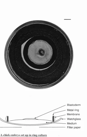
Figure 2.1 A chick embryo set up in ring culture 76
Figure 3.1 Profile of sox 3 expression during gastrulation and neurulation 94 Figure 3.2 Expression pattern of otx 2 during gastrulation and neurulation 96
Figure 3.3 Profile of early GANF expression 99
Figure 3.4 Expression profile for FGF 8 during neurulation 101
Figure 3.5 Early expression profile for BF-1 103
Figure 3.6 Expression profile for pax 6 during neurulation 105
Figure 4.1 Gene expressions in the second axis induced by a chick node graft 116 Figure 4.2 Contributions of mouse tissue to a second axis induced in chick 120 Figure 4.3 Gene expressions in the second axis induced by a mouse node graft 123 Figure 4.4 Lack of neural induction by grafts of transfected cells 127
Figure 5.3 Cell mixing and relative movement of endoderm and axial mesoderm 146
Figure 6.1 Endoderm removal operation and stage specificity of effects 157 Figure 6.2 Behaviour of lower layer after operations 160 Figure 6.3 Cell movements immediately after lower layer removal 162 Figure 6.4 Gene expressions in headfold stage definitive endoderm 164 Figure 6.5 GANF and BF-1 expression following endoderm removal 166 Figure 6.6 FGF 8 and pax 6 expression after endoderm removal 168 Figure 6.7 sox 3 and otx 2 expression following endoderm removal 170 Figure 6.8 Specification of stomodaeal ectoderm without foregut formation 172
Figure 7.1 Induction of GANF expression following endoderm grafts 184 Figure 7.2 Shh expression in axial mesoderm after endoderm removal 187 Figure 7.3 BMP 7 and goosecoid expression after endoderm removal 189 Figure 7.4 Comparing endoderm only with endoderm + axial mesoderm removals
AP antero-posterior
ANR anterior neural ridge
AVE anterior visceral endoderm
BSS balanced salt solution
BMP bone morphogenetic protein
cDNA complementary DNA
CNS central nervous system
DEPC diethylpyrocarbonate
Dil 1,1 dioctadecy 1-3,3,3,3-tetramethylindocarbocyanine perchlorate
DNA deoxyribonucleic acid
dpc days post coitum
DV dorso-ventral
EGO early gastrula organiser
EDTA diaminoethanetatraacetic acid
Fig. Figure
FGF fibroblast growth factor
HH Hamburger and Hamilton
kb kilobase
LB Luria-Bertani
M molar
ml millilitre
OD optical density
PBS phosphate buffered saline
PBT phosphate buffered saline/ Triton-X-100
PC saline Pannett and Compton bird embryo saline
PFA paraformaldehyde
RDVM rostral diencephalic ventral midline
RNA ribonucleic acid
RNase ribonuclease
rpm revolutions per minute
SDS sodium dodecyl sulphate
St. stage
TAB Tris-acetate-EDTA buffer
TE Tris-EDTA buffer
TGF transforming growth factor
Tris Tris(hydroxymethy l)aminomethane
UV ultraviolet
v/v volume per volume
w/v weight per volume
Xgal 5-Bromo-4-chloro-3-indolyl-P-D-galactoî
Pg microgram
pi microlitre
CHAPTER I
1.1 Foreword
The formation of a structured embryo from a single cell is an amazing feat of
development. The combined use of multiple model systems has enabled scientists to
make enormous advances in elucidating the developmental processes and mechanisms
involved at each step. Each model organism has particular advantages and offers
different embryological, genetic and molecular approaches, allowing a more
comprehensive understanding of early developmental processes to be achieved. The
X en opu s embryo is particularly amenable to molecular studies, to analyse the
consequences of extensive over-expression of a gene. The chick embryo, due to its
large size, planar organisation and ease of subsequent culture, allows position-specific
manipulations and tissue graftings to be made. The mouse and zebrafish embryos are
very important genetic models, with the ability to generate targeted mutations a very
powerful tool for studying the effects of loss of a gene function. As will become clear
in the following introduction, such a combinatorial approach has highlighted how
conserved many gene functions and expression patterns have been throughout
evolution. However, important differences in the timing of developmental processes,
and in the use of varying gene combinations at particular stages in the different
species, have become apparent as more information has been obtained. These
variations contribute to the formation of very different organisms from a relatively
1.2 Cell movements in amniote embrvos during gastrulation
Gastrulation is a fundamental process in vertebrate embryonic development, whereby
cells of the single-layered embryo are displaced or reshuffled and become rearranged
in a system of three concentric germ layers. Areas destined to form the internal body
structures are brought into the interior of the embryo. Tissue movements are highly
organised during gastrulation, and result in cell populations becoming correctly
positioned for future interactions. The physical proximity of precursor tissues
facilitates cell communication and inductive interactions that are critical for tissue
patterning and subsequent morphogenesis (see Balinsky, 1970 for a general
comparative review).
1.2.1 The chick embryo
The chicken blastoderm develops as a flat circular disc consisting of two parts; the
inner area pellucida surrounded by the area opaca (Fig 1.1 A). All tissues of the
embryo are formed from the majority of the area pellucida (as defined at primitive
streak stage), with the remaining area pellucida and area opaca forming only extra-
embryonic structures. Before the onset of gastrulation, the chick blastoderm has two
layers; the epiblast and the hypoblast below it. The hypoblast will later be swept to the
periphery and does not contribute to the embryo proper, but it may well have an
important patterning role.
The first sign of asymmetry in the circular area pellucida is a thickening of the
Hamilton stage2). This initiates a sweeping of epiblast cells, from lateral and
anterolateral positions, towards the midline in the posterior half of the blastoderm
(F igl.lB ). As cells converge and become concentrated at the midline, they form the
primitive streak, which begins to elongate towards the anterior of the embryo. As the
streak lengthens, the cells in its thickened walls change shape. They acquire a bottle
shape, with the neck of the bottle keeping the cells in touch with the surface, causing
the formation of a narrow furrow along the length of the streak. As the streak
approaches full length (HH stage 4; F ig l.lC ), a specialised region, known as Hensen’s
node, forms at its anterior tip.
Gastrulation constitutes a coordinated, mass immigration of individual cells down
through the furrow of the streak, then spreading out laterally and anteriorly (Fig 1. ID).
The first cells to ingress are the embryonic endoderm cells. These insert themselves
into the hypoblast beneath the anterior part of the streak, gradually pushing the
hypoblast out towards the edge of the area pellucida. Mesodermal cells then migrate
between the epiblast from which they came and the newly-formed endoderm. As these
cells delaminate, they are replaced by the adjoining areas of epiblast moving toward
the midline, and in turn ingressing through the streak. Therefore, although the
primitive streak persists, the cells which make it up do not stay in the same place and
are continually replaced.
After the full-length streak stage, the node regresses back towards the posterior of the
embryo (HH stage4-k onwards). As it does so, cells leave the front of the node to form
Wholemount embryos viewed from the dorsal aspect, anterior to the top of the page. A Short primitive streak (HH st.3). B Diagram of movements in the epiblast during formation of the primitive streak. C Full-length primitive streak (HH st.4). Dotted line shows believed boundary of the prospective neural plate. D Diagram of the anterior half of the area pellucida cut transversely to show the migration of mesodermal and endodermal cells from the primitive streak. E Head process stage (HH st.5). F Formation of the headfold and regression of Hensen’s node (HH st.6+). G Closure of the neural tube and formation of somites (HH st.9-). H Formation of the optic vesicles and three clear primary brain vesicles. Bending of fused heart tube (HH St. 11-). Scale bar = 600pm for A C; 350pm for D; 700pm for E G and 1mm for
H.
Numbers of stages after Hamburger and Hamilton, 1951.
a r e a p e llu c id a
p r i m
s tr e a k
H e n s e n ’s n o d e
m F m 'Æ
P r im itiv e^ tL «asis»-.
D
‘ """ P™,«™ »„>k
E p ib la s t
h e a d p r o c e s s
n e u ra l p l a t e fold
H en se n 's n o d e
so m ite s
H e n se n 's ; n o d e
b l o o d — l i f e : , isla n d s
folds
stre a k
y W C N e u r a l
folds
S o m ite s
H e n s e n ’s n o d e
P r im itiv e streak
(the head process; F ig l.lE ). Regression movements are a characteristic of amniote
embryos, and they co-ordinate cell activities in laying down the initial organs of the
embryonic axis in a patterned manner. Thus, anterior parts of the streak contribute to
medial areas of the embryo (the somitic mesoderm), whereas the posterior half of the
streak contributes to more lateral mesoderm (Schoenwolf et a l, 1992). The final
residue of the streak becomes partially incorporated into the tailbud, at the very
posterior of the embryo.
The rod-like structure of the notochord exerts tension on the area pellucida, stretching
the embryo along its antero-posterior (AP) axis. The part of the ectoderm that will
form the neural plate thickens and the anterior end bends ventrally to form the head
fold (HH stage 6; F ig l.lF ). As development proceeds, the neural plate closes up to
form the neural tube and begins to differentiate along its length into specialised
regions of the brain and spinal cord (Figl.lG ,H ). As the trunk region extends, the
mesoderm lying on either side of the notochord becomes segmented into paired
somites, that will form dermal, muscle and skeletal structures of the dorsal axis. Chick
development is described in detail in (Bellairs and Osmond, 1998).
1.2.2 The mouse embryo
The mouse embryo develops initially from a blastocyst comprising two parts; the inner
cells mass (ICM), most of which will mainly give rise to the embryo proper, and an
outer shell of cells forming the trophectoderm, which forms extraembryonic tissues.
The trophectoderm also encloses the blastocoel cavity. The primitive endoderm
(dpc). At this stage, the blastocyst must implant itself in the endometrial lining of the
uterus, attaching via the trophectoderm cells that overlie the ICM. During the
immediate post-implantation period (5-6dpc), the mouse embryo changes dramatically
in size and shape. The ICM rapidly grows to fill the blastocoelic cavity and the whole
embryo adopts a unique cylindrical shape (unlike most mammals). The ICM then
epithelialises into a layer of epiblast cells and a new cavity, the proamniotic cavity,
forms within the epiblast. The embryo thus acquires the shape of a cup made up of two
layers, the inner epiblast and the outer visceral endoderm (part of the primitive
endoderm). This is ‘inside-out’ in comparison to the arrangement of other mammalian
embryos and to the formation of the body organs, and so requires an inversion of the
germ layers after gastrulation. In relation to the site of implantation, the embryonic
component of the egg cylinder lies distally, while extra-embryonic regions are
proximal. Prior to gastrulation, visceral endoderm cells that are thought to establish an
essential anterior patterning centre, are found at the distal tip of the egg cylinder.
These cells then move anteriorly, just before the streak starts to form on the posterior
side of the embryo (reviewed in Beddington and Robertson, 1998).
Gastrulation in the mouse, as in the chick, involves the recruitment of epiblast cells to
a transient embryonic structure called the primitive streak. The streak arises on the
posterior proximal side of the egg cylinder (6.5dpc), at the junction of embryonic and
extra-embryonic tissue. As gastrulation progresses, the primitive streak elongates,
ultimately extending to the distal tip of the egg cylinder. The node forms at the
anterior end of the streak, but is only morphologically evident when gastrulation is
presum ptive forebrain
ANT.
anterior hypoblast
notochord
definitive node
embryonic
endoderm prim itive
streak
ANT,
anterior visceral endoderm
POST.
POST.
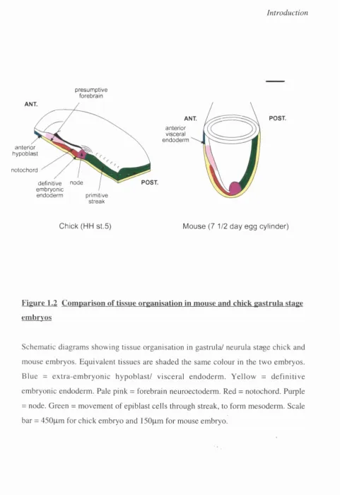
Chick (HH st.5) Mouse (7 1/2 day egg cylinder)
Figure 1.2 Comparison of tissue organisation in mouse and chick gastrula stage embryos
mesenchymal transition, then ingress and move laterally to form the new germ layers.
As mentioned for the chick, cells move through the streak in a specific progression.
Therefore, the first cells to ingress are extra-embryonic mesoderm, followed by
embryonic mesoderm and definitive endoderm. The arrangement of endoderm and
mesoderm cells is almost reversed as the new embryonic layers are formed, since the
epiblast cells initially closest to the streak ingress through it first, and then migrate the
furthest away. Definitive mesendoderm cells are inserted into the visceral endoderm,
thus displacing it to overlie extra-embryonic regions (Fig 1.2 shows tissue arrangement
at this stage).
Midline cells leaving the node become organised into the prechordal and then
notochordal plates, initially in the same layer as the endoderm, immediately anterior to
the node. These cells then move to a mesodermal position and are re-organised into a
cord (Poelmann, 1981; Sulik et a l, 1994). The neural plate becomes increasingly
defined at the anterior end of the embryo, with the neural folds elevating to form the
head folds (7.5dpc). Concomitant with development of the headfolds, the embryonic
axis rostral to the node increases rapidly in length, leading to a posterior displacement
of the node (Camus and Tam, 1999 and references therein). Development of the neural
plate in more posterior regions is less advanced, and it gradually merges into the
primitive streak region. The first somites are formed at Bdpc.
When the embryo has about 6-8 pairs of somites, the embryo ‘turns’ so that the
configuration of the germ layers is reversed. Here, the embryo effectively rolls through
the outer convex surface (of the U-shape) and the midgut onto the inner concave
surface of the embryo. The embryo also wraps itself in its extra-embryonic
membranes; the amnion and yolk sac, supporting further development of the embryo
and allowing formation of the placenta. Mouse development is described in detail in
(Kaufman and Bard, 1999).
1.3 Neural induction and the organiser
Hensen’s node in the chick and the mouse node, both found at the anterior tip of the
primitive streak, are very important signalling centres during gastrulation and have
multiple effects on the organisation of the entire embryo. They are the functional
equivalents of the amphibian dorsal blastopore lip which, as first shown by the
pioneering experiments of Spemann and Mangold (1924), can induce a complete
second axis when grafted to the ventral side of a host embryo. Most of the nervous
system in this ectopic axis developed not from the transplanted tissue, but from the
host ventral ectoderm, which in an undisturbed embryo forms epidermis. This small
piece of tissue was therefore able to influence the host cells around it, changing their
fate and arranging them into a complete second axis. Spemann named the dorsal
blastopore lip the “organiser”, and proposed that in normal development this region
induces and organises a correctly patterned nervous system in neighbouring dorsal
ectoderm.
In the 70 years since Spemann’s discovery of the organiser, an enormous amount has
Equivalent organiser structures have been identified in other vertebrates; Hensen’s
node in chick (Waddington, 1933; Storey et a l, 1992), the node in mouse
(Beddington, 1994) and the shield in zebrafish (Shih and Fraser, 1996). Conserved
patterns of gene expression between these tissues, together with the observations that
interspecies grafts in several combinations (Kintner and Dodd, 1991; Blum et al.,
1992; Hatta and Takahashi, 1996) lead to neural induction, suggest that at least some
of the signalling mechanisms, by which the organiser recruits its surrounding cells to
organise a basic body pattern, are highly conserved during vertebrate evolution.
However, it is only in the last decade that the nature of these inducing signals released
from the organiser has begun to be elucidated.
1.3.1 The ‘Default model’; inhibition of BMP signalling
For a long time, investigators thought of neural inducers as substances that actively
promote neural development, since a transplanted organiser dominates over the would-
be fate of its neighbouring cells. Instead a more convoluted double-negative
mechanism for neural induction is now favoured, based on experimental work in
Xenopus. The default state of ectoderm is neural; this pathway is actively blocked on
the ventral side of the embryo by neural inhibitors; neural inducers therefore act on the
dorsal side of the embryo to provide a permissive condition for neurogenesis, by
antagonising the neural inhibitors. This is supported by the observation that cellular
dissociation of animal caps leads to the formation of neural tissue (Harland and
Gerhart, 1997; Sasai and De Robertis, 1997; Wilson and Hemmati-Brivanlou, 1997),
suggesting that cells within the ectoderm of the Xenopus gastrula have an autonomous
The ventralising bone morphogenetic proteins (BMPs), particularly BMP2 and BMP4,
are proposed to function as epidermal inducers and neural inhibitors. The
autoneuralisation of Xenopus animal caps can be suppressed by BMP4 (Wilson and
Hemmati-Brivanlou, 1995), or by effectors of BMP4 signalling (Suzuki et al., 1997;
Wilson et a l, 1997). Further support for the default model comes from experiments
showing that not only do BM Ps induce epidermal fate in vitro, but inhibition of
endogenous BMP signalling neuralises ectodermal explants. Animal caps cut from
embryos injected with RNA encoding dominant-negative BM P receptor (Suzuki,
1995; Xu et a l, 1995), dominant negative (non-cleavable) ligand (Hawley et a l, 1995)
and anti sense BMP4 RNA (Sasai et a l, 1995) adopt a neural instead of an epidermal
fate.
Normal BMP4 expression in X enopus is also consistent with its proposed anti-
neuralising function, being widespread in the entire ectoderm at the start of
gastrulation and then cleared from the presumptive neural plate at the time when the
organiser appears (Fainsod et a l, 1994; Hemmati-Brivanlou and Thomsen, 1995;
Schmidt er a/., 1995).
Several candidate endogenous neural inducing molecules have been identified, that act
by inhibiting BMPs. Noggin (Smith and Harland, 1992), chordin (Sasai et a l, 1994)
and follistatin (Hemmati-Brivanlou et a l, 1994) are all expressed in the deep layers of
the organiser and its derivatives. They can induce neural tissue directly in ectodermal
Hemmati-Brivanlou et al., 1994; Sasai et a i, 1995). Furthermore, all three molecules have been
shown to bind directly to mature BMP2, 4 and 7 proteins and prevent them from
reaching their receptor(s) (Piccolo et a l, 1996; Zimmerman et a l, 1996; Fainsod et
al., 1997), suggesting that BMP inhibition plays a role in neural induction in vivo. This
mechanism has been highly conserved during evolution, since in Drosophila, the
product of the gene short gastrulation (sog; homologue of vertebrate chordin)
antagonises the Decapentaplegic (Dpp; Drosophila homologue of BMP4) signalling
pathway during early DV patterning of the embryo (Francois and Bier, 1995; Biehs et
al., 1996; Nguyen et a l, 1998). In addition, analysis of double mutant Drosophila
embryos has shown that both dpp and screw, a gene encoding another BMP ligand
which acts to potentiate the Dpp signal, are epistatic to sog, suggesting that all
functions of Sog involve the Dpp signalling pathway (Holley et a l, 1996; Nguyen et
a l , 1998). Indeed, no receptors have yet been identified for Noggin, Chordin or
Follistatin, suggesting that in vertebrates too, these ligands act solely to modulate
BMP signalling.
More recently, other neural inducing molecules have been described. Flik (follistatin-
like), the chick homologue of mammalian TSC36, encodes a secreted protein with a
cysteine-rich domain strongly related to those found in Follistatin (Patel et a l, 1996).
Flik is expressed in the node and node-derived axial mesoderm, as well as weaker
expression in the neural plate. The mechanism of action of Flik protein in vivo is
unknown at present. However, treatment of gastrular stage embryos with antisense
oligodeoxynucleotides to the gene causes reduced allocation of neural tissue, together
protein may somehow interact with BMP signalling pathways (Towers et a l, 1999).
Xnr3 {Xenopus nodal-related) is another gene expressed in the outer ectodermal layer
of the organiser (Smith et a l, 1995) that can induce neural tissue directly in Xenopus
when provided as RNA (Hansen et a l, 1997). X n r 3 and BMP4 have mutually
antagonistic activities (Hansen et a l, 1997), suggesting that Xnr3, itself a diverged
TGpp superfamily member, may competitively bind to BMP receptor(s) and act in a
dominant negative way to inhibit BMP signalling (Thomsen, 1997).
In Xenopus, therefore, the evidence that the actions of the organiser in neural induction
rely on modulation of BMPs by endogenous inhibitors, is compelling. However,
results from other organisms suggest that this interpretation is too simplistic, and that
neural induction is likely to involve co-operation of different classes of signal
(reviewed in Streit and Stem, 1999). BMP inhibition is not sufficient for neural
induction in the chick, since mis-expression of either noggin (A.Streit and C.D.Stem,
unpublished) or chordin (Streit et a l, 1998) does not induce expression of any neural
markers. Moreover, cell dissociation of the epiblast in chick does not lead to neural
differentiation, as it does in Xenopus, but promotes muscle development (George-
Weinstein et a l, 1996). However, Chordin-secreting cells can stabilise expression of
the pan-neural marker sox3 in non-neural ectoderm, if these cells have previously been
exposed to a node graft for five hours (too brief in itself for induction of a neural plate)
(Streit et a l, 1998), suggesting that BMP inhibition may work in conjunction with
The expression patterns of BMPs and their inhibitors in other organisms also do not
quite fit with their proposed roles in Xenopus. Follistatin is not expressed in the mouse
node (Albano et al., 1994) and only weakly in the chick (Levin, 1998), and neither
noggin nor follistatin are expressed in the zebrafish shield (Bauer et a l, 1998). Global
exposure of stage 3 chick embryos to supernatant from Noggin-expressing CHO cells
produces no observable effect, and exposure of a stage 4 node to the same supernatant
has no effect on the ability of the node to induce a second axis when grafted into an
unexposed host (Connolly et a l, 1997). Chordin expression, while present in the chick
node at the right time to take part in neural induction, persists long after the node has
virtually lost its inducing ability.
Loss of function mouse mutants also fail to show an essential function for BMP
antagonists in neural induction. Noggin mutants develop a fairly normal neural plate
and show patterning defects only at later stages (McMahon et a l, 1998). Even
noggin/chordin double knockout embryos have the majority of the neural axis,
although they do show very early anterior brain defects (Bachiller et a l, 2000). Null
mutants for follistatin (Matzuk et a l, 1995), BMP7 (Dudley et a l, 1995) or BMP2
(Zhang and Bradley, 1996) have no early neural phenotype. A proportion of mutants
lacking BMP4 die before gastrulation, but a few survive to early limb bud stages; these
do not appear to have an enlarged nervous system or absence of epidermis (Winnier et
a l, 1995). Embryos with mutations in the BMP receptor Bm pr-la die too early to be
informative (Mishina et a l, 1995). All these data therefore suggest that additional
1.3.2 Other pathways for neural induction
BMP inhibitors are not the only molecules that have been reported to have direct
neuralising activity. A novel secreted protein, Cerberus, is expressed in the extreme
anterior endomesodermal domain of the Xenopus organiser during gastrulation, and
can induce neural tissue in ectodermal explants, as well as second heads in whole
embryos (Bouwmeester et a l, 1996). Biochemically, it has been shown that Cerberus
protein can antagonise not only BMPs, but also the Wnt and Nodal signalling
pathways; it has independent sites to directly bind BMP, Wnt and Nodal proteins in
the extracellular space, and thus inhibit the actions of all three simultaneously (Glinka
et al., 1997; Piccolo et a l, 1999). Two other Wnt antagonists are expressed in the
Xenopus organiser, frzb-1 (Leyns et a l, 1997; Wang et al., 1997) and dickkopf (Glinka,
et a l, 1998). Both are secreted molecules, and Frzb-1 has been shown to bind to Wnt
proteins directly (Wang et a l, 1997), suggesting that frzb-1 and dickkopf may prevent
Wnt proteins from reaching their receptors by a functional interaction analagous to
BMPs and their inhibitors. Frzb-I is also expressed in the mouse anterior streak
(H oang et a l, 1998) and chick neural ectoderm (Baranski et a l, 2000). These
additional candidates raise the question of whether other pathways operate in parallel
with the anti-BMP mechanism of neural induction.
There is some evidence that FGFs can act as direct neural inducers in both Xenopus
(Lamb and Harland, 1995) and chick embryos (Storey et a l, 1998; Alvarez et a l,
1998). However, the role of FGFs or FGFR signalling in neural induction remains far
from clear (reviewed in Mason, 1996). Cox and Hemmati-Brivanlou (1995) found that
caps were pre-treated with a neuralising agent. Two different groups have shown that
the neuralising activity of noggin and chordin in animal caps requires an intact FGF
signalling pathway (Launay et al., 1996; Sasai et a l, 1996) suggesting that FGFs may
regulate the competence of ectoderm to respond to neural inducers. Animal caps cut
from embryos expressing a dominant-negative FGF receptor (XFD) were found to be
insensitive to neural induction by organiser grafts or by noggin (Launay et al., 1996).
However, other groups have found expression of neural markers in animal caps cut
from embryos co-injected with XFD and noggin (Bang et al., 1997). Thus, evidence
regarding the role of FGF signalling in neural induction is somewhat ambiguous.
1.3.3 Onset of neural induction
An important point that still needs to be established is when neural induction begins.
As more neural inducing molecules are discovered, it is becoming apparent that full
neural induction probably requires a sequence of multiple signals and cell states. It
may well be that each step in the hierarchy stabilises the previous ones, altering the
competence of the ectoderm to respond to future signals. Differences in the timing of
these steps between species may therefore underlie the apparently different signalling
requirements for neural induction in various species. In Xenopus, it appears that the
dorsal ectoderm is already biased in favour of a neural fate by the late blastula stage
(Savage and Phillips, 1989; (Kroll et al., 1998), and the boundary between neural and
non-neural ectoderm may be positioned before gastrulation (Zhang and Jacobson,
1993). Therefore, it could be that the dorsal ectoderm has received some neural-
inducing signals before gastrulation, and that additional permissive, or stabilising
neural direction. A ‘pre-neurulation’ state is proposed to exist in the central epiblast of
the unincubated (blastula stage) chick embryo (Callebaut et a l, 1998). Furthermore,
restricted expression of the pan-neural gene sox3 and the carbohydrate epitope L5,
both thought to reflect competence of the ectoderm to respond to neural inducing
signals, are seen very early (Rex et a l, 1997; Streit et a l, 1997). However, the
neuroectoderm is not specified until just before stage 4, and requires continued
signalling until then (Darnell et a l, 1999).
1.4 Anteroposterior patterning of the neural plate
The ectopic second axis that is formed by the actions of a graft of organiser tissue
frequently contains a correctly patterned nervous system. The organiser therefore, not
only induces multiple tissue types, but also patterns them, suggesting an intimate
relationship between the two processes.
1.4.1 Planar and vertical signalling
Basic patterning of the neural territory is likely to begin during gastrulation, once
neural induction is under way. As gastrulation proceeds, the physical relationships
between cell populations change, so that mesoderm and endoderm layers come to lie
beneath the ectoderm. Neural patterning signals from the organiser could therefore
reach the ectoderm via a planar route, while the cells still occupy the same layer, or by
the vertical route, from the involuted dorsal mesoderm to the overlying ectoderm
(reviewed in Doniach, 1993). In the chick, these two signalling routes would therefore
streak, from the prechordal tissue and notochord to the overlying ectoderm. Neural
patterning information has been shown to be transferred by both routes, mainly from
work in Xenopus. However, the extent to which vertical and planar signals function in
vivo remains unresolved, and may vary between species.
Otto Mangold proposed that neural pattern is achieved purely by vertical induction,
where the AP pattern of the mesoderm is “imprinted” onto the overlying ectoderm
(Mangold, 1933). Taking pieces of involuted dorsal mesoderm from different axial
levels and inserting them individually into the blastocoel cavities of early gastrulae, he
found that the AP level of neural tissue induced roughly corresponded to that of the
inserted mesoderm. He suggested that the different regions of the involuted mesoderm
therefore contain qualitatively different neural inducers, and that they each induce a
different AP element of the neural pattern to the ectoderm directly above. However,
the range of neural tissue induced by a given piece of mesoderm was broader than
would have been found in the ectoderm immediately above it in the intact embryo.
The evidence for generation of planar signals, mainly derived from use of Keller
explants, further suggests that a strict interpretation of the Mangold model is not likely
in vivo.
Keller explants are made by culturing dorsal mesoderm and ectoderm from early
gastrulae as a single, flat sheet, with contact only along a single edge and vertical
contact prevented. In this configuration, a range of neural marker genes can be
induced in the ectoderm, in an AP order equivalent to that in the intact embryo
(Nieuwkoop, 1952) but, more importantly, his experiments led him to propose a model
for the mechanism of AP neural patterning that is compatible with both planar and
vertical signalling, and is still widely accepted today (see next section). He inserted
folded flaps of competent ectoderm perpendicularly into the presumptive neural plates
of early gastrulae. The ectoderm therefore had contact with the host at only one end of
the fold, and any planar signals that might exist could spread into it. He consistently
found that the folds displayed posterior to anterior neural pattern from the attached
base of the fold to the outer tip. Furthermore, the type of neural tissue at the base
matched that of the region in which the fold was implanted, and significantly, was
always of a more posterior type than that at the outer tip.
1.4.2 Activation-transformation model
Based upon his ectoderm fold experiments, Nieuwkoop proposed a model for AP
neural patterning involving two sets of inducer signals (Nieuwkoop, 1952). An initial
“activator” signal, present in all organiser mesoderm, induces neural tissue with
anterior character. A second “transformer” signal, present in a gradient with a high
point in the posterior mesoderm, then progressively posteriori ses the neural plate to
generate the remaining regions of the CNS. He argued that the transformer is dominant
over the activator, since the proximal part of the fold developed with a more posterior
character, even though it must have experienced both factors. The discovery of the
neural inducer molecules noggin and chordin lend support to this model, since they
both induce neural tissue expressing anterior markers in X enopus animal caps.
Furthermore, three classes of signal- Wnts, FGFs and retinoic acid- have been shown
(reviewed in Doniach, 1995; Sasai and De Robertis, 1997). Other molecules are
probably also involved; tissue grafting experiments have shown that posterior non-
axial mesoderm (that comes to underlie the posterior neural plate) can exert a
posteriorising influence on forebrain regions, but this activity is unlikely to be
mediated by the signals just mentioned (Bang et al., 1997; Woo and Fraser, 1997).
The vitamin A metabolite retinoic acid (RA) is thought to play a role in posteriorising
the CNS by influencing the expression of Hox genes, which are involved in positional
identity along the body axis (reviewed in Maden and Holder, 1992; Conlon, 1995).
Treatment of Xenopus embryos with RA causes a reduction in forebrain volume and
corresponding increase in hindbrain volume (Durston et al., 1989; Sive et a l, 1990).
More subtle effects are also seen in the hindbrain, where anterior rhombomeres are
reduced or compressed (Papalopulu et a l, 1991). Furthermore, constitutively active
RA receptors reduce anterior neural tissue while dominant negative RA receptors
expand anterior neural structures (Blumberg et a l, 1997). Several Hox genes contain
RA response elements, and RA can induce Hox gene expression in mouse ectoderm,
while repressing expression of more anterior genes such as otx2 (Conlon and Rossant,
1992; Ang et a l, 1994). In addition, targeted mutagenesis of RA receptors can
produce homeotic alterations to the axial skeleton similar to certain Hox loss-of-
function phenotypes (Lohnes et a l , 1994), although the majority of these mutant
phenotypes are surprisingly mild. These results have led to the suggestion that there
may be an endogenous gradient of RA within the neural plate, with a high point at the
posterior end, that promotes development of posterior structures while restricting the
fine-tuned analyses of the distribution of endogenous RA during neurulation, and the
localisation of the main enzymes involved in the biosynthesis and degradation of RA,
challenge this concept (Maden et al., 1998; reviewed in Maden, 1999; Gavalas and
Krumlauf, 2000). The highpoint of the RA gradient is in fact at the hindbrain/ spinal
cord boundary, and the RA concentration gradually drops towards the posterior end of
the embryo (Maden et a l, 1998). In addition, treatment of mouse and zebrafish
embryos with RA causes a reduction of anterior rhombomeres but does not affect
forebrain and midbrain development (Holder and Hill, 1991; Morris-Kay, 1991; Woo
and Fraser, 1997), suggesting that global effects of RA on neural structures are unique
to Xenopus, and that a more conserved function of RA is in patterning the hindbrain.
A role for FGF signalling in posteriorising neural tissue is supported by many different
experiments (reviewed in Doniach, 1995; Mason, 1996). bFGF (FGF2) can transform
a frog anterior neural plate explant into posterior CNS in vitro (Cox and Hemmati-
Brivanlou, 1995). When animal caps are co-treated with bFGF and either noggin or
chordin, posterior neural markers are induced in addition to forebrain markers, at
opposite poles of the explant (Lamb and Harland, 1995; Cox and Hemmati-Brivanlou,
1995; Sasai et a l, 1996). Injection of a dominant negative receptor (XFD) at the two
cell stage results in embryos completely lacking posterior trunk and tail structures, but
with relatively normal anterior neural structures (Amaya et a l, 1991; Launay et al.,
1996). Furthermore, over-expression of XFD in Keller explants inhibits posterior
neural markers but not anterior or pan-neural markers (Holowacz and Sokol, 1999).
The authors also found they could achieve similar effects in whole embryos, by
However, in transgenic embryos expressing zygotic XFD in every cell, Kroll and
Amaya (1996) were unable to reveal a role for FGF in either neuralisation or posterior
neural patterning.
Analysis of mouse mutants further supports a role for FGF signalling in AP neural
patterning. F G F8 -/- mice show perturbed patterning of the neural plate; anterior
neuroectoderm markers are widely expressed, while posterior markers are absent (Sun
et a l, 1999). In addition, FGFR-1 deficient mouse embryos frequently exhibit
truncations or disorganisation of posterior embryonic regions (Deng et al., 1994;
Yamaguchi et a l, 1994). Finally, a large number of FGFs have expression patterns
confined to posterior axial and paraxial mesoderm, and not anterior mesoderm,
suggesting that several members of the family could be necessary for posterior
development in vivo. However, it is unlikely that a gradient of FGF signalling is
responsible for producing AP pattern, since no clear correlation between the dose of
FGF and the axial level of the neural marker induced, has been demonstrated. It is
possible instead that the effects of FGF depend upon competence of the responding
tissue, since the age of the responding tissue appears to affect the nature of the
response produced (Lamb and Harland, 1995).
Finally, members of the Wnt family are good candidates for a posterior transformation
signal. In wnt3 -/- mice, the epiblast proliferates in an undifferentiated state that lacks
AP neural patterning (Liu et a l, 1999). In addition, the function of wnt3a has been
wnt3a can synergise with noggin and follistatin to increase the expression of posterior
neural genes in Xenopus animal cap explants (McGrew et a l, 1995).
1.4.3 Head and trunk organisers
The organiser itself is a dynamic entity; its cell population, structural organisation and
profile of gene expression constantly change during gastrulation. In Xenopus and
avian embryos, changes in the cellular composition and gene activity of the organiser
can be correlated with its ability to induce the differentiation of tissues with different
AP characteristics. Transplantations of early organisers result in complete axial
duplications, including heads, whereas grafts of later organisers have reduced inducing
ability and result in only partial axes containing just trunk (Gerhart et a l, 1991; Storey
et a l, 1992). This finding, first observed by Spemann (1931), has led to the concept of
distinct organisers for head and trunk regions of the embryo (reviewed in Doniach,
1993; Harland and Gerhart, 1997; Niehrs, 1999).
The developmental fate of organiser cells also changes during gastrulation, with cells
contributing to progressively different parts of the axial mesoderm. Derivatives of the
early node/organiser contribute to the prechordal mesoderm, while cells of the late
node/organiser are destined for the more posterior notochord, and are not found in the
head region (Selleck and Stem, 1991; Selleck and Stem, 1992; Beddington, 1994;
Lane and Keller, 1997; Lemaire et a l, 1997). Since these mesoderm derivatives of the
organiser retain neural-inducing activity themselves, they may underlie the head
However, evidence for compartmentalisation of separate head and trunk patteming
activity within the organiser has recently been shown in Xenopus. The early gastrula
organiser can be subdivided into an anterior (vegetal) domain that expresses
goosecoid, and a posterior (animal) domain that expresses Xnot (Vodicka and Gerhart,
1995). Studies performed on isolated halves of the organiser show that each half not
only has a distinct fate but also induces a unique set of region-specific neural genes.
When grafted orthotopic ally to the organiser region of a host embryo, the anterior
(g5c) half develops into prechordal mesoderm while the posterior {Xnot) half develops
into notochord and somites (Zoltewicz and Gerhart, 1997). When co-cultured with
animal cap ectoderm, the anterior half induces only anterior-specific genes, while the
posterior half induces anterior and posterior neural genes (Zoltewicz and Gerhart,
1997). These results demonstrate that the organiser is already patterned by the early
gastrula stage, with segregation of region-specific neuralising activity, and these
separate functional compartments are marked by differing gene expressions.
Similar compartmentalisation of the organiser prior to gastrulation has also been
demonstrated in the zebrafish organiser (Gritsman et al., 2000 and references therein).
Differential activation of the Nodal signalling pathway is proposed to be responsible
for establishing this AP pattern within the organiser and modulating fate decisions.
Higher levels or longer exposure to Nodal signals are required for expression of gsc
and prechordal plate formation, whereas lower transient levels are sufficient to induce
flo ating head expression (the zebrafish homologue of X n o t) and notochord
development. In addition, the attenuation of Nodal signalling in zygotic
domain, followed by a fate change of cells in the prechordal plate domain to notochord
(Gritsman et a l, 2000).
Régionalisation within the avian node has also been reported (Storey et a l, 1995),
with the source of neural inducing ability localised to two sub-regions and associated
with specific cell types contained in these regions. Gene expression patterns for gsc
and cNot are similar to those observed in other species; largely restricted to prechordal
mesoderm and notochord, respectively (Pera and Kessel, 1997 and references therein).
However, other evidence suggests that determination of prechordal mesoderm fate in
the node is not complete, and further signalling is required after these cells have
extended away (Vesque et a l, 2000). Early extending axial mesoderm co-expresses
markers of both notochord and prechordal mesoderm throughout its length, only
becoming segregated at later stages. TGPp signals from the anterior endoderm are
thought to be responsible for downregulating notochord characteristics and inducing
prechordal mesoderm properties in the most anterior axial mesoderm, as it extends
(Vesque et a l, 2000).
The recent rapid increase in knowledge about molecules with head inducing activity,
has led to new insights into the molecular nature of the separate head and trunk
organisers. Cerberus is a secreted protein expressed in the anterior endoderm of the
organiser and has the unique property of inducing ectopic heads in the absence of
trunk structures (Bouwmeester et a l, 1996). It has been shown that Cerberus protein
has independent sites to directly bind BMP, W nt and Nodal proteins in the
pathways simultaneously (Glinka et a l, 1997; Piccolo et a l, 1999). Furthermore,
Piccolo et a l (1999) believe that all three inhibitory activities of cerberus are
necessary for the formation of ectopic head structures. However, it is unlikely that
cerberus interacts with the BMP pathway in the same manner as classical neural
inducers, such as noggin and chordin, since cerberus actually inhibits trunk and tail
formation and cannot dorsalise mesoderm (Bouwmeester et a l, 1996).
The discovery of two other secreted Wnt i n h i b i t o r s , ( L e y n s et a l, 1997; (Wang
et a l, 1997) and dickkopf-1 (Glinka et a l, 1998), further supports the hypothesis that
head induction is achieved by simultaneous repression of TGpp and Wnt signalling.
Injection of frzb-1 or dkk-1 alone does not induce a second axis. However, co-injection
of either of these mRNAs with that encoding the dominant negative BMP-2/4 receptor
(tBR) induces complete second axes, with forebrains, eyes and cement glands (Glinka
et a l, 1997; Glinka et a l, 1998). Interestingly, co-injection of frzb-1 and tBR mRNA
also leads to strong ectopic expression of cerberus in the ventral endoderm (Piccolo et
a l, 1999). Other combinations of Wnt and BMP inhibitors, such as chordin and frzb-1
mRNAs, or tBR and dnXwnt8 mRNAs, similarly induce complete second axes when
co-injected ventrally (Glinka et a l, 1997; Piccolo et a l, 1999). It is noteworthy that
dickkopf-1 induces heads with two eyes, unlike cerberus and frzb-1 which induce only
one; this may reflect differences in their normal domains of expression, since dkk-1 is
normally expressed in the prechordal plate (known to play a role in splitting the eye
field) whereas cerberus is not. The requirement for dkk-1 in head formation is
with microcephaly and cyclopia, lacking a head altogether in some cases, while the
trunk appears normal (Niehrs, 1999).
Nodal signalling has also been shown to inhibit head formation, and the combination
of BMP and nodal antagonists alone is sufficient for formation of an ectopic second
head (Piccolo et a l, 1999). Interestingly, nodal is actually required early on for head
formation (Varlet et al., 1997). Early nodal signalling also induces cerberus
expression; cerberus is induced by injection of X n rl mRNA, but not by X n rl DNA,
which is only expressed after the mid-blastula transition (Piccolo et al., 1999).
Therefore, it appears that early and late Nodal signalling have almost opposite roles in
neural development (like Wnt signalling); an early Nodal signal is required to pattern
the anterior endoderm and perhaps induce the organiser effectors, such as cerberus,
which then feed back negatively on Nodal signalling in order to initiate head fate and
inhibit trunk fate (Piccolo et ah, 1999). In zebrafish, nodal signalling is required to
maintain d k k l expression in the gastrula; bozozok, squint and oep mutants (all
defective in some aspect of nodal signalling) show normal initiation of d kkl in the
blastula, but this is subsequently lost in the shield at early gastrula stage (Hashimoto et
a l, 2000).
1.4.4 Summary
The organiser is not a homogeneous cell population; signals produced by different
domains of the organiser and its derivatives selectively inhibit different families of
growth factors, producing neural structures with different regional character.
balancing the inductive signals with their specific antagonists that emanate from the
organiser and organiser-derived tissues. The discovery that head induction can be
achieved by simultaneous repression of TG pp and Wnt signalling provides the basis
for a molecular model that incorporates the concepts of both Nieuwkoop’s activator
and transformer, and Spemann’s head and trunk inducer (Niehrs, 1999). According to
the model, BMP and Wnt inhibitors released from the anterior organiser and its
derivatives neutralise both BMP and Wnt signals in the ectoderm, leading to anterior
neural induction (head organiser; Nieuwkoop’s activator signal). More posteriorly,
Wnt inhibitors are progressively less active, so that a gradient of Wnt activity is
established along the AP axis. These Wnt signals are then able to posteriorise neural
fates induced by BMP inhibition alone (trunk organiser; Nieuwkoop’s transformer
signal). Other non-organiser-associated posteriorising factors, including FGFs and
retinoic acid, could then be involved in full elaboration of the AP pattern as the axis
elongates (Doniach, 1995; Sasai and De Robertis, 1997). However, even this model is
unlikely to be complete, given recent evidence proposing that inter-regulation of
activin and nodal-related factors with their antagonist, antivin, plays a role in
generating pattern along the AP axis (Thisse et a l, 2000).
1.5 Tissues involved in head induction
The cell movements of gastrulation bring the neuroectoderm into contact with several
different tissues, raising the possibility that any of these tissues could impart
patteming information to the neural plate (Fig 1.3). Organiser-derived cells form the
node
primitive streak
HH st.4
prechordal tissue
notochord
node
HH st.5
node definitive embryonic endoderm
primitive
streak migratorycells
hypoblast
presumptive forebrain
notochord
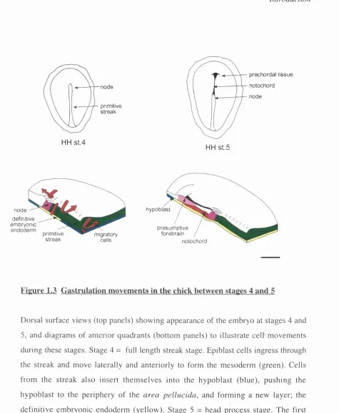
Figure 1.3 Gastrulation movements in the chick between stages 4 and 5
Dorsal surface views (top panels) show ing appearance of the em bryo at stages 4 and
5, and diagram s of anterior quadrants (bottom panels) to illustrate c e ll'm o v e m en ts
during these stages. Stage 4 = full length streak stage. Epiblast cells ingress through
the streak and m ove laterally and anteriorly to form the m esoderm (green). Cells
from the streak also insert th em s e lv e s into the h y poblast (blue), pu sh in g the
hypoblast to the periphery o f the are a p e llu c id a , and form ing a new layer; the
definitive em bryonic endoderm (yellow). Stage 5 = head process stage. The first
axial m esoderm (red) cells to leave the node form the fan o f prechordal mesoderm,
which ends up in the most anterior position. This is followed by cells which form the
neural plate and could therefore pattern it by vertical signalling. In addition, the
organiser inserts cells into the midline of the neural plate, that form floorplate. Planar
patteming signals within the ectoderm could therefore emanate from the neural
midline, or from the peripheral neural/ non-neural boundary. Finally, early anterior
patteming signals could originate in the visceral endoderm, or hypoblast. This tissue,
which is established entirely separately from the streak and organiser, is extra-
embryonic and will be swept to the periphery as it is replaced by definitive embryonic
endoderm. However, prior to gastrulation, it too underlies the area of the embryo that
will form the neuroectoderm. The evidence for signalling from each of these tissues
will be discussed in tum.
1.5.1 Role of the extra-embryonic endoderm
The mouse node (and therefore its derivatives) does not appear to contain all the
information necessary to induce a complete axis, since the duplicated axis formed
following a node graft of any age in mouse embryos, lacks the most anterior regions of
the CNS (Beddington, 1994; Tam and Steiner, 1999). Furthermore, ectopic expression
of CwntS in mouse causes duplication of the primitive streak and node, just as in
Xenopus. However, this does not result in formation of a second brain or heart in the
mouse, unlike in Xenopus (Popperl et a l, 1997). These data suggest that other signals
besides those of the organiser are also required for anterior patteming.
A growing variety of embryological, genetic and molecular studies propose that the
anterior visceral endoderm (AVE) is important for anterior embryonic development in