Analysis of the role of RXRa in
monocyte-macrophage differentiation
and function using U937 monoblastoid
cells.
Timothy James Stonehouse
A thesis submitted for the degree of
Doctor of Philosophy
1998
University College
ProQuest Number: U 6 4 2 3 9 3
All rights reserved
INFORMATION TO ALL USERS
The quality of this reproduction is dependent upon the quality of the copy submitted.
In the unlikely event that the author did not send a complete manuscript and there are missing pages, these will be noted. Also, if material had to be removed,
a note will indicate the deletion.
uest.
ProQuest U642393
Published by ProQuest LLC(2015). Copyright of the Dissertation is held by the Author.
All rights reserved.
This work is protected against unauthorized copying under Title 17, United States Code. Microform Edition © ProQuest LLC.
ProQuest LLC
789 East Eisenhower Parkway P.O. Box 1346
In this thesis the U937 monoblast cell line has been used as a model fo r
investigation of the immunobiology of the mononuclear phagocyte system,
both in terms of differentiation, and in terms of functional properties, such
as phagocytosis and accessory cell activity.
Retinoid X receptor-a (RXRa), a member of the nuclear receptor
superfamily, was shown to be expressed in primary human monocytes, and
its expression was increased upon differentiation in response to human
serum. Expression of RXR dimeric partners, R A R a and RARp, was not
induced in the same way. Expression of these proteins was also assessed in
previously derived novel U937 transfectants that hyper-expressed (aG2S
cells), or hypo-expressed (aB5A cells), RXRa. Retinoic acid (RA) activation
of aG2S cells led to an altered pattern of expression of surface antigens
which suggested a more mature phenotype, when compared to aB 5A cells
and an empty vector control cell line (MEP). RA-treated aG2S cells were
also more efficient at phagocytosing opsonised-SRBC. This
immunophagocytic process was inhibited in all three cell lines by prior
addition of G D I3, CD 16 and CD 18 mAbs. The increase in efficiency of
phagocytosis correlated with a more intense and altered phosphotyrosine
response during phagocytosis. Phagocytosis induced activation of members of
the MAPK family of kinases in all three cell lines, and phosphoproteins from
the U937 transfectants interacted differentially with a panel of SH2 domain
fusion proteins. RA-treated aG2S cells were also resistant to ionising
radiation-induced cell death. This phenomena was RA-dependent, as vitamin
D treatment did not have the same effect.
To examine accessory cell function of both the U937 parent cell line and
developed, where the cell line provides a co-stimulatory signal for CD3-
mediated T-cell activation, independent of CD80/CD86/CD28 interaction.
A p2-integrin (C D lla/18)/IC A M -l (CD54) interaction was implicated,
and there was evidence to suggest the dominant response was in cells with
a CD8+ phenotype. This implied a reduced requirement for CD28-
mediated signalling by CD8+ T-cells, and this was confirmed since
proliferation of the CD4+ T-cell population increased significantly in
response to an added CD28 mAh. C D lla/18 mAbs inhibited U937/T-cell
cluster formation as well as proliferation. CD45 mAbs increased the size
of the U937/T cell clusters formed, suggesting that ligation of CD45 may
be important in the targeting of T-cells towards antigen presenting cells.
MAbs to CD53, CD98 and CD 147 inhibited U937 dependent T-cell
proliferation. CD53 mAbs were not inhibitory when pre-pulsed on to
U937 cells, suggesting an effect at the T-cell level. CD98 and CD 147
mAbs were inhibitory in pre-pulsing, but also inhibited an accessory cell
independent proliferation assay, suggesting these molecules play a role
both at the accessory cell level and at the level of the T-cell. The
observation that most inhibitory antibodies induce tyrosine
phosphorylation in both U937 and T-cells was consistent with this finding.
Finally, aG2S cells induced greater T-cell proliferation when used as an
accessory cell, than MEP cells, which itself was more efficient than aB5A
cells. Previous exposure to opsonised-SRBC had no effect on the
accessory cell capabilities of these cells, demonstrating that phagocytic and
_____________________________________________________
Contents
Table of Contents.
Title 1
Abstract 2
Table of contents 4
List of Tables 13
List of Figures 14
A cknow ledgem ents 17
Chapter 1 Introduction 18
1.1. The myeloid cell lineage. 18
1.1.1. Myelopoiesis: Development of myeloid cells. 18
1.1.2. Molecular mechanisms that regulate myeloid cell
differentiation. 21
1.1.3. Functions of m onocytes/m acrophages. 23
1.1.4. The U937 cell line. 25
1.1.4.1. Molecular events involved in growth arrest
of U937 cells. 26
1.1.4.2. U937 cells as a model for monoblastic
differentiation. 28
1.1.5. Differentiation of U937 cells. 28
1.1.5.1. Differentiation of U937 cells induced by PM A. 28 1.1.5.2. Differentiation of U937 cells induced by RA and DHCC. 29
1.2. Phagocytosis by monocytes and macrophages. 32
1.2.1. Receptors that mediate phagocytosis on monocytes
and macrophages. 3 3
1.2.1.1. Structure of Fey receptors. 33
1.2.1.2. Signal transduction by Fey receptors. 35
1.2.1.3. Role of complement receptors (CR)
in phagocytosis. 39
1.2.1.4. Other functional properties of monocytes and
macrophages induced on phagocytosis. 40
1.2.2. Role of FcyRs in monocyte and macrophage
differentiation and activation. 41
1.3. Signalling mechanisms. 42
1.3.1. Src family of protein-tyrosine kinases. 42
1.3.1.1. Structural features. 42
1.3.1.2. Regulation of kinase activity. 45
1.3.1.3. Signal transduction mechanisms involving
_____________________________________________________
Contents
1.3.2. The Ras/MAPK signal transduction pathway. 461.3.2.1. Adaptor molecules that interact
with cell surface receptors. 47
1.3.2.2. Ras activation, regulation and effector functions. 48
1.3.2.3. The MAPK signalling cascades. 50
1.3.2.4. Transcription factors activated by MAPKs. 53
1.3.2.5. Effects of MAPK activation. 53
1.3.3. Phosphoinositide 3-kinase (PI3-K). 54
1.3.3.1. Regulation of PI3-K activity. 54
1.3.3.2. Functions of PI3-K metabolites. 55
1.3.4. Phospholipase C (PLC). 5 6
1.3.4.1. Regulation of PLC activity. 57
1.3.4.2. Effects of DAG. 57
1.4. T-cell activation. 5 8
1.4.1. MHC-TCR interaction. 5 8
1.4.1.1. MHC - peptide complex 58
1.4.1.2. TCR and TCR mediated signalling. 59
1.4.2. Co-stimulation of T-cells. 60
1.4.2.1. Interaction between CD28 or CTLA-4 and its ligands
B7.1 (CD80) or B7.2 (CD86). 61
1.4.2.2. Interaction between CD2 and its ligands
CD58, CD59, or CD48. 64
1.4.2.3. Interaction between CD27 and its ligand CD70. 65 1.4.2.4. Interaction between p2 integrins and ICAM molecules. 66
1.4.3. U937 cells as accessory cells for T-cell activation. 67
1.5. Retinoid X receptor. 6 8
1.5.1. Isolation of retinoic acid responsive nuclear receptors. 68
1.5.2. Retinoid m etabolism . 7 0
1.5.3. Structure and function of RXR. 7 0
1.5.3.1. DNA binding domain (DBD). 71
1.5.3.2. Ligand binding domain (LBD). 75
1.5.3.3. Receptor dimérisation. 77
1.5.3.4. AF-1 and AF-2 transactivating regions. 77
1.5.4. Initiation of transcription by RXR dimers. 7 8
1.5.4.1. Ligand-independent transactivation. 78
1.5.4.2. Repressors of nuclear receptor transactivation. 79 1.5.4.3. Co-activators of nuclear receptor mediated transcription. 80
1.5.5. Expression of RXRs in foetal and adult tissue. 81
1.5.6. Expression of RXR in haemopoietic cells. 8 2
______________________________________________
Contents
1.6.1. Production of U937-derived transfectants. 8 61.6.2. Characterisation of RXRa expression in selected clones 87
1.6.3. Functional responses of the U937 transfectants to
differentiating stimuli. 88
1.6.4. Discussion of these findings. 88
1.7. Aim s. 91
Chapter 2 Materials and Methods 92
2.1. Isolation, culture, and differentiation of human
peripheral hlood monocytes. 92
2.2. Culture of cell lines. 93
2.3 Preparation of U937 transfectant cell lysates. 93
2.4. Im m unohlotting. 94
2.5. Phenotypic analysis. 96
2.6. Preparation of opsonised Sheep Red
Blood Cells (op-SRBC). 96
2.7. Radiolahelling of op-SRBC. 97
2.8. Assessm ent of im m unophagocytosis. 97
2.9. M orphological analysis o f imm unophagocytosis. 97
2.10. Regulation of immunophagocytosis
by cell surface molecules. 98
2.11. Phosphotyrosine assays. 98
2.12. Effects of binding inhibitory mAbs to the
phosphotyrosine response to op-SRBC. 99
2.13. Im m unoprécipitations (IP). 99
2.14. Isolation of glutathione-SH2 fusion proteins. 99
2.15. Glutathione-SH2 fusion protein assays. 100
2.16. Isolation of T-cells. 10 2
2.17. U937/CD3 - T-cell proliferation assay. 10 2
2.18. Addition of mAhs to the U937/CD3-T-cell
proliferation assay. 103
2.19. Cell viability studies. 103
2.20. Pre-incuhation of U937 cells with inhibitory antibodies. 103
2.21. Activation of T-cells with PMA and ionomycin. 103
2.22. Phosphotyrosine studies on the effects
of inhibitory mAhs on U937 and T-cells. 104
2.23. Proliferation assays with irradiated U937 transfectants. 104
2.24. Apoptosis of irradiated U937 transfectants. 105
_____________________________________________________
Contents
2.26. U937 transfectant CD3-dependentT-cell proliferation assays. 105
Chapter 3. The role of R X R a in U937 differentiation and activation;
effects of differential expression of R X R a in U937 derived cell lines on
the efficiency of immunophagocytosis.
3.1. R esults. 107
3.1.1. Expression of RXRa, RARa, and RARp, in
peripheral blood monocytes during differentiation. 109
3.1.2. Expression of RXRa, RARa and RARp
in U937 transfectants. 112
3.1.2.1. Expression of RXRa in the U937 transfectants. 112 3.1.2.2. Expression of RARa and RARp in the U937 transfectants. 114
3.1.3. Expression of cell surface proteins on the
U937 transfectants. 114
3.1.3.1. Expression of ICAM molecules on the U937 transfectants. 116 3.1.3.2. Expression of antigens targeted by mAbs
from the myeloid panel of the 6th HLDA. 116
3.1.4. 9-cis RA-treated U937 transfectants display
differing efficiencies of imm unophagocytosis. 119
3.1.4.1. Morphological features of differing
efficiencies for uptake of opsonised SRBC (op-SRBC). 119 3.1.4.2. Quantitation of the increase in immunophagocytosis
by aG2S cells. 122
3.1.4.3. Actin staining of U937 transfectants after two hours
of exposure to op-SRBC. 123
3.1.5. Immunophagocytosis by the U937 transfectants
was inhibited by Fc receptor y lll mAbs (CD16). 123
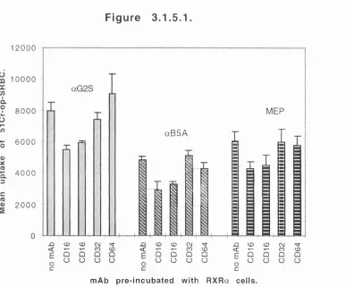
3.1.5.1. Immunophagocytosis was inhibited by Fc receptor
ylll mAbs (CD 16), but not FcR yll (CD32), or FcR yl (CD64). 125 3.1.5.2. Further investigation of Fey receptor mAb
inhibition of immunophagocytosis. 126
3.1.6. Immunonophagocytosis by the U937 transfectants is
inhibited mAbs which recognise CD13, and CD18. 126
3.1.6.1. Initial screen of effects of mAbs from the myeloid
panel on immunophagocytosis. 126
3.1.6.2. Two mAbs from the myeloid panel, CD13 (7H5), and CD18 (BU87-M2), inhibited uptake of ^’Cr-op-SRBC into the U937
_____________________________________________________
Contents
3.1.7. Expression of p47-phox, but not of p67-phox isupregulated in «G2S cells, but not in aB5A cells. 128
3.1.8. Immunophagocytosis by the U937 transfectants induces
rapid tyrosine phosphorylation of numerous proteins. 1 3 2
3.1.8.1. Pattern and intensity of tyrosine phosphorylation differs between 9-cis RA-treated U937 transfectants in response to op-SRBC. 132 3.1.8.2. Tyrosine phosphorylation responses to op-SRBC
are similar in DHCC-treated U937 transfectants. 134
3.1.9. mAbs inhibitory of immunophagocytosis do not inhibit the
phosphotyrosine response to op-SRBC. 13 6
3.1.9.1. Fey receptor mAbs did not inhibit the phosphotyrosine
response to op-SRBC. 136
3.1.9.2. The inhibitory mAbs CD13 (7H5) and CD18
(BU87-M2) do not inhibit the phosphotyrosine response to op-SRBC. 136
3.2. D iscussion. 1 4 2
3.2.1. RXRa, RARa and RARp are expressed in peripheral
blood m onocytes. 1 4 2
3.2.2. RXRa, RARa and RARp are expressed in the U937
transfectants. 14 4
3.2.3. Cell surface protein expression on U937 transfectants. 14 5
3.2.3.1. Expression of ICAM molecules. 146
3.2.3.2. Expression of integrins. 148
3.2.3.3. Expression of other cell surface molecules. 148
3.2.4. Immunophagocytosis by the U937 transfectants. 15 4
3.2.4.1. Differences in efficiency of immunophagocytosis. 154
3.2.4.2. Proteins involved in immunophagocytosis. 155
3.2.4.3. Immunophagocytic induced tyrosine phosphorylation. 157 3.2.4.4. Effects of inhibitory mAbs on immunophagocytic
induced tyrosine phosphorylation. 158
3.2.5. Expression of NADPH oxidase com ponents. 15 9
Chapter 4. Signalling processes involved in im m unophagocytosis by the
U937 transfectants.
4.1. R esults 161
4.1.1. Mitogen activated protein kinases (MAPK) are
phosphorylated in response to addition of op-SRBC. 161
_____________________________________________________
Contents
4.1.2. The activation state of the adaptor molecule She isnot effected by exposure to op-SRBC. 165
4.1.3. Expression of FRIP, a regulator of rasGAP, in the
U937 transfectants. 167
4.1.4. Tyrosine phosphorylated proteins in the three U937
transfectants have differing associations with GST-SH2 fusion
proteins of common signalling molecules. 167
4.1.5. Phosphorylation of p47-phox and p67-phox was not
induced in response to op-SRBC in the U937 transfectants. 169
4.1.6. Expression of paxillin in the U937 transfectants, and
phosphorylation of paxillin in response to op-SRBC. 173
4.2. D iscussion. 176
4.2.1. Activation of ERK-1, Jnk and p38 during
im m unophagocytosis. 176
4.2.2. Phosphorylation of She is not affected by
im m u n ophagocytosis. 177
4.2.3. FRIP is expressed in aG2S and MEP cells, but not
aB5A cells. 178
4.2.4. Interactions with GST-SH2 fusion proteins. 178
4.2.4.1. Src SH2 interactions. 179
4.2.4.2. Grb-2 and O rb-10 SH2 interactions. 179
4.2.4.3. She SH2 and PTB interactions. 180
4.2.4.4. p85 SH2 interactions. 181
4.2.4.5. PLC-yl SH2 interactions. 182
4.2.5. Phosphorylation of p47-phox and p67-phox is not
induced by immunophagocytosis. 182
4.2.6. Expression but not phosphorylation of paxillin in the
U937 transfectants. 183
4.2.7. Summ ary. 184
Chapter 5. Molecular characterisation of U937 dependent T-cell c o
stim ulation.
5.1. R esults. 187
5.1.1. U937 cells provide a co-stimulatory signal to T-cells. 188
5.1.2. U937 cells and T-cells form large cell clusters during
T-cell proliferation. 191
5.1.3. Role of C D lla /18 and CD54 in U937 induced T-cell
activation. 191
________________________________________________
Contents
5.1.3.2. Investigation of C D lla/1 8 and CD54 mAb induced
inhibition. 196
5.1.3.3. Levels of expression of C D lla/1 8 and CD54 on U937
and T-cells. 196
5.1.4. Screening panels of mAbs for effects on T-cell
proliferation. 196
5.1.4.1. Results of primary screening. 196
5.1.4.2. mAbs chosen for further studies. 199
5.1.5. Role of CD45 in U937 induced T-cell proliferation. 199
5.1.5.1. CD45 mAbs inhibit T-cell proliferation. 199
5.1.5.2. Expression of CD45 on U937 and T-cells. 199 5.1.5.3. CD45 mAbs induce the formation of larger U937 -
T-cell clusters. 203
5.1.6. Role of CD53 in U937 induced T-cell proliferation. 2 0 3
5.1.6.1. CD53 mAbs inhibit T-cell proliferation. 203
5.1.6.2. Expression of CD53 on U937 and T-cells. 203
5.1.7. Role of CD55 in U937 induced T-cell proliferation. 2 0 6
5.1.8. Role of CD98 in U937 induced T-cell proliferation. 2 0 6
5.1.8.1. CD98 mAbs inhibit T-cell proliferation. 206
5.1.8.2. Expression of CD98 on U937 and T-cells. 209
5.1.9. Role of C D w l08 in U937 induced T-cell proliferation. 2 1 0
5.1.10. Role of CD147 in U937 induced T-cell proliferation. 2 1 0
5.1.10.1. CD 147 mAbs inhibit T-cell proliferation. 210 5.1.10.2. Expression of CD 147 on U937 and T-cells. 210
5.1.11. Role of unknown antigens in U937 induced T-cell
proliferation. 2 1 2
5.1.11.1. mAbs of undefined specificity inhibit T-cell proliferation. 212 5.1.11.2. Expression of these undefined antigens on U937 and
T-cells. 212
5.1.12.Non-inhibitory mAbs that bind neither U937 or T-cells.214
5.1.13. CD53, CD55, CD98, C D w l08, CD147 and the undefined
mAbs do not inhibit U937/T-cell cluster formation. 2 1 4
5.1.14. Pre-incubation of U937 cells with mAbs inhibits
T-cell responses. 2 2 0
5.1.15. Addition of inhibitory mAbs to a PMA/ionomycin
dependent-11937 independent T-cell proliferation assay
causes inhibition of T-cell activation. 2 2 2
5.1.16. U937 cells are more efficient co-stimulators of
CD8+ T-cells, than of CD4+ T-cells. 2 2 4
_____________________________________________________
Contents
5.1.16.2. Previously described inhibitory mAbs inhibit CD4+
and CD8+ T-cells equally. 224
5.1.17. CD28 mAb stimulates proliferation of CD4+ T-cells,
but not of CD8+ T-cells. 2 2 5
5.1.18. The inhibitory mAbs previously described induce changes
in tyrosine phosphorylation in both U937 and T-cells. 2 2 9
5.1.18.1. mAbs that induce tyrosine phosphorylation in U937 cells. 229 5.1.18.2. mAbs that induce tyrosine phosphorylation in T-cells. 233
5.2. D iscu ssion . 2 3 7
5.2.1. U937 cells provide a co-stimulatory signal to T-cells. 23 8
5.2.2. U937 cells preferentially co-stimulate CD8+ T cells. 2 3 9
5.2.3. C D lla /1 8 (LFA-1)-CD54 (ICAM-1) interactions
co-stimulate T-cell responses. 2 4 0
5.2.4. CD45 mAbs inhibited T-cell proliferation. 2 4 2
5.2.5. Identification of novel molecules involved in T-cell
activation . 2 4 4
5.2.5.1. CD53 mAbs inhibited T-cell proliferation. 244 5.2.5.2. CD55 mAbs inhibited T-cell proliferation. 245 5.2.5.3. CD98 mAbs inhibited T-cell proliferation. 246 5.2.5.4. A C D wl08 mAb inhibited T-cell proliferation. 248 5.2.5.5. CD 147 mAbs inhibited T-cell proliferation. 249 5.2.5.6. Unknown mAbs inhibited T-cell proliferation. 250 5.2.5.8. Inhibitory mAbs inhibited CD4-H and CD8+ T-cells equally.250
5.2.6. PBDC-CD3-dependent T-cell proliferation assay. 2 5 0
5.2.7. Sum m ary. 251
Chapter 6. Investigation of X-irradiation induced cell death in the U 9 3 7
transfectant cell lines; ability of U937 transfectants to co-stimulate T
c e lls .
6.1. R esults 25 3
6.1.1. Proliferation of 9-cis RA-treated aG2S, aB5A and
MEP cells differs following X-irradiation. 2 5 3
6.1.2. Differences in proliferation on X-irradiated aG2S,
aB5A and MEP cells is dependent upon 9-cis RA treatment. 2 5 4
6.1.2.1. Proliferation by untreated a 0 2 S , aB5A and MEP cells
is inhibited equally following X-irradiation. 254
6.1.2.2. Proliferation by DHCC-treated a 0 2 S , aB5A and MEP
cells is inhibited equally following X-irradiation. 254
6.1.3. Proliferation by 9-cis RA-treated aG2S, aB5A and
_____________________________________________________
Contents
6.1.4. Increased numbers of viable 9-cis RA-treated aG2Scells after X-irradiation. 25 8
6.1.5. Increased numbers of 9-cis RA-treated aG2S cells
complete the cell cycle after X-irradiation. 261
6.1.6. aG2S cells are more capable co-stimulators of T cell
activation than MEP or «B5A cells. 2 6 2
6.2. D iscussion. 2 65
6.2.1. Differences in proliferation between X-irradiated U 9 3 7
transfectants are 9-cis RA dependent. 2 65
6.2.2. Differences in inhibition of proliferation was caused
by differential inducement of apoptosis. 2 66
6.2.3. Co-stimulation of T-cell activation. 2 7 2
Chapter 7. Conclusions and further objectives. 2 74
7.1. Expression of RXRa by peripheral blood monocytes. 2 74
7.2. U937 derived RXRa transfectants. 2 74
7.2.1. Assessment of differentiation. 275
7.2.2. Immunophagocytic signal transduction. 275
7.2.3. Co-stimulatory capabilities. 277
7.2.4. X-irradiation induced cell death. 277
7.3. U937 cell dependent T-cell co-stimulation. 27 8
7.3.1. U937 cells act as accessory cells. 278
7.3.2. Molecules that inhibit T-cell proliferation. 278
A p p en d ices. 2 80
I mAbs used in phenotyping of U937 transfectants. 280
II mAbs used in immunophagocytosis experiments. 283
III mAbs used in U937/CD3 dependent T-cell proliferation assay. 284 IV Expression of myeloid cell surface antigens, on 9-cis RA-treated
U937 transfectants. 289
V Screening of mAbs from the myeloid panel for effects on immunophagocytosis. 293 VI Initial screen of mAbs from the 6th HLDA for effects on the U937/CD3
T-cell proliferation assay. 294
V n mAbs that consistently inhibited T-cell proliferation. 300
V in. mAbs selected for further studies in the U937/CD3 T-cell proliferation assay. 302
IX Abbreviations 304
X Solutions 309
XI Publications arising from this thesis. 311
_______________________________________________________
TaBks
List of Tables,
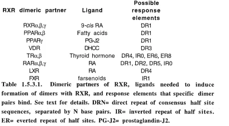
1.5.3.1. Dimeric partners of RXR, ligands needed to induce formation of dimers with
RXR, and response elements that specific dimer pairs bind. 75
2.4. Antibodies used in immunoblotting. 95
3.1.1. Normalised expression of R X R a, RA Ra, and RARp in peripheral blood
monocytes during differentiation. 111
3.1.3.1. Expression of ICAM molecules on 9-cis RA-treated and untreated U937
transfectants. 116
3.1.8.1. Proteins phosphorylated during immunophagocytosis in the
U937 transfectants. 135
5.1.3.1. C D lla , CD18 and CD54 mAbs inhibit the T-cell response. 193 5.1.3.3. Levels of expression of C D lla , CD18 and CD54 on U937 and T-cells. 198 5.1.5.2. Levels of expression of CD45 on both U937 and T-cells. 202 5.1.6.2. Levels of expression of CD53 on both U937 and T-cells. 206 5.1.7. Levels of expression of CD55 on both U937 and T-cells. 207 5.1.8.2. Levels of expression of CD98 on both U937 and T-cells. 209 5.1.10.2. Levels of expression of CD 147 on both U937 and T-cells. 212 5.1.11.2. Levels of expression of undefined mAbs on both U937 and T-cells. 214 5.1.12. Levels of expression of non inhibitory mAbs on U937 and T-cells,
and inhibition of T-cell proliferation found with these mAbs. 215
6.1.4. Increased numbers of viable 9-cis RA-treated aG2S cells after X- irradiation.261 6.1.5. Increased numbers of 9-cis RA-treated aG2S cells viable and complete
_______________________________________________________
figures
List of Figures.
1.1.1 A. Schematic representation of monocyte/macrophage development, and the factors
that determine this process. 19
1.1. IB. Schematic representation of the relative position of the human leukaemic U937
cell line in monocyte/macrophage development. 20
1.1.2. Schematic representation of transcription factors involved in monocyte/macrophage
development. 23
1.2.1.1. Schematic representation of the structures of Fey receptors. 34 1.2.1.2. Schematic representation of the probable signal transduction mechanisms
involved following FcyR cross linking. 36
1.3.2.2. Schematic representation of the probable mechanisms involved in the formation
of active Ras-GTP. 50
1.3.2.3. Schematic representation of the probable interactions involved in transduction of
signals through the MAPK pathway. 52
1.5.3. General structure of RXR. 71
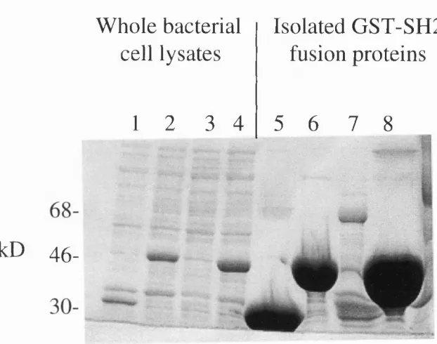
1.5.3.1. Schematic representation of the RXRa DNA binding domain. 73 2.14. Expression of GST-SH2 fusion proteins in XL2 blue cells. 101 3.1.1. Expression of R X R a, RA Ra, and RARp in peripheral blood monocytes during
differentiation. 110
3.1.2.1. Expression of R X Ra in the U937 transfectants. 113
3.1.2.2. Expression of RARa and RARp in the U937 transfectants. 115 3.1.4.1. Assessment of immunophagocytic efficiency by the U937 transfectants by
microscopy. 120
3.1.4.2. Assessment of immunophagocytic efficiency by the U937 transfectants by
uptake of radiolabelled opsonised SRBC (^’Cr-op-SRBC). 122
3.1.4.3. Visualisation of actin after two hours exposure to op-SRBC. 124
3.1.5.1. Blockade of immunophagocytosis by Fey receptor mAbs. 125
3.1.5.2. Blockade of immunophagocytosis by a titration of Fey receptor mAbs. 127 3.1.6.2. Immunophagocytosis by the U937 transfectants is inhibited by a CD13 mAb
(M50 - 7H5), and a CD18 mAb (M16 - BU87-M2). 129
3.1.7. Expression of p47-phox, and p67 phox in the U937 transfectants. 131 3.1.8.1. Immunophagocytosis induces differential patterns of tyrosine phosphorylation in
the U937 transfectants. 133
3.1.8.2. DHCC-treatment does not induce differential tyrosine phosphorylation responses
to op-SRBC in the U937 transfectants. 137
3.1.9.1. Effects of binding Fey receptor mAbs on the phosphotyrosine response to op-
SRBC. 138
3.1.9.2. Effects of binding anti-CD 13 (7H5), and anti-CD 18 (BU87-M2) mAbs on the
_______________________________________________________
Tyures
3.2.3. Schematic representation of cell surface protein expression during monoblast
(U937) differentiation. 146
4.1.1.1. Phosphorylation of ERK-1 in the U937 transfectants upon exposure
to op-SRBC. 163
4.1.1.2. The p38 stress kinase is phosphorylated in the U937 transfectants upon
exposure to op-SRBC. 164
4.1.1.3. The Jnk stress kinase is phosphorylated in the U937 transfectants upon exposure
to op-SRBC. 166
4.1.2. Activation of She is not affected in the U937 transfectants upon exposure to op-
SRBC. 168
4.1.3. Expression of FRIP in the U937 transfectant cell lines. 168
4.1.4. Tyrosine phosphorylated proteins from the U937 transfectants differentially
interact with SH2 domains of signalling molecules. 170
4.1.5. Phosphorylation of p47-phox and p67-phox is not induced in the U937
transfectants upon exposure to op-SRBC. 174
4.1.6. Expression of paxillin in the U937 transfectants; paxillin is not phosphorylated in
response to op-SRBC. 175
4.2.7. Schematic representation of possible signalling pathways utilised following
immunophagocytosis by the U937 transfectants 186
5.1.1. U937 cells co-stimulate T-cells in a CD3 dependent proliferation assay. 189 5.1.2. T-cell proliferation can be observed visually through the formation of
large U937 / T-cell clusters. 192
5.1.3.1. CD 11 a/CD 18 and CD54 mAbs inhibit the formation of U937
-T-cell clusters. 194
5.1.3.2. C D lla/CD 18 and CD54 mAbs inhibit T-cell proliferation in a dose-dependent
manner and are not cytotoxic. 197
5.1.5.1. Inhibition of T-cell proliferation induced by CD45 mAbs is dose-dependent and
not due to cytotoxicity. 200
5.1.5.3. The CD45 antibody 4.14 caused formation of large U937-T-cell clusters. 204 5.1.6.1. Inhibition of T-cell proliferation induced by CD53 mAbs is dose-dependent and
not due to cytotoxicity. 205
5.1.7. Inhibition of T-cell proliferation induced by CD55 mAbs is dose-dependent and
not due to cytotoxicity. 207
5.1.8.1. Inhibition of T-cell proliferation induced by CD98 mAbs is dose-dependent and
not due to cytotoxicity. 208
5.1.10.1. Inhibition of T-cell proliferation induced by CD 147 mAbs is dose-dependent
and not due to cytotoxicity. 211
5.1.11.1. Inhibition of T-cell proliferation induced by mAbs of unknown specificity is
_____________________________________________________________________________
l^yures
5.1.13. Inhibitory CD53, CD55, CD98, CDwlOS, CD 147 and undefined mAbs do noteffect U937 / T-cell cluster formation. 216
5.1.14. Inhibitory activity of antibodies when bound selectively to U937 cells. 221 5.1.15. Some mAbs are inhibitory of T-cell proliferation in a U937 independent
proliferation assay. 223
5.1.16.1. U937 cells preferentially stimulate CD8-I- T-cells. 226 5.1.16.2. Inhibitory mAbs affect both CD4+ and CD84- T-cells equally. 227 5.1.17. An agonistic CD28 antibody co-stimulates CD4-f- T-cells, but has no effect on
CD8-K T-cells. 228
5.1.18.1. The effects of inhibitory mAbs on tyrosine phosphorylation of
U937 cells. 230
5.1.18.2. The effects of inhibitory mAbs on tyrosine phosphorylation of purified
T-eells. 235
6.1.1. 9-cis RA-treated aG2S cells proliferate more, and aB5A cells less than MEP cells,
following X-irradiation. 255
6.1.2.1. Proliferation by untreated aG 2S, «B5A and MEP cells is inhibited equally
following X-irradiation. 256
6.1.2.2. Proliferation by DHCC-treated aG2S, aB5A and MEP cells is inhibited equally
following X-irradiation. 257
6.1.4. Increased numbers of viable 9-cis RA activated aG2S cells
after X- irradiation. 259
6.1.6. aG2S cells are more capable co-stimulators of T cell activation than
Acknowledgements.
The supervision, advice and guidance received over the past three years
from Prof. David Katz, Prof. Paul Brickell and Prof. Benny Chain, has
been very much appreciated. I would particularly like to thank Prof.
David Katz for his constant enthusiasm and willingness to talk over
problems. I would like to thank all other members of the Dept, of
Immunology for all their help, but in particular Patrick, Ness, Tom,
Debbie, Louise, Charles and Pete, who made the a lab such a great place
to work in, and who have become excellent friends and eaters of curry. I
was “helped” during this project by various summer and BSc students,
thanks to Paul Herridge for testing some of the adhesion panel of mAbs in
the U937/CD3 T-cell assay, Joel Branch, for doing FACS analysis on the
U937 transfectants, Ben Kendrick, for doing the mAb-phosphotyrosine
studies on U937 and T-cells, and (the incomparable) Hutan Ashrafian,
who did some of the FACS analysis of U937 and T-cells. I also
grudgingly acknowledge “Captain Rhodesia”, the miserable bar manager,
for serving vast amounts of flat lager and completely undrinkable bitter.
Thanks also to Dr. Darrin Smith for the GST-SH2 fusion proteins,
various antibodies and for originally giving me a job in Cambridge which
got me into science.
On a home note, I would like to thank Mike and Jo for being top
housemates, putting up with me, and making living in London a great
experience. Of course, many thanks to Caroline for all her love, support
and determination that I should get this thing finished in three years! !
This thesis is dedicated to my Mum and Dad, without who’s guidance and
support, even when thousands of miles away, nothing would have been
Cfiüfterl Introduction
C flap ter 1
In trodu ction
1.1. The m yeloid cell lineage.
This section will give a brief introduction to the myeloid cell lineage, and
the monoblastoid U937 cell line.
1.1.1. M yelopoiesis: D evelopm ent of m yeloid cells.
Haemopoietic stem cells, which are located in the bone marrow of adults,
are the precursors of erythroid, megakaryocytoid, lymphoid and myeloid
cell lineages. The eventual fate of each stem cell is determined by factors
such as interleukins (IL), colony stimulating factors (CSF), and certain
hormones which induce clonal proliferation and differentiation of stem
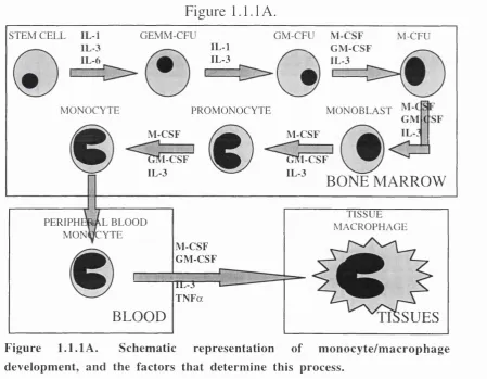
cells. Figure 1.1.1 A. shows the progression of differentiation towards the
monocyte/macrophage lineage. This process, from stem cell to a mature
monocyte, takes six days in the bone marrow (Groopman and Golde,
1981). In bone marrow, IL-1, IL-3, and IL-6 induce heteromitosis of
stem cells, which gives rise to one new stem cell and to a pluripotent
granulocyte-erythrocyte-megakaryocyte-macrophage colony forming unit
(GEMM-CFU). From this stage, the further presence of IL-1 and IL-3
commits these cells to become a common precursor of both granulocyte
and monocyte/macrophage lineages, hence known as a granulocyte-
macrophage colony forming unit (GM-CFU).
These GM-CFU precursors can then be differentiated sequentially to a
macrophage colony forming unit (M-CFU), to a monoblast, to a p ro
monocyte, and then to a mature monocyte. This differentiation is
controlled by IL-3, granulocyte-macrophage CSF (GM-CSF), and
macrophage CSF (M-CSF). IL-3 and GM-CSF are thought to be involved
in proliferation of the immature cells, and M-CSF involved in
Cfiapter 1 Introduction
lineage (review ed by Ogawa, 1993). All the above processes occur in the
bone m arrow . Following these events, m ature m onocytes enter the blood
stream. M onocytes are generally smaller than their im m ediate p rec u rso rs,
and have well developed lysosomes and an enhanced phagocytic abilities.
F rom the bloodstream , m onocytes enter body tissues. Once present in
tissues m onocytes differentiate to m acrophages under the influence o f
factors that include M -CSF, GM -C SF, IL-1, IL-3, and tu m o u r necrosis
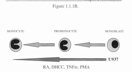
factor a ( T N F a ) . Figure 1.1.IB. shows an exam ple o f this rep resen tin g
the differentiation of a m onoblastic cell line U937, to becom e m onocytes,
and the factors that have been used to stimulate differentiation o f these
cells.
Figure 1.1.1 A.
STEM CELL IL-1 IL-3 IL-6
GEMM-CFU GM-CFU M -CSF
IL-1 G M -C SF
IL-3 / - A IL-3
L f [
M-CFU
MONOCYTE PROMONOCYTE
M -C SF M -CSF
M ONOBLAST GM
[-CSF
IL-3
1-CSF
BONE M ARROW
PERIPHl MON(
1 l b l o o d
jfCYTE
M -C SF G M -C SF
1 ... V
lL -3 1
T N F a
BLOOD
TISSUE MACROPHAGE
SUES
Figure 1.1.lA . Schematic representation of monocyte/macrophage
cfiapter 1 Introduction
Figure 1.1. IB.
M O N O C Y T E PR O M O N O C Y T E M O N O B L A S T
RA, DHCC, T N F a, PMA
U 9 3 7
Figure 1 .1 .IB . Schem atic rep resen tatio n of the relative position of th e hum an leukaem ic U937 cell line in m onocyte/m acrophage d e v e lo p m e n t, and facto rs that have been shown to d ifferen tiate this cell line to b ecom e m o n o c y te s.
H um an m onocytes have a half life of three days in the blood stream.
These cells bind to endothelium either random ly or during recru itm en t to
areas o f inflam m ation. A variety o f adhesion molecules are involved in
the interaction o f m onocytes with endothelium , including selectins, and
integrins (review ed by Springer, 1990). The m onocytes will extravasate
through the endothelium and m igrate into the surrounding tissue, w h ere
they differentiate into tissue m acrophages. The exact phenotype of these
m ature cells is dependent on the tissue that they have entered and the
context of that tissue at the time of maturation. Dendritic cells, w hich are
the m ost potent know n antigen presenting cells (A PC) and act as
“sentinels” o f the im m une system; microglia, which develop in b rain
tissue; osteoclasts, which develop in bone; Kupffer cells, w hich develop in
liver; and L angerhans cells; w hich develop in skin, are all o f the
m onocyte/m acrophage lineage and these exam ples dem onstrate the
versatility and im portance of the lineage. Tissue m acrophages can
differentiate further to form both epitheloid cells and m ulti-nucleated
cfiapter 1 Introduction
giant cells, which are features of granulomatous inflammatory diseases
such as tuberculosis (Weinberg et ah, 1984).
1.1.2. M olecular m echanisms that regulate m yeloid c e ll
d iffe r e n tia tio n .
In recent studies, some of the molecular mechanisms behind myelopoiesis
have been unravelled by the investigation of transcription factors
implicated in myeloid cell development, and of the genes which these
factors control (as reviewed by Valledor et ah, 1998). Figure 1.1.2. gives
a schematic representation of transcription factors involved in
monocyte/macrophage development. Survival and proliferation of stem
cells is regulated by a number of transcription factors. These include
GATA 2 and PLZF, both members of the zinc finger family, SCL, which
is a member of the basic helix loop helix (bHLH) family, and the
homeobox proteins, H0XB3 and H0XB4. The transcription factors
GATA 1, GATA 2, SCL and Myc are implicated in regulating
differentiation to GEMM-CFU and expression of PLZF, H0XB3 and
H 0X B 4 is down regulated during this step. However, for cells to progress
beyond GEMM-CFU, expression of GATA 1, GATA 2, SCL and c-myc
must be repressed. For example, over-expression of SCL leads to
repression of essential monocyte proteins, such as C D l lb and lysozyme,
and the lack of inhibition of proliferation associated with mature
monocytes.
Myb has been shown to control the survival and proliferation of GM-
CFU, and the commitment of these cells to M-CFU. Myb is also able to
activate expression of monocyte-specific proteins such as Mim-1 and
lysozyme. Survival and proliferation of M-CFU cells is dependent on M-
CSF, and expression of M-CSF receptor is controlled by P U .l, a member
of the Ets family, and a myeloid-specific transcription factor. Recently,
CfLaj)terl Introduction
phosphatidylinositol 3-kinase (PI3-K). When this interaction was blocked,
M-CSF-induced differentiation was abrogated (Bourette et ah, 1997), thus
showing the importance of signalling via the M-CSF receptor, and PU .L
Furthermore, PU .l in co-operation with other factors, activates
transcription of GM-CSF receptors. During subsequent maturation of M-
CFU, PU .l is essential for the transactivation of genes involved in the
functional properties of macrophages. These include C D l lb, CD18,
CD 14, the high and low affinity receptors for IgG (FcyRI and FcyRIII
respectively), and the scavenger receptors 1 and II. CCAAT enhancer-
binding protein (3 (C/EBP(3) is also thought to play a role in
transactivation of functionally important macrophage genes. The
characteristic reduction in cell proliferation associated with a mature
monocyte phenotype required two mechanisms. Firstly, Myc and Myb,
are down-regulated as over-expression of either proteins at this stage will
block monoblast differentiation. Secondly, interferon regulatory factor-1
(IRF-1) is induced in monocytes, this transactivates the transcription of
interferon a and p (IF N a and P), which act as autocrine signals that
promote inhibition of proliferation.
Other factors implicated in the maturation of monocytes to macrophages
are EG R l (a member of the zinc finger family), HOXB7, and NF-Y.
Expression of EG R l is restricted to monocytes and macrophages,
although EG Rl null mutant mice still develop macrophages, questioning
the importance of this factor in development of this lineage. NF-Y has
been implicated in the expression of ferritin, a protein involved in iron
metabolism, which controls the expression of MHC class II, thus allowing
the mature macrophage to act as an iron storage compartment and as an
Cfiaj)ter 1 Introduction
Figure 1.1.2.
5 T E M C E L L A ^ ^ ^ ^ ^ GEMM-CFU
GATA 2
GM -CFU M -CFU
SCL Myc
PLZF HOXB3+B4
-G A T A I GATA 2
▼sCL Myc PU.l
C/EBPP
M ONOCYTE PROMONOCYTE M O NO BLAST
IRF-1
BONE M A R R O W
TISSUE MACROPHAGE PER IPH K iy\L BLOOD
M O N 0C Y T E E G R l
HOXB7 NF-Y
BLOOD TISSU ES
F ig u re 1.1.2. Schem atic re p resen tatio n of tra n sc rip tio n factors involved in m onocyte/m acrophage developm ent, black u p w ard arro w s indicates facto rs ex p ressed at th a t point, black dow nw ard arro w s indicates facto rs d o w n - reg u lated at th a t point.
1.1.3. Functions of monocytes/macrophages.
O nce the cells have m atured into tissue m acrophages, there is a f u rth e r
potential developm ental step, which involves the conversion o f resting
m acrophages to an activated state. This activation is m ediated by
inflam m atory m ediators such as IFNy (Black et ah, 1987), G M -C S F (Reed
et ah, 1987), and T N F a (De Titto et ah, 1986) which are secreted by cells
su rrounding the resting m acrophage, or as a direct response to bacterial
products, such as lipo-polysaccheride (LPS). This reflects the principal
function o f activated m acrophages in host defence against a range o f
pathogens. It is these m acrophages that have the ability to phagocytose and
CHapterl Introduction
immune system by secreting inflammatory factors such as T N F a, IL-1,
IF N a and p, and reactive oxygen species (as reviewed by Johnston, 1988).
Monocytes and macrophages, along with neutrophils, are professional
phagocytes. Phagocytosis is mediated by several receptors expressed on
both monocytes and macrophages. The mannose receptor is an example of
a protein that is involved in phagocytosis via the recognition of
biochemical features of pathogens. This receptor will interact with
terminal mannose or fucose saccharides on the capsule of invading
bacteria. Fey receptors and complement receptors C R l, CR3, and CR4 are
examples of proteins that are involved in phagocytosis via interaction with
host proteins that have previously bound to the pathogen, in a process
known as opsonisation. Fey receptors recognise IgG coated particles,
whilst C R l, CR3, and CR4 recognise particles coated with components of
the complement pathway. There are three classes of Fey receptors
expressed on monocytes and macrophage. These are FcyRI (CD64), the
high affinity IgG receptor, FcyRII (CD32), a low affinity IgG receptor,
and FcyRIII (CD 16), a second low affinity IgG receptor. Phagocytosis
induces monocytes and macrophages to secrete pro-inflammatory factors,
induces the respiratory burst, and also induces antibody dependent cellular
cytotoxicity (ADCC) activity. Phagocytosis mediated by monocytes and
macrophages is discussed in detail in section 1.2. of this chapter.
Another key property of monocytes and macrophages is their ability to
adhere to lymphatic and vascular endothelium, and to extracellular matrix
proteins such as laminin and fibronectin (Pawlowski et al., 1985;
Pawlowski et al., 1988). Adhesion is also necessary for many of the
monocyte/macrophage functions previously mentioned, such as
phagocytosis, cytotoxicity, and the production of inflammatory factors.
There are three families of cell surface proteins that mediate adhesion.
These are integrins, selectins, and the immunoglobulin superfamily
Cfiapter 1 Introduction
heterodim eric glycoproteins responsible for cell-cell and cell-m atrix
interactions. Integrins are classified into sub-families according to the (3
chain involved, (31 integrins com prise a CD 29 (3 chain and a series o f
possible a chains (CD49a, b, c, d, e, or f). These (31 integrins are also
know n as very late antigens, or VLA-1 to V LA-6. (32 integrins com prise
a CD 18 (3 chain with either G D I l a (LFA-1), GDI lb (Mac-1), or G D I Ic
( p i 50/95) a chains. There are also other integrins such as the (33
(cytoadhesins) and (37 subfamilies. (31 and (33 subfamilies bind mainly to
m atrix proteins such as collagen, laminin, fibronectin and vitronectin,
w hereas the (32 subfamily interact with cellular proteins. F or example,
LFA -1 interacts with im m unoglobulin family adhesion molecules such as
IGAM-1 (GD54), IGAM-2 ( G D I 02), and IGAM-3 (GD50). The selectins,
com prising of L-selectin (GD62L), P-selectin (GD62P) and E-selectin
(GD 62E), are involved in leukocyte-endothelium interactions, and T%ts
been shown to be vital in leukocyte rolling along vascular endothelium ,
which is the first stage o f leukocyte extravasation. Selectins bind to
oligosaccharides such as G D I 5 (L e ^ and G D I5 s (sialyl Le"") (Prieto et al.,
1994).
1.1.4. The U937 cell line.
The U937 cell line was first isolated from a patient with diffuse histiocytic
ly m p h o m a and has proven to be a useful m odel for the studies o f the
m o no cyte/m acrophage lineage (Sundstrom and Nillson, 1976). The cell
line is already com m itted to the m onocyte lineage. Figure 1 .1.IB. shows
how U937 can be induced to differentiate from its “norm al” m onoblastoid
state, in sequential steps (see figure 1.1.1 A.), to becom e a m onocyte.
D ifferentiation can be induced with agents such as phorbol 12-myristate
13-acetate (PM A ) (Forsbeck et al., 1985), IF N y and T N F a (Harris et al.,
1985), retinoic acid (RA) (Olsson and Breitman, 1982), and the active
vitam in D m etabolite 1,25-dihydroxycholecalciferol (DHGG) (A m ento et
chapter 1 Introduction
differentiation of monocytes/macrophages, as well as monocyte/
macrophage functions.
U937 cells proliferate in suspension culture in the presence of growth
factors supplied by foetal calf serum. Undifferentiated U937 cells have a
doubling time of between 20 and 48 hours, and express few Fc, C3 or
chemotactic peptide receptors (Sundstrom and Nillson, 1976). The
enormous amount of literature about U937 cells reflects the extensive use
of this cell line as model system, and also the stability of the cell line over
the twenty years of its existence.
1.1.4.1. Molecular events involved in growth arrest of U937 cells.
Following differentiation towards a monocyte phenotype, induced by a
variety of factors, U937 cells shift to a non-proliferative state indicative
of a monocyte phenotype. Many proteins have been implicated in this
switch to a non-proliferative state.
Myc has been shown to dimerise with Max, and this heterodimer binds to
response elements and activates transcription of specific genes. Max may
also dimerise with Mad, and in this case act as a repressor of transcription
(reviewed by Marcu et al., 1992). After PMA treatment of U937 cells,
Myc expression is reduced, whilst Mad levels increase. Furtherm ore, the
ratio of Myc-Max dimers to Mad-Max is changed in favour of the latter,
to the extent that no Mad-Max dimers are found in untreated U937 cells,
and no Myc-Max dimers are found in differentiated U937 cells (Ayer and
Eisenman, 1993). These changes correlate with the switch from
proliferation to non-proliferation.
Another important group of proteins involved in the cell cycle are the
cyclin dependent kinases (cdk). After PMA treatment, cdk2 is
dephosphorylated, which reduces its activity. Furthermore, this decrease
CHapter 1 Introduction
cdk2 is complexed with cyclin E (Asiedu et al., 1997). Other evidence for
the involvement of cdk in U937 cell growth arrest is that levels of a cdk
inhibitor, known as p21, W A Fl, or C IP l, is increased upon
differentiation induced by both RA and DHCC, and that this increase is
mediated by RA and Vitamin D response elements in the p21 prom oter
(Liu et al., 1996a; Liu et al., 1996b).
GTA is a tyrosine kinase that has been associated with the cell cycle,
having a sequence similar to the cell cycle control gene, cdc2. PMA
treatment of U937 cells induced increases in GTA levels and activity.
GTA is thought to act by controlling the phosphorylation, and activity, of
(3-1,4-galactosyltransferase, an enzyme found in the golgi that has been
implicated in cell cycle control (Kraft et al., 1992).
Interferon regulatory factor 1 (IR Fl) is a transcription factor first found
to respond to interferons (IFN) and regulate the cell cycle. Growth arrest
induced by dl\-trans RA treatment of U937 cells has been correlated with
increased expression of IR F l, and this response is not mediated by signal
transducers and activator of transcription (STAT) molecules, which are
normally associated with IFN signalling, suggesting that the IR Fl gene
promoter contains an RA response element (Matikainen et al., 1996).
F G R l has been shown to be restricted to the monocyte lineage, and has
been shown to be expressed in PMA-treated U937 cells. HL-60 cells are
equivalent to GM-CFU cells (see figure 1.1.1 A.) that can be induced to
differentiate along either a monocyte or granulocyte lineage. HL-60 cells
that were induced to become granulocytes did not express FG R L In fact
HL-60 cells transfected to constitutively express FG R l could only be
induced to differentiate to the monocyte lineage (Nguyen et al., 1993).
Both U937 and HL-60 cells, when treated with anti sense oligonucleotides
Cfiapterl Introduction
mice had normal numbers of functional macrophages (Lee et al., 1996),
questioning the importance of this molecule in monocyte development,
and suggesting that in vivo other molecules can replace it.
1.1.4.2. U937 cells as a model for monoblastic differentiation.
Although the U937 cell line is regarded as a monoblast, expression of
adhesion molecules by the cell line resembles that of peripheral blood
monocytes (Prieto et al., 1994). When U937 cells are induced to
differentiate with PMA, they acquire several other macrophage
phenotypic properties, such as adherence, expression of Fc receptors, the
ability to phagocytose, ADCC, and the ability to produce lysozyme and
alkaline phosphatase (Harris and Ralph, 1985). These properties
demonstrate how useful U937 cells have been as an in vitro model of
monocyte/macrophage differentiation and function.
1.1.5. D ifferen tiation of U937 cells.
Although many substances have been shown to induce terminal
differentiation of U937 cells, in the interests of being concise, this section
concentrates on the actions of PMA, RA and DHCC, as these substances
are used in this project, and have been used previously in the laboratory
for the study of U937 cells (Brown et al., 1997).
1.1.5.1. Differentiation of U937 cells induced by PMA.
PMA activates protein kinase C (PKC), and this activation has many
downstream signalling effects. PKC activation leads to phosphorylation of
IkB, a regulatory protein of the NFkB transcription factor. This leads to
dissociation of NFkB from IkB, allowing NFkB to translocate to the
nucleus and activate transcription of target genes (Baeuerle and
Baltimore, 1988). PKC also activates the AP I binding proteins Fos and
Jun. The transcriptional activity of Jun has been shown to be regulated by
being phosphorylated by a member of the mitogen activated protein
Clïa^ter 1 Introduction
regulated by phosphorylation, and PKC activates this pathway by
activating upstream Raf-1. PMA has also been shown to down-regulate
expression of phospholipase Cy-1 (PLCy-1), but to leave levels of PLCy-2
unchanged during differentiation of U937 cells (Lee Y.H. et al., 1995),
and to induce the activation of STAT5 (Woldman et ah, 1997). This
STAT response involves a specific STAT5 isoform, with STATSa, but not
STAT5b being activated (Meinke et al., 1996).
Other important effects of PMA-induced differentiation include the
synthesis of cytokines such as I L - la , T N Fa, CSF-1, and IF N a+ p (Indoh
et al., 1996; Knudsen et al., 1986; Liu and Wu, 1992), adherence to
plastic, increased expression of GDIS and CD54 (ICAM-1), increased
expression of cyclo-oxygenase; and production of lysozyme and oxygen
free radicals (reviewed by Harris and Ralph, 1985).
1.1.5.2. Differentiation of U937 cells induced by RA and DHCC.
K\\-trans RA or 9-cis RA, induce partial growth arrest and differentiation
of U937 cells. RA is necessary for LPS-stimulated induction of IL -ip and
T N F a synthesis (Taimi et al., 1993), and has been shown to be involved
in the down-regulation of expression of both forms of the TNF receptor
(p60 and p80) in U937 cells (Totpal et al., 1995). Addition of RA has
been shown to induce an increase in expression of CD 18 and CD 11b,
which is consistent with monocyte differentiation, but RA is not capable
of inducing CD14 expression (James et al., 1997b; Sellmayer et al., 1994).
This suggests that RA is not capable of inducing a full monocyte
phenotype, but does differentiate the cells to an intermediate stage. The
addition of DHCC to U937 cells induces a more profound level of growth
arrest and differentiation than RA, but RA synergises with DHCC, as
measured by expression of CD 11b, CD 18 and CD 14; and an increase in
CHa^ter 1 Introduction
Another aspect of the role of RA in U937 cell biology is that PMA-
induced differentiation of U937 cells induce increased expression of two
members of a class of retinoic acid receptor, the retinoid X receptors
(RXR), known as R X R a and RXR(3 (Brown et ah, 1997). DHCC also
induces expression of R X R a protein (Defacque et ah, 1997). Both PMA
and DHCC treatment also induce increased expression of vitamin D
receptors (VDR) (Hewison et al., 1989). However, in another report,
addition of RA to U937 cells led to a decrease in transcription of the
R X R a gene, and this was antagonised by addition of DHCC (Nakajima et
al., 1996). Research in our laboratory has produced a U937-derived cell
line that expresses reduced levels of VDR. A major property of these cells
is that they are prone to apoptosis, suggesting that VDR expression may
act as a protective mechanism against programmed cell death (Hewison et
ah, 1996).
The synergistic differentiating effects of DHCC with other agents such as
TGFp and GM-CSF on U937 cells has been established (James et al.,
1997a; Testa et al., 1993; Zuckerman et al., 1988). RA and DHCC will
synergise in inducing cytokine secretion to the same level as addition of
DHCC and (Taimi et al., 1993). As previously described, DHCC, but not
RA, induces expression of CD 14 in U937 cells. The reason for this is that
ÆE there is a vitamin D responsive element (although not a consensus
vitamin D response element, see section 1.5), present in the prom oter
region of the gene coding for CD14 (Zhang et al., 1994). In one report,
DHCC and RA had antagonistic effects on the expression of CD 14 and the
low affinity Fc receptor for IgE (CD23) in U937 cells. PMA- and RA-
induced expression of CD23, whilst PMA- and DHCC-induced CD 14
expression. Furthermore, this report found that dX\-trans RA inhibited the
PMA- or DHCC-induced expression of CD 14, and DHCC inhibited the
PMA- or RA-induced expression of CD23 (Oberg et al., 1993). However,
in another more recent report, addition of 9-cis RA co-operatively
Cfia-pter 1 Introduction
(Nakajima et al., 1996). With the use of specific retinoic acid receptor
(RAR) or RXR agonists and antagonists, the synergism between RA and
DHCC activity was shown to be mediated by both RAR and RXR
(Defacque et al., 1997).
The antagonistic roles of DHCC and RA are reflected in the differential
effects that these factors have on the HL-60 cell line. DHCC induces HL-
60 cells to differentiate to become monocytes, whilst RA induces these
cells to become granulocytes. This RA-induced differentiation of HL-60
cells requires R A R a (Collins et al., 1990). The antagonistic effects of RA
and DHCC have parallels in animal models. These include studies using
chick yolk sac macrophages. When these cells are grown in prim ary
culture they are able to differentiate into multi-nucleated giant cells
(MNGC), and then, sequentially, into osteoclasts. DHCC promotes the
formation of the MNGCs, whilst RA promotes the proliferation of the
macrophages, thus inhibiting the formation of DHCC-induced MNGC
(Woods et al., 1995).
In summary, the formation of heterogeneous populations of monocytes
and macrophages throughout the body is dependent on numerous factors,
that include RA, DHCC, bacterial products such as LPS, free radicals and
cytokines. The precise control of differentiation is regulated by the
concentration of the factor and the point in development where the cell
comes into contact with any factor. The studies described represent a
selection of many hundreds of studies where the U937 cell line has been
used as a model for monocyte/macrophage differentiation and activation.
As this model is so well-established, the U937 cell line was chosen as the
CHa^terl Introduction
1.2. Phagocytosis by m onocytes and m acrophages.Phagocytosis is the process of recognition and engulfment of pathogenic
organisms and cell debris that accumulate during infection, inflammation
or wound repair.
Phagocytosis is carried out mainly by professional phagocytes, which
include monocytes, macrophages and neutrophils. The process is mediated
by proteins expressed on phagocytes that recognise either common
pathogenic structural motifs, such as bacterial cap components, or host
components that have already bound to pathogens, such as IgG or
complement. The phagocytic process is a critical element in eliminating
pathogenic organisms, and is also important for induction of specific
immunity. The process of engulfing pathogens ensures that potentially
destructive processes used to kill the pathogen, such as lysozyme release,
generation of reactive oxygen species, and generation of nitric oxide
(NO), are focused into internal compartments of the phagocytes, thus
limiting the damage that can be inflicted on invaded tissues. Phagocytosis
mediated by any of the receptors mentioned involves an interaction
between receptor and ligand, which leads to signal transduction and, in
turn, to cytoskeletal rearrangement and engulfment. The signals that
precede, and are associated with, engulfment are limited to the area
around the ligand-receptor complex, which is therefore associated with a
localised cytoskeletal rearrangement within the cell.
In this section, the emphasis will be on phagocytosis by cells of the
monocyte/macrophage lineage. Phagocytosis by neutrophils will not be
discussed, although these cells do express some of the same receptors
involved in phagocytosis, and others present on neutrophils, such as
Chg-pter 1 Introduction
1.2.1. Receptors that mediate phagocytosis on m onocytes andm acrop h ages.
Cells of the monocyte/macrophage lineage express several proteins that
are able to mediate phagocytosis. These include the Fey Receptors, which
are proteins that recognise IgG, complement receptor 1 (C R l), CR3 and
CR4, which are receptors that recognise components of the complement
pathways, and mannose receptors that recognise terminal mannose or
fucose residues (reviewed by Brown E.J., 1995).
1.2.1.1. Structure of Fey receptors.
Fey receptors are members of the immunoglobulin superfamily. These
proteins recognise IgG-coated particles. There are three classes of Fey
receptors expressed on monocytes and macrophages. These are FcyRI
(CD64) the high affinity IgG receptor which recognise monomeric IgG,
FcyRII (CD32) and FcyRIII (CD 16), which are low affinity IgG receptors
that recognise immune complexes. Several genes encode the FcyRs. Three
genes (known as A, B, and C) exist for both FcyRI, and FcyRII. Two
genes (known as A and B) exist for FcyRIII (Ravetch and Kinet, 1991).
Figure 1.2.1.1. shows the domain structure of the FcyRs that are
expressed on monocytes or macrophages. All three FcyRs have highly
homologous extracellular portions containing binding domains that
recognise the Fc portion of IgG. The most divergent parts of each
receptor are the transmembrane and intracellular portions, suggesting that
the response to IgG will depend on which FcyR it interacts with, as each
receptor may signal through different signalling pathways. In order to
transduce the phagocytic signal some forms of FcyR interact with dimers
of homologous disulphide-linked y or Ç chains. These subunits were first
identified as part of the signalling mechanisms of FceRI, and the T-cell
receptor (TCR), respectively (Brooks and Ravetch, 1994). Different