OncoTargets and Therapy 2016:9 3861–3871
OncoTargets and Therapy
Dove
press
submit your manuscript | www.dovepress.com 3861
O r i g i n a l r e s e a r c h
open access to scientific and medical research
Open access Full Text article
The clinical significance of
γ
-catenin in acute
myeloid leukemia
Jiadai Xu1
Wei Wu1
Wenyi shen1
Peng liu2
1Department of Hematology, The First
Affiliated Hospital of Nanjing Medical University, Nanjing, 2Department of
Hematology, Zhongshan Hospital, Fudan University, Shanghai, People’s Republic of China
Abstract: Dysregulation of γ -catenin may function as an oncogenic factor in various
malignancies. We investigated γ -catenin expression in acute myeloid leukemia (AML) and
explored its role in the pathogenesis of AML. γ -Catenin was significantly overexpressed in
AML patients compared to healthy donors. The γ -catenin expression in AML patients with lower
white blood cells (,30×109/L) was significantly higher than those with higher white blood cells
($30×109/L). The expression levels of γ -catenin in AML patients with mutated CEBPα were
significantly higher than those with unmutated CEBPα. AML patients with lower γ -catenin
levels were more likely to achieve complete remission compared with patients who have higher
γ -catenin levels. In K562 cells, γ -catenin knockdown suppressed cellular proliferation, while
the cellular migration was greatly enhanced. Moreover, knocking down of γ -catenin enhanced
the cytotoxicity of decitabine in K562 cells. Our investigation has indicated a potential role of
γ -catenin in the pathogenesis of AML.
Keywords:γ -catenin, acute myeloid leukemia, AML, decitabine, prognosis, bone marrow, shRNA
Introduction
Acute myeloid leukemia (AML) consists of a heterogeneous group of diseases. This disease results from abnormal self-renewal and suppressed differentiation of hematopoi-etic progenitor cells, which leads to replacement of normal marrow elements.1 It has been well established that various clinical features, including old age, high white blood cells (WBC) count at diagnosis, and past history of chemotherapy, radiotherapy, or other hematological diseases, are associated with unfavorable treatment outcomes for AML patients. Furthermore, according to the current World Health Organization categoriza-tion of AML, cytogenetic and molecular analyses play an important role in prognostic stratification for AML patients. Prognosis of cytogenetically normal AML (CN-AML) that stratified into intermediated-risk group can be further potentiated by molecular abnormalities, ie, CN-AML with NPM1 mutation or isolated CEBPα mutation in the absence of internal tandem duplications of FLT3-ITD implies low risk. Meanwhile, CN-AML with FLT3-ITD mutation in the absence of NPM1 implies high risk.2 However, numerous underlying genetic abnormalities as well as unknown prognostic markers of AML remain to be discovered for prospective novel therapeutic strategies for AML.
Both β-catenin and γ -catenin belong to the catenin family. γ -Catenin, also known as plakoglobin, which is a cytoskeletal protein and intracellular signal transduction protein, shares approximately 80% sequence homology with β-catenin.3 Human
γ -catenin gene is located on q12 of chromosome 17, with a protein molecular weight
of 82 kDa.4 Abnormal expression of γ -catenin gene reportedly occurs in a variety correspondence: Peng liu
Department of Hematology, Zhongshan Hospital, Fudan University,
180 Fenglin Road, Shanghai 200032, People’s Republic of China Tel +86 21 6026 7405 Fax +86 21 6026 7405 email liupeng9098@163.com
Journal name: OncoTargets and Therapy Article Designation: Original Research Year: 2016
Volume: 9
Running head verso: Xu et al
Running head recto: Role of γ -catenin in acute myeloid leukemia
DOI: 105514
OncoTargets and Therapy downloaded from https://www.dovepress.com/ by 118.70.13.36 on 25-Aug-2020
For personal use only.
Number of times this article has been viewed
This article was published in the following Dove Press journal: OncoTargets and Therapy
Dovepress
Xu et al
of hematologic malignancies and numerous solid tumors. However, reports on the roles of γ -catenin in different types of malignancies are varied or even contradictory. Our current study investigated the expression levels of γ -catenin gene in mononuclear cells in bone marrow (BM) from AML patients. The prognostic significance of γ -catenin level in de novo AML patients is also discussed. Finally, we explored the function of γ -catenin in the pathogenesis of AML, with the expectation that γ -catenin can serve as a potential therapeutic target for AML.
Materials and methods
Patients and samples
Between May 2008 and August 2012, BM samples were obtained from 71 patients with primary AML without treatment and 16 healthy donors at the First Affiliated Hospital of Nanjing Medical University. Informed consent were pro-vided by all patients or their legally authorized representatives according to the Declaration of Helsinki for Cryopreservation and Medical Research, and all the methods of this study were approved of by the Institutional Review Board of The First Affiliated Hospital of Nanjing Medical University. A total of 16 healthy Chinese volunteers of Han nationality were recruited, who were unrelated residents in Jiangsu Province, People’s Republic of China. All the recruited AML patients were aged between 13 and 85 years (median age: 47 years) and consisted of 40 males and 31 females. Diagnosis of AML was based on the morphologic and cytochemical criteria of the French American British classification. Mononuclear cells were isolated by the Ficoll Hypaque density gradient centrifugation method and stored at -80°C for use. Follow-up data of the patients were provided by the hospital.
cytogenetic analyses
Regular cytogenetic analysis was conducted at diagnosis. In accordance with the criteria of the National Comprehensive Cancer Network guidelines of AML (version 1, 2012), all the patients were allocated to three groups according to their cytogenetic risks. Low-risk group: 16 or t (16; 16), t (8; 21), t (15; 17); high-risk group: -5/5q-, -7/7q-, t (6; 9), t (9; 22), inv (3), t (3; 3), 11q23-non t (9; 11) or complex aberrations ($ three independent clonal chromosomal abnormalities); and intermediate-risk group: +8, t (9; 11), normal or other nondefined cytogenetics.
Molecular analyses
Analyses of FLT3/ITD, NPM1, CEBPα, as well as C-kit mutations expression, were conducted.5,6 Fusion genes,
AML1/ETO and PML/RARα, were assayed by quantitative
reverse transcriptase-polymerase chain reaction.
Quantitative real time-polymerase chain
reaction
The expression of γ -catenin mRNA was determined by a quantitative RT-PCR. β-Actin was amplified to normalize the relative levels of γ -catenin. Total RNA was extracted from stored, frozen mononuclear AML cells using TRIZOL reagent. Moloney Murine Leukemia Virus reverse transcriptase was used to synthesize cDNA from total RNA. Each reaction mix-ture consisted of 2 µL cDNA, 10 µL SYBR Green PCR Mas-ter Mix, 1 µL γ -catenin primers (5 nmol/mL) or 1 µL β-actin primers (5 nmol/mL), and deionized water, making up to a total volume of 20 µL. The sequences of primers were as follows: 5′-TCGCCATCTTCAAGTCGGG-3′ (forward primer) and 5′-AGGGGCACCATCTTTTGCAG-3′ (reverse primer) for
γ -catenin; 5′-AAGCCACCCCACTTCTCTCTAA-3′
(for-ward primer) and 5′-AATGCTATCACCTCCCCTGTGT-3′ (reverse primer) for β-actin. The amplification cycling was performed with a denaturation step at 95°C for 10 minutes, then 40 cycles at 95°C for 30 seconds, 62°C for 40 seconds, and 72°C for 45 seconds. Experiments were performed in triplicate. The relative expression level was calculated using the 2-ΔΔCT method.
cell culture
To investigate whether γ -catenin was implicated in AML or not, two cell lines were involved: the acute monocytic leuke-mia line THP-1 and the human erythroleukeleuke-mia line K562. THP-1 and K562 were cultured in Dulbecco’s Modified Eagle’s Medium and Roswell Park Memorial Institute-1640 medium, respectively. All culture media (Thermo Fisher Sci-entific, Waltham, MA, USA) were supplemented with 10% fetal bovine serum (FBS, Thermo Fisher Scientific). All cells (KeyGEN BioTECH, Nanjing, People’s Republic of China) were maintained at 37°C in 5% CO2/95% O2 environment.
Western blotting
Western blotting was performed according to widely established protocols. The antibodies were as follows: anti-γ -catenin (Becton, Dickinson and Company, BD, Oxford, UK), anti-glyceraldehyde-3-phosphate dehydroge-nase antibody (Cell Signaling Technology, Danvers, MA, USA), and the secondary horseradish-peroxidase-conjugated antibodies (Zhongshan Golden Bridge Biotechnology Co., Ltd., Beijing, People’s Republic of China).
OncoTargets and Therapy downloaded from https://www.dovepress.com/ by 118.70.13.36 on 25-Aug-2020
Dovepress Role of γ -catenin in acute myeloid leukemia
Establishment of K562/KD
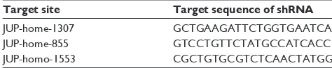
On the basis of Western blot analysis, γ -catenin protein was found to be highly expressed in K562 cells in comparison to THP-1 cells. Thereby, K562 was selected for further investi-gation. The parent cell line K562 was transfected with short hairpin RNA (shRNA) to produce a K562/KD cell model with diminished γ -catenin expression. To minimize off-target effects, three types of shRNA plasmids were used for transient transfection into K562 cells (Table 1).
Cell proliferation and migration analysis
Cell proliferation analysis was conducted with a Cell Count-ing Kit-8 (CCK-8) kit (Dojindo, Kumamoto, Japan) accord-ing to the manufacturer’s instructions. After establishaccord-ing the K562-shRNA model, cells were divided into three groups for further study, including shRNA-γ -catenin cells (knock-down, K562/KD), shRNA-NC cells (transfection negative control, K562/NC), and normal cells (K562 without trans-fection, K562/WT). In every group, the cell viability was determined at 0, 12, 24, 36, 48, and 60 hours posttransfection using the CCK-8 reagent. Each assay was run in triplicate and repeated three times.Relative cell viability (%)
OD of K562/KD or K562/NC
OD of K56
450
450 =
22 WT/ (1)
Inhibitor rate OD Treated
OD ntreated
450
450
= - -
-
×
1 U 100%
(2)
Cell migration experiment was conducted with the Tran-swell kit (Becton, Dickinson and Company) according to the instructions of manufacturer.
statistical analysis
The definition of complete remission (CR) followed the recommended criteria,7 and we defined relapse-free sur-vival (RFS) as the interval from the date of documented CR until relapse or death in CR (failure), or alive in CR at final follow-up (censored). Overall survival (OS) was defined as the interval from the date of initial diagnosis
until death (failure) or alive at final follow-up (censored). Mann–Whitney U-test was employed to evaluate quantitative parameters, and χ2 test was used for qualitative parameters. Statistical analyses were performed using Student’s t-test. The cumulative survival rate was calculated by the Kaplan– Meier method, and statistical significance was analyzed by the log-rank test. Multivariate Cox proportional hazard model was adopted to explore the prognostic effect of the various clinical variables. A P-value less than 0.05 was considered statistically significant, and all reported P-values were two-sided. All the statistical analyses were performed using the Statistical Program for Social Sciences (SPSS Inc., Chicago, IL, USA; 16.0). Graphs were plotted using GraphPad Prism 5.0 (GraphPad Software, Inc., La Jolla, CA, USA).
Results
The expression of
γ
-catenin
in all samples
An overview of the clinical characteristics of 71 patients is summarized in Table 2. γ -Catenin gene transcript levels were examined in BM mononuclear cells from 71 AML patients and 16 healthy individuals by RT-PCR. A statistically sig-nificant difference was found in γ -catenin expression levels between these two groups (P=0.020, Figure 1A). Compared with the healthy donors, AML patients exhibited significantly higher expression levels of γ -catenin.
correlation between
γ
-catenin
expression and clinical characteristics
in AML
We further explored the relationship between γ -catenin expression and the clinical features as well as known prog-nostic factors in AML patients. We compared the γ -catenin expression level among different subtypes (M0–M6) of AML and found significant overexpression in patients with AML-M2 in comparison to the other subtypes (P=0.053, Figure 1B). The expression levels of γ -catenin gene in patients with lower WBC (,30×109/L) were significantly higher than those with higher WBC ($30×109/L; P=0.032, Figure 1C). In addition, patients with CEBPα mutation had higher γ -catenin levels compared to those in the unmutated group (P=0.047, Figure 1D). In terms of age, sex, cyto-genetics, extramedullary presentation (lymphadenectasis and/or hepatosplenomegaly), mutation status of NPM1 and FLT3-ITD, and fusion genes AML1-ETO, no significant correlations with γ -catenin expression levels (all P.0.05, Figure 2A–H) were established. Because of the limited number of samples tested for PML-RARα, we were unable Table 1 Three types of shRNA plasmids
Target site Target sequence of shRNA
JUP-home-1307 gcTgaagaTTcTggTgaaTca
JUP-home-855 gTccTgTTcTaTgccaTcacc
JUP-homo-1553 cgcTgTgcgTcTcaacTaTgg
Abbreviation: shRNA, short hairpin RNA.
OncoTargets and Therapy downloaded from https://www.dovepress.com/ by 118.70.13.36 on 25-Aug-2020
Dovepress
Xu et al
to further explore the relationship between γ -catenin expres-sion and PML-RARα.
γ
-Catenin
expression level in patients
achieving cr
Apart from six patients who received alleviative treatment and nine patients diagnosed as M3, the remaining 56 patients, according to their conditions, received various standard induc-tion regimens, including idarubicin and cytarabine (Ara-C), daunorubicin and cytarabine, mitoxantrone and cytarabine,
aclacimomycin and cytarabine, homoharringtonine plus cytarabine and granulocyte-colony stimulating factor (G-CSF), and decitabine combined with half-dose CAG (cytarabine, aclarubicin, G-CSF). Among the 56 patients, 38 achieved CR after one to two therapy cycles. The rate of CR in the group of low γ -catenin expression was 71.4%, compared with 64.3% in the high-expression group. Fifty-six patients were divided into CR group (n=38) and not CR (n=18) group;
the γ -catenin level in CR group was markedly lower than that
in the not-CR group (P=0.047, Figure 3).
correlations between
γ
-catenin
expression levels and Os and rFs in
AML patients with intermediate-risk
cytogenetics
To identify the clinical prognostic effect of γ -catenin, we analyzed the prognosis of 56 patients with non-M3 AML. The median follow-up duration for OS was 12.22 months (ranging from 0.07 to 51.17 months). Between patients with high and
low γ -catenin expressions, there was no statistically
signifi-cant difference in OS and RFS (Figure 4A and B). We further analyzed the prognostic significance of γ -catenin expression level in subgroups with intermediate-risk cytogenetics, and no difference was revealed in OS and RFS (Figure 4C and D). Moreover, the effects of γ -catenin on OS and RFS in patients under 60 years of age and with intermediate-risk cytogenetics still remained unknown (Figure 4E and F).
Knockdown of
γ
-catenin
expression
suppressed K562 viability
On the basis of Western blot analysis, γ -catenin protein was highly expressed in K562 cells in comparison to THP-1 cells (Figure 5A). So, K562 was selected for further investiga-tion. The transfection efficiency was determined at 24, 48, and 72 hours posttransfection by fluorescent microscopy (Figure 5B). Consequently, the peak time in transfection efficiency was at 72 hours. The protein expression was fur-ther analyzed by Western blotting at 72 hours, showing up to
80% γ -catenin knockdown in K562/KD cells in comparison
to K562/NC cells (Figure 5C). K562/KD cells exhibited significantly lower viability than K562/NC cells, indicating that the suppression of γ -catenin expression inhibited cellu-lar viability of K562 cells. Thus, γ -catenin may be involved in cell proliferation of K562 cells (P,0.05, Figure 6A). Moreover, the cell migration experiment was performed in Transwell to compare the migration abilities among K562/ KD, K562/NC, and K562/WT cell groups. K562/KD cells exhibited enhanced migration ability over the other two Table 2 Patient characteristics
Characteristic Value P-valuea
Age, years (median, range) 47 (13–85) 0.945
,60, n (%) 50 (70.4)
$60, n (%) 21 (29.6)
Sex, n (%) 0.066
Male 40 (56.3)
Female 31 (43.7)
WBC at diagnosis, ×109/L (median, range) 20.8 (0.7–299.7) 0.032
,30, n (%) 39 (56.5)
$30, n (%) 30 (43.5)
Missing data, n 2
Hemoglobin at diagnosis, g/L (median, range)
81.0 (11.0–159.0) 0.111
Normal (male 120–160, female 110–150), n (%)
11 (17.7)
Anemia (male ,120, female ,110), n (%)
51 (82.3)
Missing data, n 9
Platelet at diagnosis, ×109/L (median, range) 40.0 (2.0–279.0) 0.284
Normal (100–300), n (%) 9 (14.3)
Thrombocytopenia (,100), n (%) 54 (85.7)
Missing data, n 8
BM blasts at diagnosis, % (median, range) 73.6 (20.4–99.0)
French American British subtype, n (%) 0.292
M0 2 (2.8)
M1 17 (23.9)
M2 26 (36.6)
M3 9 (12.7)
M4 3 (4.2)
M5 8 (11.3)
M6 5 (7.0)
Unclassified 1 (1.4)
FLT3-ITD mutation status, mutated +/total (%)
5/56 (8.9) 0.605
NPM1 mutation status, mutated +/total (%) 11/52 (21.2) 0.831
CEBPα mutation status, mutated +/total (%) 13/46 (28.3) 0.047
Cytogenetics, n (%) 0.114
Favorable 13 (21.0)
intermediate 44 (70.9)
Unfavorable 5 (8.1)
Missing data, n 9
Extramedullary presentation, n (%) 0.286
Yes 19 (26.8)
no 52 (73.2)
Notes:aγ -Catenin mRNA levels were compared among different groups. Mann–
Whitney U-test was employed to evaluate quantitative parameters, and χ2 test was
used for qualitative parameters. Statistical analyses were performed using Student’s t-test. all reported P-values were two-sided.
Abbreviations: WBC, white blood cells; BM, bone marrow.
OncoTargets and Therapy downloaded from https://www.dovepress.com/ by 118.70.13.36 on 25-Aug-2020
Dovepress Role of γ -catenin in acute myeloid leukemia
groups (P,0.05, Figure 6B), indicating that γ -catenin is a potential inhibitor for K562 cell migration.
Suppression of
γ
-catenin
expression
sensitizes K562 cells to decitabine
To understand how the knockdown of γ -catenin affects K562 cells, we treated K562/KD, K562/NC, and K562/WT cells with decitabine (4.4, 8.8, and 17.6 µM) and examined the inhibitory effects of this agent using CCK-8 assay kit, and then the inhibi-tory rate and half maximal inhibiinhibi-tory concentration (IC50) were calculated. The results showed that the inhibitory rate of decit-abine in K562/KD cells (IC50=6.24±0.77 µM) was significantly higher than that in K562/NC cells (IC50=12.14±1.38 µM) or K562/WT cells (IC50=22.51±3.83 µM), indicating that the downregulation of γ -catenin could sensitize K562 cells to decitabine (P,0.05, Figure 7A).
Benzyloxycarbonyl-Val-Ala-Asp (zVAD) as a caspase inhibitor can block the apoptotic pathway. Necrostatin-1 (Nec-1) is an inhibitor of programmed necrotic pathway. To investigate the pathway of decitabine-induced cell death, cells were treated separately with 1) growth media, 2) decitabine, 3) zVAD (20 µM) + decitabine, and 4) Nec-1 (30 µM) + decitabine. From this, we observed that the cytotoxicity of decitabine was inhibited by either zVAD or Nec-1 in any cell group, indicating that decitabine could cause cell death by both apoptotic and programmed necrotic pathways (*P=0.001, **P=0.000, ***P=0.001, #P=0.000, ##P=0.002, ###P=0.000, Figure 7B). Meanwhile, the cell viability of
K562/KD was significantly lower than that of K562/NC in the same processing method (P,0.05), indicating once again that the downregulation of γ -catenin could sensitize K562 cells to decitabine.
Figure 1 γ -Catenin expression and clinical characteristics.
Notes: (A) The level of γ-catenin gene compared between the 71 AML patients and the 16 healthy controls. (B) The level of γ-catenin gene compared between patients with AML-M2 and the others. (C) The expression level of γ-catenin gene in patients with lower WBC (,30×109/L) and patients with higher WBC ($30×109/L). (D) The expression
level of γ-catenin gene in patients with mutated CEBPα gene in contrast with patients with nonmutated CEBPα gene.
Abbreviations: AML, acute myeloid leukemia; mRNA, messenger RNA; WBC, white blood cells.
$0/ &RQWURO
3
γ
&DWHQLQ
P51$
H[SUHVVLRQOHYHO
$
:%&î/ :%&î/
3
γ
&DWHQLQ
P51$
H[SUHVVLRQOHYHO
&
3
0 2WKHUW\SHV
γ
&DWHQLQ
P51$
H[SUHVVLRQOHYHO
%
3
&(%3α± &(%3α
γ
&DWHQLQ
P51$
H[SUHVVLRQOHYHO
'
OncoTargets and Therapy downloaded from https://www.dovepress.com/ by 118.70.13.36 on 25-Aug-2020
Dovepress
Xu et al
Figure 2
γ
-Catenin
expression and clinical characteristics.
Notes: c orrelation between γ -catenin expression and other clinical characteristics including age ( A ), sex ( B ), cytogenetics ( C and D ), extramedullary presentation (lymphadenectasis and/or hepatosplenomegaly) ( E ), mutation status of NPM1 ( F ) and FLT3-ITD ( G
), and fusion genes AML1-ETO (
H
) in AML.
Abbreviations:
AML, acute myeloid leukemia; mRNA, messenger RNA.
γ &DWHQLQP51$ H[SUHVVLRQOHYHO
(
([WUDPHGXOODU\SUHVHQWDWLRQ ± 3$
&DWHQLQP51$ γH[SUHVVLRQOHYHO \HDUV \HDUV 3 γ &DWHQLQP51$ H[SUHVVLRQOHYHO
)
130 ± 130 3 γ &DWHQLQP51$ H[SUHVVLRQOHYHO%
0DOH )HPDOH 3 γ &DWHQLQP51$ H[SUHVVLRQOHYHO+
$0/(7 2 ± $0/(7 2 3 γ &DWHQLQP51$ H[SUHVVLRQOHYHO'
/RZULVN F\WRJHQHWLFV 2WKHUV 3 γ &DWHQLQP51$ H[SUHVVLRQOHYHO*
)/ 7,7 ' ± )/ 7,7 ' 3 γ &DWHQLQP51$ H[SUHVVLRQOHYHO&
+LJKULVN F\WRJHQHWLFV 3 2WKHUVOncoTargets and Therapy downloaded from https://www.dovepress.com/ by 118.70.13.36 on 25-Aug-2020
Dovepress Role of γ -catenin in acute myeloid leukemia
Discussion
Wnt signaling pathway plays a critical role in the regula-tion of early embryogenesis and is progressively shut off in differentiated mature cells.8 The aberrant activation of Wnt pathway has been found to play a role in the pathological pro-gresses in various malignant diseases, including AML.9 Wnt signaling pathways are generally termed as “canonical” path-ways, which are β-catenin dependent, and “noncanonical” pathways, which are β-catenin independent. The mechanism of the canonical pathway has been well defined, while the noncanonical one still remains unclear.
Cytoplasmic β-catenin, the central mediator, is degraded by the APC/Axin-GSK-3β complex. In this complex, APC and GSK-3β’s function is to facilitate the phophorylation of β-catenin.10 In the presence of Wnt signaling, the pathway is initiated via binding of Wnt proteins and the dimeric cell surface receptors composed of frizzled protein (Fzd) and
Figure 3 The expression level of γ-catenin gene compared between patients (n=38) who achieved CR following one to two cycles of standard induction chemotherapy and those who could not achieve CR after two cycles of induction chemotherapy (n=18).
Abbreviation: CR, complete remission.
3
&5
γ
&DWHQLQ
P51$
H[SUHVVLRQOHYHO
1RW&5
Figure 4 γ-Catenin expression and OS and RFS in AML.
Notes: (A and B) Correlations between γ-catenin expression levels and OS and RFS in AML patients. (C and D) Correlations between γ-catenin expression levels and Os and RFS in subgroups with intermediated-risk cytogenetics. (E and F) Correlations between γ-catenin expression levels and Os and rFs in subgroups who were younger than 60 years and with intermediated-risk cytogenetics.
Abbreviations: OS, overall survival; RFS, relapse-free survival; AML, acute myeloid leukemia.
$
0RQWKV
5HODSVHIUHHVXUYLYDO
5HODSVHIUHH VXUYLYDO 5HODSVHIUHH VXUYLYDO
2YHUDOOVXUYLYDO
2YHUDOOVXUYLYDO
2YHUDOOVXUYLYDO
Q
Q Q
Q
Q Q
0RQWKV
0RQWKV
0RQWKV
0RQWKV
Q Q
Q
Q Q
Q
3
3 3 3
3 3
0RQWKV
%
&
'
(
)
+LJKγFDWHQLQ
/RZγFDWHQLQ
+LJKγFDWHQLQ
/RZγFDWHQLQ
+LJKγFDWHQLQ
LQWHUPHGLDWHGULVNF\WRJHQHWLFV /RZγFDWHQLQ
LQWHUPHGLDWHGULVNF\WRJHQHWLFV
+LJKγFDWHQLQ
LQWHUPHGLDWHGULVNF\WRJHQHWLFV /RZγFDWHQLQ
LQWHUPHGLDWHGULVNF\WRJHQHWLFV
+LJKγFDWHQLQ
LQWHUPHGLDWHGULVNF\WRJHQHWLFV \
/RZγFDWHQLQ
LQWHUPHGLDWHGULVNF\WRJHQHWLFV \
+LJKγFDWHQLQ
LQWHUPHGLDWHGULVNF\WRJHQHWLFV \
/RZγFDWHQLQ
LQWHUPHGLDWHGULVNF\WRJHQHWLFV \
OncoTargets and Therapy downloaded from https://www.dovepress.com/ by 118.70.13.36 on 25-Aug-2020
Dovepress
Xu et al
Figure 5 γ-Catenin protein level in K562 and THP-1 cell line and the process of transfection in K562.
Notes: (A) The expression level of γ-catenin protein was determined by Western blot in K562 cell line and THP-1 cell line. (B) The transfection efficiency was determined posttransfection at 24, 48, and 72 hours by fluorescent microscopy (×10). (C) shRNA was transfected into K562 cell line to suppress the expression of γ-catenin protein.
Abbreviations: GAPDH, glyceraldehyde 3-phosphate dehydrogenase; KD, knockdown; h, hours; NC, negative control; shRNA, short hairpin RNA. γ&DWHQLQ
$
%
&
γ&DWHQLQ
7+3 .
±N'D
K
K
K
±N'D
±N'D
±N'D
.1& ..'
*$3'+
*$3'+
Figure 6 The cell viability and ability of migration.
Notes: (A) The cell viability of K562 cell compared between K562/KD and K562/NC after the suppression of γ-catenin protein expression. (B) The ability of migration of K562 cell line after the suppression of γ-catenin protein expression.
Abbreviations: KD, knockdown; NC, negative control; WT, wild type.
..'
.1& .:7
&HOOYLDELOLW\ &HOOQXPEHUQ
+RXUV &HOOOLQHV
3
3
3
3
..'
.1&
$
%
OncoTargets and Therapy downloaded from https://www.dovepress.com/ by 118.70.13.36 on 25-Aug-2020
Dovepress Role of γ -catenin in acute myeloid leukemia
low-density pipoprotein receptor-related protein 5 or 6, lead-ing to the recruitment and activation of the disheveled protein. This process turns the low-density pipoprotein receptor-related protein 5 or 6 into phosphorylated form and thereby leads to the dissociation of APC/Axin-GSK-3β complex. As a result, the phosphorylation and degradation of β-catenin are inhib-ited. Subsequently, β-catenin in the dephosphorylated form translocates into the nucleus, where it accumulates and acts as the ultimate effector by binding to TCF/LEF-1 to form a com-plex and activates the downstream target genes, such as cyclin D1 and C-myc,11–13 which are well-established oncogenes, and regulates the cellular proliferation and differentiation.14
Several previous studies have reported that in various hematological malignancies, including chronic myelog-enous leukemia,15 chronic lymphocytic leukemia,16 acute lymphoblastic leukemia,17 as well as AML,9 dysregulation of Wnt signaling has been identified as one of the key roles in leukemogenesis.
γ -Catenin belongs to the catenin family, which includes α-, β-, and γ -catenin. Sharing approximately 80% sequence homology, γ -catenin is closely related to β-catenin. β-Catenin and γ -catenin not only play directly an important role in cell–cell adhesion along with transmembranous E-cadherin18 but also influence the signal transduction through the Wnt canonical cascade. Unlike the clearly defined onco-genic effect of β-catenin, the role of γ -catenin in the aspect of signal transduction remains confusing. There are reports suggesting that γ -catenin may possess indirect oncogenic activity by blocking the degradation of β-catenin and in
turn increasing the level of β-catenin in the cytoplasm.19,20 Recently, in 2013, Morgan et al21 concluded that γ -catenin was overexpressed in AML and could promote the stabiliza-tion and nuclear localizastabiliza-tion of β-catenin. Moreover, it has been reported that in the absence of endogenous β-catenin expression, γ -catenin had transcriptional regulatory activity, which is TCF/LEF-family dependent.22 Moreover, in 2004, Zheng et al23 reported that the AML-associated translocation products, including PML/RAR, promyelocytic leukemia zinc finger/RAR(X-RAR), and AML-1/ETO, activated the Wnt pathway via upregulating γ -catenin and in turn encouraged the self-renewal of hematopoietic stem cells and contributed to the leukemogenesis in AML. However, from 2000 to 2006, several groups demonstrated that γ -catenin acted as a suppres-sor of tumors and metastasis. Decreased level of γ -catenin might result in poorer prognosis in several solid tumors, such as bladder, pituitary, oral, skin, and lung cancers.24–28
In this study, we compared γ -catenin mRNA levels in marrow mononuclear cells between naive AML patients and healthy donors, investigated the potential correlation between
γ -catenin and various clinical parameters, and analyzed the
clinical significance of γ -catenin in AML. γ -Catenin mRNA expression level was significantly higher in AML patients than in healthy donors, which is consistent with previous studies.21,23,29 Therefore, elucidating the role of γ -catenin in AML would help to understand the pathological mechanism of the disease and help develop a potential biomarker for clinical diagnosis and prognosis. Our data further showed that patients who had achieved CR in one to two cycles
Figure 7 The effect of decitabine.
Notes: (A) The sensitivity of K562 cell line to decitabine therapy after the suppression of γ-catenin protein expression. (B) The cell viability of K562 cell line after treatment separately with 1) growth media, 2) decitabine, 3) zVAD (20 µM) + decitabine, and 4) Nec-1 (30 µM) + decitabine.
Abbreviations: KD, knockdown; Nec-1, Necrostatin-1; NC, negative control; WT, wild type; zVAD, Benzyloxycarbonyl-Val-Ala-Asp.
$
%
'HFLWDELQHPRO/ &HOOOLQHV
,QKLELWRU\UDWH
&HOOYLDELOLW\
3
..' .1& .:7
3 3 3
3 3 DQG3
..' .1& .:7
'HFLWDELQH 'HFLWDELQH]9$' 'HFLWDELQH1HF 1HJDWLYHFRQWURO
OncoTargets and Therapy downloaded from https://www.dovepress.com/ by 118.70.13.36 on 25-Aug-2020
Dovepress
Xu et al
had significantly lower γ -catenin mRNA expression level in comparison to refractory patients with slow response.
So, γ -catenin mRNA level in naive AML patients might act as
an indicator for therapeutic efficacy. Lower level of γ -catenin suggested better response to chemotherapies. However, in all the 71 AML patients, we did not find any correlation between
γ -catenin expression and OS or RFS. Surprisingly, γ -catenin
mRNA was highly expressed in AML patients with CEBPα
mutation or low WBC count at first visit. It is well known that CEBPα mutation indicates better prognosis, while high WBC number at first-visit acts as an adverse prognostic indicator for AML. Thus, further investigation is necessary to better clarify the specific relationship between γ -catenin and AML development.
Furthermore, we initiated in vitro study with two cell lines: the acute monocytic leukemia line THP-1 and the chronic myeloid leukemia-derived erythroleukemia line K562. On the basis of Western blot analysis, γ -catenin pro-tein was highly expressed in K562 in comparison to THP-1, and so K562 was chosen for further investigation. At pres-ent, K562 is widely used as one of the model cell lines for hematopoietic study. It has the potential to differentiate into granulocyte, erythrocyte, and megakaryocyte lineages.30
Our results showed that downregulation of γ -catenin reduced the viability and growth rate of K562 cell, which is consistent with the study led by Niu et al.31 It has been demonstrated that suppression of γ -catenin can inhibit the expression of two β-catenin downstream oncogenes, c-Myc and cyclin D1. As suggested by Morgan et al,21 the over-expression of γ -catenin could stabilize β-catenin. So we speculate that γ -catenin could stimulate leukemia cellular growth by affecting β-catenin in Wnt signal pathway.
We found that downregulation of γ -catenin enhanced the toxicity of decitabine to K562 cells. Decitabine (5-aza-2′-deoxycytidine) is a cytosine analog and a DNA demethy-lation agent. It can induce leukemia cell differentiation and apoptosis and is the first-line therapeutic agent for AML showing good tolerance. The detailed mechanism of action of decitabine is as yet unclear. However, it was reported in 2007 and 2008 that the cytotoxicity of decitabine may rely on the demethylation of target genes, thus accelerating the expression of tumor suppressor genes.32,33 To our knowledge, this is the first study which established that downregulation of
γ -catenin can sensitize K562 cells to decitabine and enhance
the drug’s activity. The results suggested that γ -catenin may play a role in the cytotoxicity of decitabtine and may serve as a potential therapeutic target for AML. We also found that the cytotoxicity of decitabine was inhibited by either zVAD or Nec-1. zVAD is a caspase inhibitor, which can block the
apoptotic pathway. Nec-1 is an inhibitor of programmed necrotic pathway. This finding indicates that decitabine could cause both apoptosis and programmed necrosis, in accordance to a study by Steinhart et al.34
In several solid tumors, γ -catenin was found to regulate cellular invasion and migration. Mukhina et al35 have found that the downregulation of γ -catenin increased cell migration and invasion in MCF-7 cells, which express membrane-localized E-cadherin and γ -catenin. To discern the specific role of γ -catenin in these processes, the authors reexpressed γ -catenin in MCF-7 cells, which resulted in decreased migration and invasiveness of MCF-7 cells. The metastasis suppression activity of γ -catenin has also been described in bladder carcinomas.36 In the study by Franke,37 different types of classic and new junction proteins/structures were detected in K562 cells, including γ -catenin in the des-mosome junction. These cell–cell junctions are maintained with passages. In our investigation, knockdown of γ -catenin enhanced K562 cell migration, presumably by weakening intracellular junctions.
Conclusion
γ -Catenin gene was significantly overexpressed in AML
patients compared with healthy donors. The patients who had lower level of γ -catenin expression were more likely to achieve CR in comparison to those with higher γ -catenin level. γ -Catenin protein might accelerate the growth of AML cells. Knocking down the γ -catenin gene could enhance the cytotoxicity of decitabine in K562 cells. However, γ -catenin seemed to be capable of suppressing the migration of K562 cell line, and this needs further investigation.
Acknowledgments
This work was supported by the National Natural Sci-ence Foundation of China (81170486, 81570123, and 81400079), Jiangsu Province Health Agency Foundation (Z201402), and Jiangsu Province Natural Science Foundation (BK20141028).
Disclosure
The authors report no conflicts of interest in this work.
References
1. Estey EH. Acute myeloid leukemia: 2013 update on risk-stratification and management. Am J Hematol. 2013;88(4):318–327.
2. Vardiman JW, Thiele J, Arber DA, et al. The 2008 revision of the World Health Organization (WHO) classification of myeloid neoplasms and acute leukemia: rationale and important changes. Blood. 2009; 114(5):937–951.
3. Aktary Z, Pasdar M. Plakoglobin: role in tumorigenesis and metastasis.
Int J Cell Biol. 2012;2012:189521.
OncoTargets and Therapy downloaded from https://www.dovepress.com/ by 118.70.13.36 on 25-Aug-2020
OncoTargets and Therapy
Publish your work in this journal
Submit your manuscript here: http://www.dovepress.com/oncotargets-and-therapy-journal
OncoTargets and Therapy is an international, peer-reviewed, open access journal focusing on the pathological basis of all cancers, potential targets for therapy and treatment protocols employed to improve the management of cancer patients. The journal also focuses on the impact of management programs and new therapeutic agents and protocols on
patient perspectives such as quality of life, adherence and satisfaction. The manuscript management system is completely online and includes a very quick and fair peer-review system, which is all easy to use. Visit http://www.dovepress.com/testimonials.php to read real quotes from published authors.
Dovepress
Dove
press
Role of γ -catenin in acute myeloid leukemia
4. Whittock NV, Eady RA, McGrath JA. Genomic organization and ampli-fication of the human plakoglobin gene (JUP). Exp Dermatol. 2000;9(5): 323–326.
5. Fu Y, Zhu H, Wu W, et al. Clinical significance of lymphoid enhancer-binding factor 1 expression in acute myeloid leukemia. Leuk Lymphoma. 2014;55(2):371–377.
6. Qiao C, Zhang R, Hong M, et al. Heterogeneous leukemic clones identified by NPM1 mutation analysis in patient with acute monocytic leukemia. Leuk Lymphoma. 2012;53(5):886–890.
7. Cheson BD, Bennett JM, Kopecky KJ, et al. Revised recommenda-tions of the International Working Group for diagnosis, standardiza-tion of response criteria, treatment outcomes, and reporting standards for therapeutic trials in acute myeloid leukemia. J Clin Oncol. 2003; 21(24):4642–4649.
8. Staal FJ, Clevers HC. WNT signalling and haematopoiesis: a WNT-WNT situation. Nat Rev Immunol. 2005;5(1):21–30.
9. Lane SW, Wang YJ, Lo Celso C, et al. Differential niche and Wnt requirements during acute myeloid leukemia progression. Blood. 2011; 118(10):2849–2856.
10. McCubrey JA, Steelman LS, Bertrand FE, et al. Multifaceted roles of
GSK-3 and Wnt/β-catenin in hematopoiesis and leukemogenesis:
oppor-tunities for therapeutic intervention. Leukemia. 2014;28(1):15–33.
11. Tetsu O, McCormick F. β-Catenin regulates expression of cyclin D1
in colon carcinoma cells. Nature. 1999;398(6726):422–426.
12. Behrens J. Control of β-catenin signaling in tumor development. Ann
N Y Acad Sci. 2000;910:21–33; discussion 33–35.
13. van de Wetering M, Sancho E, Verweij C, et al. The β-catenin/TCF-4
complex imposes a crypt progenitor phenotype on colorectal cancer cells. Cell. 2002;111(2):241–250.
14. Polakis P. Wnt signaling in cancer. Cold Spring Harb Perspect Biol. 2012;4(5):a008052.
15. Zhang B, Li M, McDonald T, et al. Microenvironmental protection of CML stem and progenitor cells from tyrosine kinase inhibitors
through N-cadherin and Wnt-β-catenin signaling. Blood. 2013;121(10):
1824–1838.
16. Lu D, Zhao Y, Tawatao R, et al. Activation of the Wnt signaling path-way in chronic lymphocytic leukemia. Proc Natl Acad Sci U S A. 2004; 101(9):3118–3123.
17. Yang Y, Mallampati S, Sun B, et al. Wnt pathway contributes to the protection by bone marrow stromal cells of acute lymphoblastic leu-kemia cells and is a potential therapeutic target. Cancer Lett. 2013; 333(1):9–17.
18. Wijnhoven BP, Dinjens WN, Pignatelli M. E-cadherin-catenin cell– cell adhesion complex and human cancer. Br J Surg. 2000;87(8): 992–1005.
19. Li L, Chapman K, Hu X, Wong A, Pasdar M. Modulation of the
oncogenic potential of β-catenin by the subcellular distribution of
plakoglobin. Mol Carcinog. 2007;46(10):824–838.
20. Teuliere J, Faraldo MM, Shtutman M, et al. β-catenin-dependent
and -independent effects of ΔN-plakoglobin on epidermal growth and
differentiation. Mol Cell Biol. 2004;24(19):8649–8661.
21. Morgan RG, Pearn L, Liddiard K, et al. γ-Catenin is overexpressed
in acute myeloid leukemia and promotes the stabilization and nuclear
localization of β-catenin. Leukemia. 2013;27(2):336–343.
22. Maeda O, Usami N, Kondo M, et al. Plakoglobin (γ-catenin) has TCF/
LEF family-dependent transcriptional activity in β-catenin-deficient
cell line. Oncogene. 2004;23(4):964–972.
23. Zheng X, Beissert T, Kukoc-Zivojnov N, et al. γ-catenin contributes to
leukemogenesis induced by AML-associated translocation products by increasing the self-renewal of very primitive progenitor cells. Blood. 2004;103(9):3535–3543.
24. Clairotte A, Lascombe I, Fauconnet S, et al. Expression of E-cadherin
and α-, β-, γ-catenins in patients with bladder cancer: identification
of γ-catenin as a new prognostic marker of neoplastic progression
in T1 superficial urothelial tumors. Am J Clin Pathol. 2006;125(1): 119–126.
25. Tziortzioti V, Ruebel KH, Kuroki T, Jin L, Scheithauer BW, Lloyd RV.
Analysis of β-catenin mutations and α-, β-, and γ-catenin expression
in normal and neoplastic human pituitary tissues. Endocr Pathol. 2001;12(2):125–136.
26. Ueda G, Sunakawa H, Nakamori K, et al. Aberrant expression of β- and
γ-catenin is an independent prognostic marker in oral squamous cell
carcinoma. Int J Oral Maxillofac Surg. 2006;35(4):356–361. 27. Tada H, Hatoko M, Tanaka A, Kuwahara M, Muramatsu T. Expression
of desmoglein I and plakoglobin in skin carcinomas. J Cutan Pathol. 2000;27(1):24–29.
28. Bremnes RM, Veve R, Gabrielson E, et al. High-throughput tissue microarray analysis used to evaluate biology and prognostic significance of the E-cadherin pathway in non-small-cell lung cancer. J Clin Oncol. 2002;20(10):2417–2428.
29. Muller-Tidow C, Steffen B, Cauvet T, et al. Translocation products in acute myeloid leukemia activate the Wnt signaling pathway in hematopoietic cells. Mol Cell Biol. 2004;24(7):2890–2904.
30. Lozzio CB, Lozzio BB. Human chronic myelogenous leukemia cell-line with positive Philadelphia chromosome. Blood. 1975;45(3):321–334.
31. Niu CC, Zhao C, Yang ZD, et al. Downregulation of γ-catenin inhibits
CML cell growth and potentiates the response of CML cells to imatinib
through β-catenin inhibition. Int J Mol Med. 2013;31(2):453–458.
32. Ashkenazi A. Targeting the extrinsic apoptosis pathway in cancer.
Cytokine Growth Factor Rev. 2008;19(3–4):325–331.
33. Kroemer G, Galluzzi L, Brenner C. Mitochondrial membrane permea-bilization in cell death. Physiol Rev. 2007;87(1):99–163.
34. Steinhart L, Belz K, Fulda S. Smac mimetic and demethylating agents synergistically trigger cell death in acute myeloid leukemia cells and overcome apoptosis resistance by inducing necroptosis. Cell Death Dis. 2013;4:e802.
35. Mukhina S, Mertani HC, Guo K, Lee KO, Gluckman PD, Lobie PE. Phenotypic conversion of human mammary carcinoma cells by auto-crine human growth hormone. Proc Natl Acad Sci U S A. 2004;101(42): 15166–15171.
36. Rieger-Christ KM, Ng L, Hanley RS, et al. Restoration of plakoglobin expression in bladder carcinoma cell lines suppresses cell migration and tumorigenic potential. Br J Cancer. 2005;92(12):2153–2159. 37. Franke WW, Rickelt S. Mesenchymal-epithelial transitions:
spontane-ous and cumulative syntheses of epithelial marker molecules and their assemblies to novel cell junctions connecting human hematopoietic tumor cells to carcinomatoid tissue structures. Int J Cancer. 2011; 129(11):2588–2599.
OncoTargets and Therapy downloaded from https://www.dovepress.com/ by 118.70.13.36 on 25-Aug-2020