Research article Available online www.ijsrr.org
ISSN: 2279–0543
International Journal of Scientific Research and Reviews
Clinico-radiological assessment of alveolar socket preservation using
autogenous and alloplastic bone graft
Deepak Tanwar
1, Stuti Gupta
2* and Gurjap Singh
3MDS,Oral And Maxillofacial Surgeon,Consultant and Private Practitioner
Email-deepaktanwarwb@gmail.com,9999608619
2*
MDS,Periodontist and Oral Implantologist,Lecturer,School Of Dental Sciences,Sharda University,Greater Noida Mob.: 9953742797 Email: Stutigupta0901@gmail.com
3
MDS - Professor & Head , Department of Oral & Maxillofacial Surgery – bankeybihari Dental College Hospital & Research Centre ,Ghaziabad , India
Mob.: 9899027775Email: Gurjapsingh04@rediffmail.com
ABSTRACT:
The aim of the study is to compare the efficacy of autogenous and alloplastic bone graft in preserving alveolar socket at future implant site and to compare their comparative effects of in terms of Healing time and formed bony Architecture.Twenty patients were randomized into two groups.8 females and 12 males were included in the study. Autogenous bone graft from mandibular symphysis in group 1and Alloplastic graft nova bone putty in group 2 was used for the patients included in the study.Soft tissue healing evaluation was done as per criteria given by Landry et al. Hard tissue evaluation was done according to Kelly et al. Clinical & Mean Radiographic Score for Assessment of Bone Healing was done at baseline,1st,3rd and 6th month for both the groups. Analysis was done using software SPSS 22.0 (IBM, Chicago, USA). Chi square test, Paired t test, Independent t test and Wilcoxontest was applied for both the intergroup and intergroup comparisons. Intergroup comparison was calculated at baseline 1,3 and 6 month. It reveals that both autogenous and alloplastic bone grafts significantly result in effective healing over a period of 6 months where asautogenous bone graft proved to be significantly better than alloplast. This study reveals that both autogenous and alloplastic bone grafts significantly increase the bone density over a period of 6 months, howeverautogenous bone graft is better in increasing bone density when compared to alloplastic bone graft. Autogenous bone graft has shown to effectively increase the trabeculations in alveolar bone as compared to alloplastic bone graft.
KEYWORDS
- Alloplastic, Autogenous, Socket Preservation.Authors declare No conflict of Interest.
*Corresponding author:
Stuti Gupta
MDS, Periodontist and Oral Implantologist, Lecturer,
School Of Dental Sciences,Sharda University,Greater Noida
INTRODUCTION
Alveolar bone loss is an inevitable consequence that occurs after tooth extraction.
Transformation and resorption of the alveolar crest can create deformity.1 Narrowing of the alveolar
ridge can cause many problems in terms of cosmetic, functionality and dental implantation2 As the
controlled clinical studies have reported, the average horizontal alveolar bone loss during the first
year after atraumatic tooth extraction is 5 to 7 mm3,4. The bone loss after tooth extraction will occur
rapidly in the first 6 months in which up to 40% of the height and 60% of the width of the alveolar
bone can be lost during this time.5These problems can be minimized following procedures of socket
preservation of the extracted tooth. The best time to preserve the alveolar ridge is at the time of
extraction6. The existing methods of ridge preservation include the use of autogenous, allogenic,
xenogenous grafts and alloplasts with or without placement of absorbable and non –absorbable
membranes.7,8
The principles behind implant site development, including ridge preservation and guided
bone regeneration, originated from the principles of guided tissue regeneration. The concept of
selective cell repopulation has been useful in theorizing about enhancing site development for
implant placement. Bone can be regenerated by using a barrier membrane at an extraction site or
deficient alveolar ridge. At the time of tooth extraction, the socket can be augmented with a graft
material and sealed with a barrier membrane or a membrane may be used without graft material in
the socket. The membranes can non-resorbable or resorbable membranes.7,8
Autogenous bone graft is obtained from same individual on whom the bone grafting has to perform.
However, they are now less popular because of the necessity of harvesting from a secondary surgical
site and the possible morbidity associated with the procedures.8
Alloplasts are a synthetic graft material which is inert and implanted into tissue.
Hydroxyapatite, tricalcium phosphate, calcium sulfate and bioactive glass polymers are common
examples of synthetic bone graft materials. This graft material is inert, osteoconductive filler
material, which serves as a nidus or scaffold for new bone formation. Alloplasts have been shown to
result in defect fill, stabilization of the remaining osseous structure, clinical attachment gain, and
decreased probing depths. Allografts used by dental surgeons include: DFDBA, (decalcified freeze –
Figure 1: ALLOPLAST
Since there are few studies of quantitative and qualitative methods to investigate the effect of
socket preservation techniques after tooth extraction for dental implant treatment, this scientific and
cost-effective study with the purpose of comparing the effect of Nova boneputty and autogenous
graft from chin was undertaken for the patients who were referred for implant surgery in our
department
MATERIALS AND METHODS:
The present study was done with Patients reporting to the Department of oral and
maxillofacial surgery, I.T.S Dental College, Hospital and Research Centre, Greater Noida for 6
months’ time duration.
This study was approved by the Ethics Committee of I.T.S Dental College, Hospital and
Research Centre (Ref nos. ITS/OS//1762:2017) and is in accordance with the 1964 Helsinki Declaration and its later amendments or comparable ethical standards.
• Sample size- 20 patients (10 in each group).
• Age group-18 to 40years.
• Period of Study-6 months
• Autogenous bone graft will be harvested from : Mandibular symphysis region
• Alloplastic graft: Calcium Silocophosphate (Novabone)
It is a randomized split mouth study where 2 test sites were taken for comparative analysis.20
patients with age group-18 to 55 years were selected. This study was undertaken as a pilot study to
initially compare the bone graft procurement technique from chin with alloplastic bone graft so that
further elaborate research could be planned. Inclusion Criteria were mandibular and maxillary teeth
that are indicated for extraction because of presence of periapical and periodontal pathology. Written
informed consents were taken after explanation of purpose of study. Exclusion criteria were patient
tobacco and betel nut chewing, smoking, and patient with hypersensitivity to implant material,
patients who are undergoing current radiotherapy or chemotherapy.
Autogenous bone graft from mandibular symphysis was procured using a standard extraction
procedure by doing ostectomy using trephinebur. Socket preservation was done at the future implant
site by loading it with procured graft and secured by screws and a GTR membrane.
Alloplastic graft-ANova bone putty was used in study.
Soft tissue healing evaluation was done as per criteria given by Landry et al. at 1st,3rd and 6th
month.11,.12
Hard tissue evaluation was also made by criteria given by Kellyet al13
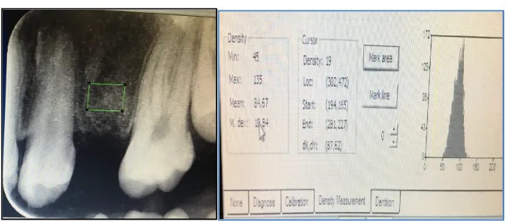
Mean Radiographic Score for Assessment of Bone Healing at Different Time Points
(baseline,1st,3rd and 6th month ) was done between Groups(Figure 2)
Figure 2: RADIOGRAPHIC EVALUATION
STATISTICAL ANALYSIS:
Analysis was done using Wilcoxon test by using software SPSS 22.0 (IBM, Chicago, USA)
for both the intergroup and intragroup comparisons. Chi square test, Paired t test, Independent t test
and Wilcoxon test was applied for both the intergroup and intragroup comparisons. Intergroup
comparison was calculated at baseline 1, 3 and 6 month.
RESULTS:
Table 1 and Graph 1- Reveals that both autogenous and alloplastic bone grafts significantly
increases the bone density over a period of 6 months. It further shows that autogenous bone graft is
better in increasing bone density when compared to alloplastic bone grafts which is also statistically
90
85
80
75 AutogenousBoneGraft
Alloplastic Bone Graft 70
65
60
Baseline 1months 3months 6months
Table 1: Mean values of density of autogenous and alloplastic bone grafts
DENSITY GROUPS MEAN SD P<0.05 INTERPRETATION
At 0 day Autogenous Group 72.9 0.468 0.000 Difference significant
Alloplastic Group 71.5 0.408
At 1 month
Autogenous Group 77.3 0.198 0.000
Difference significant
Alloplastic Group 73.7 0.211
At 3 month Autogenous Group 80.6 0.177 0.000 Difference significant
Alloplastic Group 74.3 0.18
At 6 month Autogenous Group 84.7 0.277 0.000 Difference significant
Alloplastic Group 76.9 0.212
*
Wilcoxon Test (p<0.05 significant)
Graph 1: Mean values of density of autogenous and alloplastic bone grafts
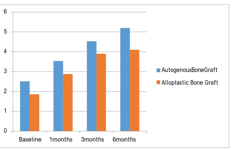
Table 2 and Graph 2 - Reveals that both autogenous and alloplastic bone grafts significantly
decreases the value of healing index over a period of 6 months. It further shows that autogenous bone
graft is better in decreasing the value of healing index when compared to alloplastic bone grafts
Baseline 1months 3months 6months 0
1 2
AutogenousBoneGraft
Alloplastic Bone Graft 3
4 5 6
Table 2: Mean values of healing index of autogenous and alloplastic bone grafts
HEALING INDEX GROUPS MEAN SD P<0.05 INTERPRETATION
At 0 day Autogenous Group 2.51 0.121 0.000 Difference significant Alloplastic Group 1.86 0.109
At 1 month Autogenous Group 3.53 0.233 0.000 Difference significant Alloplastic Group 2.87 0.219
At 3 month Autogenous Group 4.53 0.157 0.000 Difference significant Alloplastic Group 3.9 0.118
At 6 month Autogenous Group 5.2 0.214 0.000 Difference significant Alloplastic Group 4.1 0.192
*
Wilcoxon Test (p<0.05 significant)
Graph 2: Mean values of healing index of autogenous and alloplastic bone grafts
Table 3 & Graph 3- Reveals that both autogenous and alloplastic bone grafts significantly
increases the alveolar bone trabeculation over a period of 6 months. It further shows that autogenous
bone graft is better in increasing the alveolar bone trabeculation when compared to alloplastic bone
grafts which is also statistically significant (P<0.05)
Table 3: Mean values of trabeculation of autogenous and alloplastic bone grafts
TRABECULAR PATTERN MEAN SD P<0.05 INTERPRETATION
At 0 day Autogenous Group 0.6 0.378 0.000 Difference significant Alloplastic Group 0.62 0.808
At 1 month Autogenous Group 0.82 0.378 0.000 Difference significant Alloplastic Group 0.76 0.431
At 3 month Autogenous Group 1.82 0.377 0.000 Difference significant Alloplastic Group 0.81 0.48
At 6 month Autogenous Group 1.84 0.377 0.000 Difference significant Alloplastic Group 0.93 0.482
*
Graph 3: Mean values of trabeculation of autogenous and alloplastic bone grafts
DISCUSSION
This study was conducted in the Department of oral and maxillofacial surgery at I.T.S Dental
College, Hospital and Research Centre, Greater Noida for a comparative evaluation of autogenous
and alloplastic bone graft in socket preservation after extraction at future implant sites. This is a
randomized, controlled, clinical pilot study comparing ridge preservation with conventional
Calcium-Phosphosilicate (Novabone) alloplast and ATG from symphysisregion. It is a prospective
study having 4 months follow‑upperiod. Intraoperative procedure was mentioned. (Figure 3)
Figure 3: INTRAOPERATIVE
Postoperative
Radiographically and statistically significant differences were found in both the experimental
groups. This was assessed based on the primary outcome variables of hard tissue such as a change in
density, bone trabaculae pattern and soft tissue healing outcome variables like response to probing,
suppuration present and epithelisation.(Table &Graph 1,2,3 respectively) Significant resorption and
healing of calcium silicophosphate particles are expected in 3–6 months after its placement14.Most of
the calciumphosphosilicate gets biodegraded by both osteoclastic activities subsequent to the particle
disaggregation and/or chemical dissolution of the molecule in the calcium and phosphate
components followed by replacement with healthy bone.15 ATG resorbs within 4–6 months after
grafting. The remodeling process with new bone formation continues up to 1–2 years.16,17 Based on
these references, follow‑up period of 0 day, 1month, 3 month and 6 month was selected for the
current study. (Figure 2)
CBCT scans were taken on the day before extraction after 6 month post treatment.
Radiographic density was measured with the help of RVG done at 0 day,1month, 3 month, 6 month
post operatively with the help of Soredex Digora software CBCT scan being
threedimensionalinnaturehashelpedustoevaluatechangesinalveolarboneinallpossible dimensions and
measure them precisely.(Figure 2) After 6 months post treatment, the mean bone loss was more for
The change in mean width was statistically significant when ATG‑grafted sites were
compared with other grafted sites (P < 0.05). When vertical bone resorption was compared, at ATG
sites mean height showed consistently least reduction with respect to
calciumsilicophosphate.Thischangeinalveolarheightwasstatisticallysignificantfor ATG sites (P <
0.05).Literature has provided evidence in support to the fact that ridge preservation procedures
reduce the bone dimensional changes compared with extraction without ridge preservation
procedures.18,19 However, systematic review demonstrates, in spite of employing evidence‑based
ridge preservation techniques, a complete prevention of vertical and horizontal bone resorption is an
unpredictableevent.17
Figure 4: Study flow chart
( N=20)
BASELINE EVALUATION
(INCLUSION CRITERIA-PATIENTS WHO HAVE UNDERGONE EXTRACTION AND PLANNED FOR FUTURE IMPLANT PLACEMENT
RANDOMIZATION
CLINICAL PARAMETERS
1.Soft tissue eva lua tion by criteria given by La ndry et al.
2,Ha rd tissue evaluation by criteria given by Kelly
RADIOLOGICAL
Hard tissue evaluation by criteria given by Kelly
GROUP 1 GROUP 2
GROUP 1
ALLOPLASTIIC GRAFT PLACEMENT
GROUP 2 AUTOGENOUS GRAFT
PLACEMENT SOCKET PRESERVATION
AT 1 MONTH
EVALUATION OF CLINICAL PARAMETERS AT 1 MONTH
STATISTICAL ANALYSIS AND INTERPRETATION
RESULTS
EVALUATION OF CLINICAL PARAMETERS AT BASELINE
AT 3 MONTHS
EVALUATION OF CLINICAL AND RADIOLOGICAL PARAMETERS
AT 6 MONTHS
EVALUATION OF CLINICAL AND RADIOLOGICAL PARAMETERS
The results of this study are in accordance with these findings as there was some loss of ridge
Thus, ridge preservation procedures do not result in complete dimensional stability but are designed
to reduce the loss of ridge dimension compared to sites left to heal naturally after tooth extraction. In
our study, within the ridge preservation groups, ATG sites showed consistently minimum vertical, as
well as horizontal bone resorption, as evidenced by least change in alveolar width and height 6
months after therapy.
Study Flow Chart (Figure 4)was suggested and it recommends that Radio graphical analysis was performed to validate clinical results. Long term follow-up with major samples need to be done
for better results.
CONCLUSION:
This study reveals that both autogenous and alloplastic bone grafts significantly increase the
bone density over a period of 6 months, howeverautogenous bone graft is better in increasing bone
density when compared to alloplastic bone graft. Alloplastic graft also showed in effective healing
over a period of 6 months whereas autogenous bone graft proved to be significantly better than
alloplast. Autogenous bone graft has shown to effectively increase the trabeculations in alveolar bone
as compared to alloplastic bone graft.
ETHICS APPROVAL:
This study was approved by the Ethics Committee of I.T.S Dental College, Hospital and
Research Centre (Ref nos. ITS/OS//1762:2017).Study was conducted in accordance with the Declaration of Helsinki.
REFERENCES
1. Mecall RA, Rosen Feld AL. Influence of residual ridge resorption pattern on implant fixture
placement and tooth position. Int J perisdontics Restoratire Dent 1991;11: 8-23.
2. Wang HL, Carroll MJ. Guided bone regeneration using bone grafts and collagen membranes.
Quintessence Int. 2001;Jul-Aug; 32(7):504-15.
3. Froum S, Orlowski.Ridge preservation utilizing an alloplast prior to implant
placement--clinical and histological case reports. Pract Periodontics Aesthet Dent. 2000May;
12(4):393-402-404
4. Camargo PM, Lekovic V, Weinlaender M, Klokkevold PR, Kenney EB, Dimitrijevic B, et
al.Influence of bioactive glass on changes in alveolar process dimensions after exodontia.
5. Fotek PD, Neiva RF, Wang HL Comparison of dermal matrix and polytetrafluoroethylene
membrane for socket bone augmentation: a clinical and histologic study. JPeriodontol.
2009;80: 776-785.
6. Lekovic V, Kenney EB, Weinlaender M, Han T, Klokkevold P, et al.A bone regenerative
approach to alveolar ridge maintenance following tooth extraction - Report of 10 cases.J
Periodontol.1997;68: 563-570
7. Lekovic V, Camargo PM, Klokkevold PR, Weinlaender M, Kenney EB, et al. Preservation of
alveolar bone in extraction sockets using bioabsorb able membranes. J Periodontol. 1998;69:
1044-1049.
8. Shi B, Zhou Y, Wang YN, Cheng XR. Alveolar ridge preservation prior to implant placement
with surgical – grade calcium sulfate and platelet-rich plasma: a pilot study in canine
model.Int J Oral Maxillfac Implants. 2007;22: 656-665.
9. Irinakis T. Rationale for socket preservation after extraction of a single-rooted tooth when
planning for future implant placement. J Can Dent Assoc. 2006;72: 917-922.
10.Landry RG, Turnbull RS, Howley T. Effectiveness of benzydamyneHCl in the treatment of
periodontal post-surgical patients. Res Clin Forums. 1988;10:105-11867.
11.Masse JF, Landry RG, Rochette C. et al. Effectiveness of soft laser treatment in periodontal
surgery. Int Dent J. 1993;43:121-127.
12.Kelly WH, Mirahmadi MK, Simon JH, Gorman JT. Radiographic changes of the jawbones in
end stage renal disease.Oral Surg Oral Med Oral Pathol. 1980 Oct;50(4):372-81
13.Nemcovsky CE, Serfaty V. Alveolar ridge preservation following extraction of maxillary
anterior teeth. Report on 23 consecutive cases. J Periodontol. 1996;67: 390-395.
14.Stanley HR, Hall MB, Clark AE, King CJ, Hench LL. Using 45S5 bioglass cones as
endosseous ridge maintenance implants to prevent alveolar ridge resorption: a 5-year
evaluation. Int J Oral Maxillofac Implants.1997;12: 95-105.
15.Brugnami F, Then PR, Moroi H, Leone CW. Histologic evaluation of human extraction
sockets treated with demineralized freeze-dried bone allograft (DFDBA) and cell occlusive
membrane. J Periodontol1996;67: 821-825.
16.Sclar AG. Strategies for management of single-tooth extraction sites in aesthetic implant
therapy. J Oral MaxillofacSurg2004;62: 90-105.
17.Iasella JM, Greenwell H, Miller RL, Hill M, Drisko. Ridge preservation with freeze-dried
bone allograft and a collagen membrane compared to extraction alone for implant site