Programmed cell death in the developing limb
VANESSA ZUZARTE-LUÍS and JUAN M. HURLÉ*
Departamento de Anatomía y Biología Celular, Facultad de Medicina, Universidad de Cantabria, Santander, Spain
ABSTRACT The sculpturing of shape in the developing limb together with the regression of the tail in anuran tadpoles constitute, perhaps, the most paradigmatic processes of programmed cell death. The study of these model systems has been of fundamental importance to support the idea that cell death is a physiological behavior of cells in multicellular organisms. Furthermore, different experimental approaches, including comparative analyses of the pattern of cell death in different avian species (i.e. chick interdigits versus duck interdigital webs) and in chick mutants with different limb phenotypes, provided the first evidence for the occurrence of a genetic program underlying the control of cell death. Two well known research groups in the field of limb development, the USA group headed first by John Saunders and next by John Fallon and the group of Donald Ede and Richard Hinchliffe in the U.K. provided a remarkable contribution to this topic. In spite of the historical importance of the developing limb in establishing the concept of programmed cell death, this model system of tissue regression has been largely neglected in recent studies devoted to the analysis of the molecular control of self-induced cell death (apoptosis). However, a considerable amount of information concerning this topic has been obtained in the last few years. Here we will review current information on the control of limb programmed cell death in an attempt to stimulate further molecular studies of this process of tissue regression.
KEY WORDS:
apoptosis, tissue regression, BMP, FGF, retinoic acid
0214-6282/2002/$25.00 © UBC Press
Printed in Spain www.ijdb.ehu.es
*Address correspondence to: Dr. Juan M. Hurlé. Departamento de Anatomía y Biología Celular, Facultad de Medicina, C/ Cardenal Herrera Oria s/n, 39011 Santander, Spain. Fax: +34-942-201-903. e-mail: [email protected]
Abbreviations used in this paper: AER, apical ectodermal ridge; ANZ, anterior
necrotic zone; BMP, bone morphogenetic protein; INZ, interdigital necrotic zone; OP, opaque patch; PCD, programmed cell death; PNZ, posterior necrotic zone.
Patterns of Apoptosis in the Developing Limb
In the developing limb, major areas of programmed cell death occur in the undifferentiated mesoderm in association with the establishment of the prechondrogenic condensations of the skel-eton and in the ectoderm of the AER. In addition cell death is also observed during the formation of the joints (Mitrovic, 1977; Mori et al., 1995), in the establishment of the axon pathways (Tosney et al., 1988) and during the remodeling of the vascular pattern (Hurle et al., 1985; Feinberg, 1991). It must be mentioned that the areas of cell death in the limb have been formerly termed necrotic areas but they occur by apoptosis (Garcia-Martinez et al., 1993). This contra-diction is explained because the classical studies on limb pro-grammed cell death have been performed prior to the introduction in the literature of the term apoptosis.
The areas of mesodermal cell death are related with the estab-lishment of the shape and skeletal pattern of the limb and exhibit significant differences between species. A remarkable feature is that mesodermal cell death is a characteristic feature of amniota. In amphibians, limb develops without programmed mesodermal cell death (Cameron and Fallon, 1977). The biological significance of this difference between amniota and anamniota embryos remains to be explained.
In the early avian limb there are two areas of cell death, the Anterior Necrotic Zone (ANZ; Fig. 1C), and the Posterior Necrotic Zone (PNZ; Fig.1B), which have been related with the reduced number of digits in birds (three digits in the wing and four in the leg). These areas are absent in polidactylous avian mutants (Hinchliffe and Ede, 1967). The absence of wings in the wingless chick mutant is correlated with a dramatic increase in ANZ (Hinchliffe and Ede, 1973). In mammals ANZ and PNZ similar to those of the avians are not present (Milaire and Rooze, 1983).
The formation of free digits in all amniota vertebrates is accompa-nied by massive apoptosis of the interdigital mesoderm. These areas of cell death have been termed the Interdigital Necrotic Zones (INZs; Fig. 1D,F), and serve the function of sculpturing the shape of the digits (see reviews by Saunders 1966 and Hurle et al., 1996). In species with free digits, such as the chick, quail, lizard, mouse or human, apoptosis extends through all the interdigital space. In species with webbed digits, such as the duck or turtle, apoptosis is limited to the distal part of the interdigit. In species with autopods of singular morphology, such as the digits with lateral membranous lobulations present in the moorhen (Gallinula chloropus) and in the coot (Fulika atra), or the splited autopod (zygodactyly) present in Chamaleons, the pattern of INZ correlates closely with their specific phenotypes.
The correlation between INZ and the phenotype of the digits is also observed in syndactylous mutant species (Hinchliffe and Thorogood, 1974) and in experimental limbs treated with drugs which inhibit cell death (see review by Hurle et al., 1996). Interest-ingly, the inhibition of INZ is often followed by the presence of digit fusions or even by the formation of an ectopic digit (Gañan et al., 1996). This finding indicates that the interdigital mesoderm has the potential to form digits.
In addition to mesodermal cell death, apoptosis is also an important feature of the ectoderm of the AER (Fig. 1E). In the chick limb bud ectodermal apoptosis appears to exert the function of controlling the extension of that structure (Todt and Fallon, 1986). In mammalians, apoptosis in the AER is more prominent than in birds and forms well-defined foci of cell death (Milaire and Roze, 1983). The inhibition or delay of this ectoder-mal cell death causes an enlargement of the AER followed by an increase in the amount of subridge mesenchyme which results in the induction of polydatyly (Naruse and Kameyama, 1982, 1986). In serpentiform reptiles with rudimentary limbs the
regression of the limb primordium is mediated by massive cell death in the AER (see Raynaud, 1990).
BMPs are the Triggering Signal for Limb Programmed
Cell Death
Evidence obtained mainly with the chick embryo indicates that programmed cell death in the limb bud shares many control mechanisms with those regulating proliferation and differentiation. BMPs have been identified as the triggering apoptotic signal for both the ectoderm of the AER (Gañan et al., 1998) and the mesodermal cells (Gañan et al., 1996; Zou and Niswander, 1996; Yokouchi et al., 1996; Macias et al., 1997). According to their pattern of expression BMP-2, BMP-4 and BMP-7 are the most likely physiological signals triggering apoptosis in the limb bud. However, these BMPs are also involved in the control of limb patterning (Pizette et al., 2001) and in the regulation of chondro-genic differentiation (Macias et al., 1997). In fact, local treatments with any of the above mentioned BMPs induce intense growth and differentiation in the prechondrogenic mesenchyme and massive apoptosis in the undifferentiated mesoderm (Macias et al., 1997). The chondrogenic effect of BMPs appears to be mediated by the type Ib receptor (Yi et al., 2000). However, the receptor implicated in the control of apoptosis remains to be identified. Inhibition of apoptosis have been obtained in over-expression experiments using dominant negative type IB and type IA BMP receptors (Zou and Niswander, 1996; Yokouchi et al., 1996). However, since, these receptors are not expressed in the areas of programmed cell death at levels detectable by in situ hybridization the most likely explanation for these results is that the phenotype was secondary to depletion of BMPs which bind to the overexpressed receptors. Furthermore, interdigital induction of the type IB BMP receptor gene by application of TGFβ-1-beads is followed by inhibition of
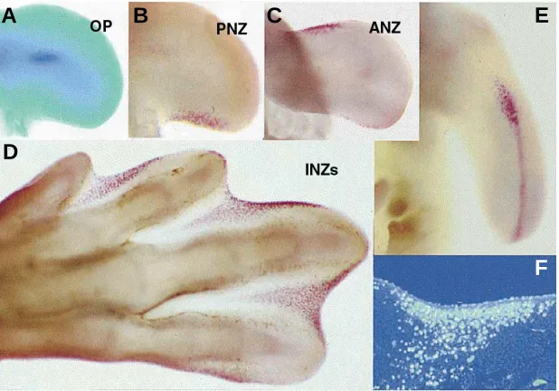
Fig. 1. The areas of cell death in the developing chick limb bud. (A) Wing bud showing the OP vital stained with nile blue.(B-E) Neutral red vital staining showing the PNZ (B), ANZ
(C) INZs (D) and cell death in the AER (E). (F) Longitudinal section of the second interdigital space of a leg bud of the same stage to that showed in (D), illustrating the appearance of INZ after TUNEL labeling.
apoptosis and formation of an ectopic digit (Merino et al., 1998).
The intracellular pathway activated by BMPs during apoptosis also remains unknown. However, there is evidence suggesting that the apoptotic effect of BMPs in the limb bud and in other develop-ing model systems is mediated by the activation of cytoplasmic kinase TAK-1 rather than by the ca-nonical intracellular pathway of BMPs involving phosphorylation of Smad proteins (Grotewold and Ruther, 2002).
In most embryonic models, BMP-signaling is additionally regulated by different types of BMP antagonists that play the function of modulating the intensity and/or the spatial distribution of the BMP signal. In the developing limb different BMP an-tagonists control the function of BMPs. One of these antagonists, Gremlin, is expressed in pat-terns that fit with a role in protecting the undifferen-tiated mesoderm from the apoptotic influence of BMPs (Merino et al., 1999). It is remarkable, that Gremlin is expressed in the duck interdigital webs while in the chick interdigital expression of this BMP antagonist is down-regulated prior to the onset of INZ. Furthermore, interdigital implantation of beads bearing Gremlin in the chick leg bud inhibits INZ
A
B
C
D
E
and induces a membranous syndactyly similar to that found in the duck (Merino et al., 1999). We have also observed that Bambi, a characteristic BMP antagonist acting at the level of the cell mem-brane, is expressed and regulated in the limb bud in the same fashion than cell death (unpublished observations)
Regulation of Programmed Cell Death by FGF-, TGF
β
s,
and RA-Signaling
It is now clear, that the regulation of limb programmed cell death by BMPs is closely integrated with other signaling pathways implicated in the control of outgrowth and differentiation of the limb bud.
In the limb bud, FGF signaling is currently considered as responsible for outgrowth. However, experiments of gain-of- and loss-of-function have demonstrated that FGFs cooperate with BMPs in the control of mesodermal apoptosis (Montero et al., 2001). When FGF-signaling is blocked by local application of FGF inhibitors, BMPs are not sufficient to trigger apoptosis. Further-more, we have provided evidence suggesting that the reduced pattern of interdigital apoptosis observed in the interdigital webs of the duck might be due to a decrease in FGF-signaling rather than caused by absence of BMPs. The fashion by which BMPs and FGFs cooperates in the control of apoptosis awaits clarification, but the expression of several genes potentially involved in apoptosis requires the integrity of both FGF- and BMP-signaling (Montero et al., 2001 and see below).
TGFβ2 has been implicated in the formation of digits. This factor is expressed in the developing digital both in birds and mice. Furthermore interdigital implantation in chick embryos of beads incubated either with TGFβ1, β2 or β3 proteins causes the forma-tion of ectopic digits. However, Dünker et al. (2002) have recently observed that interdigital cell death is inhibited in Tgfβ2(-/-)Tgfβ3(-/-) double knockouts mice.
Retinoic acid signaling exerts major roles in limb patterning, including the control of apoptosis. In mouse, inhibition of interdigital cell death and subsequent syndactyly, has been reported in a variety of mutations of retinoic acid receptor genes (see Dupe et al., 1999). Furthermore, the phenotype of the hammertoe mutant caused by defective apoptosis can be partially rescued by admin-istration of retinoic acid to the pregnant females (Ahuja et al., 1997). In the chick, we have observed that retinoic acid act in concert with BMPs to establish the interdigital regions (Rodriguez-Leon et al., 1999). The function of RA-signaling consists of promoting the apoptotic effect of BMPs and at the same time inhibiting the chondrogenic effect of these factors. This may be of considerable importance for normal morphogenesis since in the developing autopod BMPs not only induce apoptosis but also promote a dramatic growth of the cartilage.
The Apoptosis Molecular Cascade
The molecular machinery responsible for apoptosis exhibits a high degree of conservation in the course of evolution. Four functional groups of genes have been identified in the regulation of apoptosis in the C. elegans (Ced-3, Ced-4, Ced-9 and Egl-1). In vertebrates, these functional groups are conserved but each group includes many different genes. The homologous of Ced-3 in vertebrates is the large family of caspases, which are the direct
effectors of the death program. The homologous of Ced-4 is Apaf-1 (Apoptotic Protease-Activating Factor) which is the prototype of a family of pro-apoptotic factors with the role of activating caspases. Ced-9 in C.elegans inhibits both Ced-3 and Ced-4. In vertebrates Ced-9 is represented by the large Bcl-2 gene family, which includes inhibitors of cell death and pro-apoptotic factors. Egl-1 in C. elegans promotes apoptosis by inhibiting Ced-9. The homolo-gous of Egl-1 in vertebrates include several members which repress the anti-apoptotic activity of Bcl-2 (see review by Hurle and Merino, 2002).
During limb programmed cell death members of the different groups of apoptic regulators have been identified. As in other models of apoptosis, the final step of limb programmed cell death consists of the activation of caspases (Milligan et al., 1995; Jacobson et al., 1996; Mirkes et al., 2001). Associated with the pathway of caspases are the pro-apoptotic factors DIO-1 (Death Inducer-Obliterator-1; Garcia-Domingo et al., 1999), Gas1 and Gas2 (Growth Arrest Specific; Lee et al., 1999, Lee et al., 2001). The involvement of Apaf-1 in limb programmed cell death is supported by the occurrence of a reduced pattern of interdigital apoptosis and persistence of interdigital webs in mice mutant for this gene (Cecconi et al., 1998). Bax, a proapototic member of the Bcl-2 family, is expressed in the areas of cell death (Dupe et al., 1999) and Bax (-/-)Bak (-/-) double knockout mice display persis-tence of interdigital webs (Lindsten et al., 2000). In addition, several antiapoptotic members of this family, including 2, Bcl-x and A1, are eBcl-xpressed in the digital rays but not in the interdigital spaces of the mice autopod (Novack and Korsmeyer, 1994; Carrio et al., 1996) while the interdigital regions prior to the onset of apoptosis express Bag-1 which encodes an antiapoptotic protein which binds to Bcl-2 (Crocoll et al., 2002). Another antiapoptotic factor, Dad-1 (Defender Against apoptotic cell Death) has been implicated in the control of limb programmed cell death since heterozygous mutant mice for this gene display soft-tissue syndac-tyly (Nishii et al., 1999).
Many additional genes are expressed and regulated in the limb bud in patterns that overlap with apoptosis. Some of these genes are associated with events accompanying the apoptotic process, including the arrest of cell proliferation (Tone et al., 1988) or the lost of cell adhesion. In other cases their functional significance re-mains unknown.
interdigital webs. Similarly, Msx expression in ANZ and PNZ is inhibited in polydactylous mutants lacking these areas of cell death (Coelho et al., 1993 and review by Chen and Zhao, 1998). The expression of Msx genes is positively regulated by FGFs, BMPs and RA, in parallel with the pattern of apoptosis induced by those factors. Furthermore over-expression of Msx-2 in the limb bud is followed by apoptosis (Ferrari et al., 1998).
The implication of Msx genes in the control of limb programmed cell death appears to be related with the NF- kB family of transcrip-tion factors. It has been suggested that c-rel, a member of this family, is a positive regulator of apoptosis in the developing limb (Abbadie et al., 1993). At the molecular level, NF-kß members appear to connect the FGF-signaling pathway with the expression of Msx-genes (Bushdid et al., 2001).
Snail has been implicated in limb apoptosis by inhibiting cell adhesion (Montero et al., 2001). Snail transcripts are detected in the limb mesoderm of ANZ, PNZ and INZ. In the duck, where interdigital cell death is reduced Snail expression is restricted to the areas of cell death. Moreover, induction of cell death is accompa-nied by the up-regulation of Snail.
Dickkopf-1 (Dkk-1) is a secreted protein that inhibits Wnt signal-ing. It is expressed in the limb bud in domains overlapping ANZ, PNZ and INZ. Furthermore it is regulated by FGFs and BMPs in the same fashion than apoptosis and its over-expression results in limb truncation (Grotewold and Rüther, 2002).
Other factors of potential importance in the control of apoptosis, expressed in the areas of limb programmed cell death include: the lysosomal membrane glycoprotein, LAMP-1 (Stewart et al., 2000); tissue transglutaminase (Moallem and Hales, 1996; Dupe et al., 1999); TNFa-like proteins (Wride et al., 1994); insulin growth factor (van Kleffens et al., 1998); polyamines (Gritli-Linde et al., 2001); reactive oxygen species (Salas-Vidal et al., 1998); the Ft1 gene related to ubiquitin-conjugating enzymes (Lesche et al., 1997) and testosterone-repressed prostate message-2 gene, (TRPM-2; Keino et al., 1994).
Phagocytic Removal of Apoptotic Cells
A controversial question about the processes of programmed cell death concerns the origin of the phagocytic cells responsible for eliminating the apoptotic corps. In classical studies this function was assigned to the neighboring healthy mesenchymal cells. However, Cuadros et al. (1993) found evidence for the involvement of circulating macrophages in this process. More recently, Wood et al. (2000) showed that both ‘professional phagocytes’ and local mesenchymal cells are able to remove the apoptotic cells. Accord-ing to their findAccord-ings the circulatAccord-ing macrophages are the main responsible for the elimination of apoptotic cells, but the neighbor-ing mesenchymal cells are also able to act as stand-in phagocytes. Thus, in macrophageless mutant embryos the regression of interdigital tissue is only slightly retarded, indicating that the compensatory phagocytosis by local mesenchymal cells is effi-cient in removing all apoptotic cells. Surprisingly, none of the engulfment genes, characteristic of professional macrophages are up-regulated in the non-professional mesenchymal phagocytes suggesting that these cells activate a distinct molecular cascade for recognition, engulfment and digestion of apoptotic cells.
Interdigital Tissue Regression
An important feature, often forgotten, concerning the areas of interdigital cell death is that these areas constitute zones of full tissue regression (Hurle et al., 1986). The interdigits prior to the onset of cell death is covered by ectoderm and consist of a core of mesodermal cells rich in blood vessels and a complex extracellular matrix scaffold. In the avian embryos, the degenerative process occurs only in the mesodermal cells, but in subsequent stages the extracellular matrix is totally disintegrated (Fig.2E; Hurle and Fernandez-Teran, 1983), the blood vessels regress (Fig. 2 A-C; Hurle et al., 1985) and the ectodermal tissue become detached into the amniotic fluid (Fig. 2 D-E: Hurle and Fernandez-Teran, 1983). The molecular basis underlying this complex process of tissue regression awaits clarification but most evidence point to a major
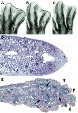
Fig. 2. Before the onset and during INZ. (A-C) Limb buds microinjected with indian ink to show the progressive regression of blood vessels in the third interdigit of chick leg buds prior to the onset of INZ (A), during INZ (B)
and at the end of the degenerative process (C). (D,E) Longitudinal sections of the third interdigit during INZ (D) and at the end of the degenerative process (E). Note in (E) that blood vessels are no longer present in the interdigit, the ectoderm is in the course of being detached into the amniotic sac (arrowheads) and the extracellular matrix appears in large clumps located between the degenerating mesodermal cells (arrows).
A
B
C
D
role of the extracellular matrix. According to this interpretation syndactyly has been found both in mice lacking the laminin α5 chain (Miner et al., 1998) and in mice null for fibrillin 2 gene (Arteaga-Solis et al., 2001). It is also likely that matrix metalloproteinases in coordination with their tissue inhibitors exert an important role in remodeling the interdigital matrix. Stromelysin-3 is expressed in the developing limb bud and in other processes involving apoptosis (Lefebvre et al., 1992; Dupe et al., 1999) and tissue inhibitor of metalloproteinases-3 (TIM-3) is specifically ex-pressed in the interdigital regions prior to cell death (Zeng et al., 1998).
Other factor of potential importance for the regression of the interdigital tissue may be the involvement of Slit2 gene in prevent-ing the incorporation of axons into the interdigital regions (Yuan et al., 1999).
Acknowledgements
This work was supported by a grant, DGESIC/PM98-0061 to J.M.H. V. Z.-L. was supported by a grant from the Portuguese Foundation, Fundação para a Ciência e Tecnologia, and the Gulbenkian Foundation. Thanks are given to Juanjo Sanz Ezquerro and Juan Antonio Montero for some of the figures illustrating this manuscript.
References
ABBADIE, C., KABRUN, N., BOUALI, F., SMARDOVA, J., STEHELIN, D., VANDENBUNDER, B., and ENRIETTO, P.J. (1993). High levels of c-rel expres-sion are associated with programmed cell death in the developing avian embryo and in bone marrow cells in vitro. Cell. Dec. 75: 899-912.
AHUJA, H.S., JAMES, W. and ZAKERI, Z. (1997). Rescue of the limb deformity in hammertoe mutant mice by retinoic acid-induced cell death. Dev Dyn. 208: 466-481.
ALLES, A.J. and SULIK, K.K. (1989). Retinoic-acid-induced limb-reduction defects: perturbation of zones of programmed cell death as a pathogenetic mechanism. Teratology 40: 163-171.
ARTEAGA-SOLIS, E., GAYRAUD, B., LEE, S.Y., SHUM, L., SAKAI, L. and RAMIREZ, F. (2001). Regulation of limb patterning by extracellular microfibrils. J Cell Biol. 154: 275-281.
BARNA, M., HAWE, N., NISWANDER, L. and PANDOLFI, P.P. (2000). Plzf regulates limb and axial skeletal patterning. Nat Genet. 25: 166-172.
BUSHDID, P.B., CHEN, C.L., BRANTLEY, D.M., YULL, F., RAGHOW, R., KERR, L.D. and BARNETT, J.V. (2001). NF-kappaB mediates FGF signal regulation of msx-1 expression. Dev Biol. 237: 107-115.
CAMERON, J.A. and FALLON, J.F. (1977). The absence of cell death during development of free digits in amphibians. Dev Biol. 55: 331-338.
CARRIO, R., LOPEZ-HOYOS, M., JIMENO, J., BENEDICT, M.A., MERINO, R., BENITO, A., FERNANDEZ-LUNA, J.L., NUNEZ, G., GARCIA-PORRERO, J.A. and MERINO, J. (1996). A1 demonstrates restricted tissue distribution during embryonic development and functions to protect against cell death. Am J Pathol. 149: 2133-2142.
CECCONI, F., ALVAREZ-BOLADO, G., MEYER, B.I., ROTH, K.A., and GRUSS, P. (1998). Apaf1 (CED-4 homolog) regulates programmed cell death in mammalian development. Cell 94: 727-737.
CHEN, Y. and ZHAO, X. (1998). Shaping limb by apoptosis. J. Exp. Zool. 282: 691-702. COELHO, C.N., UPHOLT, W.B. and KOSHER, R.A. (1993). The expression pattern of the chicken homeobox-containing gene GHox-7 in developing polydactylous limb buds suggests its involvement in apical ectodermal ridge-directed outgrowth of limb mesoderm and in programmed cell death. Differentiation 52: 129-137. CROCOLL, A., HERZER, U., GHYSELINCK, N. B., CHAMBON, P., and CATO,
A.C.B. (2002). Interdigital apoptosis and downregulation of BAG-1 expression in mouse autopods. Mech. Dev. 111: 149-152.
CUADROS, M.A., MARTIN, C., COLTEY, P., ALMENDROS, A. and NAVASCUES J. (1993). First appearance, distribution, and origin of macrophages in the early development of the avian central nervous system. J Comp Neurol. 330: 113-129.
DAWD, D.S. and HINCHLIFFE, J.R. (1971). Cell death in the “opaque patch” in the central mesenchyme of the developing chick limb: a cytological, cytochemical and electron microscopic analysis. J Embryol Exp Morphol. 26: 401-424. DUNKER N, SCHMITT K, and KRIEGLSTEIN K. (2002) TGF-beta is required for
programmed cell death in interdigital webs of the developing mouse limb. Mech Dev. 113:111-120.
DUPE, V., GHYSELINCK, N.B., THOMAZY, V., NAGY, L., DAVIES, P.J., CHAMBON, P. and MARK, M. (1999). Essential roles of retinoic acid signaling in interdigital apoptosis and control of BMP-7 expression in mouse autopods. Dev Biol. 208: 30-43.
FEINBERG, R.N. and NODEN, D.M. (1991). Experimental analysis of blood vessel development in the avian wing bud. Anat Rec. 231: 136-144.
FERRARI, D., LICHTLER, A.C., PAN, Z., DEALY, C.N., UPHOLT, W.B. and KOSHER, R.A. (1998). Ectopic expression of Msx2 in posterior limb bud mesoderm impairs limb morphogenesis while inducing Bmp4 expression, inhibiting cell proliferation, and promoting apoptosis. Dev. Biol. 197: 12-24. GAÑAN, Y., MACIAS, D., DUTERQUE-COQUILLAUD, M., ROS, M.A. and HURLE,
J.M. (1996). Role of TGFßs and BMPs as signals controlling the position of the digits and the areas of interdigital cell death in the developing chick limb autopod. Development 122: 2349-2357.
GAÑAN Y., MACIAS D., BASCO R.D., MERINO R. and HURLE JM. (1998). Morphological diversity of the avian foot is related with the pattern of msx gene expression in the developing autopod. Dev Biol. 196:33-41.
GARCIA-DOMINGO, D., LEONARDO, E., GRANDIEN, A., MARTINEZ, P., ALBAR, J.P., IZPISUA-BELMONTE, J.C. and MARTINEZ, A C. (1999). DIO-1 is a gene involved in onset of apoptosis in vitro, whose misexpression disrupts limb development. Proc. Natl. Acad. Sci. USA 96: 7992-7997.
GARCIA-MARTINEZ, V., MACIAS, D., GAÑAN, Y., GARCIA-LOBO, J.M., FRANCIA, M.V., FERNANDEZ-TERAN, M.A. and HURLE JM. (1993). Internucleosomal DNA fragmentation and programmed cell death (apoptosis) in the interdigital tissue of the embryonic chick leg bud. J Cell Sci. 106: 201-208.
GRITLI-LINDE, A., NILSSON, J., BOHLOOLY-Y, M., HEBY, O. and LINDE, A. (2001). Nuclear translocation of antizyme and expression of ornithine decar-boxylase and antizyme are developmentally regulated. Dev Dyn. 220: 259-275. GROTEWOLD, L. and RUTHER, U. (2002). The Wnt antagonist Dickkopf-1 is regulated by Bmp signaling and c-Jun and modulates programmed cell death. EMBO J. 21: 966-975.
HINCHLIFFE, J.R. and EDE, D.A. (1967). Limb development in the polydactylous talpid3 mutant of the fowl. J Embryol Exp Morphol. 17: 385-404.
HINCHLIFFE, J.R. and EDE, D.A. (1973). Cell death and the development of limb form and skeletal pattern in normal and wingless (ws) chick embryos. J Embryol Exp Morphol. 30: 753-772.
HINCHLIFFE, J.R. and THOROGOOD, P.V. (1974). Genetic inhibition of mesen-chymal cell death and the development of form and skeletal pattern in the limbs of talpid3 (ta3) mutant chick embryos. J Embryol Exp Morphol. 31: 747-760. HURLE, J.M., and FERNANDEZ-TERAN, M.A. (1983). Fine structure of the
regressing interdigital membranes during the formation of the digits of the chick embryo leg bud. J Embryol Exp Morphol. 78: 195-209.
HURLE, J.M., COLVEE, E. and FERNANDEZ-TERAN, M.A. (1985). Vascular regression during the formation of the free digits in the avian limb bud: a comparative study in chick and duck embryos. J Embryol Exp Morphol. 85: 239-250.
HURLE, J.M., FERNANDEZ-TERAN, M.A. and COLVEE, E. (1986). Regression of the interdigital tissue during the formation of the digits. Acta Histochem Suppl. 32: 165-169.
HURLE, J.M., ROS, M.A., CLIMENT, V. and GARCIA-MARTINEZ, V. (1996). Morphology and significance of programmed cell death in the developing limb bud of the vertebrate embryo. Microsc Res Tech. 34: 236-426.
HURLE, J.M. and MERINO, R. (2002). Apoptosis. In: Encyclopedia of Evolution (Ed. M. Pagel), Oxford University Press, New York. (in press)
JACOBSON, M.D., WEIL, M., and RAFF, M.C. (1996). Role of Ced-3/ICE-family proteases in staurosporine-induced programmed cell death. J. Cell Biol. 133: 1041-1051.
LEE, K.K., TANG, M.K., YEW, D.T., CHOW, P.H., YEE, S.P., SCHNEIDER, C. and BRANCOLINI, C. (1999). Gas2 is a multifunctional gene involved in the regulation of apoptosis and chondrogenesis in the developing mouse limb. Dev Biol. 207: 14-25.
LEE, K.K., LEUNG, A.K., TANG, M.K., CAI, D.Q., SCHNEIDER, C., BRANCOLINI, C. and CHOW, P.H. (2001). Functions of the growth arrest specific 1 gene in the development of the mouse embryo. Dev Biol. 234: 188-203.
LEFEBVRE, O., WOLF, C., LIMACHER, J.M., HUTIN, P., WENDLING, C., LEMEUR, M., BASSET, P. and RIO, M.C. (1992). The breast cancer-associated stromelysin-3 gene is expressed during mouse mammary gland apoptosis. J Cell Biol. 119: 997-1002.
LESCHE, R., PEETZ, A., VAN DER HOEVEN, F. and RUTHER,U. (1997). Ft1, a novel gene related to ubiquitin- conjugating enzymes, is deleted in the Fused toes mouse mutation. Mammalian genome 8: 879-883.
LINDSTEN T, ROSS AJ, KING A, ZONG WX, RATHMELL JC, SHIELS HA, ULRICH E, WAYMIRE KG, MAHAR P, FRAUWIRTH K, CHEN Y, WEI M, ENG VM, ADELMAN DM, SIMON MC, MA A, GOLDEN JA, EVAN G, KORSMEYER SJ, MACGREGOR GR, and THOMPSON CB.(2000). The combined functions of proapoptotic Bcl-2 family members bak and bax are essential for normal development of multiple tissues. Mol Cell. 6:1389-1399.
MACIAS, D., GAÑAN, Y., SAMPATH, T.K., PIEDRA, M.E., ROS, M.A. and HURLE, J.M. (1997). Role of BMP-2 and OP-1 (BMP-7) in programmed cell death and skeletogenesis during chick limb development. Development 124: 1109-1117. MERINO, R., GAÑAN, Y., MACIAS, D., ECONOMIDES, A.N., SAMPATH, K.T. and HURLE, J.M. (1998). Morphogenesis of digits in the avian limb is controlled by FGFs, TGFbetas, and noggin through BMP signaling. Dev Biol. 200: 35-45. MERINO, R., RODRIGUEZ-LEON, J., MACIAS, D., GAÑAN, Y., ECONOMIDES,
A.N., and HURLE, J.M. (1999). The BMP antagonist Gremlin regulates out-growth, chondrogenesis and programmed cell death in the developing limb. Development. 126: 5515-5522.
MILAIRE, J. and ROOZE, M. (1983). Hereditary and induced modifications of the normal necrotic patterns in the developing limb buds of the rat and mouse: facts and hypothesis. Arch. Biol. (Bruxelles) 94: 459-490.
MILLIGAN, C.E., PREVETTE, D., YAGINUMA, H., HOMMA, S., CARDWELL, C., FRITZ, L.C., TOMASELLI, K.J., OPPENHEIM, R.W. and SCHWARTZ, L.M. (1995). Peptide inhibitors of the ICE protease family arrest programmed cell death of motoneurons in vivo and in vitro. Neuron. 15: 385-393.
MINER, J.H., CUNNINGHAM, J. and SANES, J.R. (1998). Roles for laminin in embryogenesis: exencephaly, syndactyly, and placentopathy in mice lacking the laminin alpha5 chain. J Cell Biol. 143: 1713-1723.
MIRKES, P.E., LITTLE, S.A. and UMPIERRE, C.C. (2001). Co-localization of active caspase-3 and DNA fragmentation (TUNEL) in normal and hyperthermia-induced abnormal mouse development. Teratology 63: 134-143.
MITROVIC, D.R. (1977). Development of the metatarsophalangeal joint of the chick embryo: morphological, ultrastructural and histochemical studies. Am J Anat. 150: 333-347.
MOALLEM, A. and HALES, B.F. (1996). Transglutaminase and clusterin induction during normal and abnormal limb development in the mouse. Biol. Repro. 55: 281-290.
MOALLEM, A. and HALES, B.F. (1998). The role of p53 and cell death by apoptosis and necrosis in 4-hydroperoxycyclophosphamide-induced limb malformations. Development 125: 3225-3234.
MONTERO, J.A., GAÑAN, Y., MACIAS, D., RODRIGUEZ-LEON, J., SANZ-EZQUERRO, J.J., MERINO, R., CHIMAL-MONROY, J., NIETO, M.A. and HURLE, J.M. (2001). Role of FGFs in the control of programmed cell death during limb development. Development 128: 2075-2084.
MORI, C., NAKAMURA, N., KIMURA, S., IRIE, H., TAKIGAWA, T., and SHIOTA, K. (1995). Programmed cell death in the interdigital tissue of the fetal mouse limb is apoptosis with DNA fragmentation. Anat. Rec. 242: 103-110.
NARUSE, I. and KAMEYAMA, Y. (1982). Morphogenesis of genetic preaxial polydactyly, polydactyly Nagoya, Pdn, in mice. Cong. Anom. 22: 137-144. NARUSE, I. and KAMEYAMA, Y. (1986). Prevention of polydactyly manifestation
in Polydactyly Nagoya (Pdn) mice by administration of cytosine arabinoside during pregnancy. Teratology 34: 283-289.
NISHII, K., TSUZUKI, T., KUMAI, M., TAKEDA, N., KOGA, H., AIZAWA, S., NISHIMOTO, T., and SHIBATA, Y. (1999). Abnormalities of developmental cell death in Dad1-deficient mice. Genes Cells. 4: 243-252.
NOVACK, D.V., and. KORSMEYER, S. J. (1994). Bcl-2 protein expression during murine development. Am. J. Pathol 145: 61-73.
PIZETTE S, ABATE-SHEN C, and NISWANDER L.(2001).BMP controls proximodistal outgrowth, via induction of the apical ectodermal ridge, and dorsoventral patterning in the vertebrate limb. Development 128: 4463-4474.
RAYNAUD, A. (1990). Developmental mechanisms invoved in the embryonic reduction of limbs in reptiles. Int. J. Dev. Biol. 34: 233-243.
RODRIGUEZ-LEON, J., MERINO, R., MACIAS, D., GAÑAN, Y., SANTESTEBAN, E., and HURLE, J.M. (1999). Retinoic acid regulates programmed cell death through BMP signalling. Nat Cell Biol. 1: 125-126.
SALAS-VIDAL, E., LOMELI, H., CASTRO-OBREGON, S., CUERVO, R., ESCALANTE-ALCALDE, D. and COVARRUBIAS, L. (1998). Reactive oxygen species participate in the control of mouse embryonic cell death. Exp Cell Res. 238: 136-147. SAUNDERS J.W. Jr (1966). Death in embryonic systems. Science 154:604-612. STEWART, S., YI, S., KASSABIAN, G., MAYO, M., SANK, A., and SHULER, C. (2000).
Changes in expression of the lysosomal membrane glycoprotein, LAMP-1 in interdigital regions during embryonic mouse limb development, in vivo and in vitro. Anat Embryol (Berl) 201: 483-490.
TODT, W.L. and FALLON, J.F. (1986). Development of the apical ectodermal ridge in the chick leg bud and a comparison with the wing bud. Anat Rec. 215: 288-304. TONÉ, S., TANAKA, S. and KATO, Y. (1988). The cell cycle and cell population kinetics
in programmed cell death in the limb-buds of normal and 5-bromodeoxyuridine treated chick embryos. Develop. Growth & Differ. 30: 261-270.
TOSNEY, K.W., SCHROETER, S., and POKRZYWINSKI, J.A. (1988). Cell death delineates axon pathways in the hindlimb and does so independently of neurite outgrowth. Dev. Biol. 130: 558-572.
VAN KLEFFENS, M., GROFFEN, C., ROSATO, R.R., VAN DEN EIJNDE, S.M., VAN NECK, J.W., LINDENBERGH-KORTLEVE, D.J., ZWARTHOFF, E.C., and DROP, S.L. (1998). mRNA expression patterns of the IGF system during mouse limb bud development, determined by whole mount in situ hybridization. Mol Cell Endocrinol. 138: 151-161.
WANG, B (2001). Involvement of p53-dependent apoptosis in radiation teratogenesis and in the readioadaptative response in the late organogenesis of mice. J. Radiat.Res (Tokyo) 42: 1-10.
WOOD, W., TURMAINE, M., WEBER, R., CAMP, V., MAKI, R.A., MCKERCHER, S.R. and MARTIN, P. (2000). Mesenchymal cells engulf and clear apoptotic footplate cells in macrophageless PU.1 null mouse embryos. Development 127: 5245-5252. WRIDE, M.A., LAPCHAK, P.H. and SANDERS, E.J. (1994). Distribution of TNF alpha-like proteins correlates with some regions of programmed cell death in the chick embryo. Int J Dev Biol. 38: 673-682.
YANO, H., OHTSURU, A., ITO, M., FUJII, T., and YAMASHITA, S. (1996). Involvement of c-Fos proto-oncogene during palatal fusion and interdigital space formation in the rat. Develop. Growth Differe 38: 351-357.
YI, S.E., DALUISKI, A., PEDERSON, R., ROSEN, V., and LYONS, K.M. (2000). The type I BMP receptor BMPRIB is required for chondrogenesis in the mouse limb. Development. 127: 621-630.
YUAN, W., ZHOU, L., CHEN, J.H., WU, J.Y., RAO, Y. and ORNITZ, D.M. (1999). The mouse SLIT family: secreted ligands for ROBO expressed in patterns that suggest a role in morphogenesis and axon guidance. Dev Biol. 212: 290-306.
YOKOUCHI, Y., SAKIYAMA, J., KAMEDA, T., IBA, H., SUZUKI, A., UENO, N. and KUROIWA, A. (1996). BMP-2/-4 mediate programmed cell death in chicken limb buds. Development. 122: 3725-3734.
ZENG. Y., ROSBOROUGH, R.C., LI, Y., GUPTA, A.R., and BENNETT, J. (1998). Temporal and spatial regulation of gene expression mediated by the promoter for the human tissue inhibitor of metalloproteinases-3 (TIMP-3)-encoding gene. Dev. Dyn. 211: 228-237.
ZHANG, Q., AHUJA, H.S., ZAKERI, Z.F., and WOLGEMUTH, D.J. (1997). Cyclin-dependent kinase 5 is associated with apoptotic cell death during development and tissue remodeling. Dev Biol. 183: 222-233.