The origin and evolution of the nervous system
ALAIN GHYSEN*
Laboratory of Neurogenetics, INSERM E343, Université Montpellier II, France
ABSTRACT The nervous systems of animals as diverse as flies and mice share many conserved features, suggesting that such features were already present in their last common ancestor. As our knowledge of neural development increases, so does the list of conserved features, pointing to the existence of a highly sophisticated, single species as the origin of most extant nervous systems. Possible reasons for this unexpected monophyly are discussed, leading to the conclusion that the appearance of very different life forms, lifestyles and habitats requires the previous attainment of a neural circuitry that is sufficiently robust to cope with large changes without losing its overall coherence.
KEY WORDS:
Urbilateria, conservation, tolerance, adaptation
0214-6282/2003/$25.00 © UBC Press
Printed in Spain www.ijdb.ehu.es
*Address correspondence to: Dr. Alain Ghysen. Lab. of Neurogenetics, INSERM E343, cc103, Université Montpellier II, Place E. Bataillon, 34095 Montpellier, France. Fax: +33-467-143-928. e-mail: [email protected]
Note 1: "Nothing is dead but that which lives not yet / Next to the shining past, tomorrow is colorless / It is shapeless too next to the accomplished / Which reveals at the same time both effort and effect". [Translation by A. Ghysen].
Rien n’est mort que ce qui n’existe pas encore Près du passé luisant demain est incolore Il est informe aussi près de ce qui parfait Présente tout ensemble et l’effort et l’effet
From "Cortège" by Guillaume Apollinaire1
Prologue
Twelve years ago, The International Journal of Developmental Biology published a special issue on "Developmental Biology in Belgium". On that occasion I was asked to write a review on the development of the nervous system (Ghysen, 1992). At the time, the nervous systems of epineurians (chordates) and hyponeurians (annelids/arthropods) were still considered highly dissimilar, and were supposed to have diverged independently from the diffuse nerve net present in diploblasts (cnidarians and ctenarians). My review developed the idea that, on the contrary, the nervous systems of all triploblasts share many essential features, and must have derived from a common ancestor that already enjoyed a fairly sophisticated nervous system. This idea has now gained large support, and the ancestor has gained a name: Urbilateria (de Robertis and Sasai, 1996).
Many discoveries have been made over the past decade that brought substantial changes in our view of phylogeny. On one hand, the classical distinction between diploblasts and triploblasts is questioned by recent discoveries demonstrating not only that typical mesoderm derivatives such as smooth and striated muscles
do exist in so-called diploblasts, but also that their formation depends on a conserved set of mesodermal determinants, includ-ing the gene twist (Spring et al., 2000, 2002; Muller et al., 2003). Furthermore, patterns of gene expression during ctenarian em-bryogenesis reveal a clear plane of bilateral symmetry (Yamada and Martindale, 2002), and ablation experiments demonstrate that the radial symmetry supposedly typical of diploblasts is a derived character superimposed on a basically bilateral body plan (Houliston
et al., 1993). This re-appraisal of the «lowly diplobasts» is only just beginning, however, and too few data are yet available to draw meaningful conclusions about their neural development. Cnidarians and ctenarians will therefore not be considered in this paper.
The classical views about triploblast phyletic relations have also changed dramatically during the past decade, with the conven-tional separation between acoelomates, pseudocoelomates and coelomates being replaced by a subdivision that separates ecdysozoans (including nematodes and arthropods) and lophotrochozoans (including annelids, molluscs and platyhelm-inths) (Aguinaldo et al., 1997; rev. in Adoutte et al., 2000). Thus model organisms that were once thought to be very distantly related, e.g. Caenorhabditis and Drosophila, are now considered to belong to the same superphylum.
devel-opment than was possible ten years ago. In this paper I will review some of these features, to illustrate the unexpected sophistication of Urbilateria’s neural equipment.
As the special issues of the Int. J. Dev. Biol. are a privileged place to venture into somewhat speculative grounds, I will also explore possible explanations for the puzzling conclusion that a single, highly evolved species is the ancestor of all living triploblasts, while one would have thought that the earliest metazoans had many evolutionary avenues open to them.
1. The nervous system of Urbilateria
Sensory nervous system: mechanosensory organs
The sensory systems of all triploblasts comprise various types of sense organs, underlying distinct sensory modalities. Obvious similarities have been noted between sense organs of arthropods and of vertebrates, highly suggestive of a common origin. Here I will concentrate on the mechanoreceptors, as I know this system best. The case of olfaction and gustation is equally compelling, however, and the case of the photoreceptors will be discussed in another chapter of this issue.
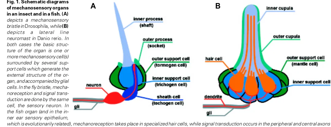
The mechanosensory bristles of flies (Fig. 1A) are composed of a bipolar sensory neuron with a specialized microtubular body at the tip of its dendrite. The dendrite is surrounded by different types of support cells, one of which produces the stiff shaft that amplifies the touch sensitivity of the neuron. The axon is accompanied by a glial cell. The entire set of cells is generated from a precursor cell by a fixed lineage. Precursor cells arise within an epithelial field of cells made competent through the expression of one or more control genes of the bHLH type (proneural genes). The choice of the precursor cell involves a process of lateral inhibition mediated by the Notch-Delta interaction.
The most primitive mechanosensory organs of vertebrates are the lateral line organs (neuromasts) that are found on the body of fish and amphibians (Fig. 1B). The lateral line is a wave-detecting system closely related to the inner ear. The neuromasts are composed of sensory hair cells, which form an elongated microtu-bular process called the kinocilium. The hair cells are surrounded by two types of support cells, which secrete the stiff cupula that ensheathes the kinocilia and modulates their sensitivity. They are innervated by bipolar neurons which are accompanied by glial cells. The different types of cells are not generated by a fixed lineage, but they all arise from an epithelial field, or placode. The formation of this field, and the choice of cell fates within the placode, involves the expression of one or more proneural genes and the action of the Notch system.
The remarkable similarities between the fly bristles and the vertebrate octavolateral system (inner ear + lateral line) have already been noted, and leave little doubt that both are derived from a common ancestor (Adam et al., 1998, Eddison et al., 2000, Fritzsch et al., 2001). Differences like the fixed lineage that gener-ates the bristle organs in arthropods, or the specialized hair cells that mediate wave detection in vertebrates, are presumably late additions to the original system. The latter may have consisted of superficial sensory neurons endowed with a mechanosensory dendrite, of support cells that would both protect and modulate the sensitivity of the dendrite, and of glial cells. All three cell types probably originated from epithelial cell patches rendered compe-tent by the local expression of proneural genes. The allocation of
cell fates within these patches may have involved cell interactions mediated by the Notch system.
It is still not clear whether the ancestral sensory neuron evolved into the specialized hair cell, or whether the hair cell arose de novo
(as did the ciliary photoreceptor cells in the vertebrate retina, whose ganglion cells seem to be homologous to the rhabdomeric photoreceptors of invertebrates, Arendt et al., 2002). One possibil-ity is that the ancestral sensory neuron duplicated, with one copy becoming specialized in the sensory function (the hair cell), and the other retaining the signal transmitting function (the neuron). In-deed, the hair cell retains many properties of a true neuron, including its presynaptic specialization. Not only could this separa-tion of funcsepara-tions have facilitated cell specializasepara-tion, but it would also open the possibility of coupling a single neuron to a larger number of sensory cells, thereby increasing the sensitivity of the system. Interestingly the same situation occurs in the visual system, where single ganglion cells are also coupled to a large number of ciliated photoreceptors.
Central nervous system: orthogonality
The central nervous system of all triploblasts is essentially a three-dimensional structure derived from a two-dimensional epi-thelium. It is built along three axes, antero-posterior, dorso-ventral, and apico-basal. Development along each axis relies on its own developmental mechanisms, and generates its own range of cell specificities. The central nervous sytem is, therefore, basically an orthogonal structure.
The vertebrate and arthropod nervous systems have long been considered to be extremely different due to their position in the body plan (dorsal in vertebrates and ventral in arthropods). Back in the XIXth century, however, it had already been suggested that the difference was more superficial than profound (for a scholarly review of this long-standing dispute, see Nübler-Jung and Arendt, 1994). The idea that the nervous systems of protostomians and deuterostomians are homologous in spite of their different posi-tions has been revived more recently (Arendt and Nübler-Jung, 1994, Ghysen, 1992). This view is completely supported by the discovery that the genetic system underlying the D/V axis is actually conserved between the two phyla, with the neural (dorsal) region of deuterostomians being truly homologous to the neural (ventral) region of protostomians (De Robertis and Sasai, 1996). Thus, what has long been considered as an inversion of the body plan may turn out to be a trivial consequence of the formation of a new mouth in the deuterostomian lineage (Nübler-Jung and Arendt, 1996). In this context it is interesting to note that when the chordate Amphioxus is swimming it has its neural side up, as decent chordates do, but when it lies buried in the sand it has its neural side down, much as a legitimate protostomian would, whereby its mouth opens in free water (Nübler-Jung, pers. comm.).
Central nervous system: connectivity
A
B
shown recently that the same system is also involved in the settingup of longitudinal fiber tracts at various distances from the midline (Rajagopalan et al., 2000, Zlatic et al., 2003). Whether this function is conserved in vertebrates has not yet been established. What-ever the case, the universal implication of the robo/slit system in everything that concerns the relation between neurons and midline indicates that this system must have been firmly rooted in the development of the urbilaterian nerve chord.
Likewise, the roles of the netrin/DCC system have been con-vincingly demonstrated in protostomes (fly, nematode) as well as in deuterostomes (chick, mouse, zebrafish). The molecular and functional conservation of axon guidance mechanisms suggests that a robust orthogonal organization already characterized the urbilaterian nerve chord. Whether elementary functional circuits were already genetically designed within this orthogonal net, and could have been inherited by all derived animals, has not been demonstrated but seems plausible enough.
Central nervous system: cell diversity
The previous sections strongly suggest that the urbilaterians already possessed the molecular mechanisms that underly axon guidance relative to the body axes. Their axons were able therefore to build the orthogonal scaffold of fibers that is so typical of all triploblastic nervous systems. Such design, however, requires that different neuronal cell types be specified during development. Is there any indication that such a diversity of neuronal identities already existed in the urbilaterians?
An important difference between arthropods and vertebrates is the mode of formation of their nerve chord. While in arthropods neuroblasts delaminate from the epithelium, in vertebrates the entire neuroepithelium invaginates to form the neural tube. This difference may be more impressive than profound, however (rev. in Arendt and Nübler-Jung, 1999). Indeed, in both cases neurons are produced by unequally dividing cells, where one of the two progenies continues dividing in a stem-cell mode, while the other becomes neuronal (or undergoes a very limited number of addi-tional divisions).
Given the major importance of the capability to generate defined types of neurons in a defined succession, it may be that no complex nervous system could evolve without the ability to program and control asymmetric divisions. In flies, where the mechanism of this asymmetry has been best analysed, one of its major protagonists is the product of the gene numb. The Numb protein is localized at one pole of the dividing cell as a cortical crescent, and determines the fate of the daughter cell by which it is inherited (Rhyu et al., 1994). There is now good evidence that this process has been conserved in vertebrates (Shen et al., 2002, Cayouette and Raff, 2002), indicating that mechanisms to program cell diversity already existed in the urbilaterian central nervous system.
Neuronal cell diversity depends on the differential expression of a large number of transcriptional regulators which are common to all triploblasts. The corresponding genes have been grouped in families according to their type of DNA-binding domain. Most families already comprised many members in the Urbilaterian
ancestor, suggesting a high level of genetic complexity in neuronal specification. To take but one example, it has been estimated that the urbilaterian genome already comprised 35 distinct bHLH genes, each of which is still recognizable in the genomes of flies, nematodes and vertebrates (Ledent and Vervoort, 2001). Most of these bHLH genes are involved in the determination of cell diversity within the nervous system.
Antero-posterior organization of the central nervous system
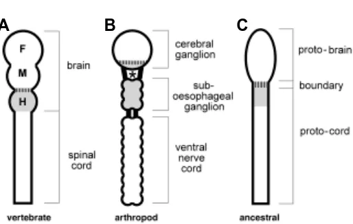
A prominent feature of the central nervous system is its longitu-dinal subdivision into distinct domains. Based on morphological and embryological grounds, the vertebrate CNS is traditionally subdivided in two major parts, the brain and the spinal cord, with the brain itself being subdivided in an anterior (forebrain), an interme-diate (midbrain) and a posterior (hindbrain) region (Fig. 2A). The arthropodian CNS is subdivided in an anterior part, the cerebral ganglion (also called brain), an intermediate part extending from the oesophagus to the neck (suboesophageal ganglion) and a caudal part, the ventral nerve cord made of a succession of segmental ganglia (Fig. 2B).
Fig. 1. Schematic diagrams of mechanosensory organs in an insect and in a fish. (A)
depicts a mechanosensory bristle in Drosophila, while (B)
depicts a lateral line neuromast in Danio rerio. In both cases the basic struc-ture of the organ is one or more mechanosensory cell(s) surrounded by several sup-port cells which generate the external structure of the or-gan, and accompanied by glial cells. In the fly bristle, mecha-noreception and signal trans-duction are done by the same cell, the sensory neuron. In the fish organ (and in the in-ner ear sensory epithelium,
A
B
C
The search for homologies between these two systems, and with other type of CNS such as the molluscan or the echinodermal, has long seemed doubtful at best, meaningless at worst. The situation has now changed with the recognition of a special region called midbrain-hindbrain boundary (MHB; vertical stripes in Fig. 2A), which plays a major organizing role in the development of the vertebrate brain (rev. in Wurst and Bally-Cuif, 2001). Not only is this «boundary» essential for the patterning of the CNS on either side, but it also defines the transition between two regions with very different genetic requirements.
The region anterior to the MHB expresses the Otx genes, which are homologous to the orthodenticle (otd) gene of the fly. Otx genes are necessary for the development of this region (rev. in Simeone, 1998), much as the otd gene is required for the development of the anteriormost part of the fly CNS, and indeed the mammalian Otx can functionally substitute for the fly gene in otd mutants (Leuzinger
et al., 1998, Nagao et al., 1998). Homeotic genes, on the other hand, are expressed in the region of the nervous system posterior to the MHB, but are excluded from the anterior region both in mammals and in flies (rev in Hirth and Reichert, 1999).
The MHB region itself (striped section in Fig. 2A) is character-ized by the expression of the Pax2/5/8 genes (rev. in Wada and Satoh, 2001). Hirth et al. (2003) have recently shown that the fly counterparts of the Pax 2/5/8 genes, pax2 and poxn, do not have adjacent expression domains except at one place: the boundary between the second and third subdivision of the fly brain (striped section in Fig. 2B). As the anteriormost homeotic gene labial is expressed in the third subdivison of the fly brain, the pax2/poxn
stripe separates an anterior domain expressing otd from a poste-rior domain expressing the homeotic genes. There is little doubt, therefore, that the Otx - Pax - Hox subdivision of the mammalain brain is strictly homologous to the similar subdivision now re-ported in flies, and therefore was in all likelyhood present in urbilaterians (Hirth et al., 2003).
In order to facilitate the discussion, and since neither the mammalian nor the fly traditional subdivisions of the CNS take
into account this major organisational feature, we will adopt the nomenclature proposed in Fig. 2C, where the region of the CNS anterior to the MHB will be called protobrain, and the posterior region will be called protocord. The case of the anterior, cephalized part of the protocord (the mammalian hindbrain; light gray in Fig. 2) will be discussed separately.
Protobrain
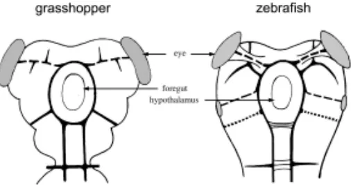
The overall design of brain connectivity is surprisingly similar in arthropod and vertebrate representatives (Fig. 3). The details of the processes by which such early tracts are established, and the genes that are responsible for this establishment, have not yet been elucidated. It is therefore difficult to decide whether or not the apparent similarity of wiring diagrams reflects a shared ancestry or not. Nevertheless what has been learned about axonal pathfinding in general, and about the formation of commis-sural tracts in particular, suggests that similarities in the basic connectivity may well represent homology rather than analogy, and therefore that a pattern not too dissimilar from what is shown in the figure may have existed in the urbilaterian and have been inherited by arthropods and chordates alike.
An important component of the brain are the olfactory centers: the antennal lobes in flies, and the olfactory bulbs in vertebrates. In both cases, the basic organization of the olfactory projection relies on the convergence of sensory axons that share the same odorant receptor onto a single projection center - a glomerulus (Hildebrand and Shepherd, 1997). This type of projection is relatively unusual in that most other sensory systems establish a somatotopic organization where the position of each axon termi-nal reflects the position of the sense organ, rather than its specificity. The similarity in glomerular organization between flies and mice suggests that the major circuitry of the olfactory system, based on the convergence of similar inputs to specific glomeruli, already existed in the urbilateria.
Another important component of the protobrain is the visual system, which is associated to this region both in vertebrates and in arthropods (Fig. 3). There seems to be an important difference between the two types of visual systems, as the vertebrate retina arises as a part of the CNS, while the arthropod retina develops peripherally. Yet, structures as dissimilar as the vertebrate and the many types of invertebrate eyes are now recognized to have derived from a simple, prototypic eye already present in urbilaterians. The apparent differences in the visual systems of arthropods and vertebrates may therefore have to be reexamined when we know better the genetic networks involved in setting up the neuronal circuitry that underlies vision.
Hindbrain genes
Just posterior to the MHB boundary region lies a region which is called hindbrain in chordates, and sub-oesophageal ganglion in insects. As mentioned previously, this region differs from the brain in that it is clearly segmented, and its segmental diversity is patterned by the Hox genes (which are excluded from the protobrain). This region also differs from the more caudal part of the nerve chord in that it is massively involved in the control of head-related and vegetative functions. It can therefore be consid-ered as a cephalized part of the protocord (light gray in Fig. 2).
In arthropods this region comprises three segments, each subdivided in two compartments. The recent discovery that the Fig. 2. Basic structure of the mammalian (A) and arthropodian (B)
central nervous systems. At the right of each system are indicated its traditional subdivisions. The common (ancestral) structure is deduced in
most posterior part of the fly brain, the tritocerebrum, is posterior to the boundary and expresses a homeotic gene, suggests that the cephalized cord actually comprises four segments. In chor-dates there is some doubt as to whether the intermediate region comprises 8 segments, as usually considered, or 4 segments each subdivided in two compartments, as many features suggest. While it may be that this specialized region appeared indepen-dently in arthropods and in chordates as a cephalization of the anteriormost part of the segmented nerve chord, it would seem more economical to imagine that urbilaterians already possessed a four-tiered CNS with a brain proper (protobrain), a boundary (MHB) region, a segmented hindbrain, and a nerve chord.
Hindbrain function
In vertebrates the hindbrain is largely devoted to the afferent and efferent innervation of the branchial arches. As the mouth-parts are derived from the branchial arches, gustation is well represented in the hindbrain, as is facial touch perception. The chordate hindbrain is also involved in the control of many essen-tial vegetative functions such as breathing (that is, forced aeration of the lungs), blood circulation (heart action and blood pressure), nutrition, and other reflex mechanisms such as coughing and vomiting (Kuhlenbeck, 1975).
In spite of major changes in habitat and physiology, major features of the hindbrain organization have remained constant throughout vertebrates. For example, the afferent projection of the lateral line of fish is very similar to the projection of one of its mammalian derivatives, the cochlea (Alexandre and Ghysen, 1999), even though the former is involved in a sense of «distant touch» (Dijkgraaf, 1989) and has disappeared in terrestrial tetra-pods, while the latter is involved in sound perception. Likewise, the control center maintaining the respiratory rythm in man still occupies its primitive position in the medulla, even though the respiratory mechanism of air breathing vertebrates is completely different from that of gilled fishes (Herrick, 1926). Thus, many features of the hindbrain are highly conserved in all vertebrates, in spite of large functional changes in the peripheral organization. Since branchial arches are considered to be a typical chordate structure, one could imagine that the hindbrain is a chordate invention without counterpart in insects. This may be untrue, however. In insects, the corresponding region of the central ner-vous system comprises the three gnathal segments, which to-gether form the sub-oesophageal ganglion. These segments are associated with the mouthparts, and are correspondingly special-ized. They are largely concerned with gustation and facial touch perception (Mitchell and Itagaki, 1992), and they receive the afferent projection of the taste-sensitive neurons on the proboscis. As for the control of vegetative functions such as breathing and circulation, there would seem again to be little common ground between chordates and insects, as in the latter breathing mostly involves passive diffusion through the tracheal system. It has recently become clear that breathing is more sophisticated in arthropods than previously assumed, however (e.g. Westneat et al., 2003), and subject to a tight CNS control which however remains to be elucidated. It will be interesting to see if some of the centers involved in the control of arthropod vegetative functions are located in the suboesophageal ganglion.
In summary, then, it seems at least possible that the anteriormost section of the protochord was already specialized in Urbilateria,
dealing with gustation and «facial» sensitivity, as well as with the control of rythmic vegetative functions.
2. Conservation and change in evolution
In this second, more speculative section, I will explore the idea that all extant triploblasts arose from a single, highly sophisticated ancestral species. The proposal I will argue for is that this unexpected monophyly derives at least in part from two linked factors. First, a major constraint of metazoan evolution is that, because of the complexity of the developmmental programme of any organism, changes are not allowed (rule of conservative changes). Second, only those developmental processes that have become extremely stable can withstand substantial varia-tion without collapsing.
The rule of conservative changes
The rule of conservative changes states that only those changes can be tolerated, that change essentially nothing. This rule applies to any set of interacting elements, where changes in any one component will alter all the interactions in which this compo-nent is involved, and adversely affect the function of the entire set. The stringency of this rule will obviously increase with the number of interactions, as it becomes more and more unlikely that a single change in one element can improve, or at least not harm, the result of the total sum of all interactions.
Biological organisms all undergo a developmental process that leads from the zygote to the reproductive stage. The rule of conservative changes implies, therefore, that any mutational change is first and foremost screened for its compatibility with every step of the developmental program. This is because the only absolute requirement is that the entire program remains functional, i.e. retains its coherence.
Given the complexity of the nervous system, the requirement for maintaining coherence must have been most stringent during the evolution of neural development, and the rule of conservative changes must have had particular relevance for this process. It is no surprise, therefore, that this rule seems to pervade all aspects of developmen-tal neurobiology, from molecules to mind. This I will illustrate by two examples, one at either end of the spectrum of application.
At the level of molecules, to take but one example, the majority (8/14) of the most conserved amino-acids of the homeodomain
are not involved in the biological function of the domain - binding to DNA. Of the 14 most conserved residues of a set of 346 homeodomain sequences, only 3 bind to bases, and 3 bind to the sugar-phosphate backbone. The 8 remaining residues ensure the structural stability of the domain by contributing to the hydropho-bic internal core (Duboule, 1994). Thus many changes can be (and have been) accepted and eventually fixed, as long as they do not affect the internal coherence of the homeodomain.
At the other extreme, that of mental processes, any new information appears to be instantly screened by the human brain for its coherence with previous information, a pre-attentive pro-cess which in the auditory system is associated to an electrical brain response called mismatch negativity (Näätänen, 1992, Koelsch et al., 2002). Interestingly, this process seems to be accentuated in autistic patients, and the acute awareness of discordances may be at the root of their inability to adjust to changing conditions (Seri et al., 1999, Gomot et al., 2002).
Conservative development and tolerance
Coming back to the issue of the developmental programme, one is led to the idea that once a coherent organization has been produced, it is further stabilized by the progressive accumulation of additional mechanisms. Because all changes are screened for their consistency with the existing organization, such additional mechanisms will necessarily reinforce and ensure developmental stability in a wide range of adverse situations. The result of this evolution would be an ever increasing robustness of the system, and an increased capability to follow, or revert to, a relatively normal course in the presence of perturbations - what Waddington called «canalization».
As pointed out recently, the progressive reinforcement and stabilization of developmental processes will buffer the individual from the effect of mutation, and the population as a whole may therefore carry more genetic variation (Kirschner and Gerhart, 1998). Populations with a wider polymorphism may diversify faster under selective conditions, thus improving the «evolvability» of the species, and the success of its genome. This potential for rapid diversification would of course be particularly advantageous during the extensive radiations that followed massive extinctions. The rule of conservative changes puts the emphasis on nega-tive selection - screening for the adaptation of each new element to all other elements with which it interacts, and removing any change that alters the pattern of interactions (and thereby the developmental programme). This view is complementary to the more usual view of evolution as based on positive selection -selection for the fittest. While the latter focuses on the interaction of the individual with the external world, the former focuses on the adaptation of individual gene products to the internal world. Given that all internal interactions are optimized in any given species (for, if they were not, positive selection would quickly ensure that they are), it follows that any non-conservative change will be detrimental and can only be, at best, tolerated (Garcia-Bellido, 2000).
Thus the concept of tolerance to genetic changes that reduce the fitness of the individual may be an important factor to under-stand evolution, in particular in the case of the extensive radia-tions that follow massive extincradia-tions. It may be urgent to incorpo-rate the massive knowledge gained over the past 20 or so years in our understanding of the genetic bases of development, into our current views of evolution, selection and adaptation.
Odd numbers
The accumulation of largely (but not entirely) redundant mecha-nisms will lead, as a corollary, to the puzzling observation that seemingly arbitrary patterns appear inordinately resistant to change. This conclusion is somewhat different from the classical comment «if it works, why fix it»? According to the latter, once the number of fingers settled on five (for whatever reason, and more likely than not without any reason), it has remained so ever since because there was no pressing reason for it to change. My proposal differs from this simple inertia in one important feature: the progressive accumulation of developmental additions that are consistent with the original pattern will make it ever more resistant to change, much like «plug-ins» may add to the performance of a system but can never change its basic structure, and indeed generally contribute to its stability.
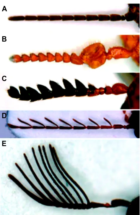
Pushing the argument one step further, one can speculate that it is only those processes that are highly robust, which can then be modified to extraordinary extents without loosing their develop-Fig. 4. Eleven-segmented antennae in various coleopter species.(A)
Cupedidae: Tenomerga cinereus; (B) Melyridae: Malachiinae; (C)
Chrysomelidae: Anomoea sp.; (D) Ptilodactylidae: Ptilodactyla sp.; (E)
Eucnemidae: Isorhipis obliqua. The Ptylodactylidae (D) are unique among all insects in that the antenal segments 4 to 10 have a basal articulated segment, rather than a simple expansion as in e.g. the Eucnemidae (E). Anterior is up in all panels. All micrographs and identificaitons kindly provided by Dr. N. Gompel.
A
B
C
D
mental stability. Indeed the five fingers of tetrapods are famed for the incredible range of structures they can give rise to, from wings to fins through toes and our agile hands. Likewise the nonsensical fixation of most coleopters for 11-segmented antennae is accom-panied by large variations in the identities of the various segments (shape, length, color, sense organs present). Roughly speaking one can apparently get the most insane-looking antenna, as long as it counts 11 segments (Fig. 4). It is tempting to invert the argument, and to imagine that it is because the 11-odd develop-mental system had reached an extreme robustness in some ancient coleopter that it could accept large variations in many of its components without collapsing. From that stage on, this develop-mental system could then generate antennae with many different morphologies - yet necessarily obeying the rule of eleven.
Robustness and adaptation in the nervous system
Let us now consider how this general idea could be applied to the nervous system. In a penetrating short paper published in IJDB, Michael Bate made the important point that «neurons are born and differentiate in ways that are not conditioned by their future func-tions as elements of neural circuits» (Bate, 1998). He went on to say that «To understand how functions ... can emerge from these beginnings, it is worth remembering that fundamental attributes of the nervous system such as the circuitry underlying locomotion or escape behavior are probably also present as a rather stereotyped and evolutionarily conserved set of cells and connections. It is at least possible to envisage that there is a fundamental framework of circuitry just as there is a scaffolding of initial pathways».
One would imagine that there has been a strong selective pressure to make such «fundamental frameworks of circuitry» as stable as possible from a developmental point of view. This involves not only stabilizing the formation of the individual circuits, but also providing for general means to adapt them to unforesee-able perturbations, i.e. general mechanisms of functional plasticity (e.g., learning) and of developmental plasticity (e.g., elimination). Both types of plasticity are indeed found in all triploblasts, and seem to rely on similar mechanisms, suggesting again that they were already present in our urbilaterian ancestors (e.g. Goda, 1995, Friedrich, 2000). It has been argued that there must also be a mechanism to assess and adjust the functional connectivity of the circuit to optimize its performance (Bate, 1992). The nature of this mechanism is still not understood, however.
The central nervous system would then be largely composed of a combination of proven, well-tried, trustworthy elementary cir-cuits, many of which were already developed in our urbilaterian ancestors. Such circuits may have become integrated in various physiological functions as diversification proceeded, a diversifica-tion that presumably took place both in space and in time. For example, mutations in the HoxA1 gene, which in mice is expressed only transiently in rhombomeres r3 and r4 at a very early stage of hindbrain development, result in the formation of a supernumerary respiratory center (del Toro et al., 2001). This result reveals the presence of groups of interconnected cells in r3, that can form a fully active respiratory center provided it is not modified into some other function by HoxA1.
In the previous section I developed the argument that it is only after a process has become developmentally stabilized that it can undergo large changes without compromising its function. Apply-ing this argument to the nervous system, one is led to the
conclu-sion that once the «fundamental frameworks of circuitry» have become stable enough, changes and adjustments can be introduced in individual circuits without perturbing the functioning of the entire system. This, of course, requires that there are genetic means to single out one circuit among all others (Brunet and Ghysen, 1999). The patterned expression of transcriptional regulators with homeotic, bHLH, paired-box and other conserved motifs in the central nervous system may be a simple way to single out neural subsets. Once genetically identified, each subset can evolve in its own way: neural subsets become «uncoupled» from each other, and freed to explore new directions without impairing the development and function of the entire system - provided, again, that such changes are compatible with the basic structure common to all subsets.
The extreme variability of behaviors and survival strategies among triploblasts would then be subordinate on the previous attainment by the urbilaterians of a high level of developmental stability in the building of elementary functional circuits. According to this view, the initial triploblast radiation may have been contingent upon reaching this highly evolved stage of neural development. Conversely, once this stage of neural development had been reached, and once the radiation had become possible, it did not leave much room for a reiteration of the entire process. It follows that the major radiation is bound to have occurred only once, and only from one ancestor: the one whose neural development had reached a level of complexity, and of developmental stability, robust enough to withstand large changes without loosing sense.
Acknowledgements
I am grateful to A. Garcia-Bellido and M. Bate, who are at the origin of many of the views expressed in this review. I thank Nicolas Gompel for fruitful discussions about coleopteran antennae and other fascinating subjects, and for providing the micrographs of Fig. 4, E. Houliston for updating my information on the so-called diploblasts and M. Bate for help with the translation of Apollinaire´s verses. I also thank M. Bate, C. Dambly-Chaudière, A. Garcia-Bellido, N. Gompel, N. König, K. Nübler-Jung and M. Vervoort for comments on early versions of this manuscript.
References
ADAM, J., MYAT, A., LE ROUX, I., EDDISON, M., HENRIQUE, D., ISH-HOROWICZ, D. and LEWIS, J. (1998) Cell fate choices and the expression of Notch, Delta and
Serrate homologues in the chick inner ear: parallels with Drosophila sense-organ
development. Development 125: 4645-4654.
ADOUTTE, A., BALAVOINE, G., LARTILLOT, N., LESPINAT, O., PRUD’HOMME, B. and DE ROSA, R. (2000) The new animal phylogeny: reliability and implications.
Proc Natl Acad Sci USA 97: 4453-4456.
AGUINALDO, A.M.A., TURBEVILLE, J.M., LINFORFD, L.S., RIVERA, M.C., GAREY, J.R., RAFF, R.A. and LAKE, J.A. (1997) Evidence for a clade of nematodes,
arthropods and other moulting animals. Nature 387: 489-493.
ALEXANDRE, D. and GHYSEN, A. (1999) Somatotopy of the lateral line projection in
larval zebrafish. Proc Nat Acad Sci USA 96: 7558-7562.
ARENDT, D. and NÜBLER-JUNG, K. (1994) Inversion of dorsoventral axis? Nature
371: 26.
ARENDT, D. and NÜBLER-JUNG, K. (1996) Common ground plans in early vertebrate
brain development in mice and flies. BioEssays 18: 255-259.
ARENDT, D., TESSMAR, K., MEDEIROS DE CAMPOS-BAPTISTA, M.I., DORRESTEIJN, A. and WITTBRODT, J. (2002) Development of pigment-cup eyes
in the polychaete Platynereis dumerilii and evolutionary conservation of larval eyes
in Bilateria. Development 129: 1143-1154.
BATE, M. (1998) Making sense of behavior. Int. J. Dev. Biol. 42: 507-509.
BRUNET, J.F. and GHYSEN, A. (1999) Deconstructing cell determination: proneural
CAYOUETTE, M. and RAFF, M. (2002) Asymmetric segregation of Numb: a
mecha-nism for neural specification from Drosophila to mammals. Nat. Neurosci. 5:
1265-1269.
DEL TORO ED, BORDAY V, DAVENNE M, NEUN R, RIJLI FM, CHAMPAGNAT J.
(2001) Generation of a novel functional neuronal circuit in Hoxa1 mutant mice. J
Neurosci. 21: 5637-5642.
DE ROBERTIS, E.M. and SASAI, Y. (1996) A common plan for dorsoventral
patterning in Bilateria. Nature 380: 37-40.
DIJKGRAAF, S. (1989) A short personal review of the history of the lateral line research. in: The mechanosensory lateral line. S. Coombs, P. Görner, H. Münz eds, Springer Verlag, New York, pp. 7-14.
DUBOULE, D. (1994) Guide to the homeobox genes. Oxford University Press 1994, pp. 28-29.
EDDISON, M., LE ROUX, I. and LEWIS, J. (2000) Notch signaling in the development
of the inner ear: lessons from Drosophila. Proc Natl Acad Sci USA 97:
11692-11699.
FRIEDRICH, M.J. (2000). Research with Drosophila provides clues to enhancing
human memory. JAMA 284: 2857-2858.
FRITZSCH, B. and BEISEL, K.W. (2001) Evolution and development of the vertebrate
ear. Brain Res Bull 55: 711-21.
GARCIA-BELLIDO, A. (2000) Los Genes del Cámbrico. Discurso inaugural del año académico 1999-2000 de la Real Academía de Ciencias exactas, físicas y naturales. Madrid. (english translation available on request at [email protected])
GHYSEN, A. (1992) The developmental biology of neuronal connectivity. Int. J. Dev.
Biol. 36: 47-58.
GODA, Y. (1995) Memory mechanisms. A common cascade for long-term memory.
Curr. Biol. 5: 136-138.
GOMOT, M., GIARD, M.H., ADRIEN, J.L., BARTHELEMY, C. and BRUNEAU, N. (2002) Hypersensitivity ot acoustic changes in children with autism:
electrophysi-ological evidence of left frontal cortex dysfunctioning. Psychophysiology 39:
577-584.
HERRICK, C.J. (1926) Brain of Rats and Men. University of Chicago Press, Chicago.
HILDEBRAND, J.G. and SHEPHERD, G.M. (1997) Molecular mechanisms of olfac-tory discrimination: converging evidence for common principles across phyla.
Annu Rev Neurosci 1997, 20: 593-631.
HIRTH, F., KAMMERMEIER, L., FREI, E., WALLDORF, U., NOLL, M. and REICHERT, H. (2003) An urbilaterian origin of the tripartite brain: developmental genetic insight
from Drosophila. Development 130: 2365-2373.
HIRTH, F. and REICHERT, H. (1999) Conserved genetic programs in insect and
mammalian brain development. BioEssays 21: 677-684.
HIVERT, B., LIU, Z., CHUANG, C.Y., DOHERTY, P. and SUNDARESAN, V. (2002) Robo1 and Robo2 are homophilic binding molecules that promote axonal growth.
Mol Cell Neurosci. 21: 534-45.
HOULISTON, E., CARRÉ, D., JOHNSTON, J.A. and SARDET, C. (1993) Axis
establishment and microtubule-mediated waves prior to first cleavage in Beroe
ovata. Development 117: 75-87.
KIRSCHNER, M. and GERHART, J. (1998) Evolvability. Proc Natl Acad Sci USA 95:
8420-8427
KOELSCH, S., SCHROGER, E. and GUNTER, T.C. (2002) Music matters: preattentive
musicality of the human brain. Psychophysiology 39: 38-48.
KUHLENBECK, H. (1975) The central nervous system of vertebrates, vol 4. Spinal cord and deuterencephalon. S. Karger, Basel.
LEDENT, V. and VERVOORT, M. (2001) The basic helix-loop-helix protein family:
comparative genomics and phylogenetic analysis. Genome Res 11: 745-770.
LEUZINGER, S., HIRTH, F., GERLICH, D., ACAMPORA, D., SIMEONE, A., GEHRING, W.J., FINKELSTEIN, R., FURUKUBO-TOKUNAGA, K. and
REICHERT, H. (1998) Equivalence of the fly orthodenticle gene and the human
OTX genes in embryonic brain development of Drosophila. Development 125:
1703-1710.
MITCHELL, B.K. and ITAGAKI, H. (1992) Interneurons of the subesophageal
ganglion of Sarcophaga bullata responding to gustatory and mechanosensory
stimuli. J. Comp. Physiol. 171: 213-230.
MULLER, P., SEIPEL, K., YANZE, N., REBER-MULLER, S., STREITWOLF-ENGEL, R., STIERWALD, M., SPRING, J. and SCHMID, V. (2003) Evolutionary aspects of developmentally regulated helix-loop-helix transcription factors in
striated muscle of jellyfish. Dev Biol. 255: 216-29.
NÄÄTÄNEN, R. (1992) Attention and brain function. Erlbaum, Hillsdale (New-Jersey, USA).
NAGAO, T., LEUZINGER, S., ACAMPORA, D., SIMEONE, A., FINKELSTEIN, R., REICHERT, H. and FURUKUBO-TOKUNAGA, K. (1998) Developmental
res-cue of Drosophila cephalic defects by the human Otx genes. Proc Natl Acad Sci
USA 95: 3737-3742.
NÜBLER-JUNG, K. and ARENDT, D. (1994) Is ventral in insects dorsal in verte-brates? A history of embryological arguments favouring axis inversion in
chordate ancestors. Roux’s Arch Dev Biol 203: 357-366.
NÜBLER-JUNG and K., ARENDT, D. (1996) Enteropneusts and chordate
evolu-tion. Curr Biol 6: 352-353.
RAJAGOPALAN, S., VIVANCOS, V., NICOLAS, E. and DICKSON, B. (2000) Selecting a longitudinal pathway: Robo receptors specify the lateral position of
axons in the Drosophila CNS. Cell 103: 1033-1045.
RHYU, M.S., JAN, L.Y. and JAN, Y.N. (1994) Asymmetric distribution of numb protein during division of the sensory organ precursor cell confers distinct fates
to daughter cells. Cell 76: 477-91.
SERI, S., CERQUIGLINI, A., PISANI, F. and CURATOLO, P. (1999) Autism in tuberous sclerosis: evoked potential evidence for a deficit in auditory sensory
processing. Clin Neurophysiol. 110: 1825-1830.
SHEN, Q., ZHONG, W., JAN, Y.N. and TEMPLE, S. (2002) Asymmetric Numb distribution is critical for asymmetric cell division of mouse cerebral cortical stem
cells and neuroblasts. Development 129: 4843-53.
SIMEONE, A. (1998) Otx1 and Otx2 in the development and evolution of the
mammalian brain. EMBO J 17: 6790-6798.
SPRING, J., YANZE, N., JOSCH, C., MIDDEL, A.M., WINNINGER, B. and SCHMID, V. (2002) Conservation of Brachyury, Mef2, and Snail in the
myogenic lineage of jellyfish: a connection to the mesoderm of bilateria. Dev
Biol. 244: 372-84.
WADA, H. and SATOH, N. (2001) Patterning the protochordate neural tube. Curr
Opin Neurobiol 11: 16-21.
WESTNEAT, M.W., BETZ, O., BLOB, R.W., FEZZAA, K., COOPER, W.J. and LEE, W.K. (2003) Tracheal respiration in insects visualized with synchrotron x-ray
imaging. Science 299: 558-560.
WURST, W. and BALLY-CUIF, L. (2001) Neural plate patterning: upstream and
downstream of the isthimic organizer. Nat Rev Neurosci 2: 99-108.
YAMADA, A. and MARTINDALE, M.Q. (2002) Expression of the ctenophore Brain Factor 1 forkhead gene ortholog (ctenoBF-1) mRNA is restricted to the pre-sumptive mouth and feeding apparatus: implications for axial organization in the
Metazoa. Dev Genes Evol. 212: 338-348.
ZLATIC, M., LANDGRAF, M. and BATE, M. (2003) Genetic specification of axonal
arbors: atonal regulates robo3 to position terminal branches in the Drosophila