Evidence that Platelet derived growth factor (PDGF) action is
required for mesoderm patterning in early amphibian
(Xenopus laevis) embryogenesis
JUNG-SUN GHIL and HAE-MOON CHUNG*
Laboratory of Developmental Biology, Department of Biology Education, Seoul National University, Seoul, Republic of Korea
ABSTRACT Mesoderm induction is one of the major events of early vertebrate embryonic patterning. It appears to be controlled by sequential and combinatorial actions of several kinds of peptide growth factors. These include activin, fibroblast growth factor (FGF), and transforming growth factor-β (TGF-β), among others. In the present study, the function of platelet-derived growth factor (PDGF) in early Xenopus laevis embryogenesis was investigated. In the animal-cap assay, PDGF caused pre-ectodermal tissue to develop a mesoderm specific morphology (elongation) and to express the mesoderm marker genes, MyoD family and α-cardiac actin. In addition, two other genes were expressed -related serum response factor SL1 (a dorsal mesodermal marker) and myosin light chain (MLC2-heart marker). A role for PDGF in normal (in vivo) mesoderm induction is implicated because injection of PDGF receptor α antisense RNA into 2-cell embryos erased the animal cap’s mesoderm marker expression. Those injected embryos also exhibited morphological abnormalities including incomplete gastrulation, failure of neural fold closing, and abnormal somitogenesis.
KEY WORDS: mesoderm induction, mesoderm patterning, PDGF, PDGF receptor
α
0214-6282/99/$15.00 © UBC Press
Printed in Spain www.ehu.es/ijdb
Original Article
*Address for reprints: Laboratory of Developmental Biology, Department of Biology Education, Seoul National University San 56-1, Shinlim-dong, Kwanak-ku Seoul 151-742, Republic of Korea. FAX: 82 2 886 2117. e-mail: hmchung@snu.ac.kr
Introduction
In amphibian embryos, the induction and patterning of the mesoderm lie at the heart of early development. During the blastula stage, the presumptive mesoderm area is specified in the marginal zone which is located between the vegetal and animal hemisphere (Woodland, 1989; Heasman, 1997). Much recent work on meso-derm induction has taken advantage of the animal cap assay in that animal cap can be induced into mesoderm under the influence of either vegetal pole explants or various soluble factors. The molecules for mesoderm induction include members of the fibroblast growth factor (FGF) family and members of the transforming growth factor-β (TGF-β) family (Kimelman and Kirschner, 1987; Sive, 1993; Cornell and Kimelman, 1994; Kessler and Melton, 1994). It is widely believed that these and other growth factors might act in combination to induce and pattern the embryo. Other intercellular signaling molecules have been identified that pattern the mesoderm by altering the competency of the cells to meso-derm-inducing agents (Christian et al., 1992). These include mem-bers of the Wnt family (Christian et al., 1991), noggin (Lamb et al., 1993; Smith et al., 1993), and chordin (Sasai et al., 1994). Although the mesoderm is known to be formed during blastula stages by
Abbreviations used in this paper: PDGF, Platelet derived growth factor; TGF-β, transforming growth factor beta; FGF, Fibroblast growth factor; SL1, related serum response factor; MLC2, myosin light chain.
Especially PDGF A-chain mRNA has been detected as a maternally encoded transcript in Xenopus oocytes and is known to be expressed in the animal cap and marginal zone at the blastula stage. In contrast, Platelet derived growth factor receptor α (PDGFRα) has been found to be expressed in the primitive streak of the mouse which is analogous to the involuting marginal zone of Xenopus (Orr-Urtreger and Lonai, 1992; Schatteman et al., 1992). In Xenopus, PDGFRα has been found to be expressed in marginal zone cells at gastrula stage, presomitic region at neurula stage and many mesenchymal tissues at various stages of development (Ho et al., 1994). In the mouse embryo, PDGFRα transcript has been detected throughout mesenchymal tissues including the splanchnic mesoderm, surrounding the neural tube, somites, and the developing vasculature (Graham et al., 1992; Schatteman et al., 1992). Recent studies on the PDGFR imply that the downstream components of the PDGFR signaling pathway are p21ras, raf-1, etc. (Wagner and
Cochran, 1993; Bowen-Pope and Seifert, 1994). In Xenopus, it has been previously demonstrated that dominant negative mutants of c-ras or c-raf inhibit the ability of FGF and activin to induce mesoderm. Curiously, overexpression of activated p21ras or raf-1
is sufficient to induce mesoderm in animal cap assay (Whitman and Melton, 1992). The downstream molecule of both ras and raf is MAP kinase (Blenis, 1993). MAP kinase phosphatase (MKP-1) was used to inactivate endogenous MAP kinase and to prevent the induction of early and late mesodermal markers by both FGF and activin (Gotoh et al., 1995; Labonne et al., 1995, 1997; Rose and Busa, 1998). PDGFRα, as well as FGF receptor, is a tyrosine kinase receptor to activate c-ras, c-raf, and MAP kinase in several cell lines (Ullrich and Schlessinger, 1990; Blenis, 1993; Claesson-Welsh, 1995). These data suggest that PDGF may function in mesoderm induction. In order to investigate this possibility, we carried out animal cap assay to check if PDGF treatment leads to mesodermal specific morphological change and gene expression.
Results
PDGF induces mesoderm from animal cap explants – morpho-logical study
When mesoderm is induced, a critical morphological change-namely elongation occurs. The animal cap explants treated with PDGF showed clear-cut elongation (Fig. 1B), while the control did not show any sign of elongation (Fig. 1A). In addition to that
morphological change, whole-mount in situ hybridization detected the expression of α-cardiac actin in the animal cap explants treated with PDGF. This data confirm that the elongated explants repre-sent authentic induced mesoderm tissue. Histological analysis showed that untreated animal caps formed atypical epidermis (Fig. 2A,B) but PDGF treated animal caps formed a ventral mesodermal vesicle (Fig. 2C,D) and a mass of tissue containing clumps of muscle-like cells (Fig. 2E,F), similar to those formed in XBra-injected animal caps (O’Reilly et al., 1995).
PDGF activates the expression of mesodermal marker genes
To determine whether PDGF is mesoderm inducer, we exam-ined the expression of mesoderm specific markers, MyoD family of genes including XMyoD, XMyf5, and XMRF4 by RT-PCR. In normal Xenopus embryos, the expression of XMyoD and XMyf5 begins weakly at the early gastrula stage (Hopwood et al., 1992), whereas XMRF4 expression is first detected only in somites at stage 18 (neural fold stage) and becomes maximum at stage 22-23 (Jennings, 1992). The XMyoD family of genes was expressed in the animal caps treated with PDGF. Similar results were ob-tained in the cases of activin and FGF, which are well known as mesoderm inducers (Fig. 3). The expression of α-cardiac actin was also assayed by RT-PCR. This is supporting evidence for the in situ study (Fig. 1). Somite mesodermal markers, such as related serum responsive factor (RSRF) family, SL1 (MEF2D), and a cardiac mesodermal marker, Xenopus myosin light chain (XMLC2) were studied by RT-PCR. SL1 and XMLC2 were both expressed in the animal cap explants treated with PDGF, activin and FGF (Fig. 3). With the mesoderm induction from the PDGF treated animal caps, neural induction also occurred. Microinjection of the XPDGF receptor antisense RNA inhibits the expression of mesoderm-specific genes in animal cap explants. To confirm the above results, the signal of XPDGF receptor was blocked by microinjection of XPDGF receptor antisense RNA. After microinjection at 1 cell stage into Xenopus embryos, the animal caps were dissected at the blastula stage (st. 8-9) and RT-PCR assay was carried out. When the control animal caps microinjected with neo pa RNA (microinjection control) were treated with PDGF, XMyoD, XMyf5, XMRF4 and α-cardiac actin were expressed. On the contrary, the animal caps microinjected with antisense RNA of XPDGF receptor and cultured with PDGF did not express any of these genes (Fig. 4).
Fig. 1. PDGF causes elongation and induces the expression of α -cardiac actin in animal caps. Animal caps were removed at the blastula stage, cultured for 25 h, then assayed by whole-mount in situ hybridization to detect the expression of α-cardiac actin. The result of in situ hybridization using sense probe (not shown) is the same as that in (A). (A) The control untreated animal caps were not elongated and did not express α-cardiac actin. (B) The PDGF-treated animal caps were elongated and α-cardiac actin expressed (white arrow head).
Fig. 2. Histological analysis of animal caps treated with PDGF. Animal caps were dissected from embryos at the mid-blastula stage and cultured to the equivalent of neural fold stage, when they were fixed, sectioned at 1 µm. An untreated animal cap forms a typical epidermis (A,B). The animal caps treated with PDGF show ventral tissue (C,D) and muscle-like tissue
XPDGFR signaling is required in the formation of somitic mesoderm
At the 2 cell stage, the antisense RNA was injected into one blastomere, while the control embryo was injected with neo pa RNA (Boehringer Mannheim). The side originated from the blast-omere into which antisense RNA was injected had less expression of α-cardiac actin and showed poorer development in the somitic mesoderm than the other side (Fig. 5C,D,E). Although the function of intercellular activin and FGF existed, the expression of α-cardiac actin was reduced by the injection of PDGFRα antisense mRNA. Muscle is largely absent from the dorsal and lateral regions. The neural fold has not closed, or the neural tube was severely distorted. The control embryos injected with neo pa RNA were normal. These results provide evidence that XPDGFRα signaling is necessary for the formation of dorsal mesoderm.
Discussion
The experiments presented here demonstrate that PDGF con-verted ectodermal cells into mesodermal cells and induced the expression of the dorsal (MyoD family genes), lateral mesodermal genes (SRF family and MLC2 genes) and the α-cardiac actin gene. The animal cap explants treated with PDGF showed an elongated morphology (Fig. 1B). Even though the elongation is not long as in the case of activin treated animal cap, it is a good sign of mesoderm induction because mesodermal cells go through convergent extention after gastrulation in normal development (Yamada and Modak, 1998). Although PDGF’s capability for mesoderm induc-tion may be weaker than activin, the histological analysis reveals that PDGF may be a mesoderm-inducer (Fig. 2B).
Expression of XMyoD, XMyf5, XMRF4, and the α-cardiac actin genes was induced by PDGF (Fig. 3). XMyoD and XMyf5 begin to
be expressed at the early gastrula stage (st. 10-11), then they are actively expressed in the dorsal (somitic) mesoderm at neurula (st. 18). When XMyoD and XMyf5 mRNA are microinjected together into Xenopus animal caps full myogenesis fails, but when XMRF4 is microinjected with them it succeeds to occur (Gurdon et al., 1992). The gene is expressed in the somitic mesoderm at neural fold stage and the gene product functions in late mesodermal differentiation (Jennings, 1992). Since MyoD family genes were induced by PDGF, we suggest that PDGF plays a role in the pattern formation of dorsal mesoderm. This notion is supported by the fact that PDGF induced the expression of SL1 (Fig. 3). The RSRF gene, belonging to the MEF2 family, is expressed in the dorsal mesoderm of the early Xenopus embryo (Chambers et al., 1992). The MEF2 protein family activates XMyoD transcription and XMEF2 expression in myogenic cells and contributes to the activation and stabilization of XMyoD transcription during muscle cell differentiation (Wong et al., 1994). SL1 (MEF2D) lies downstream of the myogenic factors in the skeletal myogenic pathway and is a dorsal mesoderm marker. SL1 regulates the cardiac muscle-specific transcription of XMLC2 in Xenopus embryos. XMLC2 is a direct target for trans-activation by the SL1 protein and a marker for terminal differentiation of cardiac muscle in the Xenopus embryo. Its expression is detected first in stage 28/29 tadpoles in the presumptive heart region and is subsequently confined to the developing heart tube (Chambers et al., 1992, 1994). In our experiment, both SL1 and XMLC2 were expressed in animal cap cells treated with PDGF, activin and FGF. This indicates that PDGF is involved in the formation of cardiac muscle originated from lateral mesoderm. However, the possibility of the PDGF inducing ventral mesoderm needs more research.
Fig. 3. Expression of muscle specific genes in animal caps treated with PDGF. Animal caps were dissected at the blastula stage and treated with PDGF or activin or FGF. In the animal cap explant of neural fold stage equivalent, the expression of a set of dorsal mesodermal markers was assessed by RT-PCR. In the animal caps treated with PDGF the expression of the general dorsal mesodermal marker XMyoD family and α-cardiac actin was induced. Somite mesodermal markers, SL1 and cardiac meso-dermal marker, XMLC2 were also expressed. However, in untreated caps the expression of any of those markers was not induced. The Ef1-α control demonstrates that a comparable amount of RNA was assayed in each set. Lane 1, non-treated control; lane 2, 50 ng/ml PDGF; lane 3, 50 ng/ml Activin; lane 4, 50 ng/ml FGF.
To confirm that PDGF induces mesoderm, the signals of XPDGFR’s signals were blocked by the microinjection of XPDGFR antisense RNA. The activation of PDGF receptor tyrosine kinase is known to depend on the dimerization of this receptor in response to ligand binding and this dimerization is thought to be mediated principally by the extracellular domain of the receptor (Ullrich and Schlessinger, 1990). To block ligand-receptor binding, Xenopus PDGFRα antisense (XDPGFRαAS) RNA complementary to the extracellular domain of the receptor was synthesized and capped with 5’7meGppp5’G to minimize degradation in cytoplasm. The results of the microinjection of XPDGFR antisense RNA suggest that the blockade of PDGFR inhibited PDGF mesoderm-inducing ability from pre-ectodermal cells in Xenopus. This was identical with the previous results-mesoderm induction by PDGF. The results indicate that the blockade of PDGFR signaling would result in abnormal mesodermal patterning in the whole embryo. As shown in Figure 5, in embryos where the PDGFR signaling was blocked, many abnormal features were obvious. Most of the injected embryos had unclosed neural folds (Fig. 5E and Table 2). In some embryos, the neural fold was opened toward the posterior side. In other embryos, the cement gland was developed but the tail was not. Furthermore, in that case axial formation was abnormal and the arrangement of somites was in a ring form due to abnormal gastrulation (Atalios et al., 1995). The expression of the α-cardiac actin gene shows some interesting phases. Abnormal somites developed in the region originated from the XPDGFRα antisense RNA-injected side of the embryo. The size of the somites was about half, compared with that of a normal embryo and the distance of the somites was wider than that of somites in a normal embryo. In those embryos, the axis was distorted, thus arrangement of the somites was crooked (Fig. 5C) but that of somites in control embryo was in a straight line along the anterior-posterior axis (Fig. 5A,B). This result reveals that PDGF relates to mesoderm formation, which requires the expression of MyoD family genes. As the embryo in which the signal of PDGF receptor was blocked still has the other resident signals such as activin and FGF receptors, muscle formation cannot be interrupted completely. Nonetheless, the reason why the extent of malformation was severe might be that PDGF compensates for the role of
TGF-β. TGF-β would only be mitogenic for cells which express PDGF
receptors (Leof et al., 1985), thus pattern formation was signifi-cantly abnormal. Further information on the role of PDGF receptor
α during normal patterning of the somites has been obtained from studies in mouse embryos. Mice carrying a targeted null mutation had a deficiency in myotome formation (Soriano, 1997). Unclosing of the neural fold is known to be due to the virtual absence of presumptive sclerotomal cells (Schatteman et al., 1992). This reflects that the initial migration and proliferation of sclerotomal precursors requires PDGFR expression. Our results suggest that PDGF mediates mesoderm induction and plays an important role in early development of Xenopus laevis. This PDGF study is expected to contribute to understanding the complexities of the mechanism of mesoderm induction in Xenopus laevis.
Materials and Methods
Embryos
Xenopus embryos were fertilized and chemically dejellied using 2% cysteine-HCl, pH 7.8, then maintained in 10% Marc’s modified Ringer’s (0.1xMMR) containing 50 units/ml of antibiotics mixture at 20-24°C until they had reached the appropriate stages for injection or analysis. Microinjection was performed in a solution of 3% Ficoll in 0.1xMMR using bevelled glass capillary needles. The number of embryos used and the amount of RNA injected are shown in Table 2. For the animal cap assay, the vitelline membrane of the embryos was removed manually, then animal caps were explanted at stage 8 using a tungsten needle and maintained in 75% MMR/0.1% bovine serum albumin until control embryos had reached
TABLE 1
OLIGONUCLEOTIDE PRIMERS USED IN THIS STUDY FOR RT-PCR ASSAY
Markers Annealing
Sequences Temperature (° C) Reference
XMyoD 60.9
U 5'-CCT TCC CGA CCC CCG ACG ACT TC-3' Hopwood et al., 1989 D 5'-CCT TGG GGA GCC TTT GGT TGG GG-3'
XMyf5 54.3
U 5'-AGC CCT CAA TGG TCT GGA AG-3' Hopwood et al., 1991 D 5'-GCG GAA GGG AGT CAG TGC TA-3'
XMRF4 55.7
U 5'-TGT GCG GAT GGG GTT TTA TT-3' Jennings, 1992 D 5'-TGG TCC CTC TCC TGC TGG TT-3'
α-cardiac actin 55.0
U 5'-GCT GAC AGA ATG CAG AAG-3' Mohun et al., 1986 D 5'-TGT GTG AGG AGG TCC CGT CA-3'
SL1 55.0
U 5'-CTC ACA AGG GGG AAA AGG GTT-3' Chambers et al., 1992 D 5'-AAC TGC CGG GAG AAC TGA ACG-3'
XMLC2 55.0
U 5'-GTG GGC ATG TCG GAG AAA-3' Chambers et al., 1994 D 5'-CCA CAC ATC CAG CCA CCC-3'
XPDGFRα 55.0
U 5'-GTT ACG GTC CAT GAT GCC TG-3' Jones et al., 1993 D 5'-GCC GCT GTT GTT TTC TTC AC-3'
EF1-α 55.0
U 5'-CAG ATT GGT GCT GGA TAT GC-3' Krieg et al., 1989 D 5'-GAT CCT CAG TAG TTC CGT CA-3'
U and D refer to upstream and downstream primers. Most primers were designed by us. Some primers were designed after published papers.
Fig. 5. XPDGF receptor antisense RNA inhibited the normal develop-ment of mesoderm. Embryos were microinjected with XPDGFR antisense RNA (0.7 ng) at 2 cell stage, cultured until control embryo reached to stage 40, were then assayed by whole-mount in situ hybridization for the expression of α-cardiac actin. While the neo pa RNA injected control embryo showed normal appearance (A,B), the embryos injected with XPDGFRα antisense RNA showed that axis was distorted (C, white arrow head), α-cardiac actin gene was partially expressed on the non-injected
the required stage. Growth factors (50 ng/ml PDGF-AA, Boehringer Mannheim, 50 ng/ml FGF2, Gibco-BRL, 50 ng/ml activin, kindly provided by Asashima, Tokyo university) were added directly to the culture. The stage followed the normal table of Xenopus laevis by Nieuwkoop and Faber (1967).
RT-PCR assay
Total RNA was prepared from animal cap explants using UltraspecTM-II kit (KDR). Cellular DNA was digested by treatment with DNase (Boehringer Mannheim) for 30 min at 37°C. Reverse transcription was carried out on RNA from 15 animal cap equivalents using AMV reverse transcriptase (20 units, Promega) at 40°C for 60 min in a 20 µl reaction mixture containing 50 mM Tris pH 8.0, 30 mM KCl, 5 mM MgCl2, 1 mM DTT, 1 mM of dNTP, 50
units RNAsin (Promega), 0.1 mg/ml BSA and 1.6 µl oligo(dT) (IDT). Two µl of RT sample was used per PCR reaction. PCR reactions were carried out in a 25 µl reaction volume and carried out as described previously by Hemmati-Brivanlou et al., 1992. The primers used for this study are listed in Table 1.
In vitro transcription
To generate XPDGFRα antisense RNA, PCR generated partial cDNA (406-636) of XPDGFRα was cloned, transcribed, and capped with 5’7 meGppp5’G. The quality of the synthetic RNA was assessed by 6% polyacrylamide gel electrophoresis and the concentration volume was determined spectrophotometrically.
Whole-mount in situ hybridization
Partial length α-cardiac actin antisense and sense probes were gener-ated in the presence of dUTP-digoxygenin by PCR. For the antisense probe, the ratio of antisense primer versus sense primer was 50: 1 and the ratio for sense probe was 1: 50 (Boehringer Mannheim). Whole-mount in situ hybridization of albino embryos was carried out as described by Harland (1991). A mixture of 4-nitro blue tetrazolium chloride (NBT) and X-phosphate was used as substrate for the color reaction. The animal cap explants were sectioned, then the results were examined using Olympus IMT-2 inverted microscopes.
Acknowledgments
This paper was supported by NON DIRECTED RESEARCH, Korea research Foundation, 1996-98. We are extremely grateful to Drs. G. Malascinski and M. Asashima for critical readings of earlier versions of this manuscript and Yoon Chul-Jung for his help with the embryo section.
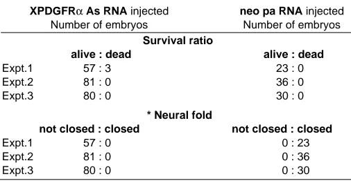
TABLE 2
DEFECTS IN NEURAL FOLD CLOSURE BY THE INJECTION OF XPDGFR ANTISENSE RNA
XPDGFRα As RNA injected neo pa RNA injected
Number of embryos Number of embryos
Survival ratio
alive : dead alive : dead
Expt.1 57 : 3 23 : 0
Expt.2 81 : 0 36 : 0
Expt.3 80 : 0 30 : 0
* Neural fold
not closed : closed not closed : closed
Expt.1 57 : 0 0 : 23
Expt.2 81 : 0 0 : 36
Expt.3 80 : 0 0 : 30
Embryos were injected with 0.7ng (expt. 1, 2, 3) of XPDGFRα antisense RNA at 2cell stage and scored at the time when control embryos reached stage 40.
* In that experiment, the embryos injected with Myf5 antisense used as injection control showed the same result that was identical with control embyo.
References
AMAYA, E., MUSCI, T.J. and KISCHNER, M.W. (1991). Expression of a dominant negative mutant of the FGF receptor disrupts mesoderm formation in Xenopus embryos. Cell 66: 257-270.
ATALIOS, P., SYMES, K., CHOU, M.M., HO, L. and MERCOLA, M. (1995). PDGF signaling is required for gastrulation of Xenopus laevis. Development 121: 3093-3110.
BLENIS, J. (1993). Signal Transduction via the MAP kinases: Proceed at your own RSK. Proc. Natl. Acad. Sci. USA 90: 5889-5892.
BOWEN-POPE, D.F. and SEIFERT, R.A. (1994). Platelet-derived growth factor in development. In Growth factors and signal transduction in development. Wiley-Liss, Inc, pp. 51-73.
CLAESSON-WELSH, L. (1994). Platelet-derived growth factor receptor signals. J. Biol. Chem. 269: 32023-32026.
CORNELL, R.A and KIMELMAN, D. (1994). Activin-mediated mesoderm induction requires FGF. Development 120: 453-462.
CHAMBERS, A.E., KOTECHA, S., TOWERS, N. and MOHUN, T.J. (1992). Muscle-specific expression of SRF-related genes in the early embryo of Xenopus laevis. EMBO J. 11: 4981-4991.
CHAMBERS, A.E., LOGANM, M., KOTECHA, S., TOWERS, N., SPARROW D., and MOHUN, T.J. (1994). The RSRF/MEF2 protein SL1 regulates cardiac muscle-specific transcription of a myosin light-chain gene in Xenopus embryos. Genes Dev. 8: 1324-1334.
CHRISTIAN, J., OLSON, D.J. and MOON, R. (1992). Xwnt-8 modifies the character of mesoderm induced by FGF in isolated Xenopus ectoderm. EMBO J.11: 33-41.
CHRISTIAN, J.L., MCMAHON, J.A., MCMAHON, A.P. and MOON, R.T. (1991). Xwnt-8, a Xenopus Wnt-1/int-1-related gene responsive to mesoderm inducing factors may play a role in ventral mesodermal patterning during embryogenesis. Development 111: 1045-1056.
DE ROBERTIS, E.M., BLUM, M., NIEHRS, C. and STEINBEISSER, H. (1992). Goosecoid and the organizer. Development 116 (Suppl.): 167-171.
GOTOH, Y., MASUYAMA, N., SUZUKI, A., UNEO, N. and NISHIDA, E. (1995). Involvement of the MAP kinase cascade in Xenopus mesoderm induction. EMBO J. 14: 2491-2498.
GRAHAM, K.M., SCHATTEMAN, G.C., BORK. T., BOWEN-POPE, D.F. and WESTON, J.A. (1992). A PDGF receptor mutation in the mouse (patch) perturbs the development of a non-neuronal subset of neuronal crest-derived cells. Develop-ment 115: 133-142.
GURDON, J.B., KAO, K., KATO, K. and HOPWOOD, N.D. (1992). Muscle gene activation in Xenopus requires intercellular communication during gastrula as well as blastula stages. Development (Suppl.): 137-142.
HARLAND, R.M. (1991). In situ hybridization: an improved whole mount method for Xenopus embryos. Methods Cell Biol. 36: 685-695.
HEASMAN, J. (1997). Patterning the Xenopus blastula. Development 124: 4179-4191.
HEMMATI-BRIVANLOU, A., KELLY, O.G. and MELTON, D.A. (1992). A truncated activin receptor dominantly inhibits mesoderm induction and formation of axial structures in Xenopus embryos. Nature 359: 609-614.
HO, L., SYMES, K., YORDAN, C., GUDAS, L.J. and MERCOLA, M. (1994). Localiza-tion of PDGFRα mRNA in Xenopus embryos suggests signaling from neural ectoderm and pharyngeal endoderm to neural crest cells. Mech. Dev. 48: 165-174.
HOPWOOD, N.D., PLUCK, A. and GURDON, J.B. (1989). MyoD expression in the forming somies is an early response to mesoderm induction in Xenopus embryos. EMBO J. 8: 3409-3417.
HOPWOOD, N.D., PLUCK, A. and GURDON, J.B. (1991). Xenopus Myf5 marks early muscle cells and can activate muscl genes ectopically in early embryos. Develop-ment 111: 551-560.
HOPWOOD, N.D., PLUCK, A., GURDON, J.B. and DILWORTH, S.M. (1992). Expression of XMyoD protein in early Xenopus laevis embryos. Development 114: 331-338.
JENNINGS, C.G.B. (1992). Expression of the myogenic gene MRF4 during Xenopus development. Dev. Biol. 150: 121-132.
demonstration that mesoderm induction establishes the lineage-specific pattern of ligand and receptor expresssion. Dev. Genet. 14:185-193.
KESSLER, D.S. and MELTON, D.A. (1994). Vertebrate embryonic induction: Meso-dermal and neural pattering. Science 266: 596-604.
KIMELMAN, D. and KIRSCHNER, M. (1987). Synergistic induction of mesoderm by FGF and TGF-β and the identification of an mRNA coding for FGF in the early Xenopus embryo. Cell 51: 869-877.
KRIEG, P.A., VARNUM, S.M., WORMINGTON, W.M. and MELTON, D.A. (1989). The mRNA encoding elongation factor1-α (Ef1-α) is a major transcript at the midblastula transition in Xenopus. Dev. Biol. 133: 93-100.
LABONNE, C., BURKE, B. and WHITMANN, M. (1995). Role of MAP kinase in mesoderm induction and axial patterning during Xenopus development. Develop-ment 121: 1475-1486.
LABONNE, C., BURKE, B. and WHITMANN, M. (1997). Localization of MAP kinase activity in early Xenopus embryos: Implications for endogenous FGF signaling. Dev. Biol. 183: 9-20.
LAMB, T.M., KNECHT, A.K., SMITH, W.C., STACHEL, S.E., ECONOMIDES, A.N., STAHL, N., YANCOPOLOUS, G.D. and HARLAND, R.M. (1993). Neural indution by the secreted polypeptide noggin. Science 262: 713-718.
LEOF, E.B., PROPER J.A., GOUSTIN, A.S., SHIPLEY, G.D., DICORLETP, P.E. and MOSES, H.L. (1985). Induction of c-sis mRNA and platelet-derived growth factor-like material by transforming growth factor; type-β: a proposed model for indirect mitogenesis involving autocrine activity. Proc. Natl. Acad. Sci. USA 85: 1524-1528.
MATZUK, M.M., KUMAR, T.R., VASSALL, A., BICKEN BACH, J.R., ROOP, D.R., JAENISCH, R. and BRADLEY, A. (1995). Functional analysis of activins in mammalian development. Nature 374: 354-356.
MOHUN, T.J., BRENNAN, S., DATHAN, N., FAIRMAN, S. and GURDON, J.D. (1986). Upstream sequences required for tissue-specific activation of the cardiac actin gene in Xenopus laevis embryos. EMBO J. 12: 3185-3193.
NIEUWKOOP, P.D. and FABER, J. (1967). Normal table of Xenopus laevis (Dandin), Amsterdam: North Holland.
O’REILLY, M.A.J., SMITH, J.C. and CUNLIFFE, V. (1995). Patterning of the meso-derm in Xenopus: dose-dependent and synergistic effects of Brachyury and Pintallavis. Development 121: 1351-1359.
ORR-URTREGER, A. and LONAI, P. (1992). Platelet-derived growth factor-A and its receptor are expressed in separate, but adjacent cell layers of the mouse embryo. Development 115: 1045-1058.
ROSE, L. and BUSA, W.B. (1998). Crosstalk betwwen the phosphatidylinositol cycle and MAP kinase signaling pathways in Xenopus mesoderm induction. Dev. Growth Differ. 40: 231-241.
SASAI, Y., LU, B., STEINBEISSER, H., GEISSERT, D., GONT, L.K. and DE ROBERTIS. E. (1994). Xenopus Chordin: a novel dorsalizing factor activated by organizer-specific homeobox genes. Cell 79: 779-790.
SCHATTEMAN, G.C., GRAHAM K.M., KOPPEN, A., WESTON, J.A. and BOWEN-POPE, D.F. (1992). Regulation and role of receptor α-subunit expression during embryogenesis. Development 115: 123-131.
SIVE, H.L., (1993). The frog prince-ss; a molecular formula for dorsoventral patterning in Xenopus. Genes Dev. 7: 1-12.
SMITH, W.C., KNECHT, A.K., WA, M. and HARLAND, R.M. (1993). Secreted noggin protein mimics the Spemann organizer in dorsalizing Xenopus mesoderm. Nature 361: 547-549.
SORIANO, P. (1997). The PDGFα receptor is required for neural crest cell develop-ment and for normal patterning of the somites. Developdevelop-ment 115: 1045-1058.
ULLRICH, A. and SCHLESSINGER, J. (1990). Signal transduction by receptors with tyrosine kinase activity. Cell 61: 203-212.
WAGNER, B.J. and COCHRAN, B.H. (1993). Growth factors: The PDGF paradigm. Molecular Genetics of Nervous System Tumors. Wiley-Liss, Inc 101-121.
WHITMANN, M. and MELTON, D.A. (1992). Involvement of p21ras in Xenopus mesoderm induction. Nature 357: 252-254.
WONG, M.W., PISEGNA, M., LU, M.F., LEIBHAM, D. and PERRY, M. (1994). Activation of Xenopus MyoD transcription by members of the MEF2 protein family. Dev. Biol. 166: 683-695.
WOODLAND, H.R. (1989). Mesoderm formation in Xenopus. Cell 59: 767-770.
YAMADA, T. and MODAK, S.P. (1998). Genetic evidence for posterior specification by convergent extension in the Xenopus embryo. Dev. Growth Differ. 40: 125-132.