A tight control over Wnt action
MIRANDA MOLENAAR and OLIVIER DESTRÉE*
Hubrecht Laboratory, Netherlands Institute for Developmental Biology, Utrecht, The Netherlands
ABSTRACT Here, we review the WNT pathway and its regulation at different levels. We focus on the transcriptional regulation of WNT target genes, in light of the recently identified negative regulators, i.e. relatives of groucho and CBP.
KEY WORDS:
WNT,
β
-Cat, TCF, groucho related gene
0214-6282/99/$15.00
© UBC Press Printed in Spain
www.ehu.es/ijdb
*Address for reprints: Hubrecht Laboratory, Netherlands Institute for Developmental Biology, Uppsalalaan 8, 3584 CT Utrecht, The Netherlands. FAX: +31 30 2516464. e-mail: [email protected]
Abbreviations used in this paper: wg, wingless; WNT, Wg/Wnt-1; Fz, Frizzled; CRD, Cysteine Rich Domain; Dsh, Dishevelled; GSK-3, Glycogen Synthase Kinase 3; β -Cat, β -Catenin; APC, Adenomatous Polyposis Coli; GBP, GSK-3 Binding Protein; TCF, T Cell Factor; β -TrCP, β -Transducin repeat Containing Protein; HMG, High Mobility Group; LEF, Lymphoid Enhancer Factor; Pan, Pangolin; Pop-1, Posterior Pharynx defective 1; Ubx, Ultrabithorax; Sia, Siamois; CREB, cAMP Responsive Element Binding; CBP, CREB Binding Protein; Grg, Groucho related gene; TLE, Transducin-like Enhancer of split; AES, Amino Enhancer of split; nr-3, nodal related 3.
Wg/Wnt gene family
In 1980, Nüsslein-Volhard and Wieschaus reported a Droso-phila null mutation of the gene wingless (Wg), first identified as a weak mutation disrupting wing patterning (Sharma and Chopra, 1976; Nüsslein-Volhard and Wieschaus, 1980). The null muta-tion leads to embryonic lethality and severe patterning defects.
A few years later, Nusse and Varmus discovered that inappro-priate activation of the Int-1 gene could induce tumours in the murine mammary gland. Int-1 turned out to be the orthologue of Wg and was later on called Wnt-1 (Nusse and Varmus, 1982; Rijsewijk et al., 1987). Wg/Wnt-1 relatives now form a multigene family with at least 20 different members identified in man, mouse, chicken, Xenopus laevis, Drosophila and C. elegans (for overview and references, see the Wnt webpage (http://www-leland.edu/~rnusse/wntwindow.html). In vertebrates, Wnt genes are expressed largely in the nervous system and mesoderm derivatives, and appear to be essential players in embryogenesis and carcinogenesis. Wnts can be divided into two main classes, the Wg/Wnt-1 (WNT) and the Wnt-5A class, based on their different functions and downstream signalling pathways (re-viewed in Moon et al., 1997).
Based on the epistatic interactions known in Drosophila, several vertebrate orthologues for the different components of the Wg signalling pathway were identified and assayed for their functional interactions. A number of recent reviews have dis-cussed the WNT signalling pathway (Miller and Moon, 1996; Cadigan and Nusse, 1997; Cavallo et al., 1997; Clevers and van de Wetering, 1997; Han, 1997; Dale, 1998). Here, we will focus on the recently discovered genes that regulate transactivation of WNT target genes. A schematic view of the WNT pathway is shown in Figure 1.
WNT perception
Genes of the Wnt family encode secreted glycoproteins, which probably act as ligands that bind to specific receptors. The
candidate receptors for WNTs belong to the family of Frizzled (Fz) proteins (Bhanot et al., 1996). Frizzled genes encode seven transmembrane proteins with an extracellular cysteine-rich do-main (CRD), which is responsible for interaction with the WNT ligand. Once secreted, the availability of WNTs for their receptor is regulated. First, glycosaminoglycans are able to enhance WNT activity, possibly by regulating the level of reactive protein and/or adjusting the affinity for Fz (reviewed in Cumberledge and Reichsman, 1997). Second, there is a competition for interaction with WNT between Fz receptors and Frzb’s. The Frzb genes encode a CRD, which is related to the extracellular domain in Fz receptors as well as a short stretch of charged residues. For instance, Xenopus Frzb-1 binds XWnt-8, preventing it from inter-acting with its receptor and antagonising its ventralising effect (Leyns et al., 1997; Wang et al., 1997).
Regulation of cytosolic
β
-catenin levels
Once WNT molecules have bound to their receptors, the cytosolic phosphoprotein Dsh becomes activated, which, in its turn, leads to inactivation of GSK-3 β. Inhibition of GSK-3 leads to elevated levels of cytosolic β-Cat. GSK-3 inhibits the WNT path-way, by phosphorylating N-terminal β-Cat residues, directing β -Cat towards the degradation pathway.
known as conductin) and GBP. APC contains a β-Cat as well as a GSK-3 binding domain.
APC functions as a tumour suppressor (Polakis, 1997). Colorectal carcinoma cell lines containing mutant APC showed elevated levels of β-Cat and constitutively activated a synthetic TCF reporter (Munemitsu et al., 1995; Korinek et al., 1997). Overexpression of wt APC in these cell lines significantly reduced the level of free β-Cat and suppressed the transactivation of the reporter. Overexpressing wt APC in Xenopus embryos and dis-ruption of the function of an APC-related gene in C. elegans produced a phenotype as expected for a positive regulator of the WNT pathway (Rocheleau et al., 1997; Vleminckx et al., 1997). Recently, APC2 has been identified in mammals and flies (Nakagawa et al., 1998; van Es et al., 1999). APC2 resembles APC in its structure, and can downregulate the level of free β-Cat both in APC-/- colon carcinoma cells (van Es et al., 1999) and in
the early Xenopus embryo (unpublished results).
Besides a role in transcriptional activation, β-Cat has a function in cell adhesion. A large proportion of the total content of β-Cat in a cell is associated with cadherin, a molecule that has a role in establishing cell-cell contacts. The rest of the β-Cat pool is captured in a multimolecular complex with APC, GSK-3 and the scaffold protein axin (Zeng et al., 1997; Behrens et al., 1998; Hart et al., 1998; Itoh et al., 1998; Hamada et al., 1999). Without axin, the complex cannot form; β-Cat is no longer phosphorylated and therefore stabilised, which can lead to e.g., the formation of additional axes in mice (Zeng et al., 1997).
To date, only one molecule has been described, that can directly inhibit the action of GSK-3 and that is GSK-3 binding protein (GBP) (Yost et al., 1998). GBP is a maternal protein that inhibits the in vivo phosphorylation by GSK-3. Ectopic expression of GBP caused stabilisation of β-Cat leading to the induction of an ectopic axis in Xenopus embryos. Depletion of maternal GBP
mRNA demonstrated that GBP is required for endogenous axis formation (Yost et al., 1998).
Recently, another regulator of cytosolic levels of β-Cat was described, both in Drosophila and in Xenopus. Loss of function of the Drosophila gene slimb and its vertebrate orthologue β-TrCP resulted in a cell-autonomous accumulation of high levels of β-Cat and the expression of ectopic WNT-responsive genes (Jiang and Struhl, 1998; Marikawa and Elinson, 1998). Slimb and β-TrCP genes encode a conserved F-box/WD-40 repeat protein related to yeast cdc4, a protein that targets cell-cycle regulators for degra-dation by the ubiquitin/proteosome pathway. Overexpression studies of β-TrCP constructs in Xenopus embryos, placed β-TrCP at the level of GSK-3, facilitating the degradation of β-Cat (Marikawa and Elinson, 1998).
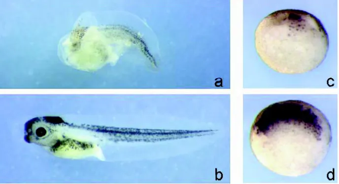
Fig. 2. ∆N-XTcf-3 suppresses, while β-Cat induces axis formation in Xenopus embryos. Injection of mRNA in 4-cell stage embryos. LacZ mRNA was co-injected to trace the cells that received the mRNA. At stage 25, the embryos were fixed and stained for β-galactosidase activity. (a)
Injection of ∆N-XTcf-3 into two dorsal blastomeres caused complete ventralisation of the embryo. (b) Injection of LacZ mRNA alone has no phenotypic effect. (c) Injection of β-Cat into one ventral blastomere caused axis duplication.
Fig. 1. Model for WNT signalling. (a) When no WNT signal is perceived, GSK-3 phosphorylates cytosolic β-Cat. Phosphorylated β-Cat is degraded via the ubiquitination pathway. In the nucleus, WNT target genes are repressed. (b) When WNT binds the receptor, GSK-3 is inhibited, cytosolic
Interaction of
β
-Cat with TCF
When a WNT signal causes elevation of cytosolic β-Cat, β-Cat is transported to and accumulates in the nucleus. We and others found that β-Cat interacts with HMG box transcription factors of the TCF/LEF family to activate transcription of WNT target genes (Behrens et al., 1996; Huber et al., 1996; Molenaar et al., 1996). The genes of the Tcf/ Lef family encode four different proteins: TCF-1, LEF-1, TCF-3 and TCF-4 (TCF). TCF factors were originally identified as lym-phoid-specific enhancers and were later on shown to be present in many different tis-sues during murine embryogenesis. Al-though Tcf genes encode DNA binding pro-teins, when transfected together with a re-porter gene, they fail to activate transcrip-tion. In Xenopus, the maternally expressed XTcf-3 was found to act directly downstream of β-Cat in embryonic axis specification (Molenaar et al., 1996). Transcriptional acti-vation of reporter genes containing
recogni-target genes of WNTs began. The previously identified consen-sus motif for TCF binding (van de Wetering et al., 1997) is found in a large number of genes. Studies in Drosophila and Xenopus identified several potential WNT target genes with functional TCF sites in their promoter region. In Drosophila, a minimal wingless response element in the midgut enhancer of Ultrabithorax (Ubx) is recognised by Lef-1. In complex with ARM, Lef-1 can stimulate transcription of this Ubx enhancer (Riese et al., 1997).
In Xenopus, one of the events triggered by the WNT pathway is dorsal mesoderm induction. Directly after mid blastula transi-tion (MBT), a number of genes are induced that are responsible for the specification of dorsal mesoderm. In the promoter regions of three of these genes, Siamois, Twin and nodal-related-3 (Lemaire et al., 1995; Smith et al., 1995; Laurent et al., 1997), functional binding sites for TCF factors were identified (Brannon et al., 1997; Laurent et al., 1997; McKendry et al., 1997; Fan et al., 1998). The XSia promoter, for example, contains three TCF sites capable of regulating its transcription (Brannon et al., 1997). Mutation of these sites eliminated the β-Cat/XTcf-3 activation of a reporter. The promoter was much more active in the dorsal than in ventral blastomeres. Ventral expression of β-Cat eliminates this difference in transcriptional activity, dependent on the presence of functional TCF/LEF sites. Interestingly, mutating the TCF sites elevated the ventral expression of the reporter gene, compared to the non mutated version (Brannon et al., 1997). This derepression by mutating TCF sites, in the absence of β-Cat, was also shown for the Ubx enhancer (Riese et al., 1997). Thus TCF factors can mediate both repression and activation of the same promoter.
TCF as a repressor of transcription
Recently, binding partners have been identified, both in Droso-phila and Xenopus, which are proposed to be responsible for the repressive effects of TCF factors. Waltzer and Bienz (1998) reported that a Drosophila CREB binding protein (dCBP) inter-acted with a region in the HMG box of dTCF. In the midgut, dCBP
Fig. 3. XGrg-4 suppresses Xnr-3 expression and axis formation in Xenopus embryos. Injection of XGrg-4 mRNA into the two dorsal blastomeres at 4-cell stage resulted in suppression of the endogenous axis (a). (b) A non-injected sibling of (a). Whole-mount in situ hybridisation with anti-sense Xnr-3 RNA of stage 9 embryos showed a suppressed expression in embryos injected dorsally with XGrg-4 (c) when compared to non-injected controls (d).
tion sites for TCF, depended on complex formation between XTcf-3 and β-Cat. Deletion of the N-terminus of XTcf-3 abrogated formation of this complex. This dominant negative ∆NXTcf-3, inhibited the activation of transcription mediated by the wt β-Cat/ XTcf-3 complex, resulting in suppression of axis formation in Xenopus embryos (Fig. 2). To date, XTcf-3 is the only family member found to be expressed maternally (Molenaar et al., 1998). Similar results, obtained by loss of function genetics, con-firmed TCF to be downstream of β-Cat in the WNT pathway. Null mutations of Drosophila dTcf, also named pangolin (pan), showed a wg-like segment polarity phenotype, indicating that dTCF is genetically downstream of Armadillo (ARM) (Brunner et al., 1997; van de Wetering et al., 1997). Pop-1, the TCF orthologue in C. elegans, is involved in WNT dependent asymmetric cell division of the EMS blastomere. However, unlike dTcf in Drosophila, pop-1 has the opposite phenotype to that of the WNT components of the mom class (Lin et al., 1995; Rocheleau et al., 1997; Thorpe et al., 1997). A possible solution to this seeming controversy in function of TCF will be described below.
In the absence of β-Cat, TCF may occupy the regulatory sites of target genes, but cannot activate transcription. The presence of β-Cat in a complex with TCF at the same sites does induce transactivation. Since TCF itself is transcriptionally inert, β-Cat must be the activator. Van de Wetering et al. (1997) showed that the C-terminus of β-Cat by itself can act as a transactivation domain, when fused to a GAL-4 DNA binding domain. Moreover, the fusion protein ARM-XTcf-3, a chimera of the C-terminus of ARM and ∆NXTcf-3, was able to activate transcription and induce an ectopic axis in Xenopus embryos (Roose et al., 1998). This indicates that the C-terminus of β-Cat is necessary to activate transcription of WNT target genes.
WNT target genes
loss of function mutants mimic the wg gain of function phenotype implying dCBP to be a negative regulator of the WNT pathway. dCBP repressed the Ubx midgut enhancer in a dTCF site depen-dent manner. Acetylation of dTCF by dCBP lowered the binding affinity of dTCF to ARM in vitro. The authors proposed that high concentrations of ARM overcome this acetylation block of dTCF and predicted that a balance between ARM and dCBP is particu-larly critical in cells near a low signalling threshold.
Another binding partner of TCF was identified performing a yeast two-hybrid screen for proteins interacting with human TCF-1 (Roose et al., TCF-1998). This TCF partner is the product of the murine Groucho-related gene 5 (mGrg-5) and belongs to the Groucho family of transcriptional repressors. Drosophila Groucho is a widely expressed co-repressor, proposed to be involved in numerous developmental processes (Hartley et al., 1988; Paroush et al., 1994; Fisher and Caudy, 1998; Parkhurst, 1998). In verte-brates, multiple homologues of groucho have been identified. These are termed TLE 1-4 in man and mGrg-1, 3, 4 in mouse. mGrg-5 encodes a naturally truncated product containing only the amino-terminal two domains of the long forms of Grg. Using mGrg-5 as a probe, the Xenopus orthologues of Grg-mGrg-5 [XGrg-mGrg-5 or XAES (Choudhury et al., 1997)] and Grg-4 (XGrg-4), were cloned. These Xenopus Grg’s are expressed maternally and throughout embryo-genesis (Molenaar et al., 1999). Both XGrg-4 and 5 interact with XTcf-3 in a region upstream of the HMG box, but downstream of the β-Cat binding domain. In transfection assays, the long form
(XGrg-4) inhibited β-Cat/XTcf-3 induced activation of transcription of a synthetic TCF reporter and the XSia promoter. In contrast, the short version (XGrg-5) enhanced the β-Cat/XTcf-3 induced tran-scriptional activation. Dorsal injection of XGrg-4 into 4-cell stage Xenopus embryos repressed transcription of XSia and Xnr-3 and suppressed formation of the endogenous axis (see Fig. 3). Ectopic axis formation, induced by a dominant positive ARM-XTcf-3 fusion protein, was inhibited by XGrg-4 and enhanced by XGrg-5.
The functional relevance of this interaction was also shown for Drosophila Tcf and groucho (Cavallo et al., 1998). dTcf and Groucho physically interact. A reduction in both dTcf and Groucho expression caused a suppression of the wg and arm segment polarity phenotype. Hence, dTcf in complex with Groucho acts as a repressor of transcription, in the absence of ARM.
Therefore, the actual transcription of TCF target genes de-pends on the balance between the constitutive repressive effects, mediated by long forms of Grg’s, possibly counteracted by short forms of Grg’s, and the activating effects of β-Cat. This dual function of TCF may also explain the observation that the C. elegans homologue, Pop-1, has opposite effects to that of WNT. Pop-1 possibly functions as a repressor of transcription in the asymmetric cell division of the EMS blastomere.
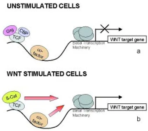
The dual role of TCF
The action of TCF factors in transactivation of WNT target genes is illustrated in the model shown in Figure 4. In unstimulated cells, TCF will bind the promoter of WNT target genes, together with other cell-type-specific factors, such as activin for the XSia promoter (Crease et al., 1998) or dpp for the Ubx enhancer (Riese et al., 1997). Binding of TCF in the absence of β-Cat allows repressors, like Grg or CBP, to interact with TCF and prevent transcription. Following WNT signalling, β-Cat translocates to the nucleus and associates with TCF, thereby activating transcription of WNT target genes, and with the help of short forms of Grg, β -Cat counteracts repression.
The combined repression and activation through TCF secures a tight control over WNT driven developmental decisions. At this point, it is important to learn what signals put the repressors in place, what the molecular events underlying derepression are and which molecules activate and repress the transcriptional machinery.
Acknowledgements
We thank H. Clevers, R. Morgan and members of the Destrée labo-ratory for carefully reading the manuscript.
References
BEHRENS, J., VON KRIES, J.P., KUHL, M., BRUHN, L., WEDLICH, D., GROSSCHEDL, R. and BIRCHMEIER, W. (1996). Functional interaction of β– catenin with the transcription factor LEF-1. Nature 382: 638-642.
BERHRENS, J., JERCHOW, B.A., WURTELE, M., GRIMM, J., ASBRAND, C., WIRTZ, R., KUHL, M., WEDLICH, D. and BIRCHMEIER, W. (1998). Functional interaction of an axin homolog, conductin, with β-Catenin, apc, and gsk3β. Science 280: 596-599.
BHANOT, P.M., BRINK, M., HARRYMAN-SAMOS, C., HSIEH, J.C., WANG, Y.S., MACKE, J.P., ANDREW, D., NATHANS, J. and NUSSE, R. (1996). A new member of the frizzled family from Drosophila functions as a wingless receptor. Nature 382: 225-230.
Fig. 4. Model for the action of TCF factors on WNT target genes.
BIENZ, M. (1998). TCF: transcriptional activator or repressor? Curr. Opin. Cell Biol. 10: 366-372.
BRANNON, M., GOMPERTS, M., SUMOY, L., MOON, R.T. and KIMELMAN, D. (1997). A β-Catenin/XTcf-3 complex binds to the siamois promoter to regulate dorsal axis specification in Xenopus. Genes Dev. 11: 2359-2370.
BRUNNER, E., PETER, O., SCHWEIZER, L. and BASLER, K. (1997). Pangolin encodes a Lef-1 homologue that acts downstream of armadillo to transduce the wingless signal in Drosophila. Nature 385: 829-833.
CADIGAN, K.M. and NUSSE, R. (1997) Wnt signalling: a common theme in animal development. Genes Dev. 11: 3286-3305.
CAVALLO, R.A., COX, R.T., MOLINE, M.M., ROOSE, J., POLEVOY, G.A., CLEVERS, H., PEIFER, M. and BEJSOVEC, A. (1998). Drosophila Tcf and Groucho interact to repress Wingless signalling activity. Nature 395: 604-608. CAVALLO, R., RUBENSTEIN, D. and PEIFER, M. (1997). Armadillo and dTCF: a
marriage made in the nucleus. Curr. Opin. Genet. Dev. 7: 459-466.
CHOUDHURY, B.K., KIM, J., KUNG, H.-F. and LI, S.S.-L. (1997). Cloning and developmental expression of Xenopus cDNAs encoding the Enhancer of split groucho and related proteins. Gene 195: 41-48.
CLEVERS, H.C. and VAN DE WETERING, M. (1997). TCF/LEF factors earn their wings. Trends Genet. 13: 485-489.
CREASE, D.J., DYSON, S. and GURDON, J.B. (1998). Cooperation between the activin and Wnt pathways in the spatial control of organizer gene expression. Proc. Natl. Acad. Sci. USA 95: 4398-4403.
CUMBERLEDGE, S. and REICHSMAN, F. (1997). Glycosaminoglycans and WNTs: just a spoonful of sugar helps the signal go down. Trends Genet. 13: 421-423.
DALE, T.C. (1998). Signal transduction by the Wnt family of ligands. Biochem. J. 329: 209-223.
FAN, M.J., GRÜNING, W., WALZ, G. and SOKOL, S.Y. (1998). Wnt signaling and transcriptional control of Siamois in Xenopus embryos. Proc. Natl. Acad. Sci. USA 95: 5626-5631.
FISHER, A.L. and CAUDY, M. (1998). Groucho proteins: transcriptional corepres-sors for specific subsets of DNA-binding transcription factors in vertebrates and invertebrates. Genes Dev. 12: 1931-1940.
HAMADA, F., TOMOYASU, Y., NAKAMURA, M., NAGAI, S., SUZUKI, A., FUJITA, F., SHIBUYA, H., TOYOSHIMA, K., UENO, N. and AKIYAMA, T. (1999). Negative regulation of wingless signaling by D-Axin, a Drosophila homolog of Axin. Science 283: 1739-1742.
HAN, M. (1997). Gut reaction to Wnt signalling in worms. Cell 90: 581-584.
HART, M.J., DE LOS SANTOS, R., ALBERT, I.N., RUBINFELD, B. and POLAKIS, P. (1998). Downregulation of β-Catenin by human Axin and its association with the APC tumour supressor, β-Catenin and GSK-3β. Curr. Biol. 8: 573-581.
HARTLEY, D.A., PREISS, A. and ARTAVANIS-TSAKONAS, S. (1988). A deduced gene product from the Drosophila neurogenic locus, enhancer of split, shows homology to mammalian G-protein beta subunit. Cell 55: 785-795.
HUBER, O., KORN, R., MCLAUGHLIN, J., OHSUGI, M., HERRMANN, B.G. and KEMLER, R. (1996). Nuclear localization of β–catenin by interaction with transcription factor LEF-1. Mech. Dev. 59: 3-10.
ITOH, K., KRUPNIK, V.E. and SOKOL, S.Y. (1998). Axis determination in Xenopus involves biochemical interactions of axin, glycogen synthase kinase 3 and β -Catenin. Curr. Biol. 8: 591-594.
JIANG, J. and STRUHL, G. (1998). Regulation of the hedgehog and wingless signalling pathways by the F-box/WD-40 repeat protein slimb. Nature 391: 493-496.
KORINEK, V., BARKER, N., MORIN, P.J., VAN WICHEN, D., DE WEGER, R., KINZLER, K.W., VOGELSTEIN, B. and CLEVERS, H.C. (1997). Constitutive transcriptional activation by a β-Catenin-Tcf complex in APC(-\-) colon carci-noma. Science 275: 1784-1787.
LAURENT, M.N., BLITZ, I.L., HASIMOTO, C., ROTHBACHER, U. and CHO, K.W.-Y. (1997). The Xenopus homeobox gene Twin mediates Wnt induction of goosecoid in establishment of Spemann’s organizer. Development 124: 4905-4916.
LEMAIRE, P., GARRETT, N. and GURDON, J.B. (1995). Expression cloning of Siamois, a Xenopus homeobox gene expresses in dorsal-vegetal cells of blastulae and able to induce a complete secondary axis. Cell 81: 85-94.
LEYNS, L., BOUWMEESTER, T., KIM, S.H., PICCOLO, S. and DE ROBERTIS, E.M. (1997). Frzb-1 is a secreted antagonist of Wnt signalling expressed in the Spemann organizer. Cell 88: 747-756.
LIN, R., THOMPSON, S. and PRIESS, J.R. (1995). Pop-1 encodes an HMG box protein required for the specification of a mesoderm precursor in early C. elegans embryos. Cell 83: 599-609.
MARIKAWA, Y. and ELINSON, R.P. (1998). β-TrCP is a negative regulator of the Wnt/β-Catenin signalling pathway and dorsal axis formation in Xenopus em-bryos. Mech. Dev. 77: 75-80.
MCKENDRY, R., HSU, S.-C., HARLAND, R.M. and GROSSCHEDL, R. (1997). LEF-1/TCF proteins mediate Wnt-inducible transcription from the Xenopus nodal-related 3 promoter. Dev. Biol. 192: 420-431.
MILLER, J.R. and MOON, R.T. (1996). Signal transduction through β–catenin and specification of cell fate during embryogenesis. Genes Dev. 10: 2527-2539.
MOLENAAR, M., BRIAN, E., ROOSE, J., CLEVERS, H. and DESIREE, O. (1999). Differential expression of the Groucho-related genes 4 and 5 early development of Xenopus laevis. Mech. Dev. (In press).
MOLENAAR, M., ROOSE, J., PETERSON, J., VENANZI, S., CLEVERS, H. and DESTRÉE, O. (1998). Differential expression of the HMG box transcription factors XTcf-3 and XLef-1 during early Xenopus development. Mech. Dev. 75: 151-154.
MOLENAAR, M., VAN DE WETERING, M., OOSTERWEGEL, M., PETERSON-MADURO, J., GODSAVE, S., KORINEK, V., ROOSE, J., DESTREE, O. and CLEVERS, H. (1996). XTcf-3 transcription factor mediates β–catenin-induced axis formation in Xenopus embryos. Cell 86: 391-399.
MOON, R.T., BROWN, J.D. and TORRES, M. (1997). WNTs modulate cell fate and behavior during vertebrate development. Trends Genet. 13: 157-162.
MUNEMITSU, S., ALBERT, I., SOUZA, B., RUBINFELD, B. and POLAKIS, P. (1995). Regulation of intracellular β-Catenin levels by the adenomatous polypo-sis coli (APC) tumour-supressor protein. Proc. Natl. Acad. Sci.USA 92: 3046-3050.
NAKAGAWA, H., MURATA, Y., KOYAMA, K., FUJIYAMA, A., MIYOSHI, Y., MONDEN, M., AKIYAMA, T. and NAKAMURA, Y. (1998). Identification of a brain-specific APC homologue, APCL, and its interaction with β-Catenin. Cancer Res. 58: 5176-5181.
NUSSE, R. and VARMUS, H.E. (1982). Many tumors induced by the Mouse Mammary Tumour Virus contain a provirus integrated in the same region of the host genome. Cell 31: 99-109.
NÜSSLEIN-VOLHARD, C. and WIESCHAUS, E. (1980). Mutations affecting seg-ment number and polarity in Drosophila. Nature 287: 795-801.
PARKHURST, S.M. (1998). Groucho: making its Marx as a transcriptional co-repressor. Trends Genet. 14: 130-132.
PAROUSH, Z., FINLEY JR., R.L., KIDD, T., WAINWRIGHT, S.M., INGHAM, P.W., BRENT, R. and ISH-HOROWITZ, D. (1994). Groucho is required for Drosophila neurogenesis, segmentation, and sex determination and interacts directly with hairy related bHLH proteins. Cell 79: 805-815.
POLAKIS, P. (1997). The adenomatous polyposis coli (APC) tumor suppressor. Biochem. Biophys. Acta 1332: F127-F147.
RIESE, J., YU, X., MUNNERLYN, A., ERESH, S., HSU, S.-C., GROSSCHEDL, R. and BIENZ, M. (1997). LEF-1, a nuclear factor coordinating signalling inputs from wingless and decapentaplegic. Cell 88: 777-787.
RIJSEWIJK, F., SCHUERMANN, M., WAGENAAR, E., PARREN, P., WEIGEL, D. and NUSSE, R. (1987). The Drosophila homolog of the mouse mammary oncogene int-1 is identical to the segment polarity gene wingless. Cell 50: 649-657.
ROCHELEAU, C.E., DOWNS, C.E., LIN, R., WITTMAN, C., BEI, Y., CHA, Y.H., ALI, M., PRIESS, J.R. and MELLO, C.C. (1997). Wnt signalling and an APC-related gene specify endoderm in early C. elegans embryos. Cell 90: 707-716. ROOSE, J., MOLENAAR, M., PETERSON, J., HURENKAMP, J., BRANTJES, H.,
MOERER, P., VAN DE WETERING, M., DESTRÉE, O. and CLEVERS, H. (1998). The Xenopus Wnt effector XTcf-3 interacts with Groucho-related tran-scriptional repressors. Nature 395: 608-612.
SMITH, W.C., MCKENDRY, R., RIBISI, S. and HARLAND, R.M. (1995). A nodal-related gene defines a physical and functional domain within the Spemann organizer. Cell 67: 37-46.
THORPE, C.J., SCHLESINGER, A., CARTER, J.C. and BOWERMAN, B. (1997). Wnt signalling polarizes an early C. elegans blastomere to distinguish endo-derm from mesoendo-derm. Cell 90: 695-705.
VAN DE WETERING, M., CAVALLO, R., DOOIJES, D., VAN BEEST, M., VAN ES, J., LOUREIRO, J., YPMA, A., HURSH, D., JONES, T., BEJSOVEC, A., PEIFER, M., MORTIN, M. and PEIFER, M. (1997). Armadillo coactivates transcription driven by the product of the Drosophila segment polarity gene dTCF. Cell 88: 789-799.
VAN ES, J.H., KIRKPATRICK, C., VAN DE WETERING, M., MOLENAAR, M., MILES, T., KUIPERS, J., DESTRÉE, O., PEIFER, M. and CLEVERS, H. (1999). Identification of APC2, a homologue of the adenomatous polyposis coli tumour suppressor. Curr. Biol. 9: 105-108.
VLEMINCKX, K., WONG, E., GUGER, K., RUBINFELD, B., POLAKIS, P. and
GUMBINER, B. (1997). Adenomatous polyposis coli tumour supressor protein has signalling activity in Xenopus laevis embryos resulting in the induction of an ectopic dorsoanterior axis. J. Cell Biol. 136: 411-420.
WALTZER, L. and BIENZ, M. (1998). Drosophila CBP represses the transcription factor TCF to antagonise Wingless signalling. Nature 395: 521-525.
WANG, S., KRINKS, M., LIN, K., LUYTEN, F.P. and MOOS JR., M. (1997). Frzb, a secreted protein expressed in the Spemann organizer, binds and inhibits Wnt-8. Cell 88: 757-766.
YOST, C., FARR III, G.H., PIERCE, S.B., FERKEY, D.M., MINGZI CHEN, M. and KIMMELMAN, D. (1998). GBP, an inhibitor of GSK-3, is implicated in Xenopus development and oncogenesis. Cell 93: 1031-1041.