Original Article
Cell lineage analysis of pattern formation in the Tubifex embryo.
II. Segmentation in the ectoderm
AYAKI NAKAMOTO, ASUNA ARAI and TAKASHI SHIMIZU*
Division of Biological Sciences, Graduate School of Science, Hokkaido University, Sapporo, Japan
ABSTRACT Ectodermal segmentation in the oligochaete annelid Tubifex is a process of separation of 50-µm-wide blocks of cells from the initially continuous ectodermal germ band (GB), a cell sheet consisting of four bandlets of blast cells derived from ectoteloblasts (N, O, P and Q). In this study, using intracellular lineage tracers, we characterized the morphogenetic processes that give rise to formation of these ectodermal segments. The formation of ectodermal segments began with formation of fissures, first on the ventral side and then on the dorsal side of the GB; the unification of these fissures gave rise to separation of a 50-µm-wide block of ~30 cells from the ectodermal GB. A set of experiments in which individual ectoteloblasts were labeled showed that as development proceeded, an initially linear array of blast cells in each ectodermal bandlet gradually changed its shape and that its contour became indented in a lineage-specific manner. These morphogenetic changes resulted in the formation of distinct cell clumps, which were separated from the bandlet to serve as segmental elements (SEs). SEs in the N and Q lineages were each comprised of clones of two consecutive primary blast cells. In contrast, in the O and P lineages, individual blast cell clones were distributed across SE boundaries; each SE was a mixture of a part of a more anterior clone and a part of the next more posterior clone. Morphogenetic events, including segmentation, in an ectodermal bandlet proceeded normally in the absence of neighboring ectodermal bandlets. Without the underlying mesoderm, separated SEs failed to space themselves at regular intervals along the anteroposterior axis. We suggest that ectodermal segmentation in Tubifex consists of two stages, autonomous morphogenesis of each bandlet leading to generation of SEs and the ensuing mesoderm-dependent alignment of separated SEs.
KEY WORDS: cell
lineage, ectodermal segmentation, Tubifex, germ band, mesodermal control.
0214-6282/2000/$20.00
© UBC Press Printed in Spain www.ehu.es/ijdb
*Address correspondence to: Dr. Takashi Shimizu. Division of Biological Sciences, Graduate School of Science, Hokkaido University, Sapporo 060-0810, Japan. FAX: +81-11-706-4851. e-mail: stak@sci.hokudai.ac.jp
Abbreviations used in this paper: GB, germ band; HRP, horseradish peroxidase; OG, Oregon Green; SE, segmental element.
Introduction
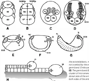
The oligochaete annelids are metamerically segmented ani-mals. The trunk portion of the body comprises multiple segments, each of which contains a similar complement of ectodermal and mesodermal tissues; the head, represented by the prostomium and containing the brain, is not a segment, nor is the terminal part of the body in which the anus is located. The trunk segmentation is visible externally as rings (or annuli) and is reflected internally not only by the serial arrangement of coelomic compartments separated from one another by intersegmental septa but also by the metameric arrangement of organs and system components. Ectodermal and mesodermal segmental structures in the oli-gochaete Tubifex arise from five bilateral pairs of longitudinal, coherent columns (bandlets) of primary blast cells that are gener-ated by five bilateral pairs of embryonic stem cells called teloblasts (M, N, O, P and Q) (Fig. 1C; Shimizu, 1982). Among these, the M lineage contributes solely to mesodermal segments; the
remain-ing lineages give rise to ectodermal segments (Goto et al., 1999b). A previous study has shown that the metameric segmen-tation in the mesoderm arises from an initially simple organization (i.e., a linear series) of primary m blast cells that serve as segmental founder cells (Fig. 1H; Goto et al., 1999a). After their birth, primary m blast cells in the M lineage undergo stereotyped sequences of cell division, and they individually generate a distinct cell cluster, which becomes a mesodermal segment. Cell clusters thus generated come to be arranged in a chain running along the anteroposterior axis. Recently, it has also been sug-gested that segmental identities in primary m blast cells of Tubifex embryos are determined according to the genealogical position in the M lineage (Kitamura and Shimizu, 2000).
1926, 1938; Devries, 1974). These authors performed cell-abla-tion experiments and reported that ablacell-abla-tion of mesoderm aborts segmentation of ectodermal tissues. Similar mesodermal control of ectodermal segmentation has also been suggested for another annelid class, leeches (Blair, 1982; Torrence, 1991). It appears that this control is widespread among annelids. At present, however, almost nothing is known about the role the mesoderm plays in ectodermal segmentation. Furthermore, presently avail-able information about early events leading to segmentation of continuous ectodermal bandlets in clitellate annelids is fragmen-tary.
In this study, we traced the process of ectodermal segmenta-tion in Tubifex embryos using lineage tracers. We also perform experiments to determine whether the early events of ectodermal segmentation occur autonomously.
Results
Summary of the early development of Tubifex
A brief review of the early development in Tubifex is presented here as a background for the observations described below (for details, see Shimizu 1982; Goto et al., 1999a, b). Precursors of teloblasts are traced back to the second (2d) and fourth (4d) micromeres of the D quadrant. At the 22-cell stage, 2d11 (resulting from the unequal divisions of 2d), 4d and 4D (sister cell of 4d) all come to lie in the future midline of the embryo (Fig. 1A). 4d divides equally to yield the left and right mesoteloblasts (Ml and Mr); 2d111
(resulting from the unequal division of 2d11) divides into a bilateral pair of ectoteloblast precursors, NOPQl and NOPQr (Fig. 1B). Ectoteloblasts N, O, P and Q arise from an invariable sequence of divisions of cell NOPQ on both sides of the embryo (Fig. 1C; Goto et al., 1999b).
After their birth, each of the teloblasts thus produced divides repeatedly, at 2.5-hr intervals (at 22°C), to give rise to small cells called primary blast cells, which are arranged into a coherent column (i.e., a bandlet; Fig. 1H). Within each bandlet, primary blast cells and their descendants are arranged in the order of their birth. Bandlets from N, O, P and Q teloblasts on each side of the embryo join together to form an ectodermal germ band (GB), while the bandlet from the M teloblast becomes a mesodermal GB that underlies the ectodermal GB (Figs. 1D and H; Goto et al., 1999a). The GBs are initially located at the dorsal side of the embryo (Fig. 1E). Along with their elongation, they gradually curve round toward the ventral midline and finally coalesce with each other along the ventral midline (Fig. 1F). The coalescence is soon followed by dorsalward expansion of GBs. The edges of the expanding GBs on both sides of the embryo finally meet along the dorsal midline to enclose the yolky endodermal tube (Fig. 1G; Goto et al., 1999a, b).
Segmentation of the ectodermal germ band (GB)
Segmentation of the ectoderm is a process of separation of
50-µm-wide blocks of cells from the initially continuous cell sheet (i.e., an ectodermal GB; Fig. 2A). This separation is mediated by
Fig. 1.Summary of Tubifex development. (A-G) Selected stages of embryonic development. (A, B) Posterior view with dorsal to the top; (C and D) dorsal view with anterior to the top; (E-G) side view with anterior to the left and dorsal to the top. (A) A 22-cell stage embryo. Cells 2d11, 4d and 4D
all come to lie in the future midline. (B) After 2d11 divides
into smaller 2d112 and larger 2d111 (not shown), 4d divides
bilaterally into left and right mesoteloblasts, Ml and Mr. About 2.5 h later, 2d111 divides equally into a bilateral pair of
ectoteloblast precursors, NOPQl and NOPQr. 4D also di-vides into a pair of endodermal precursor cells ED before
2d111 division. (C) An embryo at about 30 h after the bilateral
division of 4d. Only teloblasts are depicted. NOPQ on each side of the embryo has produced ectoteloblasts N, O, P and Q. (D) A two-day-old embryo following the bilateral division of 4d. Only teloblasts and associated structures are de-picted. At this stage, a short ectodermal germ band (EGB) extending from the ectoteloblasts N, O, P and Q is seen on either side of the embryo. A mesodermal germ band (MGB) extending from the M teloblast is located under the ecto-dermal germ band. As the M teloblasts are separated from the ectoteloblasts, mesodermal blast cells located in the vicinity of M teloblasts are not overlaid by the ectodermal germ band. (E-G) Morphogenesis of the ectodermal germ band. Embryos are shown at 2.5 (E), 4 (F) and 6 (G) days after the 4d cell division.
(E) The germ band (EGB) is associated, at its anterior end, with an anteriorly located cluster of micromeres (called a micromere cap; MC), and it is initially located at the dorsal side of the embryo. (F) Along with their elongation, the germ bands (EGB) on both sides of the embryo gradually curve round toward the ventral midline and finally coalesce with each other along the ventral midline. (G) The coalescence is soon followed by dorsalward expansion of the edge of the germ band. Pr, prostomium. (H)
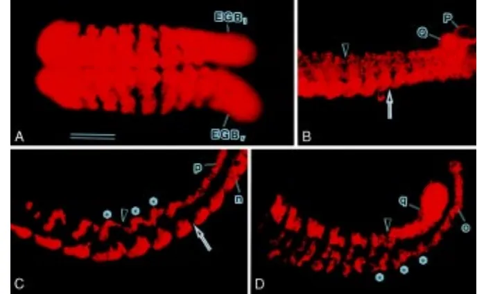
the formation of transverse fissures in the GB. The first sign of fissures emerged on the ventral side of the GB, i.e., the n bandlet, at a distance of about 20 cells from the parent N teloblast (Fig. 2B). A fissure on the dorsal side was formed in a more-anterior region, and it was united with the earlier-formed ventral fissure. As a result of this unification, a distinct clump of cells was completely separated from the remaining GB, and it was established as an ectodermal segment.
It should be noted that ectodermal segments and fissures were recognized only when ectodermal GBs were labeled with lineage tracers. In non-stained living or Hoechst-stained fixed specimens, these segmental features were difficult to be visualized.
Morphogenesis of ectodermal bandlets
To examine more closely the behavior of each ectodermal bandlet during segmentation, we labeled a single or two alternate bandlets of individual GBs with DiI and observed them under an epifluorescence microscope. Fig. 2C shows fluorescent n and p bandlets in an embryo whose left N and P teloblasts had been simultaneously injected with DiI. One of the first morphogenetic events leading to segmentation of n bandlets was the bulging of their dorsal margins, which occurred at a distance of 15-17 cells from the N teloblast (Fig. 3A). The boundaries of adjacent dorsal bulges constricted (Fig. 3A), and a cluster of cells including the bulge was separated from the following bandlet (see arrow in Fig. 2C). Each such cell cluster serves as a segmental element (SE), and SEs were integrated into a single segment.
Each p bandlet undulated to assume a wave-like shape (Figs. 2C and 3C). In other words, p bandlets appeared as a chain of S-shaped units (i.e., SEs; asterisks in Fig. 2C). The first sign of undulation was detected at a distance of 15-17 cells from the P teloblast. p bandlets became thinner at the posterior side of each dorsal “summit” of the wave; finally, the frontmost S-shaped SE
was separated from the remaining bandlet (Fig. 2C). This sepa-ration occurred at a distance of three segments from that in the N lineage (Fig. 2C).
Morphogenetic processes in o bandlets were as dynamic as those in p bandlets (Fig. 3B). Unlike the latter, however, o bandlets appeared as a chain of W-shaped units (i.e., SEs; asterisks in Fig. 2D). The first sign of morphological changes was detected at a distance of 15-17 cells from the O teloblast, and the separation of the frontmost unit from the remaining bandlet occurred at a distance of one segment from that in the N lineage. Morphogenesis of q bandlets that leads to segmentation be-gan with the formation of indentations on the dorsal side, which was first detected at a distance of 15-17 cells from the Q teloblast. Shortly after the formation of indentations, q bandlets bulged out on the ventral side opposite the indentations (Fig. 3D). At the same time, an additional indentation emerged at the “summit” between the adjacent dorsal indentations. As a result of these shape changes, SEs of the Q lineage came to assume an M shape (Fig. 2D). Since the anterior half of each element inherited a ventral bulge, it was dorsoventrally longer than the posterior half. The separation of SEs from the q bandlet occurred at a distance of two segments from that in the N lineage.
The segmentation process of the ectoderm in Tubifex is summarized schematically in Fig. 4A. As development proceeds, an initially linear array of blast cells within each bandlet gradually changes its shape, and its contour becomes indented in a lineage-specific manner, leading to the formation of distinct cell clumps, which serve as SEs. Finally, SEs are separated from the bandlets and become arranged at regular intervals of 50 µm.
Segmental distribution of blast cell progenies
To investigate the spatial relationship between clones of indi-vidual primary blast cells and ectodermal segments, primary blast
Fig. 2.Segmentation in ectodermal GBs. 2d111
cell (A), left NOPQ (B) or individual teloblasts (C, D) were injected with DiI and allowed to develop for 3 days before fixation. Wholemount preparations were viewed from the ventral side (A) or left side (B-D). In all panels, anterior is to the left; in B-D, dorsal is to the top. Bar, 100 µm (A, B); 80 µm (C, D). (A) Both the left and right germ bands (EGBl and EGBr, respectively) are labeled with DiI. Both GBs have coalesced with each other along the ventral midline in the anterior and mid regions of the embryo. Only the mid region of the embryo is in focus here. Note that GBs are divided into 50-µm-wide blocks of labeled cells by intersegmental furrows, which are recognized as non-fluorescent transverse stripes.
(B) The posterior portion of the left GB is shown. P and Q teloblasts are seen, but N and O teloblasts are out of the field. The arrow indicates the site where a fissure becomes evident in the ventralmost bandlet (i.e., n bandlet). The arrowhead indicates fissures at the dorsal side of the GB. (C) Fluorescent n and p bandlets in the left GB. These bandlets were
cells located in the vicinity of their parents were individually injected with DiI (at about 24 h after completion of teloblastogenesis), and the distribution of their progenies was examined at 6 days after DiI labeling. In this study, DiI injection was confined to cells located within a distance of 3 cells from their parents because these cells remained undivided after birth. For each lineage, more than 20 embryos were examined.
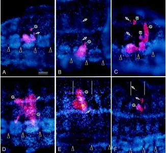
n blast cell clones. Progenies of a single primary n blast cell were found to be confined to a single segment. Two types of their distribution within a segment were discernible (Fig. 5 A,B). In some of the embryos examined, the progeny of a single primary blast cell
was distributed in both the anterior and posterior portions of a segment (Fig. 5A). This cell clone was comprised of ganglionic cells (organized in an anterior larger cluster and a posterior smaller cluster), epidermal cells located near the ventral side, and one peripheral neuron located in the posterior part of the segment (Fig. 5A). In other embryos, progeny cells of a single primary blast cell were largely distributed in the posterior part of a segment, and a few additional cells were also found in the ganglion of the next segment (Fig. 5B). This cell clone gave rise to a cluster of ganglionic cells, epidermal cells localized near the ventral midline, and two periph-eral neurons (Fig. 5B). It should be noted that when combined, clones of these two types generate one segmental complement of progeny for the N lineage (see Goto et al., 1999b).
o blast cell clones. In all of the embryos examined, progeny cells of a single primary o blast cell were found to be distributed in two consecutive segments (Fig. 5C). In each of the embryos, labeled cells in the more-anterior segment were confined to its posterior portion. This portion included a large cluster of gangli-onic cells, two peripheral neurons, and a cluster of three cells whose identity was unknown. In contrast, labeled cells in the next-more-posterior segment were distributed in its anterior and mid portions. The anterior portion included a cluster of ganglionic cells, a peripheral neuron, and a cluster of three cells of unknown identity (Fig. 5C). The central portion was comprised of epidermal cells organized in a large cluster (Fig. 5C). Judging from the abovementioned cellular compositions, it is safe to say that in the O lineage, one primary blast cell generates one segmental complement of progeny.
p blast cell clones. As in primary o blast cell clones, the progeny of a single primary p blast cell was divided into two consecutive segments (Fig. 5D). In each of the embryos examined, the more-anterior segment exhibited three centrally located ganglionic cells, three peripheral neurons in its dorsoposterior region, and dorsal epidermal cells. In the next-more-posterior segment, gan-glionic cells were organized in a relatively large cluster located in the central region of a ganglion. Epidermal cells and four periph-eral neurons were present in the dorsal region of this segment. A cluster of “deep” cells (ventral setal sac) was also observed under the epidermis in the ventral region. This observation suggests that, as in the O lineage, one primary blast cell of the P lineage gives rise to one segmental complement of progeny (see Goto et al., 1999b).
q blast cell clones. Clones of primary q blast cells were similar to n blast cell clones in that progeny cells of a single primary blast cell were confined to one segment and exhibited two types of distribution pattern (Fig. 5 E,F); when combined, the two types generate one segmental complement of progeny. In one type, progenies of a single primary q blast cell were confined to the anterior half of a segment, and they were comprised of ganglionic cells, three peripheral neurons, a cluster of “deep” cells (dorsal setal sac) with an overlying epidermis, and a few cells of unknown type in the middle region of the segment (Fig. 5E). The other type of distribution pattern was characterized by the localization of four peripheral neurons to the posterior half of a segment and the lack of contribution of ganglionic cells (Fig. 5F). In this type, however, clusters of epidermal cells were found in both the anterior and posterior halves of the segment (Fig. 5F).
In summary, we suggest that in any of the ectoteloblast lineages, there is not a one-to-one relationship between primary
blast cells and segments. In the N and Q lineages, two classes of primary blast cell exist alternately in the bandlets, and two consecutive primary blast cells give rise to one segmental comple-ment of progeny (Fig. 4B). In contrast, there is only one type of primary blast cell clone in the O and P lineages. In these lineages, one primary blast cell generates one segmental complement of progeny, but during segmentation, each of the serially homolo-gous primary blast cell clones is divided into two parts, which are inherited separately by two consecutive segments (Fig. 4B).
Morphogenesis of ectodermal bandlets derived from “soli-tary” teloblasts
As Fig. 2A shows, ectodermal segments established in the GB are uniform in size and shape. As described above, however, SEs derived from each of the four ectodermal bandlets are distinct from each other in their morphology and the timing of their separation from the bandlets. We wanted to know how such heterogeneous SEs are integrated into a morphologically uniform segment. The first question to be answered was whether
morpho-Fig. 4. Schematic summary of morphogenetic events leading to ectoder-mal segmentation.(A) Formation of SEs is followed by their separation from bandlets. This separation occurs first in the N lineage, followed by the O, Q and
P lineage in this order. Upon their integration into a discrete segment, segmental units further change their shape to intermingle with each other within the segment. (B) Segmental contribution of clones of primary blast cells. Three segments are shown; each combination of color and pattern represents an individual clone. In each lineage, the order of primary blast cells is shown to the right of the figure, together with their parent teloblasts. In the N and Q lineages, two consecutive primary blast cells give rise to one segmental complement of progeny. In the O and P lineages, each of the serially homologous primary blast cell clones is divided into two parts, which are inherited separately by two consecutive segments.
Fig. 5.Segmental distribution of clones of primary blast cells. Single primary blast cells were injected with DiI in left GBs of embryos about 6 h after completion of teloblastogenesis. The embryos were raised for 6 days before fixation. Their ectoderm plus a trace of mesoderm were dissected out from fixed specimens, stained with Hoechst 33258, and photographed by epifluorescence microscopy. Each panel shows a double exposure fluores-cence micrograph; DiI-labeled cells appear red, and cell nuclei are blue-colored. Arrowheads indicate boundaries between ventral ganglia; the pairs of vertical lines in E and F indicate the anterior and posterior margins of a segment where DiI-labeled cells are located. (The position of a segment is inferred from the position of the ventral gan-glion.) (A,B) N lineage. Two types of distribution pattern are found. In one type (A), labeled cells are distributed in both the anterior and posterior portions of a segment. In the other type (B), labeled cells were confined to the posterior portion of a segment. Arrows indicate peripheral neurons. e, epidermis. (C) O lineage. Labeled cells are distributed in two consecutive segments. Arrows indicate peripheral neurons. e, epidermis; u, cell cluster of unknown identity.
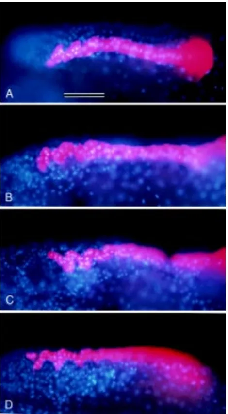
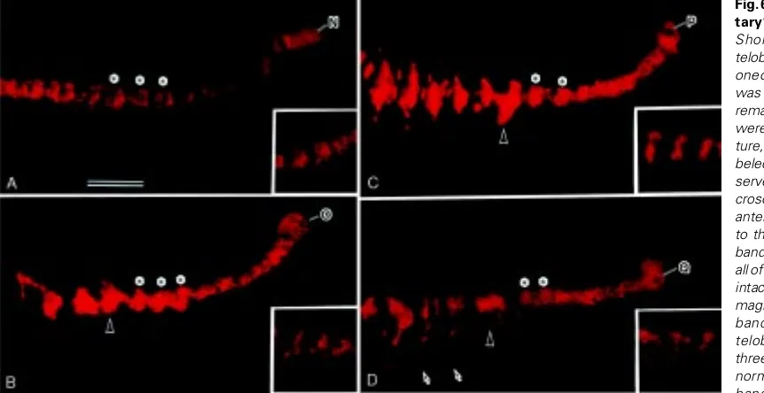
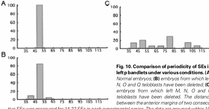
logical changes in ectodermal bandlets occur autonomously or are dependent on neighboring ectodermal bandlets. During the early phase of morphogenesis, ectodermal bandlets are in close contact with each other along their length; even in their undulating portions, there is no interstice between adjacent bandlets. This suggests the possibility that transformation of each bandlet could be a process that depends on adjacent bandlets. To test this possibility, we labeled one of the four ectoteloblasts with DiI and ablated other three ipsilateral ectoteloblasts (or their precursors) simultaneously, and then we examined the behavior of the bandlet produced from the labeled teloblast. Even when an ectoteloblast was forced to be “solitary” by removal of all of its (ipsilateral) sister teloblasts, it continued dividing at a rate comparable to that in intact embryos. There was no difference in the dividing ability and rate among the teloblasts of the four lineages. Fig. 6 shows representative “solitary” bandlets of the four lineages. All but o bandlets were very similar to the respective bandlets in intact embryos, not only in shape but also in periodicity of separated SEs (see Fig. 10B). This suggests that lineage-specific bandlet transformation in the N, P and Q lineages occurs independently of adjacent bandlets.
In contrast, “solitary” o bandlets exhibited features character-istic to the P lineage rather than the O lineage. As Fig. 6B shows, SEs produced from the “solitary” o bandlet assumed an S shape, which is normally characteristic of the P lineage. This finding suggests that “solitary” o bandlets adopt the P fate rather than the O fate.
Morphogenesis of ectodermal bandlets without the underly-ing mesoderm
We next tried to answer the question of whether the mesoder-mal GB plays a role in ectodermesoder-mal segmentation. As Fig. 7 shows, the intersegmental furrows in the ectodermal GB run parallel to
the boundary between mesodermal segments, though in wholemount preparations, these two lines do not necessarily superimpose precisely. There is a possibility that the segmented mesoderm could promote integration of ectodermal SEs into a discrete rectangular domain. To test this possibility, we ablated the left or right M teloblasts from embryos whose 2d111 cells had been injected with DiI or HRP.
Fig. 6. Morphogenesis in “soli-tary” ectodermal bandlets.
Shortly after completion of teloblastogenesis (see Fig. 1C), one of the left four ectoteloblasts was injected with DiI and the remaining three ectoteloblasts were all ablated. After 3-day cul-ture, bandlets derived from la-beled ectoteloblasts were ob-served by epifluorescence mi-croscopy. In all panels shown, anterior is to the left and dorsal is to the top. Insets show control bandlets seen in embryos where all of the left ectoteloblasts were intact. All panels are at the same magnification. Bar, 100 µm. (A) n bandlet produced by the N teloblast (N). Asterisks mark three SEs, which appear to be normal in morphology. (B) o bandlet produced by the O teloblast (O). Asterisks indicate SEs, each of which has assumed an S-shape. This morphology is apparently different from that of the control bandlet (inset) but is reminiscent of a normal p bandlet (see Figs. 2C and 4A). The arrowhead indicates an abnormally large cluster of cells. (C) p bandlet produced by the P teloblast (P). Asterisks indicate SEs, each of which has assumed a deformed S-shape. The arrowhead indicates an abnormally large cluster of cells. This cluster is probably generated through fusion of two SEs. (D) q bandlet produced by the Q teloblast (Q). Asterisks indicate SEs, which appear to be normal in morphology. The arrowhead indicates a cell cluster that is two-times larger than an SE. The arrows indicate labeled cells that have migrated ventrally.
Fig. 7. Intersegmental furrows of the ectodermal GB run parallel to the segmental boundaries of the mesoderm. A 2d11 cell of a 22-cell embryo
Figures 8A and B show representative embryos in which the right and left M teloblasts had been ablated, respectively. In contrast to normally segmented ectodermal GBs on the side with intact M teloblasts (and hence mesodermal GBs), ectodermal GBs on the mesoderm-deficient side failed to generate any segmental organization. These GBs appeared to be normal in the vicinity of the teloblasts. When blast cells proliferated, however, the GBs did not undergo the dorsalward expansion that normally accompanies ectodermal segmentation. As a result, they ap-peared as a long rod running along the ventral midline. Although some clumps of cells were discernible in the GB, they did not appear to be arranged in segmental organization (Fig. 8A). When a single bandlet on the mesoderm-deficient side was labeled with DiI, it was found that individual bandlets underwent morphogen-esis, to some extent, to form rudiments of lineage-specific SEs along their length. In most cases, however, these rudiments did not appear to be separated from bandlets (Fig. 8 D,E). In some embryos, fissures (or constrictions between cell clumps) were observed in labeled bandlets, but separating cell clumps were not uniform in shape and size (Fig. 8C).
These results suggest that ectodermal segmentation requires the presence of the mesoderm. However, this does not necessar-ily mean that the mesoderm directly regulates the segmentation process in the ectodermal GB. There is a possibility that abnormal compaction of bandlets that results from the failure of the ectoder-mal GB to expand dorsally hampers separation of SEs from each bandlet. Therefore, in the last experiment, we examined the behavior of left p bandlets in embryos from which all of ipsilateral M, N, O and Q teloblasts had been ablated. Even under this condition, p bandlets continued elongating. Furthermore, as Fig. 9 shows, cell clusters reminiscent of SEs in intact embryos, did separate from bandlets. However, unlike SEs in normal embryos, these cell clusters were not arranged regularly; the distances between two successive clusters varied and were much larger than the distance in intact embryos (Fig. 10).
Discussion
In this study, we traced the morphogenetic processes leading to segmentation in the ectoderm of Tubifex. Segmentation of the ectodermal GB is the process of separation of 50-µm-wide blocks of cells from the initially continuous GB. Our major findings are as follows: (a) the separation of ectodermal segments from the GB is mediated by formation of fissures, first on the ventral side and then on the dorsal side of the GB. The unification of these fissures give rise to the establishment of an ectodermal segment; (b) as development proceeds, an initially linear array of blast cells in each ectodermal bandlet gradually changes its shape, and its contour becomes indented in a lineage-specific manner, giving rise to the formation of distinct cell clumps, which are separated from the bandlet to serve as segmental elements (SEs); (c) SEs in the N and Q lineages are each comprised of clones of two consecutive primary blast cells. In contrast, in the O and P lineages, individual blast cell clones are distributed across SE boundaries; each SE is a mixture of a part of a more-anterior clone and a part of the next-more-posterior clone. (d) Morphogenetic events, including segmentation, in an ectodermal bandlet pro-ceed normally in the absence of neighboring ectodermal bandlets. (e) Without the underlying mesoderm, separated SEs fail to space themselves at regular intervals along the anteroposterior axis.
Two-step process
The present study showed that ectodermal segmentation in Tubifex involves three key events: (a) generation of SEs within each bandlet, (b) separation of SEs from bandlets, and (c) arrangement of
Fig. 8. Ablation of mesoderm abrogates segmentation of the ectoder-mal GB. 2d111 cells were injected with (A) HRP or (B) DiI. Shortly after the
birth of M teloblasts, the right (A) or left (B) M teloblast was ablated. The embryos were cultured for 4 days before fixation. (A) Ventral view. In contrast to normally segmented ectodermal GB on the left side, the right ectodermal GB (EGBr) does not show any sign of segmentation. Distinct cell clusters are detected in the right GB, but they are not necessarily organized in accordance with intersegmental furrows (arrowheads) in the left GB. (B)
separated SEs at 50-µm intervals along the anteroposterior axis. As was demonstrated in cell-ablation experiments, the first two events occur normally in each ectodermal bandlet in the absence of its neighboring bandlets. This suggests that morphogenetic processes leading to generation and separation of SEs are initiated autono-mously in each bandlet. In contrast, the distribution pattern of separated SEs along the anteroposterior axis apparently depends on the germ layers underlying ectodermal bandlets. It is unlikely that the separation of SEs from bandlets autonomously leads to their arrangement at 50-µm intervals. Thus, we suggest that the ectoder-mal segmentation in Tubifex is divided into two stages, autonomous morphogenesis of each bandlet leading to generation of SEs and the ensuing non-autonomous alignment of separated SEs.
Although cellular basis for shape change in ectodermal bandlets
derived from the O teloblast are specified as those equivalent to P lineage blast cells. It seems plausible that in intact embryos, primary blast cells derived from the O teloblast would be induced to assume the O fate by interactions with neighboring ectodermal bandlets. Thus, unlike those in other lineages, the cell fate decision and the morphogenetic process of the o bandlet depend on external cues. Details of the external cues remain to be explored.
It appears that separated SEs are unable to adjust the distances between themselves. Nevertheless, SEs in intact embryos are arranged at regular intervals of 50 µm. As discussed below, this is simply because ectodermal bandlets in intact embryos are underlain by the mesodermal GB, which is a linear array of 50-µm-wide cell clusters (i.e., mesodermal segments). However, it is presently un-clear whether the mesoderm is the only germ layer that generates regular arrangement of SEs in the overlying ectodermal GB. In this study, we noticed that when underlain by an endoderm, SEs came to be arranged at random intervals (Figs. 9 and 10C). However, this result does not necessarily mean that the endoderm lacks the ability to organize SEs. During developmental stages, as was observed in this study, the endoderm comprises numerous cells derived from macromeres (Shimizu, 1982) but does not show any sign of such repetitive or segmental organization as seen in the mesodermal GB (Nakamoto, unpublished data). The random arrangement of SEs underlain by the endoderm could merely be a reflection of a lack of repetitive organization in the endoderm.
Mesodermal control of ectodermal morphogenesis
During Tubifex embryogenesis, the ectodermal GB is normally underlain by the mesodermal GB (Goto et al., 1999a). The results of the present cell-ablation experiments suggest that the mesoderm plays an important role in two aspects of ectodermal morphogenesis, viz. spatial arrangement of SEs along the anteroposterior axis and dorsalward expansion of the ectodermal GB.
Only when the mesoderm was intact were separated SEs ar-ranged at regular intervals of 50 µm. Otherwise, they were spaced randomly. One of the simplest interpretations of these results is that the mesoderm serves as a framework for regular SE arrangement. In support of this notion, the mesodermal GB is a linear series of segmental founder cells, each of which gives rise to a 50-µm-wide mesodermal segment; furthermore, the boundary between
meso-Fig. 9. Morphogenesis in p bandlets in the absence of all other teloblast lineages. Left M teloblasts were ablated from embryos shortly after their birth. After about 36 h, left P teloblasts of the same embryos were injected with DiI, and the other three ipsilateral ectoteloblasts were ablated. The embryos were raised for 3 days before fixation. Two representative wholemount preparations are viewed from the left side. Anterior is to the left and dorsal is to the top. Asterisks indicate cell clusters that are similar to SEs in normal p bandlets. Arrowheads indicate relatively large clusters, which appear to have resulted from fusion of two SEs. Bar, 100 µm.
was not investigated in this study, it is likely that the lineage-specific morphology of a bandlet is generated through a lineage-specific cell division pattern of primary blast cells. Given the autonomy of morphogen-esis of bandlets leading to SE separation, it appears that primary blast cells of each lineage are specified, at birth, to undergo lineage-specific sequences of cell division. In this connection, it is intriguing to note that in the absence of neighbors, bandlets de-rived from O teloblasts look like p bandlets. A preliminary observation has shown that when allowed to develop to more advanced stages, blast cells of solitary o bandlets have differentiated according to the P fate rather than the O fate (A. Arai, unpublished data). This suggests that in the absence of neighboring bandlets, primary blast cells
Fig. 10. Comparison of periodicity of SEs in left p bandlets under various conditions.(A)
Normal embryos; (B) embryos from which left N, O and Q teloblasts have been deleted; (C)
embryos from which left M, N, O and Q teloblasts have been deleted. The distance between the anterior margins of two consecu-tive SEs was measured for 14-22 SEs in each experimental series. The data are grouped within
dermal segments is determined autonomously (Goto et al., 1999a). Conceivably, SEs separated from each bandlet undergo a process by which they align themselves to mesodermal segments.
The present study has also shown that in the absence of the underlying mesoderm, the ectodermal GB failed to expand dorsally and assumed the shape of a rod running along the ventral midline (Fig. 8A). This finding suggests that in intact embryos, the mesoder-mal GB serves as a substrate for ectodermesoder-mal expansion and that the endoderm cannot take the place of the mesoderm in this respect. As suggested previously, the mesoderm is apparently motile and can expand circumferentially toward the dorsal midline by itself (Goto et al., 1999a). There is a possibility that the mesoderm plays a role as a “conveyer” of the overlying ectodermal GB.
Comparison with other annelids
Are the features of ectodermal segmentation revealed in this study shared by clitellate annelids ? Blair (1982) and Torrence (1991) reported that when M teloblasts in leech embryos were ablated, no segmentally iterated structures formed in the ectodermal GB on the mesoderm-deficient side. On the other hand, Shain and others (2000) have recently shown that in the leech Theromyzon, the separation of ganglionic primordia (which correspond to SEs we referred to in this paper) from the N lineage bandlet occurs indepen-dently of neighboring bandlets (including mesodermal GB), suggest-ing autonomy of early events of ectodermal bandlet morphogenesis. Based on these studies, it is thought that, as in Tubifex, the ectoder-mal segmentation in leech embryos consists of an early autonomous morphogenetic process followed by a mesoderm-dependent pro-cess.
In addition, the segmental distribution pattern of primary blast cell clones in leech embryos is strikingly similar to that reported here for Tubifex (Weisblat and Shankland, 1985). This suggests that oli-gochaetes and leeches adopt similar processes that specify seg-mental boundaries in the ectoderm. Thus, it appears that key elements of the mechanisms underlying ectodermal segmentation have been conserved in oligochaetes and leeches.
Materials and Methods
Embryos
Embryos of the freshwater oligochaete Tubifex hattai were obtained according to the method of Shimizu (1982) and cultured at 22ºC. For the experiments, embryos were all freed from cocoons in the culture medium (Shimizu 1982). To sterilize their surfaces, cocoons were treated with 0.02% chloramine T (Wako Pure Chemicals, Osaka, Japan) for 3 min and washed thoroughly in three changes of the culture medium. The culture medium used in the cell-ablation experiments was autoclaved, and antibiotics (penicillin G and streptomycin, 20 units/ml each) were added shortly before use (Kitamura and Shimizu, 2000). Unless otherwise stated, all experiments were carried out at room temperature (20-22ºC).
Microinjection of lineage tracers
The cell lineage tracers used in this study were DiI (1,1'-dihexadecyl-3,3,3',3'-tetramethylindocarbocyanine perchlorate; Molecular Probes, Inc., Eugene, USA), HRP (horseradish peroxidase; Sigma, type VI-A ) and OG dextran (dextran, Oregon Green 488; 10000 MW, anionic, lysine fixable; Molecular Probes, Inc.). DiI was dissolved in ethanol at 100 mg/ml and stored at room temperature. Before use, an aliquot of this solution was diluted 20 times in safflower oil (Kitamura and Shimizu, 2000). Target cells were
injected with oil droplets containing DiI by means of micropipettes. DiI-injected embryos were kept in darkness. Preparation of solutions of HRP and OG dextran and their injection were performed according to the method described previously (Goto et al., 1999a).
Preparation of labeled embryos for observation
DiI- or OG dextran-labeled embryos were fixed with 3.5% formaldehyde in phosphate buffer (40.5 mM Na2HPO4, 9.5 mM NaH2PO4, pH 7.4) for 1 h. After being washed thoroughly in three changes of phosphate buffer, they were occasionally stained with 1 µg/ml Hoechst 33342 in phosphate buffer to observe nuclei. The specimens were then mounted in phosphate buffer and observed under a Zeiss Axioskop epifluorescence microscope. Some specimens were examined under a Molecular Dynamics Sarastro-2000 confocal laser-scanning microscope. HRP-injected embryos were fixed with 1% glutaraldehyde in phosphate buffer and processed for color development of HRP according to the method described previously (Goto et al., 1999a). Stained embryos were dehydrated in methanol and cleared in a mixture of one part benzyl alcohol and two parts benzyl benzoate, and they were observed as whole mounts in this mixture with Nomarski differential interfer-ence contrast optics.
Blastomere ablation
Embryos without vitelline membranes were placed on 2% agar in the culture medium. Blastomeres were killed by making a wound on their surface with fine forceps. Within a minute the yolk mass of these cells began to coagulate. The coagulating cells were removed by pulling them away from the remainder of the embryo. The operated embryos were allowed to develop in the culture medium containing antibiotics, which was renewed daily.
References
BLAIR, S.S. (1982). Interactions between mesoderm and ectoderm in segment formation in the embryo of a glossiphodiid leech. Dev. Biol. 89: 389-396. DEVRIES, J. (1974). Le mésodermie, feuillet directeur de l’embryogenese chez le
lombricien Eisenia foetida. III. La différenciation du tube digestif et des dérivés ectodermiques. Acta Embryol. Exp. 157-180.
GOTO, A., KITAMURA, K. and SHIMIZU, T. (1999a). Cell lineage analysis of pattern formation in the Tubifex embryo. I. Segmentation in the mesoderm. Int. J. Dev. Biol. 43: 317-327.
GOTO, A., KITAMURA, K., ARAI, A. and SHIMIZU, T. (1999b). Cell fate analysis of teloblasts in the Tubifex embryo by intracellular injection of HRP. Dev. Growth Differ. 41: 703-713.
KITAMURA, K. and SHIMIZU, T. (2000). Analyses of segment-specific expression of alkaline phosphatase activity in the mesoderm of the oligochaete annelid Tubifex: implications for specification of segmental identity Dev. Biol. 219: 214-228. PENNERS, A. (1926). Experimentelle Untersuchungen zum Determinationsproblem
am Keim von Tubifex rivulorum Lam. II. Die Entwicklung teilweise abgetöteter Keime. Z. Wiss. Zool. 127: 1-140.
PENNERS, A. (1938). Abhängigkeit der Formbildung vom Mesoderm im Tubifex-Embryo. Z. Wiss. Zool. 150: 305-357.
SHAIN, D.H., STUART, D.K., HUANG, F.Z. and WEISBLAT, D.A. (2000). Segmenta-tion of the central nervous system in leech. Development 127: 735-744. SHIMIZU, T. (1982). Development in the freshwater oligochaete Tubifex. In
Develop-mental Biology of Freshwater Invertebrates (Ed. F. W. Harrison and R. R. Cowden), Alan R. Liss, New York, pp. 286-316.
TORRENCE, S.A. (1991). Positional cues governing cell migration in leech neurogenesis. Development 111: 993-1005.
WEISBLAT, D.A. and SHANKLAND, M. (1985). Cell lineage and segmentation in the leech. Phil. Trans. R. Soc. Lond. B312: 39-56.
Received: July 2000
