Original Article
Early establishment of epithelial apoptosis in the
developing human small intestine
PIERRE H. VACHON
1,3, ÉRIC CARDIN
1, CHARLÈNE HARNOIS
1, JOHN C. REED
2and ANNE VÉZINA
11CIHR Group on the Functional Development and Physiopathology of the Digestive Tract, Centre de recherche en biologie du
développement des épithéliums, département d’anatomie et de biologie cellulaire, Faculté de médecine, Université de Sherbrooke, QC, Canada, 2The Burnham Institute, La Jolla Cancer Research Center, California, USA and 3Thématique de recherche en physiopathololgie
digestive du Centre de recherches cliniques du CHUS, Fleurimont, QC, Canada
ABSTRACT In the adult small intestine, the dynamic renewal of the epithelium is characterized by a sequence of cell production in the crypts, cell maturation and cell migration to the tip of villi, where apoptosis is undertaken. Little is known about enterocytic apoptosis during development. In man, intestinal architectural features and functions are acquired largely by mid-gestation (18-20 wks); the question whether the establishment of enterocytic apoptotic processes parallels or not the acquisition of other intestinal functional features remains open. In the present study, we ap-proached this question by examining enterocytic apoptosis during development of the human jejunum (9-20 wks gestation), using the ISEL (in situ terminal uridine deoxynucleotidyl nick-end labelling) method. Between 9 and 17 wks, apoptotic enterocytes were not evidenced. However, beginning at the 18 wks stage, ISEL-positive enterocytes were regularly observed at the tip of villi. Since the Bcl-2 family of proteins constitutes a critical checkpoint in apoptosis, acting upstream of the apoptotic machinery, we investigated the expression of six Bcl-2 homologs (Bcl-2, Bcl-XL, Mcl-1, Bax, Bak, Bad) and one non-homologous associated molecule (Bag-1). By immunofluorescence, we found that all homologs analyzed were expressed by enterocytes between 9 and 20 wks. However, Bcl-2 homologs underwent a gradual compartmentalization of epithelial expression along the maturing crypt-villus axis, to establish gradients of expression by 18-20 wks. Western blot analyses indicated that the expression levels of Bcl-2 homologs were modulated during morpho-genesis of the crypt-villus axis, in parallel to their gradual compartmentalization of expression. Altogether, these data suggest that regulatory mechanisms of human enterocytic apoptosis become established by mid-gestation (18-20 wks) and coincide with the maturation of the crypt-villus axis of cell proliferation, differentiation and renewal.
KEY WORDS:
Bcl-2 homologs, crypt-villus axis, enterocyte, gut, programmed cell death.
0214-6282/2000/$20.00
© UBC Press Printed in Spain
www.ehu.es/ijdb
*Address correspondence to: Pierre H. Vachon, Ph.D., Département d’anatomie et de biologie cellulaire, Faculté de médecine, Université de Sherbrooke, Sherbrooke (QC) J1H5N4, Canada. FAX: +819-564-5320. e-mail: phvachon@courrier.usherb.ca
Abbreviations used in this paper: FITC, fluorescein isothiocyanate; ISEL, in situ terminal uridine deoxynucleotidyl nick-end labelling; K18, cytokeratin 18; OCT, optimum cutting temperature; PBS, phosphate buffered saline; PMSF, phenylmethylsulfonyl fluoride; TdT, terminal deoxynucleotidyl transferase; SDS-PAGE, sodium dodecyl sulfate-polyacrylamide gel electrophoresis.
Introduction
Apoptosis, a form of programmed cell death, is an intricately regulated process which plays a crucial role in tissue morphogen-esis, renewal, and repair (Jacobson et al., 1997). The typical death throes of a cell undergoing apoptosis include DNA fragmentation, nuclear condensation, organelle degradation, and cell shrinkage. The Bcl-2 family of proteins constitutes a critical decisional check-point in apoptosis, acting upstream of the apoptotic machinery responsible for the irreversible degradation of cellular constituents (Reed et al., 1996a,b; Adams and Cory, 1998). At least 15 family members have been identified so far in mammalian cells, sharing homology in three or four conserved domains (BH1 to BH4), and functioning either as anti-apoptotic (e.g. Bcl-2, Bcl-XL, Mcl-1) or
expression levels; however, post-transcriptional and post-transla-tional modifications can also sway the balance in favor of either pro-or anti-apoptotic homologs (Gajewski and Thompson, 1996; Reed et al., 1996a; Adams and Cory, 1998). Furthermore, interactions with other types of molecules, such as the anti-apoptotic protein Bag-1 (Wang et al., 1994; Takayama et al., 1995; Wang et al., 1996), add another level of complexity to the regulation of Bcl-2 homolog functions (Gajewski and Thompson, 1996; Adams and Cory, 1998). Nonetheless, it is acknowledged that characterization of the expression profiles of Bcl-2 homologs in tissues constitutes a crucial step in the understanding of the regulation of apoptosis, in tissue-specific developmental and/or renewal systems (Hale et al., 1996; Moss and Holt, 1996; Reed et al., 1996a; Jacobson et al., 1997; Potten, 1997; Adams and Cory, 1998).
The small intestinal epithelium is a useful model for the in situ study of the establishment and working mechanics of tissue renewal processes, including programmed cell death. Its rapid, continuous cell renewal consists of spatially separated stem cells, proliferative and differentiated compartments, located respectively in the lower regions of the crypts and on the villi (Leblond, 1981; Jones and Gores, 1997; Potten, 1997). In human adults, the dynamic renewal of the intestinal epithelium is characterized by a
(Jones and Gores, 1997; Potten, 1997). Spontaneous crypt cell apoptosis, a rarer (less frequent) process, serves to remove defective/injured progeny cells, as well as senescent Paneth cells (Potten, 1992; Moss and Holt, 1996; Jones and Gores, 1997; Potten, 1997). A role for some 2 homologs (namely 2, Bcl-XL, Mcl-1 and Bak) has been proposed in the regulation of apoptosis in the human adult small intestine, a potential function well illus-trated by their differential patterns (or gradients) of epithelial expression along the crypt-villus axis (Hockenbery et al., 1991; Lu et al., 1993; Krajewski et al., 1994c, 1995, 1996).
However, little is known on enterocytic apoptosis during devel-opment. The morphogenesis of the small intestinal mucosa, and thus of the crypt-villus axis, has been the subject of many reviews (Ménard, 1989; Ménard and Calvert, 1991; Ménard and Beaulieu, 1994). In man, intestinal architectural features and functions are acquired early during fetal life (Ménard, 1989; Ménard and Calvert, 1991; Ménard and Beaulieu, 1994). Villus formation by mesenchy-mal infiltration of the stratified epithelium begins around 8-9 wks of gestation, proceeding distally until the entire intestine is lined by short villi covered by a simple columnar epithelium. By 15 wks, crypt formation has begun with the invagination of the intervillous epithelium into the underlying mesenchyme. Finally, by mid-gesta-tion (18-20 wks), the overall architectural and funcmid-gesta-tional organiza-tion of the crypt-villus axis, including digestive capacities, are highly similar to those of the new-born/adult intestinal mucosa (Ménard, 1989; Ménard and Calvert, 1991; Ménard and Beaulieu, 1994). Therefore, the question whether the establishment of enterocytic apoptotic processes parallels or not the acquisition of other intestinal functional features remains open.
In the present study, we approached this question by examin-ing epithelial apoptosis durexamin-ing development of the human small intestine between 9 and 20 wks of gestation. The epithelial expres-sion and localization of six Bcl-2 homologs (Bcl-2, Bcl-XL, Mcl-1, Bax, Bak, Bad), and one non-homologous associated molecule (Bag-1), were investigated as well. Herein, we find that intestinal epithelial apoptosis is detected at the tip of villi at 18-20 wks, but not in the earlier developmental stages studied. We also show that the epithelial expression of Bcl-2 homologs undergo a gradual compartmentalization of expression during intestinal develop-ment, in order to establish differential patterns (or gradients) along the crypt-villus axis by 18-20 wks. This gradual compartmentalization of Bcl-2 homolog expression parallels mirroring changes in their protein expression levels. Hence, these data altogether suggest that regulatory mechanisms of human enterocytic apoptosis be-come established by mid-gestation and coincide with the matura-tion of the crypt-villus axis.
Results
To ascertain whether the establishment of human enterocytic apoptotic processes parallels or not the morphogenesis and matu-ration of the small intestinal crypt-villus axis of cell prolifematu-ration and differentiation, we examined epithelial apoptosis during develop-ment of the human jejunum between 9 and 20 wks of gestation. In parallel, the epithelial expression and localization of Bcl-2 homologs, acknowledged as central regulators of programmed cell death, were investigated throughout the same developmental period.
Emergence of enterocytic apoptosis during morphogenesis of the crypt-villus axis
Villus-tip apoptosis, the normal fate of intestinal epithelial cells, is readily observed when using the ISEL method (Gavrieli et al., 1992; Hall et al., 1994; Moss and Holt, 1996, Aschoff et al., 1999). Using this approach, we examined the presence of epithelial apoptotic cells during the development of the human jejunum (9-20 wks). Between 9 wks and 14 wks of gestation, we failed to observe any enterocytic apoptosis either among villus cells (Fig. 1A) or intervillous cells (not shown). Absence of apoptotic enterocytes was likewise noted between 15 wks and 17 wks (not shown), although crypt formation had begun. However, beginning at 18 wks, villus-tip apoptotic enterocytes were consistently observed (Fig. 1B) and such consistent detection of villus-tip ISEL-positive enterocytes persisted at 19 and 20 wks (Fig. 1D). As shown with greater magnification in Figure 1D, typically 1-3 ISEL-positive cells were detected at the apex of villi. Interestingly, ‘spontaneous’ crypt cell apoptosis, a rarer (less frequent) process observed in the adult small intestine (Potten, 1992; Hall et al., 1994; Merritt et al., 1995; Moss and Holt, 1996; Jones and Gores, 1997; Potten, 1997), was not evidenced herein (Fig. 1C). Finally, it is of note that single ISEL-positive mesenchymal cells were occasionally observed through-out the developmental period studied (not shown).
Epithelial localization of Bcl-2 homologs during morphogen-esis of the crypt-villus axis
Since our ISEL observations indicated that villus-tip enterocytic apoptosis emerges by mid-gestation (18-20 wks), and because the Bcl-2 family of proteins exert critical regulatory functions in apoptosis (Reed et al., 1996a,b; Adams and Cory, 1998), we then investi-gated the jejunal epithelial expression of six Bcl-2 homologs (Bcl-2, Bcl-XL, Mcl-1, Bax, Bak, Bad) and one non-homologous associ-ated molecule (Bag-1). All molecules analyzed herein were readily detected in the jejunal epithelium beginning at 9 wks of gestation. Indeed, a cytoplasmic (non-nuclear) staining was observed in
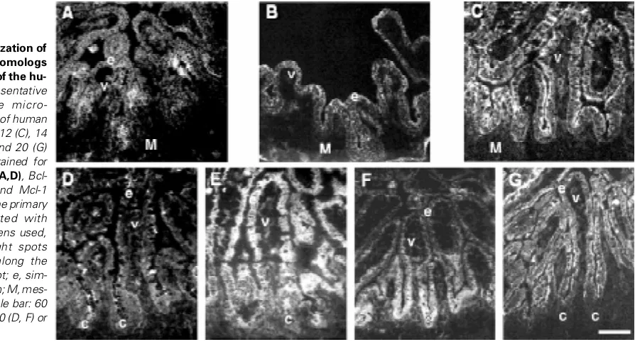
epithelial cells of both growing villi and intervillous regions for Bcl-2 (Fig. Bcl-2A), Bcl-XL (not shown), Mcl-1 (Fig. 2C), Bax (Fig. 3A), Bak (Fig. 3B) and Bad (Fig. 3C), as well as for the anti-apoptotic molecule Bag-1 (Fig. 2B). This rather homogenous staining along the intervillous-villus axis remained essentially unaltered between 9 and 14 wks of gestation (Fig. 2 A-C and Fig. 3 A-C). Between 15 and 17 wks, we observed that the epithelial expression of some of the Bcl-2 homologs underwent a compartmentalization process along the maturing crypt-villus axis. This gradual process was found to culminate by mid-gestation (Fig. 2 D-G and Fig. 3 D-E). Thus, beginning at 18 wks, staining for Mcl-1 (Fig. 2G) and Bak (Fig. 3D) were found concentrated in villus enterocytes, but poorly detectable or absent in crypt cells. Bcl-2 staining was found much weaker than in previous stages, and exhibited a decreasing crypt-to-villus gradient of staining (Fig. 2D). Likewise, Bag-1 exhibited a decreasing gradient of staining from the base of the crypts to the apex of villi (Fig. 2F). Bad also displayed a compartmentalized expression pattern along the crypt-villus axis around the 18 wks stage, staining being strong in the upper half of villi (with occasional strong staining at the base of villi as well), but weaker in the rest of the epithelium (Fig. 3E). However, Bcl-XL (Fig. 2E) and Bax (not shown) were still detected homogeneously throughout the intesti-nal epithelium. These differential crypt-villus patterns of Bcl-2 homolog expression remained unchanged from 18 to 20 wks (Fig. 2 D-G and Fig. 3 D-E).
Epithelial expression levels of Bcl-2 homologs during mor-phogenesis of the crypt-villus axis
To further characterize the crypt-villus compartmentalization process of epithelial expression of Bcl-2 homologs, we then investigated the developmental protein expression levels of these same homologs. Immunoblot analyses of lysates from jejunal mucosal scrappings demonstrated the protein expression of all molecules analyzed herein (Fig. 4). Thus, Bcl-2 (~ 26 kDa), Bcl-XL (~ 28-30 kDa), Bag-1 (~ 32-34 kDa), Mcl-1 (~ 39-42), Bax (~21
Fig. 2. Epithelial localization of anti-apoptotic Bcl-2 homologs during development of the hu-man jejunum. Representative immunofluorescence micro-graphs of cryosections of human fetal jejunums at 9 (B), 12 (C), 14 (A), 18 (E), 19 (D, F) and 20 (G) weeks of gestation stained for the detection of Bcl-2 (A,D), Bcl-XL (E), Bag-1 (B,F), and Mcl-1
kDa), Bak (~ 25-28 kDa) and Bad (~ 28-32 kDa) were detected at all developmental stages studied as protein bands migrating at their previously reported relative molecular weights (Krajewski et al., 1994a-c, 1995; Takayama et al., 1995; Krajewska et al., 1996; Krajewski et al., 1996; Packham et al., 1997; Adams and Cory, 1998; Metcalfe et al., 1999).
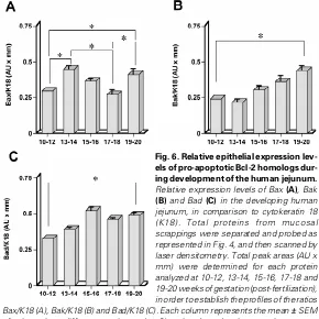
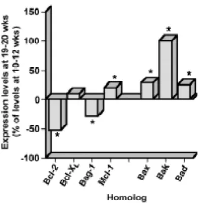
To examine the developmental epithelial expression profiles of each molecule studied in relation to the morphogenesis of the crypt-villus axis, their relative epithelial expression levels were evaluated by comparison with a reference protein, cytokeratin 18 (K18). The densitometric data presented in Figures 5-7 show that the relative epithelial expression levels of all Bcl-2 homologs analyzed (including the Bag-1 protein) are differentially modulated in parallel to the morphogenesis of the crypt-villus axis, as well as in concomitance to the establishment of their differential expres-sion patterns. In the case of anti-apoptotic proteins (Fig. 5), Bcl-2 levels increased slightly between 10-14 weeks, but decreased sharply around 15 wks (when cryptogenesis has begun) in order to stabilize at lower levels (Fig. 5A), thus resulting in a significant ~53% overall reduction (Fig. 7). Bag-1 expression levels gradually decreased between 10 and 20 wks (Fig. 5C), resulting in a significant ~30% overall reduction (Fig. 7). Although Bcl-XL de-creased gradually between 10 and 15 wks, its levels returned to those of 10-12 wks in subsequent stages (Fig. 5B), thus resulting in a slight (and non-significant) ~10% overall increment (Fig. 7). Finally, Mcl-1 epithelial expression increased gradually between 10-20 wks (Fig. 5D), resulting in a significant ~20% overall incre-ment (Fig. 7). In the case of pro-apoptotic Bcl-2 homologs (Fig. 6), Bax exhibited the most complex modulations of epithelial expres-sion levels. Indeed, it increased between 10 and 14 wks, in order to decrease gradually between 14 and 18 wks, and then increase sharply again between 18 and 20 wks (Fig. 6A). This resulted in a significant ~30% overall increment (Fig. 7). On the other hand, Bak increased gradually and steadily between 10 and 20 wks (Fig. 6B),
sion of Bcl-2 homologs which appears to culminate at 18 wks to establish differential patterns (or gradients) of expression along the crypt-villus axis, coinciding with the emergence of villus-tip apoptotic cells. Finally, we found that the expression levels of Bcl-2 homologs are modulated during morphogenesis of the crypt-villus axis, in
Fig. 3. Epithelial localization of pro-apoptotic Bcl-2 homologs during development of the human jeju-num. Representative immunofluores-cence micrographs of cryosections of human fetal jejunums at 10 (C), 12 (A), 14 (B), 18 (E), and 20 (D) weeks of gestation stained for the detection of Bax (A), Bak (B,D) and Bad (C,E). Note in (E) that the primary antibodies cross-reacted with goblet cells of specimens used, as evidenced by bright spots sparsely distributed along the crypt-villus axis. c, crypt; e, simple columnar epithelium; V, villus. Scale bar: 60 (C), 75 (A-B), 100 (E) or 125 (D) µM.
expression levels also increased gradually between 10 and 20 wks (Fig. 6C), resulting in a significant ~25% overall increment (Fig. 7).
Discussion
In this study, we examined the question whether the establishment of enterocytic apoptotic processes parallels or not the morphogenesis and maturation of the small intestinal crypt-villus axis of cell prolifera-tion and differentiaprolifera-tion. To do so, we inves-tigated the presence of intestinal epithelial cell apoptosis, as well as the localization and expression of six Bcl-2 homologs (Bcl-2, Bcl-XL, Mcl-1, Bax, Bak, Bad) and one non-homologous associated molecule (Bag-1), in the developing human small intestine between 9 and 20 wks of gesta-tion. We found that villus-tip epithelial cell apoptosis emerges only by the time of the 18 wks stage. We also observed a gradual compartmentalization of epithelial
Fig. 5. Relative epithelial expression levels of anti-apoptotic Bcl-2 homologs during development of the human jejunum. Relative expression levels of Bcl-2 (A), Bcl-XL(B), Bag-1 (C) and Mcl-1 (D) in the developing human jejunum, in comparison to cytokeratin 18 (K18). Total proteins from mucosal scrappings were separated and probed as represented in Fig. 4, and then scanned by laser densitometry. Total peak areas (AU x mm) were determined for each protein analyzed at 10-12, 13-14, 15-16, 17-18 and 19-20 weeks of gestation (post-fertilization), in order to establish the profiles of the ratios Bcl-2/K18 (A), Bcl-XL/K18 (B), Bag-1/K18 (C) and Mcl-1/K18 (D). Each column represents the mean ± SEM of at least three different specimens (n ≥ 3) analyzed per developmental age group; statistically significant (0.001 ≤ p ≤ 0.05) differences are indicated by an asterisk (*). Values on the abscissa represent weeks post fertilization.
Fig. 6. Relative epithelial expression lev-els of pro-apoptotic Bcl-2 homologs dur-ing development of the human jejunum.
Relative expression levels of Bax (A), Bak
(B) and Bad (C) in the developing human jejunum, in comparison to cytokeratin 18 (K18). Total proteins from mucosal scrappings were separated and probed as represented in Fig. 4, and then scanned by laser densitometry. Total peak areas (AU x mm) were determined for each protein analyzed at 10-12, 13-14, 15-16, 17-18 and 19-20 weeks of gestation (post-fertilization), in order to establish the profiles of the ratios Bax/K18 (A), Bak/K18 (B) and Bad/K18 (C). Each column represents the mean ± SEM of at least three different specimens (n ≥ 3) analyzed per developmental age group; statistically significant (0.001 ≤ p ≤ 0.05) differences are indicated by an asterisk (*).Values on the abscissa represent weeks post fertilization.
parallel to their gradual compartmentalization of epithelial expres-sion and to the emergence of villus-tip apoptosis.
In contrast to laboratory animals with short gestational periods (e.g. rat and mouse), human intestinal architectural and functional features are acquired as early as by mid-gestation (18-20 wks) (Ménard, 1989; Ménard and Calvert, 1991; Ménard and Beaulieu, 1994). Such features include not only a well defined crypt-villus axis, but as well as enzymatic brush border membrane digestive activities that are similar to those measured in the newborn/adult intestinal mucosa (Ménard, 1989; Ménard and Calvert, 1991; Ménard and Beaulieu, 1994). Other mid-gestation intestinal char-acteristics such as absorption/transport of lipids, sugars and amino acids, as well as basement membrane composition and integrin expression, or hormonal/growth factor responses, are likewise highly comparable to those found in the new-born/adult (Ménard, 1989; Ménard and Calvert, 1991; Ménard and Beaulieu, 1994; Beaulieu, 1999). Our observations, namely that villus-tip apoptosis and crypt-to-villus differential patterns of Bcl-2 homolog expression are already estab-lished by 18-20 wks, can be added to this seemingly growing list. Indeed, villus-tip apoptosis is the normal fate of enterocytes in the adult small intestine, and the epithelial stainings observed herein for Bcl-2, Bcl-XL, Bak and Mcl-1 at mid-gestation correlate with (or confirm) those previously observed in third-trimester and/or adult human small intes-tinal specimens (Hockenbery et al., 1991; LeBrun et al., 1993; Lu et al., 1993; Krajewski et al., 1994c, 1995, 1996) (to our knowledge, there has been no report of Bag-1, Bax or Bad expression in the human small intestine prior to the present study). In light of these considerations, and taking into account the acknowledged role of Bcl-2 homologs as decisional regulators of apoptosis, our data altogether strongly suggest that regulatory mechanisms of human enterocytic apoptosis become established by mid-gestation, as is the case for other intestinal epithelial cellular processes and functions.
Another aspect of our findings concerns the developmen-tal establishment of differential patterns of epithelial expres-sion of Bcl-2 homologs in the small intestine. The crypt-villus axis constitutes an elegant example of vectorial compartmentalization of proliferative/undifferentiated (crypts)
crypt-villus axis (Potten 1992; Moss and Holt, 1996; Jones and Gores, 1997; Potten, 1997).
In support of this, studies have consistently reported a dramatic increase of apoptosis in crypt cells, but little or no increase in villus cells, after irradiation or chemotherapeutic drug exposure (Potten, 1992; Hall et al., 1994; Merritt et al., 1995; Potten, 1997; Pritchard et al., 1999). In addition, some Bcl-2 homologs have been shown individually to exhibit gradients of expression along the crypt-villus axis, suggesting a role for these apoptotic regulators in intestinal epithelial programmed cell death (Hockenbery et al., 1991; LeBrun et al., 1993; Lu et al., 1993; Krajewski et al., 1994b,c, 1995; Merritt et al., 1995; Krajewski et al., 1996; Wilson and Potten, 1996; Aschoff et al., 1999). Our study, which analyzed six homologs at the same time, clearly illustrates a differential pattern of epithelial expression in the human small intestine, where proliferative/undif-ferentiated crypt cells exhibit a Bcl-2 homolog expression profile that differs from the one observed in differentiated villus cells. To this effect, analyses of enterocytic apoptosis in bcl-2—/— and bax— /— knockout mice have reported differential consequences for crypt
and villus cells with regards to apoptosis resistance and/or suscep-tibility after irradiation (Wilson and Potten, 1996; Potten, 1997; Pritchard et al., 1999). Finally, Bag-1, a protein known to associate with Bcl-2 and to participate in signal transduction pathways that promote cell survival (Wang et al., 1994; Takayama et al., 1995; Gajewski and Thompson, 1996; Reed et al., 1996a,b; Wang et al., 1996; Packham et al., 1997; Adams and Cory, 1998), was shown
taken together strongly support the concept that intestinal epithe-lial cell survival and apoptosis may be regulated differentially according to the state of cell differentiation.
In conclusion, the present findings provide new insights into the developmental establishment of regulatory mechanisms of epithe-lial apoptosis in the human small intestine, whereby a compartmentalized epithelial expression of Bcl-2 homologs is established along the crypt-villus axis by mid-gestation, coincident with the emergence of apoptosis at the apex of villi. The develop-mental processes responsible for such early establishment of adult-like intestinal apoptotic features in man remain to be under-stood. For instance, are the modulations of Bcl-2 homolog expres-sion levels observed herein simply consequent to the establish-ment of the differential crypt-villus patterns of epithelial expression, or could these be involved in the absence of apoptotic enterocytes until mid-gestation is reached? Further analyses, using in vitro model systems, will be required to dissect at the molecular level the mechanisms which influence the functions of Bcl-2 homologs in epithelial cells, as well as to identify the exact functions enacted by Bcl-2 homologs themselves in the regulation of epithelial pro-grammed cell death.
Materials and Methods
Tissue processing
Human fetal jejunum specimens from 39 fetuses ranging in age from 9 to 20 weeks post-fertilization (fetal ages were estimated according to Streeter, 1920) were obtained from normal elective pregnancy terminations. Only specimens obtained rapidly (60 min or less) were used. The present study was in accordance with a protocol approved by the institutional Human Research Ethical Review Committee for the use of human biological mate-rials. For immunolocalization and ISEL (in situ terminal uridine deoxynucleotidyl nick-end labelling) studies, tissues were washed in PBS (pH 7.4) and embedded in OCT (Optimum Cutting Temperature) compound (TissueTek, Miles Laboratories, Elkhart, IN), as previously described (Beaulieu et al., 1991). For analyses of protein expression levels, mucosal scrappings (Beaulieu et al., 1993; Beaulieu and Vachon, 1994) of samples were washed in PBS (pH 7.4) and homogenized in 20 mM Tris-HCl (pH 6.8) containing 0.1 mM phenylmethylsulfonyl fluoride (PMSF), 50 µg/ml leupeptin, 50 µg/ml antipain, and 0.1 mg/ml aprotinin. Total proteins were measured using the BioRad (Hercules, CA) protein assay. Aliquots of homogenates were directly solubilized in 2x solubilization buffer (2.3% [w/v] SDS, 10% [v/v] glycerol, and 0.001% [w/v] bromophenol blue in 62.5 mM Tris-HCl [pH 6.8] containing 5% [v/v] β-mercaptoethanol), boiled (105°C, 5 min), cleared by centrifugation (13000g, 5 min, room temperature), and processed for storage as described (Beaulieu and Vachon, 1994; Vachon et al., 1995, 1996).
Antibodies
Primary rabbit polyclonal antibodies used in the present study were Ab 1682, directed against human Mcl-1 (Krajewski et al., 1994a, 1995); Ab 1695, directed to human/mouse Bcl-XL (Krajewski et al., 1994c); Ab 1701 (Krajewski et al., 1994a) and Ab PC68 (Calbiochem, San Diego, CA), both directed against human Bcl-2; Ab 1712 (Krajewski et al., 1994b) and Ab PC66 (Calbiochem), both directed to human Bax; Ab 1764, directed against human Bak (Krajewska et al., 1996; Krajewski et al., 1996); Ab I-19 (Santa Cruz Biotech., Santa Cruz, CA), directed to human/mouse Bak; Ab PC67 (Calbiochem), directed to human Bcl-XL; Ab K-20 (Santa Cruz Biotech.), directed against human/mouse Mcl-1; and Ab 9292 (New England Biolabs, Beverly, MA) and Ab R-20 (Santa Cruz Biotech.), both directed to human Bad. Primary mouse monoclonals used were mAb K56C8 (Takayama et al., 1995; Wang et al., 1996), directed against human Bag-1; mAb 32 and mAb 48 (both from Transduction Labs./Biocan Scientific, Mississauga, ON, Canada), directed to human Bad; and mAb CY-90 (Sigma-Aldrich Canada Fig. 7. Differential modulations of epithelial Bcl-2 homolog expression
Ltd., Oakville, ON), directed against human cytokeratin 18 (K18). Note that antibodies Ab1682, Ab1695, Ab1701, Ab1712, Ab1764 and mAb K56C8 were developed in the laboratory of one of the authors (J.C.R.) of the present study and have been characterized extensively in previous studies (Krajewski et al., 1994a-c, 1995; Takayama et al., 1995; Krajewska et al., 1996; Krajewski et al., 1996; Wang et al., 1996).
Immunolocalization analyses
Cryosections (4-6 µm thick) of human fetal jejunum samples were fixed and stained by indirect immunofluorescence as described previously (Beaulieu et al., 1991; Beaulieu and Vachon, 1994; Vachon et al., 1997). Rabbit antisera were used at 1:100-1:1000 dilutions, and mouse monoclonals were used at 1:50-1:1000 dilutions. All dilutions were made in PBS (pH 7.4) containing 5% (w/v) non-fat powdered milk. FITC-conjugated goat anti-rabbit or anti-mouse IgG (Roche Diagnostics/Boehringer Mannheim, Laval, QC, Canada) were used as secondary antibodies. Sections were counterstained with 0.01% (w/v) Evans blue in PBS (pH 7.4), mounted in glycerol-PBS (9:1) containing 0.1% (w/v) paraphenylenediamine, and viewed with a Reichart Polyvar 2 microscope (Leica, St-Laurent, QC, Canada) equipped for epifluorescence. In all cases, no specific immun-ofluorescent staining was observed when primary antibodies were omitted or replaced by non-immune (rabbit or mouse) serum (not shown). All immunofluorescent micrographs shown herein are representative of at least three (n≥3) different specimens for each developmental stage analyzed.
In situ detection of apoptosis-associated DNA strand breaks In situ terminal deoxynucleotidyl transferase (TdT)-mediated dUTP nick-end labeling (ISEL) (Gavrieli et al., 1992) was carried out as previously described (Vachon et al., 1996, 1997) on 4-6 µm thick cryosections of human fetal jejunum samples, using the ApopTag apoptosis detection kit (Oncor, Gaithersburg, MD). Preparations where then counterstained with Evans blue, mounted and viewed with a Reichart Polyvar 2 microscope (Leica, St-Laurent, QC, Canada) equipped for epifluorescence.
Analyses of protein expression levels
Sodium dodecyl sulfate-polyacrylamide gel electrophoresis (SDS-PAGE) on 15% (w/v) acrylamide Tris-HCl gels (Bio-Rad) was performed as described previously (Vachon et al., 1996, 1997). Broad range molecular mass markers (6.8-209 kDa range; BioRad) were used as standards. Total proteins (50 µg/well) were separated by electrophoresis and then electrotransferred to nitrocellulose membranes (Supported NitroCellulose-1; Life Technologies/Gibco-BRL, Burlington, ON, Canada) for subsequent immunoblotting (Vachon et al., 1996, 1997). Rabbit antisera were used at 1:200-1:2000 dilutions, and mouse monoclonals were used at 1:100-1:5000 dilutions. Immunoreactive bands were visualized by the enhanced chemiluminescence method (ECL system; Amersham/Pharmacia Biotech., Baie D’Urfé, QC, Canada) according to the manufacturer’s instructions.
To characterize the developmental epithelial expression profiles of Bcl-2 homologs in relation to the morphogenesis of the crypt-villus axis, their relative epithelial expression levels were evaluated by comparison with a reference protein, cytokeratin 18 (K18). This epithelial cytoskeletal compo-nent is expressed in the intestinal epithelium at constant levels throughout the development of the intestine, and well as during the enterocytic differentiation process in vivo and in vitro (Fig. 4; Beaulieu et al., 1993; Vachon and Beaulieu, 1995; Vachon et al., 1995). Band intensities were quantified by laser densitometry using an Alpha Imager 1200 Documenta-tion and Analysis system (Alpha Innotech Corp., San Leondro, CA). Total peak areas (AU x mm) were determined at 10-12, 13-14, 15-16, 17-18 and 19-20 weeks of gestation (post-fertilization) in order to establish the ratios homolog/K18 for each molecule studied. Values shown represent mean ± SEM of at least three different specimens (n ≥ 3) per developmental age group; statistically significant (0.001 ≤ p ≤ 0.05) differences were deter-mined with the Student t test.
Acknowledgements
This work was supported by the Canadian Institutes of Health Research (CIHR) (grant MGC-15186). The authors would like to thank Drs. A. Bilodeau, C. Poulin, M. Morin and F. Jacot, from the Centre hospitalier
universitaire de Sherbrooke (CHUS), for their cooperation in providing specimens for this study; and J.-F. Beaulieu for reading the manuscript. PHV is a Chercheur-boursier du Fonds de la recherche en santé du Québec (FRSQ), and a Chercheur de la Fondation Canadienne pour l’innovation (FCI).
References
ADAMS, J.M. and CORY, S. (1998). The Bcl-2 protein family: arbiters of cell survival. Science 281: 1322-1326.
ASCHOFF, A.P., OTT, U., FÜNFSTÜCK, R. and STEIN, G. (1999). Colocalization of Bax and Bcl-2 in small intestine and kidney biopsies with different degrees of DNA fragmentation. Cell. Tissue Res. 296: 351-357.
BEAULIEU, J-F. (1999). Integrins and human intestinal functions. Front. Biosci. 4: D310-D321.
BEAULIEU, J-F. and VACHON, P.H. (1994). Reciprocal expression of laminin A-chain isoforms along the crypt-villus axis in the human small intestine. Gastroenterology 106: 829-839.
BEAULIEU, J-F., CHARTRAND, S. and VACHON, P.H. (1991). Immunolocalization of extracellular matrix components during organogenesis in the human small intestine. Anat. Embryol. 183: 363-369.
BEAULIEU, J-F., JUTRAS, S., KUSAKABE, M. and PERREAULT, N. (1993). Expres-sion of tenascin in the developing human small intestine. Biochem. Biophys. Res. Commun. 192: 1086-1092.
GAJEWSKI, T.F. and THOMPSON, C.B. (1996). Apoptosis meets signal transduc-tion: Elimination of a BAD influence. Cell 87: 589-592.
GAVRIELI, Y., SHERMAN, Y. and BEN-SASSON, S.A. (1992) Identification of programmed cell death in situ via specific labeling of nuclear DNA fragmentation. J. Cell Biol. 119: 493-501.
HALE, A.J., SMITH, C.A., SUTHERLAND, L.C., STONEMAN, V.E.A., LONGTHORNE, V.L., CULHANE, A.C. and WILLIAMS, G.T. (1996). Apoptosis: molecular regula-tion of cell death. Eur. J. Biochem. 236: 1-26.
HALL, P.A., COATES, P.J., ANSARI, B. and HOPWOOD, D. (1994). Regulation of cell number in the mammalian gastrointestinal tract: The importance of apoptosis. J. Cell. Sci. 107: 3569-3577.
HOCKENBERY, D.M., ZUTTER, M., HICKEY, W., NAHM, M. and KORSMEYER, S.J. (1991). Bcl-2 protein is topographically restricted in tissues characterised by apoptotic cell death. Proc. Natl. Acad. Sci. USA 88: 6961-6965.
JACOBSON, M.D., WEIL, M. and RAFF, M.C. (1997). Programmed cell death in animal development. Cell 88: 347-354.
JONES, B.A. and GORES, G.J. (1997). Physiology and pathophysiology of apoptosis in epithelial cells of the liver, pancreas, and intestine. Am. J. Physiol. 273: G1174-G1188.
KRAJEWSKA, M., MOSS, S.F., KRAJEWSKI, S., SONG, K., HOLT, P.R. and REED, J.C. (1996). Elevated expression of Bcl-X and reduced Bak in primary colorectal adenocarcinomas. Cancer. Res. 56: 2422-2427.
KRAJEWSKI, S., BODRUG, S., KRAJEWSKA, M., SHABAIK, A., GASCOYNE, R., BEREAN, K. and REED, J.C. (1995). Immunohistochemical analysis of Mcl-1 protein in human tissues: differential regulation of Mcl-1 and Bcl-2 protein production suggests a unique role for Mcl-1 in control of programmed cell death in vivo. Am. J. Pathol. 146: 1309-1319.
KRAJEWSKI, S., KRAJEWSKA, M. and REED, J.C. (1996). Immunohistochemical analysis of in vivo patterns of Bak expression, a proapoptotic member of the Bcl-2 protein family. Cancer Res. 56: Bcl-2849-Bcl-2855.
KRAJEWSKI, S., BODRUG, S., GASCOYNE, R., BEREAN, K., KRAJEWSKA, M. and REED, J.C. (1994a). Immunohistochemical analysis of Mcl-1 and Bcl-2 proteins in normal and neoplastic lymph nodes. Am. J. Pathol. 145: 515-525.
KRAJEWSKI, S., KRAJEWSKA, M., SHABAIK, A., MIYASHITA, T., WANG, H.G. and REED, J.C. (1994b). Immunohistochemical determination of in vivo distribution of Bax, a dominant inhibitor of Bcl-2. Am. J. Pathol. 145: 1323-1336.
KRAJEWSKI, S., KRAJEWSKA, M., SHABAIK, A., WANG, H-G., IRIE, S., FONG, L. and REED, J.C. (1994c). Immunohistochemical analysis of in vivo patterns of Bcl-X expression. Cancer Res. 54: 5501-5507.
LU, Q., POULSOM, R., WONG, L. and HANBY, A.M. (1993). Bcl-2 expression in adult and embryonic non-haematopoietic tissues. J. Pathol. 169: 431-437.
MÉNARD, D. (1989). Growth-promoting factors and the development of the human gut. In Human Gastrointestinal Development (Ed. E. Lebenthal). Raven Press, New York, pp. 123-149.
MÉNARD, D. and BEAULIEU, J-F. (1994). Human intestinal brush border membrane hydrolases. In Membrane Physiopathology (Ed. G. Bkaily). Kluwer Academic, Norwell, pp. 319-341.
MÉNARD, D. and CALVERT, R. (1991). Small and large intestinal fetal and postnatal development: patterns and regulation. In Growth of the Gastrointestinal Tract: Gastrointestinal Hormones and Growth Factors (Eds. T. Salomon and J. Morrisset). CRC Press, Boca Raton, pp. 147-162.
MERRITT, A.J., POTTEN, C.S., WATSON, A.J.M., YOH, D.Y., NAKAYAMA, K-I., NAKAYAMA, K. and HICKMAN, J.A. (1995). Differential expression of Bcl-2 in intestinal epithelia. Correlation with attenuation of apoptosis in colonic crypts and the incidence of colonic neoplasia. J. Cell Sci. 108: 2261-2271.
METCALFE, A.D., GILMORE, A., KLINOWSKA, T., OLIVER, J., VALENTIJN, A., BROWN, R., ROSS, A., MACGREGOR, G., HICKMAN, J.A. and STREULI, C.H. (1999). Developmental regulation of Bcl-2 family protein expression in the involut-ing mammary gland. J. Cell Sci. 112: 1771-1783.
MOSS, S.F. and HOLT, P.R. (1996). Apoptosis in the intestine. Gastroenterology 111: 567-568.
PACKHAM, G., BRIMMEL, M. and CLEVELAND, J.L. (1997). Mammalian cells express two differently localized Bag-1 isoforms generated by alternative transla-tion initiatransla-tion. Biochem. J. 328: 807-813.
POTTEN, C.S. (1992). The significance of spontaneous and induced apoptosis in the gastrointestinal tract of mice. Cancer Metast. Rev. 11: 179-195.
POTTEN, C.S. (1997). Epithelial cell growth and differentiation II. Intestinal apoptosis. Am. J. Physiol. 273: G253-G257.
PRITCHARD, D.M., POTTEN, C.S., KORSMEYER, S.J., ROBERTS, S. and HICKMAN, J.A. (1999). Damage-induced apoptosis in intestinal epithelia from Bcl-2-null and bax-null mice: Investigations of the mechanistic determinants of epithelial apoptosis in vivo. Oncogene 18: 7287-7293.
REED, J.C., MIYASHITA, T., TAKAYAMA, S., WANG, H-G., SATO, T., KRAJEWSKI, S., AIMÉ-SEMPÉ, C., BODRUG, S., KITADA, S. and HANADA, M. (1996a).
Bcl-REED, J.C., ZHA, H., AIME, S.C., TAKAYAMA, S. and WANG, H.G. (1996b). Structure-function analysis of Bcl-2 family proteins. Regulators of programmed cell death. Adv. Exp. Med. Biol. 406: 99-112.
STREETER, G.L. (1920). Weight, sitting head, head size, foot length, and menstrual age of the human embryo. Contr. Embryol. 11: 143-179.
TAKAYAMA, S., SATO, T., KRAJEWSKI, S., KOCHEL, K., IRIE, S., MILLAN, J.A. and REED, J.C. (1995). Cloning and functional analysis of Bag-1: a novel bcl-2-binding protein with anti-cell death activity. Cell 80: 279-284.
VACHON, P.H. and BEAULIEU, J-F. (1995). Extracellular heterotrimeric laminin promotes differentiation in human enterocytes. Am. J. Physiol. 268: G857-G867.
VACHON, P.H., LOECHEL, F., XU, H., WEWER, U.M. and ENGVALL, E. (1996). Merosin and laminin in myogenesis; specific requirement for merosin in myotube stability and survival. J. Cell Biol. 134: 1483-1497.
VACHON, P.H., SIMONEAU, A., HERRING-GILLAM, E. and BEAULIEU, J-F. (1995). Cellular fibronectin is down regulated at the mRNA level in differentiating human intestinal epithelial cells. Exp. Cell Res. 216: 30-34.
VACHON, P.H., XU, H., LIU, L., LOECHEL, F., HAYASHI, Y., ARAHATA, K., REED, J.C., WEWER, U.M. and ENGVALL, E. (1997). Integrins (α7β1) in muscle function and survival; disrupted expression in merosin-deficient congenital muscular dystrophy. J. Clin. Invest. 100: 1870-1881.
WANG, H-G., MIYASHITA, T., TAKAYAMA, S., SATO, T., TORIGOE, T., KRAJEWSKI, S., TANAKA, S., HOVEY III, L., TROPPMAIR, J., RAPP, U.R. and REED, J.C. (1994). Apoptosis regulation by interaction of Bcl-2 protein and Raf-1 kinase. Oncogene 9: 2751-2756.
WANG, H-G., TAKAYAMA, S., RAPP, U.R. and REED, J.C. (1996). Bcl-2 interacting protein, Bag-1, binds to and activates the kinase Raf-1. Proc. Natl. Acad. Sci. USA 93: 7063-7068.
WILSON, J.W. and POTTEN, C.S. (1996). Immunohistochemical localization of Bax and Bad in the normal and Bcl-2-null gastrointestinal tract. Apoptosis 1: 183-190.