brain
sciences
Review
Multiple Sclerosis: Immunopathology and
Treatment Update
Narges Dargahi1, Maria Katsara2, Theodore Tselios3, Maria-Eleni Androutsou4, Maximilian de Courten1 , John Matsoukas5and Vasso Apostolopoulos1,*
1 Centre for Chronic Disease, College of Health and Biomedicine, Victoria University, Melbourne VIC 3030, Australia; [email protected] (N.D.); [email protected] (M.d.C.)
2 Medical Department, Novartis (Hellas) SACI, Metamorphosis, Athens 14452, Greece; [email protected]
3 Department of Chemistry, University of Patras, Rio, Patras 26500, Greece; [email protected] 4 Vianex S.A., Metamorphosis, Attikis, Athens 14451, Greece; [email protected] 5 ELDrug S.A., Patras Science Park, Platani, Patras 26504, Greece; [email protected] * Correspondence: [email protected]; Tel.: +61-3-9919-2025
Academic Editor: Evanthia Bernitsas
Received: 25 June 2017; Accepted: 3 July 2017; Published: 7 July 2017
Abstract: The treatment of multiple sclerosis (MS) has changed over the last 20 years. All immunotherapeutic drugs target relapsing remitting MS (RRMS) and it still remains a medical challenge in MS to develop a treatment for progressive forms. The most common injectable disease-modifying therapies in RRMS includeβ-interferons 1a or 1b and glatiramer acetate. However, one of the major challenges of injectable disease-modifying therapies has been poor treatment adherence with approximately 50% of patients discontinuing the therapy within the first year. Herein, we go back to the basics to understand the immunopathophysiology of MS to gain insights in the development of new improved drug treatments. We present current disease-modifying therapies (interferons, glatiramer acetate, dimethyl fumarate, teriflunomide, fingolimod, mitoxantrone), humanized monoclonal antibodies (natalizumab, ofatumumab, ocrelizumab, alemtuzumab, daclizumab) and emerging immune modulating approaches (stem cells, DNA vaccines, nanoparticles, altered peptide ligands) for the treatment of MS.
Keywords:multiple sclerosis; immunotherapy; drug delivery; vaccine
1. Introduction
In the early 1900s, only a few cases of multiple sclerosis (MS) were reported, which quickly became a common occurrence for admission to neurological wards. Today, MS accounts over 2.5 million affected individuals with an estimated cost of US$2–3 billion per annum [1]. The distribution of MS varies according to geographic location. For example, the further north or south from the equator the higher the prevalence of MS; countries that lie on the equator have extremely low prevalence compared to Scotland, Norway, and Canada. The prevalence of MS has increased progressively over time with 30/100,000 diagnosed in 2008 to 33/100,000 diagnosed in 2013 globally. In fact, in a Norwegian cohort over 53 years (1961–2014), the prevalence increased from 20 to 203/100,000 and the incidence increased from 1.9 to 8/100,000 [2]. It is possible that the increase in prevalence is due to improved diagnostic procedures and reporting and changes in lifestyle (lack of vitamin D and increased smoking) [1]. MS is commonly diagnosed between 20 years and 40 years of age although it can affect younger and older individuals [3], and most commonly affects those with a genetic predisposition (major histocompatibility complex (MHC) class II phenotype, human leukocyte antigen (HLA)-DR2 and HLA-DR4 most commonly affected). In fact, the incidence of MS is increased 10-fold in monozygotic
twins as compared to siblings of patients with MS [4–6]. In addition, viral infections can trigger disease where parts of the virus mimics that of the myelin sheath [7]. Although usually not life-shortening, MS is a chronic neurological disease often interfering with life and career plans of an individual [8].
MS is categorized into 4 distinct types, primarily based on its clinical course, which are characterized by increasing severity: (a) Relapsing/remitting MS (RRMS), the most common form, affecting 85% of all MS patients which involves relapses followed by remission; (b) secondary progressive MS (SPMS), which develops over time following diagnosis of RRMS; (c) primary progressive MS (PPMS) affecting 8–10% of patients, noted as gradual continuous neurologic deterioration; and (d) progressive relapsing MS (PRMS) the least common form (<5%), which is similar to PPMS but with overlapping relapses [9–11]. MS leads to a wide range of symptoms with various severity involving different parts of the body. MS diagnosis is mainly clinically based however, magnetic resonance imaging (MRI) assists in diagnosis [12]. As such, examination of the cerebrospinal fluid (CSF) and visual induced potentials with MRI can assist in confirming the clinical suspicion of MS [12,13]. MS symptoms and disease progression are varied, with some individuals experiencing little disability while most (up to 60%) require a wheelchair 20 years from diagnosis [9].
Although treatments against MS are able to decrease the relapse rate in RRMS, the prevention of long-term effects remains a problem; medications for progressive forms of MS are also limited in their efficacy. Hence, new improved drugs are required to effectively treat MS. One of the major pathophysiological mechanisms of MS involves autoreactive T cells, primarily T helper (Th)-1 CD4+T cells and Th17 cells leading to cytokine secretion and activation of an inflammatory cascade resulting in demyelination within the brain and spinal cord and axonal damage; autoreactive antibodies cannot be discounted. Indeed, MS is generally known as a chronic autoimmune disorder of the central nervous system (CNS) [14,15]. MS causes breakdown of the blood brain barrier (BBB) leading to migration of immune cells (macrophages, T cells, B cells) and secretion of pro-inflammatory cytokines and chemokines [16] which induces inflammation, formation of sclerotic plaques (lesions), demyelination and neurodegeneration [17]. MS lesions may form in any location of the CNS white matter or in grey matter, often leading to physical disability and sometimes, decline in cognitive ability [16,18]. It is therefore, conceivable to target immune cells and their products in order to prevent tissue damage by modulating inflammation [9,19] while reducing potential side effects such as global immunosuppression [6,19,20]. The major constituents of the myelin sheath in which autoreactive T cells and antibodies recognize, include, myelin basic protein (MBP), myelin oligodendrocyte glycoprotein (MOG) and proteolipid protein (PLP).
2. Immunopathophysiology of MS
The brain has primarily been considered to be an organ which is highly immune-advantaged, although a number of studies have challenged this [6]. In the last 10 years an important shift has surfaced in MS research, suggesting that MS is not just a disease of the immune system, but equally involves factors contributed by the CNS [21,22]. Immune cells residing in the CNS get activated following damage to CNS tissue; notably microglial cells whereby they upregulate MHC class I and II molecules and cell surface co-stimulatory molecules and secrete cytokines and chemokines, paving entry for T (CD4 and CD8) cells, B cells, monocytes, macrophages and dendritic (DC)-like cells into CNS lesions [6]. Infiltrating immune cells secrete pro-inflammatory cytokines, nitric oxide, and matrix metalloproteinases [23,24], leading to destruction of the myelin sheath.
Brain Sci.2017,7, 78 3 of 27
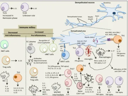
information is gathered, and transferred to the rest of the body [25,27]. MS involves 2 main steps, (i) myelin sheath damage resulting in formation of lesions in the CNS and (ii) inflammation, which together destroy the neuron tissue [25,28]. In MS, damage of oligodendrocytes and destruction of myelin sheath leads to breakdown of the nerve axon and loss of neuronal function [28]. Demyelination increases the inflammatory activation processes leading to damage of BBB and stimulation of macrophage activation and oxidative stress pathways [29]. The white matter lesions include myelin breakdown together with infiltration of monocytes, B cells, T cells and DC [30]. Microglia and macrophages are the main innate immune cells present in MS lesions where they either act together with T and B cells, or directly cause neuroinflammatory tissue damage [31]. Cells involved in the inflammatory process include those that are both in the innate and adaptive immune systems and are described below (Figure1).
Brain Sci. 2017, 7, 78 3 of 26
information is gathered, and transferred to the rest of the body [25,27]. MS involves 2 main steps, (i)
myelin sheath damage resulting in formation of lesions in the CNS and (ii) inflammation, which
together destroy the neuron tissue [25,28]. In MS, damage of oligodendrocytes and destruction of
myelin sheath leads to breakdown of the nerve axon and loss of neuronal function [28].
Demyelination increases the inflammatory activation processes leading to damage of BBB and
stimulation of macrophage activation and oxidative stress pathways [29]. The white matter lesions
include myelin breakdown together with infiltration of monocytes, B cells, T cells and DC [30].
Microglia and macrophages are the main innate immune cells present in MS lesions where they
either act together with T and B cells, or directly cause neuroinflammatory tissue damage [31]. Cells
involved in the inflammatory process include those that are both in the innate and adaptive immune
systems and are described below (Figure 1).
Figure 1. The immunological complexity of the immune/cytokine network in multiple sclerosis.
2.1. Natural Killer T (NKT) Cells
NKT cells share properties of both T cells and NK cells and recognize glycolipid antigens
presented in complex with the MHC class I‐like molecule, CD1d. Two subsets of NKT cells have
been identified (type I, invariant NKT (iNKT) cells and type II, variant NKT (vNKT) cells) and are
implicated in the pathogenesis of MS in humans and in the murine model of MS, experimental
autoimmune encephalomyelitis (EME). iNKT cells express cell surface markers characteristic of
activated or memory T cells (CD25, CD44, CD69) with the majority being CD4+ as well as markers characteristic of NK cells (NK1.1 or CD161, Ly49). Following activation of iNKT cells (via binding to α‐GalCer‐CD1d complex) an array of cytokines is secreted that are associated with both pro‐ and
anti‐inflammatory immune responses and play a role in both innate and acquired immunity. As
such, iNKT cells, (i) secrete interleukin (IL)‐4 and IL‐13 which stimulate CD4+ T cells to differentiate into anti‐inflammatory Th2 cells (IL‐4, IL‐10 producers) which inhibit Th17, Th1, CD8+ T cells in the
Figure 1.The immunological complexity of the immune/cytokine network in multiple sclerosis.
2.1. Natural Killer T (NKT) Cells
and tumor growth factor (TGF)-beta which stimulate the production of T regulatory (Treg) cells (IL-10, TGF-beta producers) which inhibit Th17, Th1 and CD8+T cells in the CNS; and (iii) secrete IL-4, IL-10, IL-13, interferon (IFN)-gamma and GM-CSF which activate suppressive myeloid derived suppressor cells (MDCs), DC and macrophages which in turn secrete IL-10 to activate Treg cells and suppress Th17, Th1 and CD8+ T cells in the CNS [32]. Due to the pleiotropic properties of iNKT cells, they play a role in protecting the host against pathogens, tumors, autoimmunity and are involved in tissue rejection, ischemia reperfusion injury and obesity related diabetes [32]; deficiency or dysfunction of iNKT cells has been shown to be linked to the development of autoimmune diseases. Indeed, iNKT cell numbers are decreased in patients with MS [32] and are restored in patients in remission [33]. Analysis of iNKT cells in MS patients in remission showed a Th2 cytokine profile, suggesting an immunoregulatory effect of iNKT cells in MS [34]. Similarly, in the EAE mouse model, protection of EAE development is associated with high levels of iNKT cells and suppression of Th1 and Th17 cells [35]. Interestingly, injections of α-Galactosylceramide (α-GalCer), and analogues thereof, have potent activities in protecting mice against, cancer, infections, inflammatory conditions and autoimmune disorders. Hence, it is possible to develop iNKT cell based modulating therapies against MS [36,37]. Like iNKT cells, variant NKT (vNKT) cells also share properties of both T cells (CD4+) and NK cells (NK1.1) and recognizeβ-linked glycolipid antigens in complex with CD1d. They are less common in mice compared to iNKT cells but are more abundant in humans. Of interest, vNKT cells recognize the self-glycolipid, sulphatide, which is abundantly expressed within the myelin sheath suggesting a role in MS although not yet established [38]. Likewise, vNKT cells recognizing sulphatide self-myelin ligand are present in high levels in mice with EAE suggesting their role in disease progression [38].
2.2. Mucosal-Associated Invariant T (MAIT) Cells
MAIT cells are a subset of T cells of the innate immune system to defend against microbial infections. They are present in the liver, lungs, mucosa and blood and make up to 25% of CD8 T cells in healthy individuals; they also support adaptive immune responses in that they have a memory like phenotype [39]. The MHC class I-like molecule, MRI, presents microbial antigens and vitamin B metabolites to MAIT cells, leading to their activation [39,40]. However, MAIT cells have also been implicated in autoimmune diseases such as MS, inflammatory bowel disease and rheumatoid arthritis where they are often noted at the site of autoimmune attack. Recently, it was reported that in MS, MAIT cells are highly present at the sites of demyelination and secrete pro-inflammatory Th1 cytokines (IFN-gamma and TNF-alpha) and activate Th17 cells (IL-17 and IL-22 cytokines) [22]; the major cytokines in the pathogenesis of chronic inflammatory and autoimmune diseases. In addition, MAIT cell have been noted in white matter inflammatory lesions [41] as well as transcription over expression of MR1 in MS lesions. Conversely, it has been reported that MAIT cells are decreased in blood of patients with RRMS [42]. It is not clear whether MAIT cells exert a protective or a non-protective role, thus a better understanding of how MAIT cells are involved in MS and of their interactions would aid in a better understanding of the pathogenesis of MS and development of therapeutic strategies.
2.3. Regulatory T Cells (Tregs)
Brain Sci.2017,7, 78 5 of 27
anti-CD28 monoclonal antibody in Lewis rats results in Treg cell expansion and reduction in EAE disease severity [48]. Interestingly, injection of anti-CD25 monoclonal antibody, which blocks the effects of Treg cells into C57BL/6 mice increased susceptibility to EAE induction [45]. In patients with MS however, the frequency of Foxp3+CD25+CD4+Treg cells does not differ to those in healthy individuals, although the function of such cells are impaired (maturation and migration) [49]. In addition, mRNA and protein levels of Foxp3 are impaired in Treg cells of patients with MS especially in RRMS and are normalized during SPMS [49]. Hence, impaired functionality of Treg cells is primarily observed in the early stages of MS but not in their chronic stage, suggesting a causative role [50]. Further studies of Treg cells in MS may aid in the understanding for why tolerance against self-antigens is broken, leading to disease. However, it is not clear whether the impaired function of Treg cells is a direct cause of MS or whether such impairment is a general outcome for all autoimmune disorders.
2.4. Macrophages and Microglia
Macrophages are divided into M1 or M2 based on their pro- or anti-inflammatory cytokine secretion phenotype [51]. M1 macrophage phenotype of mice (F4/80+CD11b+CD11c+iNOS+) and human (CD40+CD86+CD64+CD32+) is induced in the presence of interferon (IFN)-gamma and/or toll-like receptor (TLR) ligands such as lipopolysaccharide (LPS). M1 macrophages are pro-inflammatory and primarily secrete IL-1, IL-6, IL-12, TNF-alpha, iNOS and MCP-1 [51]. In general, they stimulate adaptive immune responses. The M2 macrophage phenotype of mice (F4/80+CD11c−CD301+Arg1+CD206+) and humans (CD163+CD206+) is induced in the presence of IL-4, IL-10, IL-13 and Arg1 that blocks iNOS activity [51]. M2 macrophages are anti-inflammatory and primarily secrete IL-1 receptor antagonist, IL-4, IL-10, transforming growth factor (TGF)-beta1. Macrophages play a crucial role in the pathogenesis of MS. In fact, in active demyelinating and early re-myelinating lesions, macrophages are highly present compared to inactive, demyelinated or late re-myelinated lesions [52]. However, a distinction of M1 vs M2 macrophages in human brain tissues is not so clear, with both M1 macrophages and an intermediate subtype (M1/M2, CD40+CD206+) being present [53]. Like macrophages, microglia cells are divided into M1- and M2-polarized microglia cells. M1 microglia cells are pro-inflammatory and express CD40, CD74, CD86 and CCR7, whereas, M2 microglia cells are anti-inflammatory and express mannose receptor (CD206) and CCL22. In MS brain lesions however, like macrophages, an intermediate microglia phenotype is present expressing CD40, CD74, CD86 and CCL22 but not CD206 markers [54]. Interestingly, in an EAE model it was shown that suppression of CCL22 decreased M1 macrophage accumulation in the CNS, thus therapies designed to suppress CCL22 have the potential to decrease demyelination and progression of disease. In addition, in mice M1 microglia cells have been found to switch to M2 microglia cells during remyelination, hence M2 polarization is necessary for efficient remyelination [55]. Indeed, fasudil (a selective Rho kinase inhibitor), injected into EAE bearing mice shifted M1 to M2 macrophages and ameliorated the clinical severity of EAE [56].
2.5. T Helper Cells
Th1 cells recognize MOG, PLP and MBP peptide epitopes presented in the context of MHC class II, HLA-DRB1*1501 (HLA-DR2, HLA-DR15) and HLA-DRB1*04 (HLA-DR4) alleles. As a result CD4 T cells become activated, cross the blood brain barrier and induce CNS autoimmunity. Some drug therapeutics target the MHC class II-peptide-T cell receptor (TCR) complex in an attempt to modulate or divert Th1 responses to therapeutic Th2 responses. Indeed, it was recently shown that dimethyl fumarate (DMF) injection in RRMS patients reduced Th1, Th17 and CD8 T cells and increased Th2 cells; this resulted in high levels of IL-4 and decreased levels of IFN-gamma and IL-17 [58]. In addition, we have shown that mannan conjugation of self-MBP, PLP or MOG native peptides or altered peptide ligands, are able to divert Th1 responses to Th2 responses in human PBMC from MS patients, in immunized mouse spleen cells and are able to ameliorate EAE in mice [59–73]. The role of Th9 cells in MS is not as clear although in mice, IL-9 and Th9 cells induce EAE and inflammation and IL-9 knockout mice are protected from developing EAE [74]. Th17 cells play a crucial role in the pathogenesis of MS in both mice and humans by inducing an inflammatory milieu. In fact, IL-17A is present at high levels in CNS lesions, cerebrospinal fluid and in the serum of patients with MS [75]. Th17 cells express high levels of CCR6 which binds to the ligand CCL20 on vascular endothelial cells, enabling their entry through the blood brain barrier where they secrete pro-inflammatory cytokines including IL-17A. In addition, IL-17 interferes with the remyelination process. Of interest, anti-IL-17A humanized neutralizing monoclonal antibody (AIN457 or Secukinumab) injected in patients with MS showed reduction of lesions compared to placebo-treated control subjects [75]. In addition, Th22 cells are highly present in the peripheral blood and cerebral spinal fluid of patients with active RRMS [76], and IL-22 mRNA and Th22 cells are increased in relapsing MS compared to remitting MS patients [77]. Furthermore, Th22 cells specifically recognize MBP and are resistant to IFN-beta therapy [76].
IL-27, a member of the IL-6/IL-12 cytokine family, is secreted by macrophages, dendritic cells and microglia cells, with pleiotropic roles in immunomodulation being either pro- or anti-inflammatory. IL-27 also stimulates or inhibits T cell differentiation. Th1 cells are induced by IL-27 whereas Th2, Th17 and Treg cells are inhibited by IL-27. In addition, Tr1 cells a specialized subset of T cells which secrete IL-10 are induced in the presence of IL-27 [78]. In 40 patients with RRMS, circulating plasma IL-27 levels were significantly higher compared to healthy control subjects [79]. Likewise, IL-27 and IL-27R are elevated in post-mortem MS brain lesions compared to non-MS control brains. Macrophages and microglia were identified to be the source of IL-27 and triggering infiltration of CD4 and CD8 T cells [80]. In addition, the effects of IL-27 on microglia cells showed that nitric oxide, TNF-alpha and IL-6 were secreted, promoting Th1 polarization, suggestive that IL-27 enhances microglia neuroinflammation [81]. Hence, suppressing IL-27 may be a strategy to modulate inflammatory responses in patients with MS.
2.6. CD8 T Cells
Brain Sci.2017,7, 78 7 of 27
MS lesions secrete high levels of IL-17 (classed as, Tc17 CD8 T cells) [84]. Tc17 cells secrete IL-17 and TNF-alpha and low IFN-gamma and are negative for granzyme B, perforin and cytolytic activity unlike the classical CD8 Tc1 cells. In peripheral blood of patients with SPMS and RRMS elevated levels of Tc1 and Tc17 cells are noted as well as a high percentage of TNF-alpha secreting CD8 T cells [85]; Tc21 cells are increased in the remission phase of RRMS compared to SPMS. In addition, higher levels of CD8+IFN-gamma+TNF-alpha+IL-17+T cells in the relapsing phase of RRMS compared to remission phase, SPMS and controls [85]. It is clear that CD8 T cells contribute to the pathogenesis of MS, and it is important to understand how such cells escape T cell tolerance and induce CNS autoimmunity in order to design and develop new therapeutics against MS.
2.7. B Cells
Although there is a presence of T cells in MS plaques, B cells also contribute to the pathogenesis of MS where they secrete autoantibodies and cytokines and being APC they activate T cells. In patients with MS the presence of oligoclonal bands (OCB) in cerebrospinal fluid and brain parenchyma is a consistent finding in over 95% of patients. OCB is a product of clonally expanded B cells and IgG synthesis. In MS plaques plasma cells are noted in large numbers where antigen uptake, processing and presentation takes place as well as synthesis of IgG. Interestingly, over 50 antibodies isolated from cerebrospinal fluid from patients with MS did not react to MBP, PLP or MOG [86] but some groups reporting that they bind to intracellular proteins such as, MKNK1/2, FAM84A, AKAP12A and glial potassium channel KIR4.1, or, against intracellular lipid determinants [87,88]. Moreover, anti-MOG autoantibodies is a hallmark of childhood MS as well as in some patients with neuromyelitis optical spectrum disorder. It is clear, that abnormal activation of B cells within the CNS of patients with MS, suggests that B cells play a role in the pathophysiology of the disease. Further studies are required to ascertain whether B cell depletion is able to restore immune function and hence, be used as a therapeutic target against MS.
2.8. Dendritic Cells
DC are professional APC which process and present antigenic peptide epitopes on their surface in complex with MHC class I or class II, resulting in CD4 or CD8 T cell stimulation respectively. Even though MS is generally associated with predominant auto-reactive T cells, emerging evidence indicates that DCs play an important role in the pathophysiology of MS, primarily due to their T cell activating and cytokine secreting properties. Following activation of DCs in the periphery, T cells specific to myelin epitopes are activated inducing pro-inflammatory cytokines aiding their entry through the BBB into the CNS. In the CNS resident APC and T cells are further activated leading to demyelination and motor deficits. In patients with MS, DCs are abundantly present within inflamed lesions, cerebrospinal fluid and in the circulation and produce high levels of TNF-alpha, IFN-gamma and IL-6 [89]. In addition, the expression of co-stimulatory molecules, CD40 and CD80 on DCs are increased in RRMS and SPMS patients, suggesting an activated pro-inflammatory state of DCs, hence their contributing role in the pathogenesis of MS.
2.9. Myeloid Derived Suppressor Cells
in patients with MS, leading to the failure of MDSCs to suppress autoimmune T cells, as a result of disease progression. The use of ex vivo cultured MDSCs could be a viable strategy to develop new improved treatments against MS.
3. Current Drug Therapies for Multiple Sclerosis
The majority of the treatments for MS are long term mainly suppressing the immune system however, such immune-suppressants pose increased risks for infections and cancer [27]. Alternative treatment options involve disease-modifying therapies such as, interferons, glatiramer acetate, monoclonal antibodies and sphingosine-1-phosphate receptor modulators (Table1, Figure2). These therapies have dramatically reduced the number of attacks and decreased disease progression. In fact, interferons are effective in the early relapsing phases of MS but not in the advanced phases of the disease [27]. Ultimately, induction of tolerance against self-antigens and re-establishing immune homeostasis can effectively “cure” the disease; such strategies have been the focus of recent research.
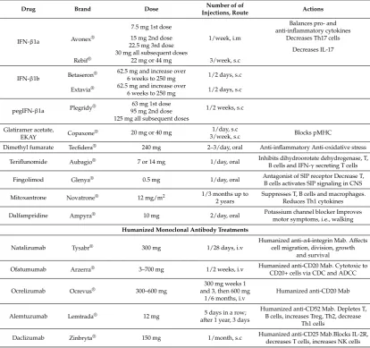
Table 1.Disease-modifying drugs available to patients with RRMS.
Drug Brand Dose Number of of
Injections, Route Actions
IFN-β1a Avonex®
7.5 mg 1st dose
1/week, i.m
Balances pro- and anti-inflammatory cytokines
15 mg 2nd dose Decreases Th17 cells
22.5 mg 3rd dose
Decreases IL-17 30 mg all subsequent doses
Rebif® 22 mg or 44 mg 3/week, s.c
IFN-β1b Betaseron®
62.5 mg and increase over
6 weeks to 250 mg 1/2 days, s.c Extavia® 62.5 mg and increase over
6 weeks to 250 mg 1/2 days, s.c
pegIFN-β1a Plegridy®
63 mg 1st dose
1/2 weeks, s.c 95 mg 2nd dose
125 mg all subsequent doses Glatiramer acetate,
EKAY Copaxone® 20 mg or 40 mg
1/day, s.c
Blocks pMHC 3/week, s.c
Dimethyl fumarate Tecfidera® 240 mg 2–3/day, oral Anti-inflammatory Anti-oxidative stress
Teriflunomide Aubagio® 7 or 14 mg 1/day, oral Inhibits dihydroorotate dehydrogenase, T,
B cells and IFN-γsecreting T cells
Fingolimod Glenya® 0.5 mg 1/day, oral Antagonist of SIP receptor Decrease T,
B cells activates SIP signaling in CNS
Mitoxantrone Novatrone® 12 mg/m2 1/3 months up to
2 years
Suppresses T, B cells and macrophages. Reduces Th1 cytokines
Dalfampridine Ampyra® 10 mg 2/day, oral Potassium channel blocker Improves
motor symptoms, i.e., walking Humanized Monoclonal Antibody Treatments
Natalizumab Tysabr® 300 mg 1/28 days, i.v Humanized
anti-α4-integrin Mab. Affects cell migration, division, growth
and survival
Ofatumumab Arzerra® 3–700 mg 1/2 weeks, i.v Humanized anti-CD20 Mab. Cytotoxic to
CD20+ cells via CDC and ADCC
Ocrelizumab Ocrevus® 300–600 mg
300 mg weeks 1 and 3, then 600 mg
1/6 months, i.v
Humanized anti-CD20 Mab
Alemtuzumab Lemtrada® 12 mg 5 days in a row;
after 1 year, 3 days
Humanized anti-CD52 Mab. Depletes T, B cells, increases Treg, Th2, decrease
Th1 cells
Daclizumab Zinbryta® 150 mg 1/month, s.c Humanized anti-CD25 Mab.Blocks IL-2R,
decreases T cells, increases NK cells
Brain Sci.2017,7, 78 9 of 27
Brain Sci. 2017, 7, 78 8 of 26
3. Current Drug Therapies for Multiple Sclerosis
The majority of the treatments for MS are long term mainly suppressing the immune system however, such immune‐suppressants pose increased risks for infections and cancer [27]. Alternative treatment options involve disease‐modifying therapies such as, interferons, glatiramer acetate, monoclonal antibodies and sphingosine‐1‐phosphate receptor modulators (Table 1, Figure 2). These therapies have dramatically reduced the number of attacks and decreased disease progression. In fact, interferons are effective in the early relapsing phases of MS but not in the advanced phases of the disease [27]. Ultimately, induction of tolerance against self‐antigens and re‐establishing immune homeostasis can effectively “cure” the disease; such strategies have been the focus of recent research.
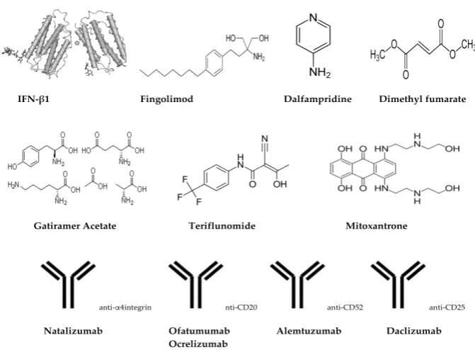
Figure 2. Chemical/schematic structures of treatments/drugs for MS.
Table 1. Disease‐modifying drugs available to patients with RRMS.
Drug Brand Dose Number of of
Injections, Route Actions
IFN‐β1a Avonex
®
7.5 mg 1st dose
1/week, i.m
Balances pro‐ and
anti‐inflammatory cytokines
15 mg 2nd dose Decreases Th17 cells
22.5 mg 3rd dose
Decreases IL‐17
30 mg all subsequent doses
Rebif® 22 mg or 44 mg 3/week, s.c
IFN‐β1b
Betaseron® 62.5 mg and increase over 6
weeks to 250 mg 1/2 days, s.c
Extavia® 62.5 mg and increase over 6
weeks to 250 mg 1/2 days, s.c
pegIFN‐β1a Plegridy
® 63 mg 1st dose 1/2 weeks, s.c 95 mg 2nd dose
125 mg all subsequent doses
Glatiramer
acetate, EKAY Copaxone® 20 mg or 40 mg
1/day, s.c
Blocks pMHC
3/week, s.c
Dimethyl
fumarate Tecfidera® 240 mg 2–3/day, oral
Anti‐inflammatory
Anti‐oxidative stress
Teriflunomide Aubagio® 7 or 14 mg 1/day, oral Inhibits dihydroorotate dehydrogenase, T, B cells and IFN‐γ secreting T cells
Fingolimod Glenya® 0.5 mg 1/day, oral
Antagonist of SIP receptor
Decrease T, B cells activates SIP
signaling in CNS
Mitoxantrone Novatrone® 12 mg/m2 1/3 months up to 2 years
Suppresses T, B cells and macrophages.
Reduces Th1 cytokines
Dalfampridine Ampyra® 10 mg 2/day, oral Potassium channel blocker Figure 2.Chemical/schematic structures of treatments/drugs for MS.
3.1. Treatment of MS Relapses
Patients with MS who present with a relapse are generally treated with corticosteroids intravenously, plasma exchange or adrenocorticotropic hormone injections [50,93]. Although effective in reducing the duration of the relapse and patients recovery faster there are no long-term neuroprotective benefits [27,94–97].
3.2. Long-Term Treatment of MS with Disease-Modifying Agents
The key therapeutic features of disease-modifying drugs are their anti-inflammatory effects in the relapsing phase of MS, although demyelination leading to chronic disability still remains a major hurdle [27,104–106]. Some studies, however, have shown that early intervention of disease-modifying drugs to patients with RRMS can reduce acute disability or death [27,107–110].
In general, disease-modifying drugs main action is by suppressing or altering the immune system. Hence, based on this theory that MS is, at least in part, a result of altered or abnormal immune response that results in attack of the myelin sheath. Current available drugs and their actions are described below (Table1, Figure2).
3.2.1. Interferons (Avonex®, Biogen, Cambridge, MA, USA; Betaseron®, Bayer, Leverkusen, Germany; Extavia®, Novartis Pharma AG, Basel, Switzerland; Rebif®, EMD Serono Inc., Darmstadt, Germany; Plegridy®, Biogen, Cambridge, MA, USA)
Interferon (IFN) type 1 consist of a group of IFNs (IFN-α, -β, -ε, -κ, -τ, -δ, -ζ, -ω, -ν) which help regulate the immune system. IFN-βis primarily produced by fibroblasts but other cells such as NK cells, B cells, T cells, macrophages also secrete IFN-β. IFN-βhas anti-viral and anti-tumor activity as well as being effective in reducing the relapse rate in patients with MS [106]. The mechanism by which IFN-βacts, is that it balances the expression of pro- and anti-inflammatory cytokines in the brain and decreases the number of inflammatory cells crossing the blood brain barrier. As a consequence, there is decreased inflammation of neurons, increases nerve growth factors and improves neuronal survival. Moreover, IFN-βreduces Th17 population and IL-17 cytokine which are known to be involved in the immunopathophysiology of MS [111]. IFN-βinjection subcutaneously or intramuscularly to patients with RRMS aims to decrease the relapse rate, duration and severity, however, there is lack of efficacy to long-term disability. Avonex was approved in 1996, the first FDA approved treatment for RRMS. To date there are 3 approaches using IFN-β; IFN-β1a low dosage (Avonex®), IFN-β1a (Rebif®) high dosage, and, IFN-β1b (Betaseron®, Extavia®) high dosage. Furthermore, pegIFN-β-1a (Plegridy®) has polyethylene glycol linked to IFN-β-1a allowing it to be active for longer in the body, hence fewer injections are required compared to Avonex®, Rebif®, Betaseron®and Extavia®. The first large scale human clinical trial in patients with RRMS using IFN-βwas published in 1993 and showed that relapse rates were reduced by 34% in high dose IFN-β1b and by 8% in lower dose compared to placebo group and severity of relapses were also reduced [112]. Subsequent 5 year follow-up data showed that IFN-β1a and IFN-β1b decreased lesions up to 30% and reduced the formation of new lesions up to 50%, however, the study failed to show any reduction in disability progression in patients [113]. IFNs have no direct neuroprotective effects, however, through their direct effect on CD4+Th1 cells and altering their profile results in decreased demyelination of neurons, which prevents further neuronal damage [114]. Despite the impact of IFN-βin disease progression in patients with RRMS there are limitations in their use, with side effects ranging from local body aches, skin reactions (swelling, redness), fever, myalgia, flu-like symptoms to more serious side effects such as suicidal thoughts, hallucinations, seizures and heart and liver problems [9]. As a result, many patients have stopped treatment and overall the benefit of using IFNs is relatively small.
3.2.2. Glatiramer Acetate (Copaxone®, Inc., Petah Tikva, Israel)
Brain Sci.2017,7, 78 11 of 27
results in side effects ranging from minor (fever, chills) to more serious (cardiovascular, digestive, muscular, respiratory issues).
3.2.3. Dimethyl Fumarate (Tecfidera®, Biogen, Cambridge, MA, USA)
Dimethyl fumarate (BG-12) is a methyl ester of fumaric acid that modulates immune responses and was approved by the FDA in 2013. BG-12 was shown in phase III clinical trials to reduce relapse rate and increase the time to disability progression in patients with RRMS [121]. BG-12 reduces the migration of inflammatory cells through the blood brain barrier and activates nuclear factor erythroid 2-related factor (Nrf2) [122]. Nrf2 regulates anti-oxidative proteins that protect cells against oxidative damage and inflammation. In fact, BG-12 protects neuronal cells from oxidative stress by increasing glutathione levels and suppressing pro-inflammatory cytokines from splenocytes in vitro [123]. Side effects of BG-12 include diarrhea, abdominal pain, nausea, abnormal liver enzymes and decreased lymphocyte counts.
3.2.4. Teriflunomide (Aubagio®, Sanofi Genzyme, Cambridge, MA, USA)
Teriflunomide is an active metabolite of leflunomide (an immunosuppressive disease-modifying drug used for rheumatoid arthritis) which inhibits the enzyme dihydroorotate dehydrogenase [124] and inhibits the proliferation of B and T cells. In addition, teriflunomide exerts anti-inflammatory properties by inhibiting IFN-gamma producing T cells while IL-4 and IL-10 producing T cells are unaffected [125]. In MS, oral administration of teriflunomide reduced relapse rates, MS lesions and decreased disability progression [126–131]. Moreover, permanent discontinuation due to side effects was substantially less common in MS patients who received teriflunomide compared to IFN-β-1a. Side effects include, reduced white blood cell count, alopecia, hepatic effects, nausea, diarrhea, numbness in hand and feet, allergic reactions, breathing issues and increased blood pressure. Teriflunomide was approved by the FDA in 2012 and by EMA in 2013 for use in patients with RRMS.
3.2.5. Fingolimod (Gilenya®, FTY720, Novartis Pharma AG, Basel, Switzerland)
Fingolimod was granted FDA approval in 2010 and was the first oral therapy (0.5 mg once daily) available for patients with relapsing forms of MS. Fingolimod is a sphingosine 1-phosphate (S1P) receptor modulator, which acts as a super agonist of S1P receptor causing receptor internalization and leading to reduced infiltration of potentially auto-reactive lymphocytes into the CNS, and as such, they remain localized in the lymph nodes [132–134]. In addition, a secondary beneficial effects of fingolimod is that it targets SIP receptors on glia cells in the CNS, activating signaling pathways within the CNS [132,135]. Based on Phase III human clinical trials in patients with RRMS (TRANSFORMS, FREEDOMS and FREEDOMS II), fingolimod was more effective compared to first line treatment IFNβ-1a and placebo, in reducing the frequency of flare-ups (clinical exacerbations), disability progression, MRI outcome measures, including brain volume loss and was associated with clearly identified adverse events [103,136,137]. More than 180,000 patients have been treated with fingolimod in clinical trials and post-marketing settings globally, and the total patient exposure now exceeds 395,000 patient-years. Side effects include bradycardia (within 6 h after treatment initiation), blurred vision, diarrhea, back pain, headache, cough and vomiting. With reasonable data showing its long-term safety and disease improvement, fingolimod is a great alternative choice for patients with highly active RRMS and who prefer the oral treatment option.
3.2.6. Mitoxantrone (Novantrone®, Immunex/Amgen, Thousand Oaks, CA, USA)
macrophages and reduces pro-inflammatory cytokines (IFN-γ, TNF-α, and IL-2) [138,139]. In patients with SPMS, intravenous injection of 12 mg/m2mitoxantrone every 3 months up to 2 years resulted in reduced disability progression by 84% [140,141]. However, several side effects are associated with mitoxantrone which range from nausea, vomiting, hair loss, to, cardiotoxicity, leukemia, infertility, infection, leukopenia and thrombocytopenia [11]. As a result, its use has significantly been reduced over time. Furthermore, due to the risk of cardiotoxicity and leukemia, there is a limit on the cumulative lifetime dose to be administered to patients [11,142].
3.3. Treatment Using Humanized Monoclonal Antibodies
3.3.1. Natalizumab (Tysabr®, Biogen, Cambridge, MA, USA)
Natalizumab is a humanized monoclonal antibody against the cellular adhesion molecule α4- integrin. Integrins are transmembrane receptors that enable cell-extracellular matrix adhesion activating cell signaling which regulate cell growth, division, survival, differentiation and migration. Integrins are expressed on T cells, B cells, monocytes, macrophages, NK cells, DC, neutrophils and eosinophils. Interfering or blockingα4-integrin affects immune cell migration across the blood brain barrier, thus, by blocking the interaction betweenα4-integrin and vascular endothelial adhesion molecule-1, inhibits transendothelial migration to the CNS [143]. Natalizumab is administered intravenously once a month [144] which reduces activated T cells within the CNS, resulting in anti-inflammatory responses and hence, neuroprotective effects [114]. In a phase III clinical trial natalizumab reduced brain lesions and the rate of disability progression up to 24 months [12,145]. In addition, natalizumab decreased by 92% of contrast-enhancing lesions, by 83% of new or expanding T2-weighted lesions, and by 76% in new T1-weighted hypointense lesions [146,147]. Natalizumab, was approved by the FDA in 2004, but was withdrawn due to 3 cases of rare brain infection, progressive multifocal leukoencephalopathy (PML; that usually leads to death or severe disability), but was re-introduced in 2006 under a special prescription program. However, by 2012 a further 212 cases (or 2.1/1000) of PML were reported to be attributed to natalizumab [148]. Despite these reports the FDA has not withdrawn natalizumab from the market as the clinical benefits outweigh the risks involved. Other side effects include, hepatotoxicity, allergic reactions and increased risks of infection. Due to the risks involved with natalizumab, there are reservations over its use as a preferred treatment option.
3.3.2. Ofatumumab (Arzerra®, Novartis Pharma AG, Basel, Switzerland)
Brain Sci.2017,7, 78 13 of 27
being further investigated in 2 Phase III trials (ASCLEPIOS I AND ASCLEPIOS II) and are recruiting patients with relapsing forms of MS (ofatumumab versus teriflunomide). The adaptive study design of both trials was recently presented by Hauser SL and colleagues at the American Academy of Neurology April 2017 in Boston, USA and results are highly anticipated [154].
3.3.3. Ocrelizumab (Ocrevus®, Genentech Inc., San Fransisco, CA, USA)
A few months ago (March 2017), the FDA approved ocrelizumab to be used in PPMS, the first drug approved by the FDA for this form of MS and phase IV clinical trials were a requirement of the FDA to be conducted in order to determine the safety of ocrelizumab in younger patients with MS, ie, risk of cancer and effects on pregnancy (study outcomes due by 2024); although clinical trials in patients with lupus and rheumatoid arthritis were halted due to high rates of infections and increased risk of progressive multifocal leukoencephalopathy [155]. In addition, in patients with MS, there was an increased risk of breast cancer (6/781 females with MS on ocrelizumab compared to 0/668 females with MS in other trials) [155].
3.3.4. Alemtuzumab (Lemtrada®, Sanofi Genzyme, Cambridge, MA, USA)
Alemtuzumab is a humanized monoclonal antibody against CD52, a cell surface molecule expressed on B and T cells; mature NK cells, plasma cells, neutrophils and importantly, hematological stem cells do not express CD52. In phase III clinical trials in patients with RRMS, alemtuzumab showed significantly lower annualized relapse rates and MRI measures (gadolinium-enhancing lesions, new or enlarging T2 lesions and brain atrophy) and were free of clinical disease longer, compared to IFNβ-1a [156,157]. Alemtuzumab can cause serious side effects including, immune thrombocytopenia, kidney problems, serious infusion problems (trouble breathing, swelling, chest pain, irregular heart beat), certain cancers (blood cancers, thyroid cancer), cytopenia and serious infections. It was approved by the FDA in 2014 to be used in RRMS patients, but due to the frequent and significant adverse events of alemtuzumab, it is generally used in patients with RRMS who have used 2 or more MS drugs and have failed to work.
3.3.5. Daclizumab (Zinbryta®, Biogen, Cambridge, MA, USA)
Daclizumab is a humanized monoclonal antibody against CD25, the IL-2 receptor expressed on the surface of T cells. The mechanism by which daclizumab works is that it blocks the IL-2 receptor on T cells, preventing the activation of T cells. It was originally approved by the FDA in 1997 to prevent acute kidney transplants (together with corticosteroids and cyclosporine) however its use was halted due to low market demand. In recent years its use has re-emerged to treat patients with RRMS, it is injected subcutaneously, once a month [158]. In human clinical trials, daclizumab showed 45% reduced annualized relapse rates and 54% lower in the number of new lesions [158]. The side effects associated with daclizumab are relatively minor compared to other MS drugs, and include infections, skin rashes and liver complications.
4. New and Emerging Immunotherapeutic Strategies against MS
Antigen/peptide specific immunotherapy or using immune cells (i.e., stems cells), aim to restore tolerance while avoiding the use of non-specific immunosuppressive drugs as describe in Section3, is a promising approach to fight autoimmune diseases including MS. As such, a number of approaches have been utilized.
4.1. Stem Cells
autoimmune diseases. The first report of a bone marrow transplant in 1997 in a chronic myelogenous leukemia patient with MS which showed marked improvements in MS brain lesions [159] quickly led to the use of HSC transplantation (HSCT) in MS patients. HSCT in patients with active RRMS, reduce progression in about 70% of patients, decrease relapses dramatically and suppresses inflammatory MRI activity [160]. MS patients who have not responded to conventional therapy, who’s disease is aggressive with relapsing-remitting course and who are not presenting with high level of disability, are considered appropriate candidates for such treatment [161]. Although the clinical efficacy of HSCT long term has not been established. The mechanism by which HSCT works is that HSCT “reboots” the immune system and thus, prevents inflammation associated with the disease.
Mesenchymal stem cells (MSC) are isolated from an adult’s bone marrow, are differentiated in vitro for 2–3 weeks and re-injected back into the patient. In recent years a vast amount of research has been conducted in MSCs to treat MS with most studies being in mice and EAE models, and more recently in human clinical trials. In fact, in a pilot study in advanced MS patients, MSC transplantation improved expanded disability scale score with stabilization in 1/7 and disease progression in 1/7 patients and vision and low contrast sensitivity test showed improvement in 5/6 patients with 1/6 showing worsening effects [162]. In a phase II randomized double-blind, placebo-controlled crossover clinical trial showed lower mean cumulative number of lesions in patients receiving MSCs compared to placebo [163]. No serious adverse events were reported. The mechanism of action of MSC includes immunomodulation, neuroprotection and neuroregeneration [162]. The use of MSCs that reduce MRI parameters is a new and emerging research focus to develop new improved treatments for MS.
4.2. DNA Vaccine Studies
BHT-3009, a DNA vaccine that encodes the full-length human MBP, was developed with the aim to tolerize patients with MS against MBP [9,164,165]. In fact, in 30 patients with RRMS or SPMS who received 4 injections of BHT-3009 on weeks 1, 3, 5, 9 with escalating doses of 0.5 mg, 1.5 mg or 3 mg was reported to be safe and conferred positive changes on brain MRI and reduced the number of CD4+T cells [9,164,166]. In addition, in a retrospective, randomized double blind, phase II study in 155 MS patients, BHT-3009 had no impact on the risk for persistent black holes (axonal loss and disability progression). However, there was a correlation to those who had generated high anti-IgM MBP antibodies to reduced risk of persistent black holes [167].
4.3. Nanoparticles
Nanoparticles have extensively been characterized and used as vaccine formulations in pre-clinical models of cancer and infectious diseases [168,169]. Polymeric biodegradable lactic-glycolic acid (PLGA) nanoparticles loaded with MOG35–55peptide together with recombinant IL-10, were partially endocytozed by dendritic cells, secreted both MOG35–55peptide and IL-10 in culture media for several weeks in vitro [170]. In mice, PLGA nanoparticles (MOG35–55+ IL-10) showed significant amelioration of EAE and reduction of IL-17 and IFN-gamma secretion by splenic T cells in vitro [170]. Recently, poly(ε-caprolactone) nanoparticles loaded with recombinant human MBP reduced IFN-gamma cytokines, reduced the clinical score and showed only mild histological changes of the myelin sheath [171]. Hence, nanoparticles as a delivery method of self-antigens are a promising tool to treat MS.
4.4. Altered Peptide Ligands
Brain Sci.2017,7, 78 15 of 27
multi-center double-blinded phase II clinical trial with NBI-5788 was suspended—Th2 responses were induced (IL-5, IL-13), however, 13/142 patients developed immediate-type hypersensitivity, who also generated anti-NBI-5788 antibodies which cross-reacted with native agonist MBP83–99peptide [172,174]. National Institute of Neurological Disorders and Stroke sponsored trial, CGP77116, was used in a MRI-controlled phase II clinical trial. CGP77116, has Ala D-amino acids of MBP83–99 peptide at positions 83, 84, 89, 91 (CGP77116) of MBP83–99peptide, in order to enhance stability [174]. However, this peptide was poorly tolerated at the dose tested, and the trial had to be discontinued. Three patients showed exacerbations to disease of which two were linked to CGP77116 injection with high IFN-gamma and low IL-4 (Th1-skewing) were secreted by activated CD4+T cells. These CD4+T cells also cross reacted with the native agonist MBP83–99peptide [175]. Accordingly, the problems noted with both NBI-5788 and CGP77116 were likely due to inadequate pre-screening of APL effects on the many clonotypes against the targeted epitopes. Thus, although the APL was highly effective at blocking or switching some clones, it activated others. Thus, further pre-clinical testing is required and new modified peptides need to be designed, or a carrier needs to be used which further changes the resulting immune response.
4.4.1. Cyclic Peptides
Cyclization of peptides increases the stability, since linear peptides are sensitive to proteolytic enzymes. In addition, cyclic peptides are an important intermediate step and a useful template towards the rational design and development of non-peptide mimetics. While mimetic strategy is a challenging perspective it is worth pursuing in particular for MBP epitope-based MS therapy as it is still in its infancy. Efforts to design semi-mimetics of MBP72–85epitope by combining non-natural amino acids as spacers and MBP epitope immunophores (Ser, Arg, Glu, Ala, Gln), led to substances that were effective to some extent in inducing the onset of EAE. Cyclic peptides are not only as a step towards non-peptide mimetics but also as putative therapeutics in MS [66].
Structure activity studies of the immunodominant agonist peptide MBP87–99, have shown that K91, P96 are important T cell receptor contact residues. Double mutation of K91, P96 to R91,A96 or single mutation of P96 to A96(APL) of either in their linear or cyclic forms, results in suppression of EAE and decreased inflammation in the spinal cord of Lewis rats [71]. Single and double cyclic[A91]MBP83–99peptide and cyclic[A91A96]MBP83–99peptides emulsified in CFA induced IL-4 cytokines in SJL/J mice [62] however conjugation to reduced mannan further enhanced IL-4 cytokines with no IFN-gamma responses [63]. In guinea pigs and Lewis rats, cyclic[A91A96]MBP83–99showed significantly reduced mechanical pain hypersensitivity compared to cyclic MBP83–99peptide. This was associated with reduced T cell and macrophage infiltration to injured nerves of the spinal cord of animals [176–178]. In addition, these APL decreased CD4+T cell line proliferation raised from a patient with MS, increased IL-10 cytokine secretion, bound to HLA-DR4 and were more stable to lysosomal enzymes (cathepsin B, D, H) compared to their linear counterparts [70]. Double mutation of K91, P96to A91, A96in either linear or cyclic forms were also shown to be active, with suppression of EAE in SJL/J mice, higher Th2 over Th1 cytokines produced, bound to HLA-DR4, the cyclic forms were more stable to lysosomal enzymes and induced high levels of IL-10 of peripheral blood mononuclear cells from patients with MS [61]. Recently, cyclic native agonist MOG35–55peptide was shown to ameliorate clinical and neuropathological features of EAE in mice compared to its linear counterpart [179]. Thus, cyclic peptides, which offer greater stability and are able to modulate immune responses, are novel leads for the immunotherapy of many diseases, such as MS [66].
4.4.2. Mannan as a Carrier to Modulate Immune Responses
trials; mannan-MUC1 induces protection against cancer recurrence at 18 years follow-up [182–185]. Furthermore, ex vivo cultured dendritic cells pulsed with mannan-MUC1 (CVacTM) and re-injection into patients induces strong cellular and clinical responses in ovarian cancer patients [186,187]. Due to the immunomodulatory properties of mannan, its effects as a carrier to MS peptides were determined.
Mutations of MBP83–99 agonist native peptide to result in mutant peptides (APL)—linear [A91]MBP
83–99, [E91]MBP83–99, [F91]MBP83–99, [Y91]MBP83–99 and [R91, A96]MBP83–99, induced IFN-gamma albeit reduced compared to the native agonist peptide, however, only the double APL [R91, A96]MBP83–99induced IL-4 secretion by T cells and antagonized IFN-gamma production in vitro by T cells against the native MBP83–99 peptide [67]. In addition, T cells against the native MBP83–99peptide cross-reacted with all peptides except [Y91]MBP83–99 and [R91,A96]MBP83–99 [68]. Conjugation of [R91,A96]MBP83–99, [A91,A96]MBP83–99, F91]MBP83–99, [Y91]MBP83–99 peptides to mannan, completed abrogated IFN-gamma responses and elicited high IL-4 (i.e., Th1 to Th2 switch) [63,69,188]. Likewise, linear double-mutant APL [L144R147]PLP139–151induces high levels of IL-4, and cyclization of this analog elicited low levels of IFN-gamma. When conjugated to mannan, [L144R147]PLP139–151peptide completely abrogated IFN-gamma, while both linear and cyclic native agonist PLP139–151 peptides stimulated IFN-gamma secreting T cells [64]. Furthermore, mannan conjugated to the immunodominant agonist MOG35–55peptide primes non-pathogenic Th1 and Th17 cells and ameliorates EAE in mice [73]; a phase I human clinical trial is planned using mannan conjugated to MOG35–55 peptide later this year. It is clear that, mannan is able to divert immune responses from Th1 to Th2 and is a promising carrier for further studies for the development of immunotherapeutics against MS.
5. Symptomatic Medication
Dalfampridine (Ampyra/Fampyra®, Acorda Therapeutics)
Dalfampridine is not intended to delay symptoms or change the course of disease, but rather, to improve motor symptoms such as walking. Dalfampridine, is a potassium channel blocker, resulting in improved potassium currents and nerve conductance. Dalfampridine is used in patients who have had MS for more than 3 years and it was approved by the FDA in 2010. Common side effects include nausea, nervousness and dizziness, which are relatively minor compared to other MS drugs.
6. Conclusions and Future Prospects
MS is an autoimmune disorder of the CNS with an array of immune cells being either activated or suppressed leading to demyelination and disease progression. In addition, genetic predisposition, viral mimicry, vitamin and mineral deficiency, geographical location are also etiological factors that contribute to disease. More recently, citrulination of myelin peptides have been shown to contribute to disease activation [59,60]. A number of treatment options are available to patients with MS, in particular those with active disease, however due to side effects, limited long term effectiveness and inability to reverse disease, new improved treatment options are required. As described here a number of new and upcoming promising therapeutic candidates are becoming available, although their effectiveness in human clinical trials remains to be determined. Recently, it was reported that non-peptide mimetics mapping the MBP83–96T cell epitope can function as T cell receptor antagonists, hence such an approach may pave the way to developing alternative and improved immunotherapeutics against MS [189]. With the plethora of information regarding the immunopathophysiology of MS and availability of treatment options and new upcoming treatments, the future holds promise for managing and treating the disease.
Acknowledgments: V.A. would like to thank Vianex S.A. Greece for support (Specific task agreement MS immunotherapeutics). V.A. and J.M. would like to thank Vianex S.A. Greece for their enthusiasm, support and helpful discussions regarding drug development and immunotherapeutics against MS.
Brain Sci.2017,7, 78 17 of 27
Conflicts of Interest:V.A. is supported by Vianex S.A. Greece in developing immunotherapeutics against MS; N.D. is supported under VU-Vianex contract 2 (specific task agreement MS immunotherapeutics) in developing immunotherapeutics against MS; J.M. is head of the scientific advisory board of ELDrug a spin off company of Vianex S.A.; M.-E.A. works for Vianex S.A. Greece; T.T. has an association with Vianex S.A. Greece in relation to supporting his research; M.K. is an employee of Novartis (Hellas) Greece; M.d.C. declares no conflicts of interest. The review represents a detailed literature search in the areas of drugs and treatments against MS with no bias towards immunotherapeutics developed by Vianex S.A.
References
1. Compston, A.; Coles, A. Multiple sclerosis.Lancet2002,359, 1221–1231. [CrossRef]
2. Grytten, N.; Torkildsen, O.; Myhr, K.M. Time trends in the incidence and prevalence of multiple sclerosis in norway during eight decades.Acta Neurol. Scand.2015,132, 29–36. [CrossRef] [PubMed]
3. Antel, J.; Antel, S.; Caramanos, Z.; Arnold, D.L.; Kuhlmann, T. Primary progressive multiple sclerosis: Part of the ms disease spectrum or separate disease entity? Acta Neuropathol. 2012,123, 627–638. [CrossRef] [PubMed]
4. Sadovnick, A.D.; Ebers, G.C.; Dyment, D.A.; Risch, N.J. Evidence for genetic basis of multiple sclerosis. Lancet1996,347, 1728–1730. [CrossRef]
5. Dai, H.; Ciric, B.; Zhang, G.X.; Rostami, A. Interleukin-10 plays a crucial role in suppression of experimental autoimmune encephalomyelitis by bowman-birk inhibitor. J. Neuroimmunol. 2012,245, 1–7. [CrossRef] [PubMed]
6. Hemmer, B.; Nessler, S.; Zhou, D.; Kieseier, B.; Hartung, H.P. Immunopathogenesis and immunotherapy of multiple sclerosis.Nat. Clin. Pract. Neurol.2006,2, 201–211. [CrossRef] [PubMed]
7. Sospedra, M.; Martin, R. Immunology of multiple sclerosis. Annu. Rev. Immunol. 2005, 23, 683–747. [CrossRef] [PubMed]
8. Rieckmann, P. Improving ms patient care.J. Neurol. Suppl.2004,251, v69–v73. [CrossRef] [PubMed] 9. Katsara, M.; Matsoukas, J.; Deraos, G.; Apostolopoulos, V. Towards immunotherapeutic drugs and vaccines
against multiple sclerosis.Acta Biochim. Biophys. Sin.2008,40, 636–642. [CrossRef] [PubMed]
10. Lublin, F.D.; Reingold, S.C. Defining the clinical course of multiple sclerosis: Results of an international survey.Neurology1996,46, 907–911. [CrossRef] [PubMed]
11. Eckstein, C.; Bhatti, M.T. Currently approved and emerging oral therapies in multiple sclerosis: An update for the ophthalmologist.Surv. Ophthalmol.2016,61, 318–332. [CrossRef] [PubMed]
12. Polman, C.H.; Reingold, S.C.; Banwell, B.; Clanet, M.; Cohen, J.A.; Filippi, M.; Fujihara, K.; Havrdova, E.; Hutchinson, M.; Kappos, L.; et al. Diagnostic criteria for multiple sclerosis: 2010 revisions to the mcdonald criteria.Ann. Neurol.2011,69, 292–302. [CrossRef] [PubMed]
13. Lunde Larsen, L.S.; Larsson, H.B.W.; Frederiksen, J.L. The value of conventional high-field mri in ms in the light of the mcdonald criteria: A literature review.Acta Neurol. Scand.2010,122, 149–158. [CrossRef] [PubMed]
14. Gafson, A.; Giovannoni, G.; Hawkes, C.H. The diagnostic criteria for multiple sclerosis: From charcot to mcdonald.Mult. Scler. Relat. Disord.2012,1, 9–14. [CrossRef] [PubMed]
15. Mahad, D.H.; Trapp, B.D.; Lassmann, H. Pathological mechanisms in progressive multiple sclerosis. Lancet Neurol.2015,14, 183–193. [CrossRef]
16. Minagar, A.; Alexander, J.S. Blood-brain barrier disruption in multiple sclerosis.Mult. Scler.2003,9, 540–549. [CrossRef] [PubMed]
17. Steinman, L. Multiple sclerosis: A coordinated immunological attack against myelin in the central nervous system.Cell1996,85, 299–302. [CrossRef]
18. Bennett, J.; Basivireddy, J.; Kollar, A.; Biron, K.E.; Reickmann, P.; Jefferies, W.A.; McQuaid, S. Blood–brain barrier disruption and enhanced vascular permeability in the multiple sclerosis model eae.J. Neuroimmunol.
2010,229, 180–191. [CrossRef] [PubMed]
19. Farjam, M.; Zhang, G.X.; Ciric, B.; Rostami, A. Emerging immunopharmacological targets in multiple sclerosis.J. Neurol. Sci.2015,358, 22–30. [CrossRef] [PubMed]
21. Jiang, J.; Kelly, K.A. Phenotype and function of regulatory t cells in the genital tract.Curr. Trends Immunol.
2011,12, 89–94. [PubMed]
22. Bianchini, E.; De Biasi, S.; Simone, A.M.; Ferraro, D.; Sola, P.; Cossarizza, A.; Pinti, M. Invariant natural killer T cells and mucosal-associated invariant T cells in multiple sclerosis.Immunol. Lett. 2017,183, 1–7. [CrossRef] [PubMed]
23. Tabarkiewicz, J.; Pogoda, K.; Karczmarczyk, A.; Pozarowski, P.; Giannopoulos, K. The role of il-17 and th17 lymphocytes in autoimmune diseases.Arch. Immunol. Ther. Exp.2015,63, 435–449. [CrossRef] [PubMed] 24. Van Hamburg, J.P.; Asmawidjaja, P.S.; Davelaar, N.; Mus, A.M.C.; Colin, E.M.; Hazes, J.M.W.;
Dolhain, R.J.E.M.; Lubberts, E. Th17 cells, but not th1 cells, from patients with early rheumatoid arthritis are potent inducers of matrix metalloproteinases and proinflammatory cytokines upon synovial fibroblast interaction, including autocrine interleukin-17a production. Arthritis Rheum.2011,63, 73–83. [CrossRef] [PubMed]
25. Dolati, S.; Babaloo, Z.; Jadidi-Niaragh, F.; Ayromlou, H.; Sadreddini, S.; Yousefi, M. Multiple sclerosis: Therapeutic applications of advancing drug delivery systems. Biomed. Pharmacother. 2017,86, 343–353. [CrossRef] [PubMed]
26. Münzel, E.J.; Williams, A. Promoting remyelination in multiple sclerosis-recent advances.Drugs2013,73, 2017–2029. [CrossRef] [PubMed]
27. Inglese, M.; Petracca, M. Therapeutic strategies in multiple sclerosis: A focus on neuroprotection and repair and relevance to schizophrenia.Schizophr. Res.2015,161, 94–101. [CrossRef] [PubMed]
28. Koriem, K.M.M. Multiple sclerosis: New insights and trends.Asian Pac. J. Trop. Biomed.2016,6, 429–440. [CrossRef]
29. Kallaur, A.P.; Lopes, J.; Oliveira, S.R.; Simão, A.N.; Reiche, E.M.; de Almeida, E.R.D.; Morimoto, H.K.; de Pereira, W.L.; Alfieri, D.F.; Borelli, S.D.; et al. Immune-inflammatory and oxidative and nitrosative stress biomarkers of depression symptoms in subjects with multiple sclerosis: Increased peripheral inflammation but less acute neuroinflammation.Mol. Neurobiol.2016,53, 5191–5202. [CrossRef] [PubMed]
30. Mirshafiey, A.; Jadidi-Niaragh, F. Prostaglandins in pathogenesis and treatment of multiple sclerosis. Immunopharmacol. Immunotoxicol.2010,32, 543–554. [CrossRef] [PubMed]
31. Fischer, M.T.; Sharma, R.; Lim, J.L.; Haider, L.; Frischer, J.M.; Drexhage, J.; Mahad, D.; Bradl, M.; Van Horssen, J.; Lassmann, H. Nadph oxidase expression in active multiple sclerosis lesions in relation to oxidative tissue damage and mitochondrial injury.Brain2012,135, 886–899. [CrossRef] [PubMed]
32. Van Kaer, L.; Wu, L.; Parekh, V.V. Natural killer t cells in multiple sclerosis and its animal model, experimental autoimmune encephalomyelitis.Immunology2015,146, 1–10. [CrossRef] [PubMed]
33. Gigli, G.; Caielli, S.; Cutuli, D.; Falcone, M. Innate immunity modulates autoimmunity: Type 1 interferon-beta treatment in multiple sclerosis promotes growth and function of regulatory invariant natural killer t cells through dendritic cell maturation.Immunology2007,122, 409–417. [CrossRef] [PubMed]
34. Araki, M.; Kondo, T.; Gumperz, J.E.; Brenner, M.B.; Miyake, S.; Yamamura, T. Th2 bias of cd4+ nkt cells derived from multiple sclerosis in remission.Int. Immunol.2003,15, 279–288. [CrossRef] [PubMed] 35. Mars, L.T.; Laloux, V.; Goude, K.; Desbois, S.; Saoudi, A.; Van Kaer, L.; Lassmann, H.; Herbelin, A.; Lehuen, A.;
Liblau, R.S. Cutting edge: V alpha 14-j alpha 281 nkt cells naturally regulate experimental autoimmune encephalomyelitis in nonobese diabetic mice.J. Immunol.2002,168, 6007–6011. [CrossRef] [PubMed] 36. Van Kaer, L. Alpha-galactosylceramide therapy for autoimmune diseases: Prospects and obstacles.Nat. Rev.
Immunol.2005,5, 31–42. [CrossRef] [PubMed]
37. Van Kaer, L.; Parekh, V.V.; Wu, L. Invariant nk t cells: Potential for immunotherapeutic targeting with glycolipid antigens.Immunotherapy2011,3, 59–75. [CrossRef] [PubMed]
38. Jahng, A.; Maricic, I.; Aguilera, C.; Cardell, S.; Halder, R.C.; Kumar, V. Prevention of autoimmunity by targeting a distinct, noninvariant cd1d-reactive t cell population reactive to sulfatide.J. Exp. Med.2004,199, 947–957. [CrossRef] [PubMed]
39. Napier, R.J.; Adams, E.J.; Gold, M.C.; Lewinsohn, D.M. The role of mucosal associated invariant t cells in antimicrobial immunity.Front. Immunol.2015,6, 344. [CrossRef] [PubMed]
Brain Sci.2017,7, 78 19 of 27
41. Abrahamsson, S.V.; Angelini, D.F.; Dubinsky, A.N.; Morel, E.; Oh, U.; Jones, J.L.; Carassiti, D.; Reynolds, R.; Salvetti, M.; Calabresi, P.A.; et al. Non-myeloablative autologous haematopoietic stem cell transplantation expands regulatory cells and depletes il-17 producing mucosal-associated invariant t cells in multiple sclerosis.Brain2013,136, 2888–2903. [CrossRef] [PubMed]
42. Miyazaki, Y.; Miyake, S.; Chiba, A.; Lantz, O.; Yamamura, T. Mucosal-associated invariant t cells regulate th1 response in multiple sclerosis.Int. Immunol.2011,23, 529–535. [CrossRef] [PubMed]
43. Kohm, A.P.; Carpentier, P.A.; Anger, H.A.; Miller, S.D. Cutting edge: CD4+CD25+ regulatory t cells suppress antigen-specific autoreactive immune responses and central nervous system inflammation during active experimental autoimmune encephalomyelitis.J. Immunol.2002,169, 4712–4716. [CrossRef] [PubMed] 44. Zhang, X.; Koldzic, D.N.; Izikson, L.; Reddy, J.; Nazareno, R.F.; Sakaguchi, S.; Kuchroo, V.K.; Weiner, H.L.
Il-10 is involved in the suppression of experimental autoimmune encephalomyelitis by CD25+CD4+ regulatory t cells.Int. Immunol.2004,16, 249–256. [CrossRef] [PubMed]
45. McGeachy, M.J.; Stephens, L.A.; Anderton, S.M. Natural recovery and protection from autoimmune encephalomyelitis: Contribution of CD4+CD25+ regulatory cells within the central nervous system. J. Immunol.2005,175, 3025–3032. [CrossRef] [PubMed]
46. Matejuk, A.; Bakke, A.C.; Hopke, C.; Dwyer, J.; Vandenbark, A.A.; Offner, H. Estrogen treatment induces a novel population of regulatory cells, which suppresses experimental autoimmune encephalomyelitis. J. Neurosci. Res.2004,77, 119–126. [CrossRef] [PubMed]
47. Weber, M.S.; Prod’homme, T.; Youssef, S.; Dunn, S.E.; Rundle, C.D.; Lee, L.; Patarroyo, J.C.; Stuve, O.; Sobel, R.A.; Steinman, L.; et al. Type ii monocytes modulate t cell-mediated central nervous system autoimmune disease.Nat. Med.2007,13, 935–943. [CrossRef] [PubMed]
48. Beyersdorf, N.; Gaupp, S.; Balbach, K.; Schmidt, J.; Toyka, K.V.; Lin, C.H.; Hanke, T.; Hunig, T.; Kerkau, T.; Gold, R. Selective targeting of regulatory t cells with cd28 superagonists allows effective therapy of experimental autoimmune encephalomyelitis.J. Exp. Med.2005,202, 445–455. [CrossRef] [PubMed] 49. Zozulya, A.L.; Wiendl, H. The role of regulatory t cells in multiple sclerosis.Nat. Clin. Pract. Neurol.2008,4,
384–398. [CrossRef] [PubMed]
50. Diebold, M.; Derfuss, T. Immunological treatment of multiple sclerosis.Semin. Hematol.2016,53(Suppl. 1), S54–S57. [CrossRef] [PubMed]
51. Mosser, D.M.; Edwards, J.P. Exploring the full spectrum of macrophage activation.Nat. Rev. Immunol.2008, 8, 958–969. [CrossRef] [PubMed]
52. Bruck, W.; Sommermeier, N.; Bergmann, M.; Zettl, U.; Goebel, H.H.; Kretzschmar, H.A.; Lassmann, H. Macrophages in multiple sclerosis.Immunobiology1996,195, 588–600. [CrossRef]
53. Vogel, D.Y.; Vereyken, E.J.; Glim, J.E.; Heijnen, P.D.; Moeton, M.; van der Valk, P.; Amor, S.; Teunissen, C.E.; van Horssen, J.; Dijkstra, C.D. Macrophages in inflammatory multiple sclerosis lesions have an intermediate activation status.J. Neuroinflamm.2013,10, 35. [CrossRef] [PubMed]
54. Peferoen, L.A.; Vogel, D.Y.; Ummenthum, K.; Breur, M.; Heijnen, P.D.; Gerritsen, W.H.; Peferoen-Baert, R.M.; van der Valk, P.; Dijkstra, C.D.; Amor, S. Activation status of human microglia is dependent on lesion formation stage and remyelination in multiple sclerosis. J. Neuropathol. Exp. Neurol. 2015, 74, 48–63. [CrossRef] [PubMed]
55. Miron, V.E.; Boyd, A.; Zhao, J.W.; Yuen, T.J.; Ruckh, J.M.; Shadrach, J.L.; van Wijngaarden, P.; Wagers, A.J.; Williams, A.; Franklin, R.J.; et al. M2 microglia and macrophages drive oligodendrocyte differentiation during cns remyelination.Nat. Neurosci.2013,16, 1211–1218. [CrossRef] [PubMed]
56. Liu, C.; Li, Y.; Yu, J.; Feng, L.; Hou, S.; Liu, Y.; Guo, M.; Xie, Y.; Meng, J.; Zhang, H.; et al. Targeting the shift from m1 to m2 macrophages in experimental autoimmune encephalomyelitis mice treated with fasudil. PLoS ONE2013,8, e54841. [CrossRef] [PubMed]
57. Apostolopoulos, V.; de Courten, M.P.; Stojanovska, L.; Blatch, G.L.; Tangalakis, K.; de Courten, B. The complex immunological and inflammatory network of adipose tissue in obesity.Mol. Nutr. Food Res.2016,60, 43–57. [CrossRef] [PubMed]