ISSN(Online): 2319-8753 ISSN (Print): 2347-6710
I
nternational
J
ournal of
I
nnovative
R
esearch in
S
cience,
E
ngineering and
T
echnology
(A High Impact Factor, Monthly, Peer Reviewed Journal) Visit: www.ijirset.com
Vol. 8, Issue 8, August 2019
Categorization of Lung Tissue Patterns in
Interstitial Lung Diseases
Kommajosyula Mahima, Pankaj Rathi
Research Scholar, M. Tech (D.C.), SITE, Nathdwara, Rajasthan, India
Associate Professor, SITE, Nathdwara, Rajasthan, India
ABSTRACT: In this analysis proposed and evaluated the convolution neural network designed for classification of ILD patterns. The 7outputs of ILD patterns: healthy, ground glass opacity (GGO), micro nodules, consolidation, reticulation, honeycombing and a combination of GGO/reticulation. To train and evaluate the CNN, we used first deep CNN designed for the specific problem. Finally we classify the performance (91%) demonstrated the potential of CNNs in analyzing lung patterns. We proposed a deep CNN to classify lung CT image patches into 7 different classes, The last layer provide different outputs ofthe tissue patterns such as healthy, Honeycombing , Ground Glass Opacity (GGO), Reticulation etc. We used a dataset of 100 CT scan which are collected from different radiology centers for training and evaluation of the system. In future we can use athree- dimensional images of the CT scans and also we can integrate this system into a computer aided diagnosis (CAD)system which will assist radiologist for better diagnosis
KEYWORDS: ILD, CNN, Deep learning, machine learning
I. INTRODUCTION
Convolution neural network gives better results of accuracy of medical image analysis. Convolution Neural Network (CNN) is a model of deep learning as classic example of deep learning. Convolution neural networks (CNN) are fully and locally connected to hidden layers unlike traditional neural network because for all fully connected networks, the operation becomes computationally intensive. CNNs use parameter sharing, pooling and dropout also which reduce the number of common features to large extent and hence addressing the computational issues[17[19][16]First convolution layer extracted features which are usually low-level features such as edges and lines and Subsequent layers extracted high-level features Size of input is N x N x D and this has to be convolved with kernels whose size k x k x D separately Convolution of an input with one kernel produces one output feature, and with H kernels independently produces . Features Each feature in the output consists of (N-k+1) x (N-k+1) elements. For each position of the kernel in a sliding window process, k x k x D elements of input and k x k x D elements of kernel are multiplied and added. The k x k x D multiply-accumulate operations are required for producing one output feature. Analysis and Comparison. Analyze the medical images based on features extracted in previous stage using suitable classifiers. The results will be finally compared with few results of conventional method of machine learning that may be ordinary neural network based learning technique. Even though the problems are superficially similar, research on image analysis for natural and medical images has traditionally been separated. Natural image analysis often refers to problems such as object detection, face recognition and 3D reconstruction, using images from normal RGB cameras. Medical image analysis entails tasks like detecting diseases in X-ray images, quantifying anomalies in MRI, segmenting organs in CT scans, etc.
ISSN(Online): 2319-8753 ISSN (Print): 2347-6710
I
nternational
J
ournal of
I
nnovative
R
esearch in
S
cience,
E
ngineering and
T
echnology
(A High Impact Factor, Monthly, Peer Reviewed Journal) Visit: www.ijirset.com
Vol. 8, Issue 8, August 2019
local connectivity and weight sharing. Every neuron of the layer is linked with a small input. Different neurons respond to different local areas of the input, which merge with each other to get a better representation of the image.
Figure 1: Block diagram of CNN system
Deep learning technology applied to medical imaging may become the most disruptive technology radiology has seen since the advent of digital imaging. Most researchers believe that within next 15 years, deep learning based applications will take over human and not only most of the diagnosis will be performed by intelligent machines but will also help to predict disease, prescribe medicine and guide in treatment. Which field in medical has revolutionized Deep learning first? ophthalmology, pathology, cancer detection, radiology or prediction and personalized medicine. Ophthalmology will be the first field to be revolutionized in health care, however, pathology, cancer diagnosis has received more attention and currently we have application with decent accuracy. Google Deep Mind Health is working with National Health Service, UK signed five year agreement to process the medical data of up to 1m patients across the trusts five hospitals. Even its early days of this project, Deep mind already has high hopes for the proposal. Researchers and vendors in medical sector are moving this field forward have a bold recommendation i.e. IBM Watson recently boosted itself through billion-dollar entry into the imaging arena by the acquisition Merge (imaging and Google Deep Mind Health is another big investment. [30][23]Even though, huge investment and interest, deep learning future in medical imaging is not that near as compare to other imaging applications due to the complexities involved in this field. The notion of applying deep learning based algorithms to medical imaging data is a fascinating and growing research area however, there are several barriers that slow down its progress. These challenges are efficient cases are also issue in case of rare disease. Another issue major issue is unbalancing of data that is very common in health sector i.e.rare diseases, by virtue of being rare, are underrepresented in the data sets. If not accounted for properly, the class imbalance that ensues
II. RELATED WORK
ISSN(Online): 2319-8753 ISSN (Print): 2347-6710
I
nternational
J
ournal of
I
nnovative
R
esearch in
S
cience,
E
ngineering and
T
echnology
(A High Impact Factor, Monthly, Peer Reviewed Journal) Visit: www.ijirset.com
Vol. 8, Issue 8, August 2019
images. Deep Learning remains in central focus of the research that would help in improving the results of conventional learning methods. The comparison is suggested regarding quantitative as well as quality statistical parameters such accuracy of medical image diagnosis, PSNR and computation time. A set of metrics of performance measures such as accuracy of CAD-based medical image analysis is recommended so that the same can be used for wide range of applications of medical image analysis
III. PROPOSED SYSTEM
The scans were produced by different CT scanners with slightly different pixel spacing so a pre-processing step was applied. The image intensity values were cropped and segmented the images. The six most relevant ILD patterns, namely GGO,reticulation, consolidation, micro nodules, honeycombing and a combination of GGO and reticulation. Healthy tissue was also added, leading to 7 classes.
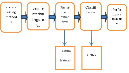
Fig.2 System Architecture
The annotators tried to avoid the bronchovascular tree which should be segmented and removed, before applying the fixed-scale classier. Annotation of the lung fields was also performed for all scans. The classification performance 85% demonstrated the potential of CNNs in analyzing lung patterns. We propose a deep CNN for the classification of ILD patterns that exploits the outstanding descriptive capability of deep neural networks. The method as been evaluated on a dataset of. To the best of our knowledge, this is the first time a deep CNN has been designed and trained for lung tissue characterization .Finally we provide empirical rules and principles on the design of CNN Architectures for similar texture classification problems. The scans were produced by different CT scanners with slightly different pixel spacing so a pre-processing step was applied, which rescale dalls can stomach a specific spacing value(i.e.,0.4mm).However ,the use of different reconstruction kernels by the scanners, still remains an open issue that complicates the Problem even further. The image intensity values were cropped within the window[-1000,200] in HU and mapped to [0,1].Experienced radiologists from the “Inselspital” annotated (or annotated) both databases by manually drawing polygons around the six most relevant ILD patterns, namely GGO, reticulation, consolidation, micro nodules, honeycombing and a combination of GGO and reticulation. Healthy tissue was also added, leading to 7 classes. The annotation focused on typical instances of the considered ILD patterns,
IMAGE SEGMENTATION: Image segmentation means division of an image into meaningful structures. It is process of extracting and representing information from the image to group pixels together with region of similarity [11]. Sonka et al. define the goal of segmentation as “to divide an image into parts that have a strong correlation with
Preproc essing method s Featur e extrac tion Classifi
ISSN(Online): 2319-8753 ISSN (Print): 2347-6710
I
nternational
J
ournal of
I
nnovative
R
esearch in
S
cience,
E
ngineering and
T
echnology
(A High Impact Factor, Monthly, Peer Reviewed Journal) Visit: www.ijirset.com
Vol. 8, Issue 8, August 2019
objects or areas of the real world contained in the image” [7]. Figure 3.1 shows a basic example of the image segmentation where Figure 3.1.is an original gray scale image and Figure 2.1b is a segmented image [19]. All the objects of the original image can be identified in segmented image with their boundaries. There are many techniques available for the image segmentation. Examples are, threshold based segmentation, edge based segmentation, region based segmentation, clustering based image segmentation, markov random field based segmentation and hybrid techniques
CONVERT IMAGE TO GRAY SCALE:-An MRI image is chosen from the file to be processed. This image is converted to gray scale image. These images have shades of gray between 0 to 255, where 0 corresponds to black and 255 to white for instance.
ENHANCEMENT AND SMOOTHING: There are different types of noise encountered by different techniques, depending on the noise nature and characteristics. In medical image processing, necessary to perform a high degree of noise reduction in an image before performing high level processing steps, so we used types of filers
HIGH PASS FILTERS: Noise presented in the image can reduce the capacity of region growing filter to grow large region or may result as a fault edges, so this gray scale image passes in to the filter. A high pass filter tends to retain the high frequency information within an image while reducing the low frequency information. So here, image filter as high pass filter is used to replace each pixel of the image with weighted average of the surrounding.
MEDIAN FILTER:Is nonlinear digital filter that is used to remove noise like salt and pepper, to smoothen and to preserve the edges of the image so it is very widely.
IV. CLASSIFICATION
A Convolution Neural Network (CNN) is a powerful machine learning technique from the field of deep learning. CNNs are trained using large collections of diverse images. From these large collections, CNNs can learn rich feature representations for a wide range of images. These feature representations often outperform hand-crafted features such as HOG, LBP, or SURF. An easy way to leverage the power of CNNs, without investing time and effort into training, is to use a pre-trained CNN as a feature extractor.
4.1. EXPERIMENTAL SETUP
(1)Evaluation: The evaluation of the different ILD patch classification approaches is based on a train-validation-test scheme. The actual training of the methods was carried-out on the training set, while the validation set was used for fine tuning Implementation:
LOCAL BINARY PATTERN (LBP)
ISSN(Online): 2319-8753 ISSN (Print): 2347-6710
I
nternational
J
ournal of
I
nnovative
R
esearch in
S
cience,
E
ngineering and
T
echnology
(A High Impact Factor, Monthly, Peer Reviewed Journal) Visit: www.ijirset.com
Vol. 8, Issue 8, August 2019
Fig.3 LBP Features
STATISTICAL FEATURE :Texture analysis techniques describe texture of regions in an image through higher order moments of their grayscale histograms. The most commonly used method for texture analysis is based on extracting various textural features from a gray level co-occurrence matrix (GLCM). The GLCM approach is based on the use of second-order statistics of the grayscale image histograms. Structural texture analysis techniques describe a texture as the composition of well-defined texture elements such as regularly spaced parallel lines. The properties and placement rules of the texture elements define the image texture. Model based texture analysis techniques generate an empirical model of each pixel in the image based on a weighted average of the pixel intensities in its neighbourhood. The estimated parameters of the image models are used as textural feature descriptors. Transform based texture analysis techniques convert the image into a new form using the spatial frequency properties of the pixel intensity variations. The success of this type lies in the type of transform used to extract textural characteristics from the image. The image processing of citrus fruit images using statistical and transform based texture analysis is explained here.
GRAY-LEVEL CO-OCCURRENCE MATRIX (GLCM):Texture Analysis Using the Gray-Level Co-Occurrence
Matrix (GLCM The GLCM functions characterize the texture of an image by calculating how often pairs of pixel with specific values and in a specified spatial relationship occur in an image, creating a GLCM, and then extracting statistical measures from this matrix.
ISSN(Online): 2319-8753 ISSN (Print): 2347-6710
I
nternational
J
ournal of
I
nnovative
R
esearch in
S
cience,
E
ngineering and
T
echnology
(A High Impact Factor, Monthly, Peer Reviewed Journal) Visit: www.ijirset.com
Vol. 8, Issue 8, August 2019
Proposed CNN In order to decide on the optimal architecture and configuration of a CNN, one should first comprehend the nature of the problem considered - in this case - the classification of ILD patterns. Unlike arbitrary objects in colour images, which involve complex, high-level.
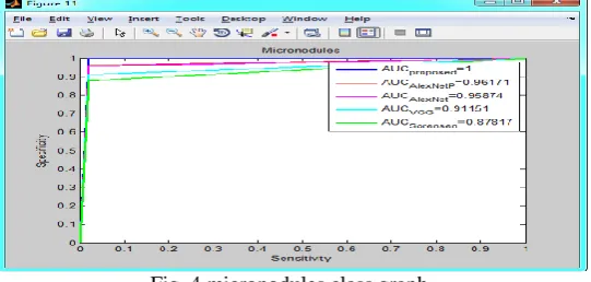
We also analysis for AlexNet, AlexNet pre-trained (AlexNetP), VGG-Net, the method by Sorensen et al. [13] and the proposed CNN., the area under the curve (AUC) was computed and the 95% confidence interval was plotted according to [41]. The comparison showed that the proposed method achieved the highest AUC on each of the 7 classes. To test the statistical significance of the AUC differences, The analysis was performed per class (one-vs-all) while the average over all classes is also presented. For each ROC, the AUC is given and the 95% confidence interval is plotted. Comparing on the most difficult patterns i.e., consolidation, reticulation, honeycombing and reticulation/GGO. For the rest of the patterns (healthy, GGO and micronodules) the difference between the proposed method and the pre-trained AlexNet was not considered significant, while for GGO the difference from VGG-Net was also non-significant. Finally, the superiority of the proposed method after averaging over all considered classes was also found to be statistically significant.
Fig.5 Ground Glass Opacity Class Graph
Fig.6. Accuracy graph
V. CONCLUSION
ISSN(Online): 2319-8753 ISSN (Print): 2347-6710
I
nternational
J
ournal of
I
nnovative
R
esearch in
S
cience,
E
ngineering and
T
echnology
(A High Impact Factor, Monthly, Peer Reviewed Journal)
Visit: www.ijirset.com Vol. 8, Issue 8, August 2019
by using data augmentation to generate more training images of rare or abnormal data, though there is risk of over fitting. Aside from data-level strategies, algorithmic modification strategies and cost sensitive learning have also been Analysis
REFERENCES
1. Syed MS Islam Hassan Mahmood Adel Ali Al-Jumaily ; Scott Claxton Deep Learning of Facial Depth Maps for Obstructive Sleep Apnea Prediction Published in: 2015 IEEE 12th International Symposium on Biomedical Imaging (ISBI) DOI: 10.1109/iCMLDE.2018.00036 Sydney, Australia,
2. Yaniv Bar Idi tDiamant Lior Wolf ; Sivan Lieberman Date of Conference: 16-19 Chest pathology detection using deep learning with non-medical training DOI: 10.1109/ICIIECS.2018.8276011
3. Ho-Shon Sarvnaz Karim Len Hamey Modality Classification and Concept Detection in Medical Images Using Deep Transfer Learning 2018 International Conference on Image and Vision Computing New Zealan(IVCNZ)DOI: 10.1109/IVCNZ.2018.8634803Teleconference Location: Auckland, New Zealand, New Zealand
4. JahanzaibLati fChuangbai Xia Medical Imaging using Machine Learning and Deep Learning Algorithms: A Review 2019 2nd International Conference on Computing, Mathematics and Engineering Technologies (iCoMET)10.1109/ICOMET.2019.8673502Sukkur, Pakistan, 5. Nirmala Singh Sachchidanand Singh Object classification to analyze medical imaging data using deep learning Published in: 2017 International
Conference on Innovations in Information, Embedded and Communication Systems ICIIECS)DOI: 10.1109/ICIIECS.2017.8276099 6. Conference Location: Coimbatore, India
7. J.selvakumar , A.Lakshmi, T.Arivoli, “Brain Tumor Segmentation and Its Area Calculation in Brain MR images using K-Mean Clustering and Fuzzy C-Mean Algorithm” , IEEE-International Conference On Advances In Engineering, Science And Management (ICAESM -2012) March 30, 31, 2017
8. Sudipta Roy, Samir K.Bandyopadhyay, “Detection and Quantification of Brain Tumor from MRI of Brain and it‟s Symmetric Analysis”, IJICTR, Volume 2 No. 6, June 2012.
9. Krishna Kant Singh , AkanshaSingh,“A Study Of Image SegmentationAlgorithms For Different Types Of Images”, IJCSI International Journal of Computer Science Issues, Vol. 7, Issue 5,September 2018
10. Qurat-Ul-Ain, L.G., Kaz mi, S.B., Jaffar, M.A ., Mirza, A.M.: „ Classificationandsegmentation of brain tumor using texture analysis‟ , Recent Adv. Artif. Intell. Knowl. Eng. Data Bases, 2015 10, pp. 147 –155
11. 2 Khalid, N.E.A., Ibrahim, S., Haniff, P.N.M.M.: „MRI brain abnormalities segmentation using K-nearest neighbors (k-NN) ‟, Int. J. Comput. Sci. Eng. (IJCSE), 2011, 3, (2), pp. 980 –990
12. 3 Aslam, H.A., Ramashri, T., Ahsan, M.I.A.: „A new approach to imagesegmentation for brain tumor detection using pillar K-means algorithm ‟, Int. J. Adv. Res. Comput. Commun. Eng., 2013, 2, (3), pp. 1429 –1436
13. Maity, A., Pruitt, A.A., Judy, K.D.: „Cancer of the central nervous system‟ (Clinical Oncology, 2008, 4th edn.) 14. Ricci, P.E., Dungan, D.H.: „Imaging of low and intermediate-grade gliomas ‟, Semradonc, 2001, 11, (2), pp. 103 –112
15. Rajini, N.H., Narmatha, T., Bhavani, R.: „Automatic classification of MR brain tumor images using decision tree ‟. Special Issue of Int. J. of Computer Applications on Int. Conf. on Electronics, Communication and Information Systems (ICECI 12), 2012, pp. 10 –13