R E S E A R C H P A P E R
TNF-a
gene expression is increased following zinc
supplementation in type 2 diabetes mellitus
Anna Chu•Meika Foster• Dale Hancock•
Kim Bell-Anderson•Peter Petocz •Samir Samman
Received: 5 September 2014 / Accepted: 28 October 2014 / Published online: 15 November 2014 ÓSpringer-Verlag Berlin Heidelberg 2014
Abstract Chronic low-grade inflammation in type 2 diabetes mellitus (DM) can elicit changes in whole-body zinc metabolism. The interaction among the expression of inflammatory cytokines, zinc transporter and metallothio-nein (MT) genes in peripheral blood mononuclear cells in type 2 DM remains unclear. In a 12-week randomized controlled trial, the effects of zinc (40 mg/day) supple-mentation on the gene expression of cytokines, zinc transporters and MT in women with type 2 DM were examined. In the zinc-supplemented group, gene expres-sion of tumour necrosis factor (TNF)-a tended to be upregulated by 27±10 % at week 12 compared to base-line (P=0.053). TNF-a fold change in the zinc-treated group was higher than in those without zinc supplemen-tation (P\0.05). No significant changes were observed in the expression or fold change of interleukin (IL)-1borIL-6. Numerous bivariate relationships were observed between
the fold changes of cytokines and zinc transporters, includingZnT7withIL-1b(P\0.01),IL-6(P\0.01) and TNF-a (P\0.01). In multiple regression analysis, IL-1b
expression was predicted by the expression of all zinc transporters and MT measured at baseline (r2=0.495, P\0.05) and at week 12 (r2=0.532, P\0.03). The current study presents preliminary evidence that zinc sup-plementation increases cytokine gene expression in type 2 DM. The relationships found among zinc transporters, MT and cytokines suggest close interactions between zinc homeostasis and inflammation.
Keywords InflammationCytokinesZinc transporters
MetallothioneinGene expressionType 2 diabetes mellitus
Introduction
Zinc is involved in the biosynthesis, storage and secretion of insulin within the pancreatic b-cells (Huang 2014). Furthermore, intracellular zinc can act directly on the insulin signalling pathway to improve glucose uptake and insulin sensitivity in peripheral tissues (Haase and Maret 2005; Foster and Samman 2010). Individuals with type 2 diabetes mellitus (DM) are suggested to be of poor zinc status due to the presentation of higher urinary zinc excretion and lower serum zinc concentrations (Jansen et al. 2009). Suboptimal zinc status can affect glycaemic control by compromising the production and secretion of insulin in the pancreas (Huber and Gershoff 1973) and impacting insulin sensitivity in peripheral tissues (Jansen et al. 2009; Kelleher et al. 2011). Hence, the persistent elevation of blood glucose concentration featured in type 2 DM may be attributed partly to perturbed zinc homeostasis. Electronic supplementary material The online version of this
article (doi:10.1007/s12263-014-0440-4) contains supplementary material, which is available to authorized users.
A. ChuM. FosterK. Bell-AndersonS. Samman Discipline of Nutrition and Metabolism, School of Molecular Bioscience, University of Sydney, Sydney, NSW 2006, Australia
D. Hancock
Discipline of Molecular Biology, School of Molecular Bioscience, University of Sydney, Sydney, NSW 2006, Australia
P. Petocz
Department of Statistics, Macquarie University, Sydney, NSW 2109, Australia
S. Samman (&)
Department of Human Nutrition, University of Otago, PO Box 56, Dunedin 9054, New Zealand
Cellular zinc homeostasis is regulated primarily by two families of zinc transporters and metallothionein (MT) (Lichten and Cousins2009). The Zrt- and Irt-like protein (Zip) (SLC39) family of transporters is responsible for increasing cytoplasmic zinc concentration by transporting zinc from intracellular organelles or the extracellular space. Conversely, the ZnT (SLC30) family of transporters functions to decrease the cytoplasmic zinc concentration by transporting zinc from the cytosol into the extracellular space or internal organelles, such as those involved in secretory pathways. MT acts as a target for zinc ion binding in the cytoplasm and is believed to assist in the trafficking of zinc ions through the cell (Babula et al. 2012). The regulatory control of cellular zinc transporters and MT is complex and has been shown to be influenced by zinc status, systemic glucose and inflammatory cytokines (Lichten and Cousins2009; Hennigar and Kelleher2012). Cellular zinc transporters in different cell types have been shown to be both up- and downregulated to meet the changing demand for zinc in inflammatory conditions (Foster and Samman2012). The redistribution of zinc from the systemic circulation into cellular compartments is thought to be crucial for immune function. During acute infection, an increase in systemic interleukin (IL)-6 can induce hepatic accumulation of zinc, which contributes to the rapid decrease in the plasma zinc concentration often seen in the acute phase response (Liuzzi et al.2005). The chronically activated innate immune system in type 2 DM (Pickup2004) also can elicit changes to whole-body zinc metabolism. Exposure to systemic cytokines, such as IL-1b, have been associated with downregulation of the zinc transporters shown to play a role in insulin production and storage in the pancreaticb-cells (Egefjord et al.2009).
The expressions of immune markers, such as 1b, IL-6 and TNF-a, often are linked to the progression of type 2 DM (Donath and Shoelson2011). Specifically, increase in IL-1b has been shown to promote pancreatic b-cell destruction (Banerjee and Saxena2012) which, in combi-nation with TNF-a and IL-6, may synergistically exacer-bate the extent ofb-cell apoptosis and disease progression (Cnop et al. 2005). Furthermore, TNF-a and IL-6 can promote insulin resistance in peripheral tissues by modu-lating the expression of key regulators in the insulin sig-nalling pathway (Mlinar et al.2007). Attenuation of CRP and inflammatory cytokine production may be achieved by dietary supplementation of anti-inflammatory nutrients, such as zinc and n-3 polyunsaturated fatty acids (PUFA) (Calder 2006; Foster and Samman 2012). In a 6-week cross-over intervention study which manipulated the composition of fatty acids, a diet high ina-linolenic acid (ALA) derived from FSO resulted in significant reductions in cytokine production and vascular inflammation in hypercholesterolemic subjects (Zhao et al. 2004, 2007).
The effectiveness of a modest level of ALA supplemen-tation on inflammation in type 2 DM is currently unknown. Similarly, the effect of zinc supplementation on inflam-matory biomarkers in type 2 DM remains largely unex-plored. While zinc supplementation appears to reduce the level of systemic CRP and IL-6 (Bao and Prasad 2010), conflicting findings are reported for the effect of zinc sup-plementation on ex vivo cytokine production in stimulated mononuclear cells (Aydemir et al. 2006; Prasad and Beck 2007). In a recent zinc supplementation trial in individuals with type 2 DM (Foster et al. 2013b), we reported that systemic inflammatory markers were associated with the gene expression of zinc transporters and MT, suggesting an interplay between systemic markers of inflammation and cellular zinc transport. Consistent with our observation, a recent study in obese women showed levels of systemic C-reactive protein (CRP) and tumour necrosis factor (TNF)-ato be inversely correlated with a range of zinc transporter gene expressions (Noh et al.2014).
Peripheral blood mononuclear cells (PBMC) have been used as a candidate target tissue to detect transcriptome changes in response to dietary modification in humans (de Mello et al.2012). The expression of zinc transporters and MT in PBMC have been described previously in a healthy population (Foster et al. 2011) and those with type 2 DM (Foster et al.2013b). However, the interactions between the gene expression of inflammatory cytokines, zinc transport-ers and MT remain unclear. To extend our previous report, specifically the relationships between systemic cytokines (IL-1b, IL-6 and TNF-a) and zinc transporters and MT (Foster et al.2013b), the gene expressions ofIL-1b,IL-6and TNF-ain PBMC were measured to provide further insight into the interactions between zinc and the immune system in type 2 DM. The present study aims to investigate the effect of zinc on the gene expression of inflammatory cytokines in PBMC and explore possible relationships between the gene expression of cytokines, zinc transporters and MT.
Materials and methods
Study design
and the inclusion of women who suffer from chronic conditions such as type 2 DM contributes biomedical knowledge that advances patient care (Kim et al. 2010). Enrolled participants were randomized into four equal groups to receive a total of 40 mg/day elemental zinc (‘Zn Group’), 2,000 mg/day flaxseed oil (‘FSO group’), both zinc and flaxseed oil (‘Zn?FSO group’), or placebo for 12 weeks. Placebo capsules that were identical in appear-ance to their active counterparts were given to the placebo groups. Zinc placebo capsules contained cellulose, while olive oil was used as the placebo for FSO. All procedures followed were in accordance with the ethical standards of the Human Research Ethics Committee of the University of Sydney. Informed consent was obtained from all partici-pants for being included in the study. The study protocol was registered atwww.clinicaltrials.gov(NCT01505803). Markers of systemic inflammation, glycaemia and zinc Venous blood samples from participants were collected at baseline, and at weeks 4, 8 and 12 for the analysis of glucose, haemoglobin A1c (HbA1c), cytokines, CRP and zinc. Serum glucose was measured by glucose hexokinase UV method using the Gluco-quant reagent kit adapted for a Modular PPE auto-analyser (Roche Diganostics, Basel, Switzerland). Serum insulin was determined by chemilu-minescent microparticle immunoassay on an Architect i2000SR Analyzer (Abbott Laboratories, Abbott Park, IL, USA). HbA1c was assayed using ion-exchange high per-formance liquid chromatography (HPLC) on a Variant II analyser equipped with the Variant II NU Program (Bio-Rad Laboratories, Hercules, CA, USA), according to the manufacturer’s protocol. Plasma zinc was determined using inductively coupled plasma mass spectrometry (Ag-ilent 7500ce ICPMS, Santa Clara, CA, USA). The human cytokine/chemokine Milliplex MAP kit (Millipore, Bille-rica, MA, USA) was used for the simultaneous quantifi-cation of serum IL-1b, IL-6 and TNF-a concentrations, according to the manufacturer’s instructions. Samples were analysed on a Luminex 100 Bioanalyser (Luminex Corp., Austin, TX, USA) using Fidis multiplex technology (Bio-medical Diagnostics, Marne la Valle´e, France). Serum CRP was measured using the Tina-quant CRP (gen.3) immu-noturbidimetric method adapted for a Roche Modular PPE analyser (Roche Diagnostics, Basel, Switzerland) accord-ing to the manufacturer’s instructions.
Zinc transporter, metallothionein and cytokine gene expressions
PBMC were isolated from blood samples collected at baseline and week 12, processed through to cDNA and stored at-80°C until quantitative real-time PCR analysis.
Unstimulated PBMC from individual samples were extracted, and total RNA was prepared using the RNAqu-eous Small Scale Phenol-Free Total RNA Isolation Kit (Applied Biosystems-Life Technologies Australia Pty Ltd, Victoria, Australia) according to the manufacturer’s instructions. Total RNA was reverse transcribed into cDNA using the Superscript VILO cDNA Synthesis System (Invitrogen-Life Technologies Australia Pty Ltd, Victoria, Australia) following the manufacturer’s protocol. Forty complete samples of cDNA from study participants were recovered for cytokine gene expression analysis. Invento-ried TaqMan gene expression assays were obtained forIL -1b, IL-6 andTNF-afor relative quantification of cytokine mRNA using TaqMan real-time PCR, as per the manufac-turer’s instructions (StepOnePlus Real-Time PCR System, Applied Biosystems-Life Technologies Australia Pty Ltd, Victoria, Australia). The selected cytokine transcripts cor-respond with measures of systemic inflammation that pre-viously showed relationships with expression of zinc transporter genes (Foster et al. 2013b). Relative quantifi-cation of zinc transporter mRNA was conducted using TaqMan real-time PCR (ABI 7500 Fast Sequence Detection System; Applied Biosystems-Life Technologies Australia Pty Ltd, Victoria, Australia). Inventoried TaqMan gene expression assays, and one custom-designed assay, were obtained for ZnT1, ZnT5, ZnT6, ZnT7,ZnT8, Zip1, Zip3, Zip7,Zip10,MT-1AandMT-2AmRNA (Applied Biosys-tems-Life Technologies Australia Pty Ltd, Victoria, Aus-tralia). Messenger RNA expression levels for all genes were normalized to 18S rRNA expression as an endogenous reference and quantified using theDCPmethod; fold change relative to baseline was quantified using theDDCPmethod. Statistical analysis
Descriptive statistics were expressed as mean±SD. Cytokine gene expressions and fold changes were descri-bed as mean±SEM. Differences in group means of baseline characteristics, cytokine expressions and fold changes were assessed by analysis of variance (ANOVA). No significant changes were observed in primary outcomes measured (glycaemia, systemic inflammatory markers or cytokine mRNA expressions) after FSO supplementation, with or without zinc. Hence, post hoc investigations using independent t-tests were used to compare groups catego-rized according to whether participants did or did not receive zinc supplementation. Post hoc analysis according to FSO supplementation was performed and confirmed no effect on cytokine gene expressions when FSO was con-sumed alone or in combination with zinc.
models were used to explore the relationships among gene expression ofIL-1b, IL-6 and TNF-a and expression of all zinc transporters and MT measured, at baseline and week 12. The residuals from the regression models were checked to see whether they satisfied the assumptions of normality and homoscedasticity: the initial regressions indicated that analyses of MT-1A and MT-2A were more appropriately conducted on a log scale. Statistical analyses were carried out using SPSS (PASW) version 18. With analysis of the primary outcomes, a value ofP\0.05 was taken to des-ignate statistical significance. In univariate outcomes within multivariate analyses, a more conservative desig-nation ofP\0.01 as statistically significant was used due to the large number of statistical tests employed.
Results
Participant characteristics
Characteristics of study participants, biomarker outcomes and zinc transporter expressions have been described pre-viously in detail (Foster et al.2013a,b). Of the 48 enrolled participants, complete sets of cDNA samples were avail-able from 40 participants for PBMC cytokine gene expression analysis. The age and BMI of participants were (mean±SD) 65.1±8.0 years and 28.3±5.0 kg/m2, respectively. The average time since type 2 DM diagnosis was 6.6±5.4 years. The baseline fasting blood glucose concentration, fasting insulin and HbA1c of the partici-pants were 6.7±1.8 mmol/L, 67.2±36.8 pmol/L and 6.7±1.0 %, respectively. The mean plasma zinc con-centration was 12.8±2.0lmol/L, which is within the reference range of 10–18lmol/L. The baseline serum concentrations of CRP, IL-1b, IL-6 and TNF-a were 1.7±2.2 mg/L, 1.0±1.9 pg/mL, 1.6±2.0 pg/mL and 10.3±3.5 pg/mL, respectively. At baseline, there were no differences in zinc transporter and MT expressions between groups. Participant characteristics and biochemical mea-sures at baseline are shown in Supplemental Table 1. Markers of systemic inflammation and glycaemia
After 12 weeks of intervention, no significant changes were detected in the measures of systemic inflammation or glycaemia as a result of zinc supplementation. The mean plasma zinc concentration was significantly higher in the zinc-supplemented group when compared to those without zinc treatment after 4 weeks (P\0.05; Supplemental Fig. 1) and remained significantly higher at weeks 8 and 12. There were no significant changes observed in zinc transporter and MT mRNA expressions between treatment groups [reported previously in (Foster et al.2013b)].
Cytokine mRNA expression and fold change
At baseline, no differences were observed in the mRNA expression ofIL-1b,IL-6orTNF-aamong the intervention groups. TNF-a was the most highly expressed mRNA transcript of the cytokines measured, expressing at almost threefold of IL-1b. Separated into the four treatment groups, no significant changes were found in cytokine gene expressions after 12 weeks of intervention (Table1).
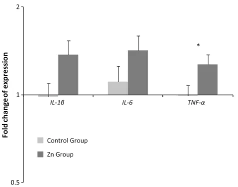
Secondary analyses were conducted with the four treatment groups differentiated according to whether par-ticipants received zinc treatment (Zn group and Zn?FSO group) or no zinc treatment (FSO group and placebo). In the zinc-treated group, mRNA expression levels at week 12 tended to be higher than baseline for TNF-a (per cent increase 27±10 %; mean±SEM; P=0.053), IL-6 (42±17 %; P =0.066), but not IL-1b (37±16 %; P=0.19). In those without zinc treatment, no significant differences were found between baseline and week 12 cytokine mRNA expressions. When the data were expres-sed as fold change, TNF-a was significantly higher (P=0.037) as a result of zinc supplementation (Fig.1). IL-1b fold change was marginally higher (P=0.054) in the zinc-treated group, while no differences were observed in the fold change ofIL-6(P=0.17).
When separated into those groups who did receive FSO (FSO and FSO?Zn group) or did not receive FSO (pla-cebo and Zn group),IL-1b, IL-6andTNF-a gene expres-sions were not significantly different between week 0 and 12. Fold change of IL-1b, IL-6 and TNF-a were not Table 1 Relative mRNA expression and fold changes ofIL-1b,IL-6 andTNF-ain PBMC (mean±SEM)
Placebo (n=10)
FSO (n=10)
Zn (n=11)
Zn?FSO (n=9) IL-1b
Week 0 0.96±0.11 1.72±0.39 1.04±0.15 0.91±0.14 Week 12 0.97±0.02 1.33±0.06 1.46±0.01 0.98±0.06 Fold change 1.08±0.24 0.88±0.43 1.51±0.30 1.21±0.35 IL-6
Week 0 0.10±0.12 0.13±0.25 0.05±0.36 0.17±0.13 Week 12 0.08±0.02 0.14±0.08 0.08±0.02 0.26±0.10 Fold change 0.97±0.30 1.24±0.29 1.46±0.48 1.38±023 TNF-a
Week 0 2.86±0.15 3.85±0.16 2.77±0.26 2.89±0.18 Week 12 3.01±0.12 3.16±0.27 3.64±0.25 3.09±0.23 Fold change 1.10±0.12 0.88±0.12 1.38±0.16 1.14±0.10 FSO flaxseed oil, IL-1b interleukin-1b, IL-6, interleukin-6, TNF-a tumour necrosis factor-a,PBMCperipheral blood mononuclear cells Relative mRNA expression expressed as copies of mRNA per 10618S rRNA
Fold change data are calculated using theDDCpmethod and expressed
significantly different in the FSO group when compared to those who did not receive FSO.
Relationships among various cytokines and zinc transporters
Bivariate correlations between fold changes of individual zinc transporters andIL-1b,IL-6 andTNF-a are shown in Table2. Positive correlations between TNF-a fold change and fold changes ofZnT5, ZnT6, ZnT7, Zip1, Zip3,Zip7, Zip10, MT-1A and MT-2A were observed when all
participants were considered (P\0.05). Similarly,IL-1b
fold change was positively correlated to the fold change of ZnT7 (r=0.44, P\0.01; Fig.2a) and Zip1 (r=0.42, P\0.01; Fig.2b). The treatment of zinc abolished a number of correlations between fold changes of cytokines and zinc transporters; in particular, fold changes of IL-6 andZnT5(control group, r=0.61, P\0.01; zinc group, r= -0.13, P[0.05; Fig. 3), ZnT6 (control group, r=0.49, P\0.05; zinc group, r=0.09, P[0.05) and ZnT7 (control group, r=0.54, P\0.05; zinc group, r=0.32,P[0.05).
Using multiple regression analysis for all participants at baseline,IL-1bexpression was predicted by expressions of all zinc transporters and MT measured (r2=0.495, P=0.04, Table3), with marginally significant univariate correlations observed with Zip1 (P=0.02) and Zip7 (P=0.01). At week 12, overall significant multivariate relationship was maintained between expressions ofIL-1b
and zinc transporters and MT (r2=0.532,P=0.02) with a single significant univariate relationship found between IL-1b and ZnT7 (P=0.002); ZnT7 expression explained 36 % of the variability in IL-1b expression. Multiple regression analyses using expression of zinc transporters and MT as predictors ofIL-6 orTNF-aexpression did not show any significant relationships (data not shown).
Discussion
The key observation in the present study is an upregulation of cytokine gene expression in PBMC after zinc
0.5 1 2
IL-1β IL-6 TNF-α
Fold
change
o
f
e
xpression
Control Group Zn Group
*
Fig. 1 Fold change ofIL-1b,IL-6andTNF-ain PBMC separated by whether participants received zinc treatment (n=20) or no zinc treatment (n=20). Fold change data (mean±SEM) are calculated using theDDCpmethod and expressed on a log scale. *P\0.05 by
independentt-test
Table 2Bivariate Pearson’s correlations between fold changes of cytokines, zinc transporters and metallothionein when analysed in all participants (n=38a) and according to whether participants received
(zinc,n=20) or did not receive (control,n=18) zinc supplements for 12 weeks
ZnT1 ZnT5 ZnT6 ZnT7 ZnT8b Zip1 Zip3 Zip7 Zip10 MT-1A MT-2A
IL-1b
All 0.28 0.28 0.33* 0.44** -0.03 0.42** 0.38* 0.24 0.14 0.19 0.36
Zinc 0.33 0.49* 0.27 0.57** -0.23 0.51* 0.31 0.27 0.24 0.10 0.37
Control 0.31 0.24 0.42 0.29 0.04 0.35 0.70** 0.33 -0.06 0.38 0.32
IL-6
All 0.07 0.21 0.29 0.43** 0.05 0.06 0.38* 0.22 0.42** 0.51** 0.14
Zinc -0.08 -0.13 0.09 0.32 -0.13 0.10 0.46* 0.31 0.43 0.59** 0.24
Control 0.29 0.61** 0.49* 0.54* 0.32 0.04 0.41 0.18 0.39 0.40 -0.01
TNF-a
All 0.31 0.39* 0.45** 0.56** 0.10 0.52** 0.34* 0.44** 0.33* 0.33* 0.60**
Zinc 0.31 0.44 0.41 0.60** 0.11 0.64** 0.49* 0.48* 0.40 0.28 0.65**
Control 0.42 0.55* 0.49* 0.57* -0.12 0.46 0.36 0.54* 0.22 0.43 0.55*
*P\0.05; **P\0.01
a Missing data for expression of zinc transporters and metallothionein for two participants
supplementation in postmenopausal women with type 2 DM. Specifically, higher fold change of TNF-a was observed, with IL-1b fold change, and IL-6 expression tending to increase after 12 weeks of zinc supplementation. A range of bivariate relationships were found among fold changes of various cytokines and zinc transporters. In addition, zinc transporter and MT gene expressions were shown to predict the expression ofIL-1b. Collectively, the observations support a relationship between zinc and immune response in type 2 DM.
The significant upregulation observed in TNF-a
expression after zinc supplementation in the present study is in agreement with several in vitro studies (Scuderi1990; Wellinghausen et al.1996; Chang et al.2006). In cultured cells, changes in gene expression and secretion of cyto-kines as a result of zinc treatment have been reported. Increases in TNF-a, IL-1b and IL-6 secretion have been shown in otherwise unstimulated PBMC incubated with high levels of zinc (C100 lM) in the media (Wellinghau-sen et al.1996; Chang et al. 2006). Similar effects were observed with zinc added at physiologically relevant lev-els;TNF-aandIL-1bmRNA expressions were shown to be upregulated with 30lM of zinc added to the incubating media (Chang et al.2006). Limited data are available from
human trials on the effects of zinc supplementation on cytokine production in non-activated PBMC. The gene expressions of TNF-a and IL-1b remain unchanged in unstimulated leucocytes extracted from healthy subjects given 15 mg/day of zinc for 4 days (Aydemir et al.2006). The present results are novel, and to our knowledge, there are no previous reports of the effect of zinc supplementa-tion on unstimulated cytokine expressions in postmeno-pausal women with type 2 DM.
Perturbation in zinc metabolism is often reported in type 2 DM (Jansen et al.2009). The increase in the plasma zinc concentration seen in zinc-supplemented participants in the present study suggests that zinc is available for intracellular zinc replenishment in previously zinc-deficient tissues. We hypothesize that an increase in cellular zinc content in PBMC may explain these observations. The classic tran-scription factor, nuclear factor-kappa B (NF-jB), has been proposed to be influenced by increased zinc availability. NF-jB represents the converging signalling molecule in immune cells; the propagation of Toll-like, IL-1 and TNF receptor signalling pathways is reliant on the activation of jB. Zinc has been proposed to enhance or inhibit NF-jB activity, dependent on the different health and/or zinc status of the studied population (Foster and Samman2012). r=0.44
P<0.01
0.125 0.25 0.5 1 2 4
2 1
0.5 0.25
IL-1β
fold
change
ZnT7 fold change a
r=0.42 P<0.01
0.125 0.25 0.5 1 2 4
2 1
0.5 0.25
IL-1β
fold
change
Zip1 fold change b
Fig. 2 Bivariate correlations between fold changes of IL-1b and ZnT7 (Fig.2a), and IL-1b and Zip1 (Fig.2b) in all participants (n=38). Fold change data are calculated using theDDCpmethod and
expressed on a log scale
r=-0.13 P>0.05
0.25 0.5 1 2 4
2 1
0.5 0.25
IL-6
fold
change
ZnT5 fold change b
r=0.61 P<0.01
0.25 0.5 1 2 4
2 1
0.5
IL-6
fold
change
ZnT5 fold change a
Fig. 3 Bivariate correlations between fold changes ofIL-6andZnT5 in the control group (n=18, Fig.3a) and in the zinc group (n=20, Fig.3b). Fold change data are calculated using theDDCpmethod and
In zinc-deficient subjects, NF-jB activation was suppressed with the consequent reduction in the cytokine production of immune cells (Prasad et al.2006), and both of these phe-nomena were reversed by zinc supplementation.
Increased plasma zinc concentration, per se, may be contributing to the observed upregulation of cytokines. Higher systemic zinc concentrations may initiate intracel-lular signalling through an extracelintracel-lular zinc receptor, GPR39. This novel zinc sensor is implicated in the regu-lation of cell proliferation and growth (Cohen et al.2014), through modulation of the extracellular regulating kinase (ERK) 1/2 and Akt pathways. Although the direct effect of GPR39 on intracellular signalling in the immune cells is unclear, it is conceivable that the activation of GPR39 by increased extracellular zinc may trigger downstream sig-nalling eventuating in upregulation of cytokine expression. Given that a state of zinc deficiency appears to co-exist with type 2 DM (Jansen et al.2009), the current findings after zinc supplementation may be evidence of improved immune response through increased efficiency in signalling pathways and thereby upregulation of cytokine gene expressions.
The multiple bivariate correlations found in the present study between fold changes of cytokine, zinc transporter and MT mRNA suggest coordination between fluxes of cellular zinc ions and cytokine production in PBMC, an important function of immune cells. These findings support previous observations of a relationship between zinc transporter gene expression and systemic inflammatory biomarkers in humans (Foster et al. 2013b; Noh et al. 2014). Systemic inflammatory biomarkers, such as CRP, IL-6 and TNF-a, were able to predict the gene expressions of zinc transporter and MT in PBMC; in particular, increases in systemic IL-6 concentration were related to increases in the expression of a cluster of zinc transporters, ZnT5, ZnT7, Zip1, Zip7andZip10, which are responsible
largely for the uptake of extracellular zinc and transport of zinc into intracellular organelles and secretory pathways in PBMC. Considering that inflammatory biomarkers in the systemic circulation are the cumulative secretion of many peripheral tissues, the present study is unique in elucidating the relationship between zinc transporter, MT and cytokine gene expression within PBMC.
At both baseline and week 12, IL-1b gene expression was predicted by zinc transporter and MT gene expres-sions. Furthermore, IL-1b expression was strongly pre-dicted by the gene expressions ofZip1andZip7at baseline and ZnT7at week 12 in univariate analyses. Considering their proposed localization within the cell (Huang and Tepaamorndech 2013; Jeong and Eide 2013), these zinc transporters are likely to be involved in either increasing cytoplasmic zinc concentration or the transport of zinc into the golgi apparatus. Taken together with the previously observed relationships between systemic IL-6 with gene expression ofZnT7,Zip1andZip7(Foster et al.2013b), we hypothesize that systemic cytokines have a regulatory control of zinc transporter transcription which influences the flux of zinc into the secretory pathway, thereby assisting in immune cell function such as IL-1b transcrip-tion. This hypothesis is supported by recent findings that suggest external stimuli such as systemic cytokines may regulate zinc transporter expression through the JAK-STAT pathways (Miyai et al. 2014). In particular, cyto-kines were found to induce the expression of Zip10, thereby providing an influx of intracellular zinc. The uptake of zinc via this particular zinc transporter appears to be critical for the survival, function and signalling of B-lymphocytes (Hojyo et al.2014). In a similar vein, we have previously reported an association between Zip10 gene expression and glycaemic control (Foster et al.2014), whereby the change in fasting glucose was correlated with the change in Zip10 expression. These observations Table 3 Multiple regression
models using expression of zinc transporters and metallothionein to predictIL-1bexpression at baseline (model 1) and week 12 (model 2) for all participants
Factors Model 1 (week 0) n=38 Pvalue Model 2 (week 12) n=37 Pvalue Regression
coefficient
Standard error
Regression coefficient
Standard error
ZnT1 -0.02 0.04 0.58 0.02 0.07 0.75
ZnT5 -0.01 0.34 0.97 -0.64 0.39 0.12
ZnT6 -0.19 0.23 0.41 -0.09 0.30 0.78
ZnT7 0.06 0.07 0.38 0.30 0.09 0.002
ZnT8 -1.38 2.31 0.55 -4.81 4.55 0.30
Zip1 0.18 0.07 0.02 -0.01 0.09 0.89
Zip3 0.11 0.11 0.35 0.12 0.14 0.41
Zip7 -0.34 0.13 0.01 -0.17 0.26 0.53
Zip10 -0.01 0.09 0.91 -0.11 0.12 0.34
MT-1A -0.11 0.10 0.28 -0.14 0.16 0.40
MT-2A 0.05 0.22 0.82 0.32 0.44 0.48
highlight the range of external stimuli that can influence zinc flux and function in the cell. Figure4 shows the hypothesized interaction between external stimuli, zinc transport and cellular function.
The PBMC transcriptome has been shown to be reflec-tive of gene expressions in metabolically acreflec-tive tissues, such as liver (Powell and Kroon1994) and muscle (Rud-kowska et al.2011), both of which play a role in glucose and zinc homeostasis. Furthermore, modifications in die-tary macronutrients and antioxidants seem to effect chan-ges in the PBMC transcriptome (de Mello et al. 2012), hence presenting it as a promising target tissue in nutri-genomic studies. From the current study, it is evident that zinc supplementation impacts on the gene expression of cytokines and affects the relationships between cytokine and zinc transporter transcription in PBMC. In particular, the relationships between fold change ofIL-6 withZnT5, ZnT6andZnT7fold changes in the control group were not observed in the zinc-treated group. Whether zinc supple-mentation will elicit similar effects in cell types other than PBMC, such as those of the pancreas and other metaboli-cally active tissues in type 2 DM, requires further investigation.
The efficacy of zinc supplementation in the management of type 2 DM has been investigated recently. Meta-analyses have shown that zinc supplementation in type 2 DM results in lower fasting glucose (Capdor et al.2013) and increased high density lipoprotein concentrations (Foster et al.2010). In the current study, zinc supplementation upregulates gene
expression of cytokines in PBMC despite no changes being observed in systemic levels of the corresponding inflam-matory markers (Foster et al. 2013b). Differences in the derivation of samples may provide an explanation in the discrepancy of results. In PBMC, the measure of cytokine gene expressions is upstream of cytokine secretion. Regu-latory processes at post-transcriptional, translational and post-translational levels are required prior to eventual secretion of cytokines into the systemic circulation. In addition, the systemic cytokine concentration is reflective not only of PBMC cytokine secretion but from a variety of sources, such as liver, muscle and adipose tissue. Hence, the effect of increased cytokine gene expressions in PBMC remains uncertain in the context of the whole-body inflammatory status in type 2 DM.
The current study presents preliminary data on the effects of zinc supplementation on the expression of cytokines and their relationships with zinc transporter gene expressions in type 2 DM. The present observations are novel in that they provide evidence of interactions between zinc transport and cytokine transcription in PBMC, which add to our previous reports (Foster et al. 2013a,b). The study is limited by the lack of proteomic and intracellular zinc analyses. The ulti-mate function and activity of transcribed genes are influ-enced by a number of post-transcriptional regulatory processes, and further research is needed to explore the multiple possible sub-cellular localization sites of zinc transporter proteins which are influenced by the host’s zinc status (Lichten and Cousins2009).
Fig. 4 Potential zinc-mediated pathways by which external stimuli (such as systemic glucose, cytokines and zinc) may act on cellular function. External stimuli such as systemic IL-6, glucose and extracellular zinc bind to respective receptors on the plasma membrane (1). This triggers intracellular signalling events (ERK 1/2, Akt and JAK/ STAT pathways;2), resulting in modulation of zinc transporters and MT gene expressions (3). Changes in intracellular zinc concentration can influence
In summary, the current study presents preliminary evidence that zinc supplementation increasesTNF-a gene expression in postmenopausal women with type 2 DM. The multiple relationships found between gene expression and fold change of zinc transporters, MT and cytokines suggest close interactions between zinc homeostasis and inflammation.
Acknowledgments Financial support obtained from The Medical Advances Without Animals (MAWA) Trust; and Sydnovate, The University of Sydney. The funding bodies did not play any role in designing the study; in the collection, analysis and interpretation of data; in the writing of the manuscript; or in the decision to submit the manuscript for publication.
Conflict of interest Anna Chu, Meika Foster, Dale Hancock, Kim Bell-Anderson, Peter Petocz, and Samir Samman declare that they have no conflict of interest.
References
Aydemir TB, Blanchard RK, Cousins RJ (2006) Zinc supplementa-tion of young men alters metallothionein, zinc transporter, and cytokine gene expression in leukocyte populations. Proc Natl Acad Sci USA 103:1699–1704. doi:10.1073/pnas.0510407103 Babula P, Masarik M, Adam V et al (2012) Mammalian
metallo-thioneins: properties and functions. Metallomics 4:739–750. doi:10.1039/c2mt20081c
Banerjee M, Saxena M (2012) Interleukin-1 (IL-1) family of cytokines: role in type 2 diabetes. Clin Chim Acta 413:1163–1170. doi:10.1016/j.cca.2012.03.021
Bao B, Prasad A (2010) Zinc decreases C-reactive protein, lipid peroxidation, and inflammatory cytokines in elderly subjects: a potential implication of zinc as an atheroprotective agent. Am J Clin Nutr 91:1634–1641. doi:10.3945/ajcn.2009.28836.1634 Calder PC (2006) N-3 polyunsaturated fatty acids, inflammation, and
inflammatory diseases. Am J Clin Nutr 83:1505S–1519S Capdor J, Foster M, Petocz P, Samman S (2013) Zinc and glycemic
control: a meta-analysis of randomised placebo controlled supplementation trials in humans. J Trace Elem Med Biol 27:137–142. doi:10.1016/j.jtemb.2012.08.001
Chang K-L, Hung T-C, Hsieh B-S et al (2006) Zinc at pharmacologic concentrations affects cytokine expression and induces apoptosis of human peripheral blood mononuclear cells. Nutrition 22:465–474. doi:10.1016/j.nut.2005.11.009
Cnop M, Welsh N, Jonas J et al (2005) Mechanisms of pancreatic beta-cell death in type 1 and type 2 diabetes. Diabetes 54:97–107 Cohen L, Sekler I, Hershfinkel M (2014) The zinc sensing receptor, ZnR/GPR39, controls proliferation and differentiation of col-onocytes and thereby tight junction formation in the colon. Cell Death Dis 5:e1307. doi:10.1038/cddis.2014.262
De Mello VDF, Kolehmanien M, Schwab U et al (2012) Gene expression of peripheral blood mononuclear cells as a tool in dietary intervention studies: what do we know so far? Mol Nutr Food Res 56:1160–1172. doi:10.1002/mnfr.201100685 Donath MY, Shoelson SE (2011) Type 2 diabetes as an inflammatory
disease. Nat Rev Immunol 11:98–107. doi:10.1038/nri2925 Egefjord L, Jensen JL, Bang-Berthelsen CH et al (2009) Zinc
transporter gene expression is regulated by pro-inflammatory cytokines: a potential role for zinc transporters in beta-cell apoptosis? BMC Endocr Disord 9:7. doi:10.1186/1472-6823-9-7
Foster M, Samman S (2010) Zinc and redox signaling: perturbations associated with cardiovascular disease and diabetes mellitus. Antioxid Redox Signal 13:1549–1573. doi:10.1089/ars.2010. 3111
Foster M, Samman S (2012) Zinc and regulation of inflammatory cytokines: implications for cardiometabolic disease. Nutrients 4:676–694. doi:10.3390/nu4070676
Foster M, Petocz P, Samman S (2010) Effects of zinc on plasma lipoprotein cholesterol concentrations in humans: a meta-analysis of randomised controlled trials. Atherosclerosis 210:344–352. doi:10.1016/j.atherosclerosis.2009.11.038 Foster M, Hancock D, Petcoz P, Samman S (2011) Zinc transporter
genes are coordinately expressed in men and women indepen-dently of dietary or plasma zinc. J Nutr 1195–1201. doi:10.3945/ jn.111.140053
Foster M, Petocz P, Caterson ID, Samman S (2013a) Effects of zinc anda-linolenic acid supplementation on glycemia and lipidemia in women with type 2 diabetes mellitus: a randomized, double-blind, placebo-controlled trial. J Diabetes Res Clin Metab. doi:10.7243/2050-0866-2-3
Foster M, Petocz P, Samman S (2013b) Inflammation markers predict zinc transporter gene expression in women with type 2 diabetes mellitus. J Nutr Biochem 24:1655–1661. doi:10.1016/j.jnutbio. 2013.02.006
Foster M, Chu A, Petocz P, Samman S (2014) Zinc transporter gene expression and glycemic control in post-menopausal women with type 2 diabetes mellitus. J Trace Elem Med Biol. doi:10. 1016/j.jtemb.2014.07.012[Epub ahead of print]
Haase H, Maret W (2005) Fluctuations of cellular, available zinc modulate insulin signaling via inhibition of protein tyrosine phosphatases. J Trace Elem Med Biol 19:37–42. doi:10.1016/j. jtemb.2005.02.004
Hennigar SR, Kelleher SL (2012) Zinc networks: the cell-specific compartmentalization of zinc for specialized functions. Biol Chem 393:565–578. doi:10.1515/hsz-2012-0128
Hojyo S, Miyai T, Fujishiro H et al (2014) Zinc transporter SLC39A10/ZIP10 controls humoral immunity by modulating B-cell receptor signal strength. Proc Natl Acad Sci USA 111:11786–11791. doi:10.1073/pnas.1323557111
Huang L (2014) Zinc and its transporters, pancreatic b-cells, and insulin metabolism. Vitam Horm 95:365–390. doi:10.1016/ B978-0-12-800174-5.00014-4
Huang L, Tepaamorndech S (2013) The SLC30 family of zinc transporters—a review of current understanding of their biolog-ical and pathophysiologbiolog-ical roles. Mol Aspects Med 34:548–560. doi:10.1016/j.mam.2012.05.008
Huber AM, Gershoff SN (1973) Effect of zinc deficiency in rats on insulin release from the pancreas. J Nutr 103:1739–1744 Jansen J, Karges W, Rink L (2009) Zinc and diabetes—clinical links
and molecular mechanisms. J Nutr Biochem 20:399–417. doi:10. 1016/j.jnutbio.2009.01.009
Jeong J, Eide DJ (2013) The SLC39 family of zinc transporters. Mol Aspects Med 34:612–619. doi:10.1016/j.mam.2012.05.011 Kelleher S, McCormick NH, Velasquez V, Lopez V (2011) Zinc in
specialized secretory tissues: roles in the pancreas, prostate, and mammary gland. Adv Nutr 2:101–111. doi:10.3945/an.110. 000232
Kim AM, Tingen CM, Woodruff TK (2010) Sex bias in trials and treatment must end. Nature 465:688–689. doi:10.1038/465688a Lichten LA, Cousins RJ (2009) Mammalian zinc transporters: nutritional and physiologic regulation. Annu Rev Nutr 29:153–176. doi:10.1146/annurev-nutr-033009-083312 Liuzzi JP, Lichten LA, Rivera S et al (2005) Interleukin-6 regulates
Miyai T, Hojyo S, Ikawa T et al (2014) Zinc transporter SLC39A10/ ZIP10 facilitates antiapoptotic signaling during early B-cell development. Proc Natl Acad Sci U S A. doi:10.1073/pnas. 1323549111
Mlinar B, Marc J, Janez A, Pfeifer M (2007) Molecular mechanisms of insulin resistance and associated diseases. Clin Chim Acta 375:20–35. doi:10.1016/j.cca.2006.07.005
Noh H, Paik HY, Kim J, Chung J (2014) The alteration of zinc transporter gene expression is associated with inflammatory markers in obese women. Biol Trace Elem Res 158:1–8. doi:10. 1007/s12011-014-9902-1
Pickup JC (2004) Inflammation and activated innate immunity in the pathogenesis of type 2 diabetes. Diabetes Care 27:813–823. doi:10.2337/diacare.27.3.813
Powell EE, Kroon PA (1994) Low density lipoprotein receptor and 3-hydroxy-3-methylglutaryl coenzyme A reductase gene expres-sion in human mononuclear leukocytes is regulated coordinately and parallels gene expression in human liver. J Clin Invest 93:2168–2174. doi:10.1172/JCI117213
Prasad A, Beck F (2007) Zinc supplementation decreases incidence of infections in the elderly: effect of zinc on generation of cytokines and oxidative stress. Am J Clin Nutr 85:837–844
Prasad AS, Bao B, Beck FWJ, Sarkar FH (2006) Correction of interleukin-2 gene expression by in vitro zinc addition to
mononuclear cells from zinc-deficient human subjects: a specific test for zinc deficiency in humans. Transl Res 148:325–333. doi:10.1016/j.trsl.2006.07.008
Rudkowska I, Raymond C, Ponton A et al (2011) Validation of the use of peripheral blood mononuclear cells as surrogate model for skeletal muscle tissue in nutrigenomic studies. OMICS 15:1–7. doi:10.1089/omi.2010.0073
Scuderi P (1990) Differential effects of copper and zinc on human peripheral blood monocyte cytokine secretion. Cell Immunol 126:391–405. doi:10.1016/0008-8749(90)90330-T
Wellinghausen N, Driessen C, Rink L (1996) Stimulation of human peripheral blood mononuclear cells by zinc and related cations. Cytokine 8:767–771. doi:10.1006/cyto.1996.0102
Zhao G, Etherton T, Martin K (2004) Dietarya-linolenic acid reduces inflammatory and lipid cardiovascular risk factors in hypercho-lesterolemic men and women. J Nutr 134:2991–2997