R E S E A R C H
Open Access
C-banding and AgNOR-staining were still
effective complementary methods to
indentify chromosomal heteromorphisms
and some structural abnormalities in
prenatal diagnosis
Jian Jiang Zhu , Hong Qi
*, Li Rong Cai, Xiao Hui Wen, Wen Zeng, Guo Dong Tang, Yao Luo, Ran Meng,
Xue Qun Mao and Shao Qin Zhang
Abstract
Background:In prenatal diagnosis, CMA has begun to emerge as a favorable alternative to karyotype analysis, but it could not identify balanced translocations, triploidies, inversion and heteromorphisms. Therefore, conventional cytogenetic and specific staining methods still play an important role in the work-up of chromosome anomaly. This study investigated the application of C-banding and AgNOR-staining techniques in prenatal diagnosis of
chromosomal heteromorphisms and some structure abnormalities.
Results:Among the 2970 samples, the incidence of chromosomal heteromorphisms was 8.79% (261/2970). The most frequent was found to be chromosome Y (2.93%, 87/2970), followed by chromosome 1 (1.65 %, 49/2970), 9 (1.52 %, 45/2970), 22 (0.77 %, 23/2970) and 15 (0.64 %, 19/2970). We compared the incidence of chromosomal heteromorphisms between recurrent spontaneous abortion (RSA) group and control group. The frequency of autosomal hetermorphisms in RSA group was 7.63% higher than that in control group (5.78%), while the frequency of Y chromosomal heteromorphisms was 4.76% lower than that in control group (5.71%). Here we summarized 4 representative cases, inv (1) (p12q24), psu dic (4;17) (p16.3;p13.3), r(X)(p11; q21) and an isodicentric bisatellited chromosome to illustrate the application of C-banding or AgNOR-staining, CMA or NGS was performed to detect CNVs if necessary.
Conclusions:This study indicated that C-banding and AgNOR-staining were still effective complementary methods to identify chromosomal heteromorphisms and marker chromosomes or some structural rearrangements involving the centromere or acrocentric chromosomes. Our results suggested that there was no evidence for an association between chromosomal heteromorphisms and infertility or recurrent spontaneous abortions. Undoubtedly, sometimes we needed to combine the results of CMA or CNV-seq to comprehensively reflect the structure and aberration of chromosome segments. Thus, accurate karyotype reports and genetic counseling could be provided.
Keywords:Prenatal diagnosis, C-banding, AgNOR-staining, Chromosomal heteromorphisms, Chromosomal structural abnormality, Recurrent spontaneous abortion
© The Author(s). 2019Open AccessThis article is distributed under the terms of the Creative Commons Attribution 4.0 International License (http://creativecommons.org/licenses/by/4.0/), which permits unrestricted use, distribution, and reproduction in any medium, provided you give appropriate credit to the original author(s) and the source, provide a link to the Creative Commons license, and indicate if changes were made. The Creative Commons Public Domain Dedication waiver (http://creativecommons.org/publicdomain/zero/1.0/) applies to the data made available in this article, unless otherwise stated. * Correspondence:[email protected]
Introduction
Currently, chromosomal microarray analysis (CMA) and copy number variation sequencing (CNV-Seq) are widely used in prenatal diagnosis due to the capability of identi-fying microdeletion and microduplication syndromes as well asde novopathogenic CNVs which may be missed by conventional karyotyping [1]. However, their drawbacks are also obvious including the incompetence of balanced translocations, triploidies, inversion and heteromorphisms identification. So far, most of the clinical experts do not support the replacement of conventional karyotyping by CMA as a first-tier clinical test or in pregnancies at low risk for chromosome anomalies [2, 3] mainly due to the detection of variant of unknown significance (VOUS) and economic reasons. Therefore, the karyotype analysis technology is still essential for chromosomal disorder diagnosis.
After different treatments such as denaturation and/or enzymatic digestion, chromosomes show light and dark bands under light microscopy in different regions. Chromosome banding techniques are generally divided into two types: (1) bands are distributed along the entire chromosome, e.g. the most frequently used Giemsa-tryp-sin banding (G-banding), Q banding and R banding; (2) bands are located in specific chromosomal regions, including C-banding (constitutive heterochromatin), Ag-NOR staining (nucleolar organizer region) and T-banding (telomere). Each chromosome has a unique band pattern that can be reliably identified according to the corre-sponding banding technique. However, a dark-stained band by a particular treatment may appear light-stained in another. G-banding is the most frequently used method in the clinical laboratories because of its stability and cost-effectiveness.
C-banding and AgNOR-staining as the main banding techniques for chromosomal heteromorphisms detection are not conventional used in many laboratories due to the chromosomal heteromorphisms are generally con-sidered to have no pathological significance and could be stably inherited to the offspring. Surprisingly, there have been increased conflicting evidence as to the possible association between heteromorphisms and the recurrent abortions [4–6], infertility or reproductive out-comes following assisted reproductive technology [7–9]. This study summarized the application of C-banding and Ag-NOR staining techniques for the prenatal diag-nosis of fetal chromosome karyotype in our hospital, and explored the relationship between heteromorphisms and recurrent spontaneous abortion (RSA).
Materials and methods Sample information
From May 2015 to April 2018, a total of 2972 cases ac-cepted karyotype analysis by invasive prenatal diagnosis
based on the indications of high-risk including advanced maternal age, positive aneuploidy screening results, or ultrasound structural anomalies in the Beijing Haidian Maternal and Child Health Hospital. The pregnant women’s obstetrical history was detail recorded, 2970 suc-cessful diagnostic samples were divided into two groups:
the RSA group (history of spontaneous abortions≥2,
Group A) and the control group (history of spontaneous abortions≤1, Group B). Samples with chromosomal het-eromorphisms, structural abnormalities involving the centromere regions or acrocentric chromosomes, as well as mark chromosomes were verified by C-banding and AgNOR-staining techniques at first. Then CMA or NGS was also used to exclude chromosome CNVs if necessary. All clinical tests were based on written informed consent of pregnant women.
Cell culture and karyotype analysis
Fetal cells obtained from villus, amniotic fluid or cord blood were cultured with double-line using standard
methodologies. Chromosomes in metaphases were
prepared from the cultured cell or lymphocytes using Giemsa-trypsin banding techniques. At least twenty metaphases chromosomes were counted and three meta-phases chromosomes were analyzed in each line by two independent laboratory technicians to avoid uncertainty and variable results.
Chromosomal heteromorphisms were described follow-ing the criteria in the International System for Chromo-some Nomenclature [10]. 1/9/16qh+ is defined as being at least twice the length of the corresponding region on the homologous chromosome, qh- is defined as being half length of the normal corresponding region. Polymor-phisms of the Y chromosome were evaluated as such that Yqh+ (>size of chromosome 18), and Yqh- (<size of
chromosome 21). For D/G–group chromosomes, the
in-crease/decrease of the centromere or short arm in length were designated as cenh+/- if the variants were also C-banding positive. Additionally, distinct variants in size or number of satellites (ps+, pss) and lengths of stalks (pstk+) of the D/G –group were also recorded. The pericentric inversion of chromosomes 9 and Y were also categorized as heteromorphisms [8].
C-banding
Ag-NOR staining
The slide was covered with 4 layers of clean lens paper (the size was slightly smaller than the slide) first. Then, it was moved to a plate floating in a 65°C water bath. 5ml of freshly prepared 10% AgNO3solution (containing
0.1% formic acid) was dropped onto the lens paper sev-eral times by a straw and the slide remained in the water bath for about 15min until the lens paper turned black or dark brown. After gently removing the lens paper with tweezers, the slide was washed with tap water and samples were finally stained with Giemsa for 1 minute in room temperature. The most important thing in the whole process of the experiment was to avoid the light.
CMA and CNV-seq
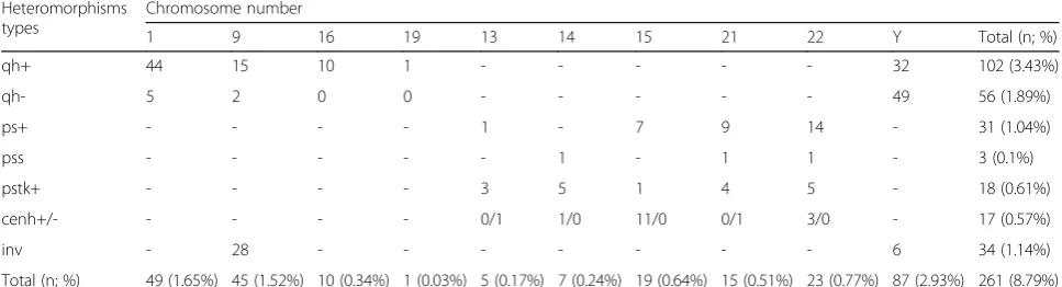
Affymetrix CytoScan 750K array platforms were used to detect copy number variants. Genomic DNA extraction was performed using Genomic DNA Extraction kit (QIAamp DNA Blood Mini Kit, QIAGEN GmBH, Hilden, Germany) according to the in-house protocols. The standard experimental procedure incorporated the following steps: digestion, ligation, polymerase chain re-action (PCR), PCR purification, fragmentation, labeling, hybridization, washing, staining and scanning. Data was analyzed using the Chromosome analysis software (Chromosome Analysis Suite version 2.1) (Affymetrix; Thermo Fisher Scientific, Inc.). Copy number variation Table 1Summary the number and frequency of all observed Chromosomal heteromorphisms
Heteromorphisms types
Chromosome number
1 9 16 19 13 14 15 21 22 Y Total (n; %)
qh+ 44 15 10 1 - - - 32 102 (3.43%)
qh- 5 2 0 0 - - - 49 56 (1.89%)
ps+ - - - - 1 - 7 9 14 - 31 (1.04%)
pss - - - 1 - 1 1 - 3 (0.1%)
pstk+ - - - - 3 5 1 4 5 - 18 (0.61%)
cenh+/- - - 0/1 1/0 11/0 0/1 3/0 - 17 (0.57%)
inv - 28 - - - 6 34 (1.14%)
Total (n; %) 49 (1.65%) 45 (1.52%) 10 (0.34%) 1 (0.03%) 5 (0.17%) 7 (0.24%) 19 (0.64%) 15 (0.51%) 23 (0.77%) 87 (2.93%) 261 (8.79%)
sequencing (CNV-seq) was performed as previously described [11,12].
The conventional genomic and phenotype public
data-bases such as UCSC Genome Browser (http://genome.
ucsc.edu/cgi-bin/hgGateway), ClinGene (http://www. clinicalgenome.org/), OMIM (http://omim.org),
DE-CIPHER (https://decipher.sanger.ac.uk), DGV (http://
dgv.tcag.ca/dgv/app/home) and PubMed (http://www. ncbi.nlm.nih.gov/pubmed) were used for retrieval and interpretation.
Statistical analysis
Data were analyzed using SPSS Statistics (version 22.0). Differences between the RSA and the control groups within the cohort were tested with Chi-squared test statistics. The significance level was set atp< 0.05.
Results
Among the 2972 prenatal diagnosis cases (including 2735 amniotic fluid, 68 villus and 169 cord blood), 2 cases of amniotic fluid failed to culture due to less cloning, and the success rate of cultivation was 99.93% (the proportion of male and female was 1533:1437). A total of 131women were investigated in the group A, and the group B consisted of 2839.
Chromosome heteromorphisms
The distribution among different chromosomes
hetero-morphisms was presented in Table 1 with total
fre-quency of 8.79% (261/2970). The most frequent was found to be chromosome Y (2.93%, 87/2970), followed by chromosome 1 (1.65 %, 49/2970), 9(1.52 %, 45/2970), 22 (0.77 %, 23/2970) and chromosome 15 (0.64 %, 19/ 2970). In 1533 male fetuses, 5.68 % (87/1533) had Y
chromosomal heteromorphisms, followed by Yqh- 3.26 %, Yqh+ 2.09 %, and inv(Y) 0.39% (Table3). The partial karyotypes of chromosomal heteromorphisms by G-banding and C/AgNOR-staining are presented in Fig.1. Parental studies had been performed for most prenatally reported chromosome heteromorphisms, particularly for pericentric inversion, confirming that these variations were stably inherited in family.
The group A had a higher frequency (7.63%) of auto-somal hetermorphisms compared with the group B
(5.78%)(Table 2), but a lower frequency (4.76%) of Y
chromosomal heteromorphisms compared with the group B (5.71%) (Table 3). While there were no statistically significant difference between two groups (P> 0.05).
Chromosome abnormal
C-banding and AgNOR-staining were important banding methods to characterize marker chromosomes or other structural rearrangements involving the centromere or acrocentric chromosomes. Here we summarized 4 repre-sentative cases, inv (1) (p12q24), psu dic (4;17)(p16.3; p13.3), r(X)(p11;q21) and an isodicentric bisatellited chromosome to illustrate the application of C-banding or AgNOR-staining. CMA or NGS was performed to detect CNVs. Among them , 2 CNVs with VOUS were detected in case#1, pathogenic CNVs were detected in case #2 and case #3, respectively (Table4).
Discussion
Chromosomal variation were mainly refers to the variations on heterochromatic segments, satellites and satellite stalks in the population, but euchromatic heteromorphisms with C-banding and Ag-NOR negative
could also be included [13–15]. Like most other
Table 3Number and frequency of Y Chromosomal heteromorphisms in different groups
Group Chromosome heteromorphisms
No. of male cases Yqh+ Yqh- inv (Y) Total of heteromorphisms (n; %) Chi-square test
Group Aa 63 0 2 (3.17%) 1 (1.59%) 3 (4.76%) Continuity Correctionχ2=0.002p> 0.05
Group Bb 1470 32 (2.18%) 47 (3.2%) 5 (0.34%) 84 (5.71%)
Total 1533 32 (2.09%) 49 (3.2%) 6 (0.39%) 87 (5.68%)
*Significant atp< 0.05
a
the recurrent spontaneous abortion group
b
the control group
Table 2Number and frequency of autosomal heteromorphisms in different groups
Group Chromosome heteromorphisms
No. of cases 1/9/16/19qh± D/G variation inv(9) Total of heteromorphisms (n; %) Chi-square test
Group Aa 131 4 (3.05%) 4 (3.05%) 2 (1.53%) 10 (7.63%) Pearsonχ2=0.783p>0.05
Group Bb 2839 73 (2.57%) 65 (2.29%) 26 (0.92%) 164 (5.78%)
Total 2970 77 (2.59%) 69 (2.32%) 28 (0.94%) 174 (5.86%)
*Significant atp< 0.05.
a
the recurrent spontaneous abortion group
b
literatures, here we mainly focused on the C-banding or AgNOR-staining positive chromosomal heteromorphisms. As anticipated, while most of chromosmal hetero-morphisms were successfully detected according to our criteria, we still occasionally encounter disputes between experienced technicians occasionally. Without a reference standard other than between homologues, it is difficult to determine whether two similar-sized homo-logues are slightly smaller or larger than the normal ones [16]. In addition, we found that the diagnostic criteria in different literatures were not uniform, although most of
the literature was consistent with ours [8, 17], some did not mention the details of the judgment criteria [9, 18]. This could be one of the reasons for conflicting reports of heteromorphisms impact human reproduction. Shivanand [16] considered that the length of short arms of chromo-some 16 was not appreciably altered by compaction, Moreover, it is intermediate in size compared to the qh re-gions of 1, 9,and 16. Also, it is easily identified in a C-banded cell, therefore provides a useful reference standard to set criteria. Frequencies of heteromorphisms in various populations showed differences due to ethnic origins, age
Fig. 2Prenatal diagnosis of chromosome 19 heteromorphism by G-banding (a) and C-banding (b). Chromosome 19 is labelled, with an arrow indicating the large heterochromatic region
Table 4C-banding or AgNOR-staining applied to the auxiliary diagnosis of 4 cases with chromosome abnormal
Case No. Brief clinical information Chromosome karyotype SNP-array/CNV-seq Pregnancy outcome
Case#1 42-year-old, G5P1,amniocentesis at 19 weeks’ gestation because of advanced age
F:46,XN,inv(1)(p12q24)mat
P:46,XX,inv(1)(p12q24)
H:46,XY
F::arr [hg19] 2q13(110,498,141_ 110,980,295)x4,
12p12.1 (23,797,551_24,076, 457)x1
P: arr [hg19] 2q13(110,498,141_ 110,980,295)x4
H: arr(1-22)x2,(XN)x1
continued pregnancy
Case#2 25-year-old, G5P1,amniocentesis at 18 weeks’ gestation because of a positive serological screening result (a high risk of Down syndrome 1:176)
F:45,XY,
der(4)dup(4)(p15.1p16) psu dic(4;17)(p16.3;p13.3),-17
P:46,XX
H:46,XY
F:arr [hg19] 2q12.3q13(107,586, 661_110,980,295)x3,
4p16.3p15.1 (68,345_32,437, 069)x3
induced abortion
Case#3 35-year-old, G4P1,puncture of umbilical vein at 26 weeks’ gestation because of advanced age and ultrasonic anomalies (coarctation of the aorta? long bone dysplasias).
F:45,X [31]/46,X,r(X)(p11; q21) [29]
P:46,XX
H:46,XY
F:
seq[GRCh37]del(X)(p22.33p11.3), del(X)(q21.31q28)
ChrX:g.60001_43660000del, 0710001_155260000del
induced abortion
Case#4 37-year-old, G2P1,amniocentesis at 19 weeks’gestation because of advanced age
F:47,XN,+mar [37]/46, XN[63]
P:46,XX
H:46,XY
F:arr(1-22)x2,(XN)x1 continued pregnancy
and geographical distribution. Like other studies of Asian populations had shown greater variation in the size of the Y than in the white population [19]. In our study, the fre-quency of Y chromosome heteromorphisms was the high-est, which was apparently higher than other literature [6] and Yqh-was slightly more common than Yqh+. Perhaps the populations selected introduced biases and the criteria used in current studies were subjective, making it difficult to directly compare frequencies. Our data showed that each frequency of chromosomal heteromorphisms does not appear to be significantly different in group A and group B, confirmed that variation in these C-banding or AgNOR-staining positive regions has no clinical signifi-cance. It was almost certain that the common heteromor-phisms were stably inherited in the family by study of the parental chromosomes, which would strengthen the argu-ment against the associated between heteromorphisms and infertility. However, further research is required to de-lineate the mechanisms impacting human reproduction, focusing on patients with unexplained infertility, poor em-bryonic development, and spontaneous abortions. Current cytogenetics has mainly focused its efforts on the identifi-cation of clonal chromosomal aberrations (CCAs).
How-ever, many investigators have demonstrated that “non
clonal chromosome aberrations,” or NCCAs are not
“noise”but rather a highly significant feature of the gen-ome system, and the significance of NCCAs will
emphasize the ultimate importance of studying heterogen-eity in biology, including heteromorphisms and euchro-matic variants [20]. Of equal importance, CNV-seq and CMA will complement the karyotyping method by allow-ing the detetion of small CNV, with more potentially pathogenic genetic candidates detected among the CNV regions, refined genetic causes of conditions like spontan-eous miscarriage and other medical syndromes could be investigated [12,21].
In recent years, more and more studies had focused
on the chromosomal heteromorphisms [22] which can
be verified by the application of C-banding and AgNOR -staining techniques. Several other centromeric variants, verified by C-banding in the course of a prenatal diagnosis [23–28], have been described for chromosomes 5, 6, 12, 18, 19 or 20. Our data presented a rare heteromorphism on chromosome 19 and it was different from the four different classes that Crossen noted [29]. A distinct dark-staining band at the long arm of chromosome 19 near the centromere region was revealed by G-banding (Fig. 2a) and it was also dark-stained by C-banding as the same as centromere (Fig. 2b). So it was most likely constitutive heterochromatin which was considered without patho-logical significance. As the fetus has no abnormal ultra-sound findings except for increased nuchal translucency (3.7mm) at 13 weeks’gestation, the parents rejected fur-ther chromosome verification and CNVs testing. Our
karyotype report was 46, XN, 19qh+. No abnormalities were found in the 6-month follow-up after birth. Indeed, the discovery of rare chromosomal polymorphisms raised the question of whether or not differences in size and banding pattern observed between homologue could account for the existence of a normal variant. Therefore, for rare chromosomal heteromorphisms, family verifica-tion and original analysis should be performed to better assess the genetic effects of the heteromorphisms. In addition, whether the heteromorphisms will change (such as changes in the length or size of heteromorphisms re-gions) during the genetic processes remains to be further studied.
In our prenatal diagnosis work, C-banding was per-formed to verify the pericentric inversion and structure rearrangement involving the centromere regions. It was convenient and easy to make diagnosis by observing the position and morphological changes of centromeres with C-banding. Our data showed that several cases of inv (9) cannot be clearly diagnosed by G-banding due to the short and poor karyotype, but it can be easily confirmed by C-banding (Fig. 1). Similarly, in the diagnosis of inv
(1)(p12q42) (Case #1, Fig 3), the qh regions of inv (1) appeared in the distal long arm of the 1 chromosome rather than in the middle were especially visible by
C-banding (Fig. 3a), two VOUS were revealed by
Array-SNP (Fig. 3b). After genetic counseling, the pregnant woman chose to continue pregnancy with some misgiv-ings. Fortunately, the child had no abnormalities since birth at term. We will continue to monitor the child's
follow-up to check whether these VOUS really didn’t
have clinical significance.
As shown in the diagnosis of case #2, C-banding can also be used to identify pseudodicentric chromosome and to determine whether a fragment of unknown origin is heterochromatin. G-banding showed an apparent unknown dark-stained band (Fig.4a, indicated by arrow) inserted at the breakpoint of the derived chromosome formed by reciprocal translocation of chromosomes 4 and 17. The C-banding demonstrated that the derivative chromosome was dicentric with only one primary
constriction (presumably the active centromere),
whereas the other regions were light stained (Fig.4b). So we confirmed that the inserted fragment was not
constitutive heterochromatin. Therefore, we conducted the CMA test and finally revealed that it was a 32.3 Mb duplication of 4p16.3p15.1, at the same time, a 3.3Mb
duplication of 2q was also detected (Fig. 4c). The
pregnancy was terminated at 23 weeks of gestation due to the chromosome abnormality.
C-banding and AgNOR-staining were also routinely
used to characterize marker chromosomes. In case #3, Karyotyping revealed mosaic of the mark chromosome with two types of cell lines 45,X and 46,X,+mar (Fig.5a). The C-banding showed that the mark chromosome had a clear dark-staining centromere and the rest of the chromosome regions were light stained (Fig.5b). There-fore, we hypothesized that the mark could not be the heterochromatic region of the Y chromosome but likely to be a circle X with a large fragment deletion, and this was later confirmed by the CNV-seq analysis (Fig. 5c). About 16% of the individuals with Tuner syndrome have
an addtional cell line with 46 chromosomes due to the
presence of an extra ring chromosome X [30]. Ring X
chromosome, with different degree of mosaicism, show-ing common clinical characteristics of Turner syndrome had been reported, and karyotype-phenotype correlation related manifestation was dependent on the degree of genetic material lost in ring (X) formation as well as
mosaicism [31]. Undoubtedly, for mosaic Tuner with
mixed cell lines, FISH tests for Xcen and Ycen should be performed since ring X is more commonly seen in mosaic Turner and present of Y material will be clinic-ally significant and requires intervention. In addition, the interphase FISH of uncultured cells can avoid the influ-ence of cell culture and thus more accurately identify the degree of mosaicism, the metaphase FISH can clearly locate the recombinant chromosome. CNV-seq or CMA analysis can reveal exact break points on both arms of X chromosomes. In contrast to case#3, the supernumerary
marker chromosomes (SMC) of case #4, which we highly speculated that it was a bisatellited metacentric micro-chromosome (Fig.6a). The further identified by AgNOR-banding showed prominent satellites on both sides of the marker (Fig.6b). About 70% of SMC are derived from ac-rocentric chromosomes and those markers derived from can be identified by FISH [32].
CMA has advantages over conventional cytogenetic, including the ability to precisely characterize CNVs associated with abnormal karyotypes. Moreover, a sig-nificant proportion of cases studied by array detected clinically significant CNVs even in samples with ap-parently normal karyotypes [33, 34], but the expensive inspection costs and VOUS still require us to con-sider its cost-effectiveness. These scenarios present a challenge for prenatal diagnosis, and genetic counsel-ing prior to prenatal CMA greatly facilitates delivery
of complex results [35]. Although VOUS results have
limited impact on parental well-being and perception
of children’s development, the initial diminished per-ception of child competency and later dissatisfaction with genomic testing indicate the need to assist par-ents in coping with ambiguous results [36]. It is sug-gested to not change the current policy of microarray application in prenatal diagnosis until more data on the clinical significance of copy number changes are available [2]. So, without professional genetic counsel-ing qualifications and informed consent of subjects, it is not appropriate to blindly expand the scope of de-tection without clinical indications. We recommend that CMA or CNV-seq used for fetal chromosomal structural abnormalities (especially de novo), ultra-sound abnormalities, and the SMCs with C-banding and Ag-NOR negative. We also strongly recom-mended de novo SMC for CNVs examination (case #4) because that a prospective study showed that 69% of de novo SMC contained euchromatin material, 95.4% of which for non-acrocentric markers [37].
Conclusion
Each technology has its limitations, we didn’t demon-strate that C-banding and/or AgNOR-staining was ab-solutely necessary above other available technologies such as FISH or CMA. But base on our work, it is suggested that C-banding and AgNOR-staining, which were more convenient, fast and economical, could still be effective complementary methods to study hetero-morphic variations, and to characterize marker chro-mosomes or other structural rearrangements involving centromere regions or acrocentric chromosomes, es-pecially when it was difficulties to make a definite diagnosis due to the short and/or poor G-banding karyotype. On the other hand, purely using G-banding without combining C-banding and AgNOR can hardly tell the difference between cenh+ and ps+, as well as pss and pstk+, and thereby causes the unreliable karyotype results.
Unsurprisingly, further studies are needed to delineate whether heteromorphisms impact human reproduction. Absolutely, there is an urgent need to establish normal-ized diagnostic criteria for chromosomal heteromor-phisms, and this is essential if any degree of comparison is to be made between these and other clinical studies. Contrary to some previous studies, we found no evi-dence for an association between chromosomal hetero-morphisms and infertility or RSA. Our study agreed that familial cytogenetic studies do not appear to be routinely necessary when a common chromosomal
het-eromorphisms is diagnosed prenatally [38]. Perhaps
most critically, followup studies are distinctly lacking. Studies of the progeny (or products of conception) of carriers of heteromorphic variants are sorely needed to better delineate the heritability and consequences of these variants [22].
In summary, in prenatal diagnosis, it is necessary make a diagnosis in a limited time according to the limited clinical manifestations of the fetus, so reasonable detection methods should be selected and avoided the unnecessary expansion of detection scope which gener-ated additional and more complex data to amplify and exacerbate some pre-existing ethical problems [39].
Abbreviations
CCAs:Clonal Chromosomal Aberrations; CMA: Chromosomal Microarry Analysis; CNV-Seq: Copy Number Variation Sequencing; FISH: Fluorescence In Situ Hybridization; NCCAs: Non Clonal Chromosome Aberrations;
NOR: Nucleolar Organizer Region; RSA: Recurrent Spontaneous Abortion; SMC: Supernumerary Marker Chromosomes; VOUS: Variant Of Unknown Significance
Acknowledgements
The authors would like to acknowledge and thank all the families for their cooperation and staff who participated in this study. Thank Annoroad Gene Technology and Be reative lab (Beijing) for their expert laboratory work and analyses.
Authors’contributions
JJZ conceived of the study and performed the statistical analysis, and helped to draft the manuscript. HQ participated in the design of the study, and participated in its design and coordination. LRC, XHW, WZ, GDT and YL carried out the cytogenetics and molecular genetic studies. RM, XQM, SQZ carried out genetic counseling and interventional prenatal diagnosis for pregnant women. All authors read and approved the final manuscript.
Funding Not applicable
Availability of data and materials
Data supporting the results reported in the published article can be found in the tables and figures.
Ethics approval and consent to participate
The sample-related data analyzed for this manuscript was entirely retrospective with no patient or patient-related identifiers included in the analysis. No waivers or institutional approval was required. Each patient received written informed consent for participation.
Consent for publication
Informed written consent was obtained from participants for publication for images and other clinical information relating to these cases to be reported for academic purpose.
Competing interests
The authors declare that they have no competing interests.
Received: 3 April 2019 Accepted: 23 August 2019
References
1. Wapner RJ, Martin CL, Levy B, Ballif BC, Eng CM, Zachary JM, et al. Chromosomal microarray versus karyotyping for prenatal diagnosis [J]. N Engl J Med. 2012;367(23):2175–84.
2. Miny P, Wenzel F, Tercanli S, Filges I. Chromosomal Microarrays in Prenatal Diagnosis: Time for a Change of Policy?[J]. Microarrays (Basel). 2013;2(4):304–17. 3. Novelli A, Grati FR, Ballarati L, Bernardini L, Bizzoco D, Camurri L, et al.
Microarray application in prenatal diagnosis: a position statement from the cytogenetics working group of the Italian Society of Human Genetics (SIGU), November 2011[J]. Ultrasound Obstet Gynecol. 2012;39(4):384–8. 4. Akbas H, Isi H, Oral D, Turkyilmaz A, Kalkanli-Tas S, Simsek S, et al.
Chromosome heteromorphisms are more frequent in couples with recurrent abortions [J]. Genet Mol Res. 2012;11(4):3847–51.
5. Wang Y, Li G, Zuo MZ, Fang JH, Li HR, Quan DD, et al. Y chromosome polymorphisms may contribute to an increased risk of male-induced unexplained recurrent miscarriage [J]. Biosci Rep. 2017;37:2. 6. Dai R, Pan Y, Fu Y, Liu Q, Han W, Liu R. Role of male genetic factors in
recurrent pregnancy loss in Northeast China [J]. Eur J Obstet Gynecol Reprod Biol. 2018;224:6–11.
7. Cheng R, Ma Y, Nie Y, Qiao X, Yang Z, Zeng R, et al. Chromosomal polymorphisms are associated with female infertility and adverse reproductive outcomes after infertility treatment: a 7-year retrospective study [J]. Reprod Biomed Online. 2017;35(1):72–80.
8. Hong Y, Zhou YW, Tao J, Wang SX, Zhao XM. Do polymorphic variants of chromosomes affect the outcome of in vitro fertilization and embryo transfer treatment?[J]. Hum Reprod. 2011;26(4):933–40.
9. Dong Y, Jiang YT, Du RC, Zhang HG, Li LL, Liu RZ. Impact of chromosomal heteromorphisms on reproductive failure and analysis of 38 heteromorphic pedigrees in Northeast China [J]. J Assist Reprod Genet. 2013;30(2):275–81. 10. International Standing Committee on Human Cytogenetic Nomenclature,
Shaffer LG, Mc Gowan-Jordan J, Schmid M. ISCN 2013 : an international system for human cytogenetic nomenclature (2013)[M]. Basel: Karger; 2013. vi, 140 p. , 141 folded sheet
11. Liang D, Lv W, Wang H, Xu L, Liu J, Li H, et al. Non-invasive prenatal testing of fetal whole chromosome aneuploidy by massively parallel sequencing [J]. Prenat Diagn. 2013;33(5):409–15.
coverage whole genome sequencing [J]. Eur J Obstet Gynecol Reprod Biol. 2018;224:21–8.
13. Jalal SM, Schneider NR, Kukolich MK, Wilson GN. Euchromatic 16p+ heteromorphism: first report in North America [J]. Am J Med Genet. 1990;37(4):548–50.
14. Webb GC, Krumins EJ, Eichenbaum SZ, Voullaire LE, Earle E, Choo KH. Non C-banding variants in some normal families might be homogeneously staining regions [J]. Hum Genet. 1989;82(1):59–62. 15. Song XH, Hsu HK, Su MT, Chang TS, Su PY, Chen M, et al. Euchromatic
variants of 8q21.2 in twins [J]. Taiwan J Obstet Gynecol. 2017;56(2):227–9. 16. Patil SR, Lubs HA. Classification of qh regions in human chromosomes 1, 9,
and 16 by C-banding [J]. Hum Genet. 1977;38(1):35–8. 17. Sun L, Chen ZH, Yang L, Yi CX, Liu J, Ou CQ. Chromosomal
polymorphisms are independently associated with multinucleated embryo formation [J].
J Assist Reprod Genet. 2018;35(1):149–56.
18. Inan C, Sayin NC, Dolgun ZN, Gurkan H, Erzincan SG, Uzun I, et al. Prenatal diagnosis of chromosomal polymorphisms: most commonly observed polymorphism on Chromosome 9 have associations with low PAPP-A values()[J]. J Matern Fetal Neonatal Med. 2017:1–8.
19. Hou JW, Wang TR. Study of human Y chromosome polymorphism in Taiwan [J]. Acta Paediatr Taiwan. 1999;40(5):302–4.
20. Heng HH, Regan SM, Liu G, Ye CJ. Why it is crucial to analyze non clonal chromosome aberrations or NCCAs?[J]. Mol Cytogenet. 2016;9:15. 21. Bagheri H, Mercier E, Qiao Y, Stephenson MD, Rajcan-Separovic E. Genomic
characteristics of miscarriage copy number variants [J]. Mol Hum Reprod. 2015;21(8):655–61.
22. Tempest HG, Simpson JL. Why are we still talking about chromosomal heteromorphisms?[J]. Reprod Biomed Online. 2017;35(1):1–2. 23. Fineman RM, Issa B, Weinblatt V. Prenatal diagnosis of a large
heteromorphic region in a chromosome 5: implications for genetic counseling [J]. Am J Med Genet. 1989;32(4):498–9.
24. Jabs EW, Carpenter N. Molecular cytogenetic evidence for amplification of chromosome-specific alphoid sequences at enlarged C-bands on chromosome 6[J]. Am J Hum Genet. 1988;43(1):69–74.
25. Chantot-Bastaraud S, Siffroi JP, Berkane N, Heim N, Herve F, Uzan S, et al. Prenatal diagnosis of a large centromeric heteromorphism of chromosome 12: implications for genetic counseling [J]. Fetal Diagn Ther. 2003;18(2):111–3.
26. Pittalis MC, Santarini L, Bovicelli L. Prenatal diagnosis of a heterochromatic 18p+ heteromorphism [J]. Prenat Diagn. 1994;14(1):72–3.
27. Friedrich U. Centromere heteromorphism in chromosome 19[J]. Clin Genet. 1985;28(4):358–9.
28. Petersen MB. Rare chromosome 20 variants encountered during prenatal diagnosis [J]. Prenat Diagn. 1986;6(5):363–7.
29. Crossen PE. Variation in the centromeric banding of chromosome 19[J]. Clin Genet. 1975;8(3):218–22.
30. Jacobs P, Dalton P, James R, Mosse K, Power M, Robinson D, et al. Turner syndrome: a cytogenetic and molecular study [J]. Ann Hum Genet. 1997;61(Pt 6:471–83.
31. Chauhan P, Jaiswal SK, Lakhotia AR, Rai AK. Molecular cytogenetic characterization of two Turner syndrome patients with mosaic ring X chromosome [J]. J Assist Reprod Genet. 2016;33(9):1161–8.
32. Dutta UR, Vempally S, Ranganath P, Dalal A. A novel combined 15q11.2 duplication and a bisatellited supernumerary marker derived from chromosome 22: molecular characterization of the marker [J]. Gene. 2014;539(1):162–7.
33. Shaffer LG, Dabell MP, Fisher AJ, Coppinger J, Bandholz AM, Ellison JW, et al. Experience with microarray-based comparative genomic hybridization for prenatal diagnosis in over 5000 pregnancies [J]. Prenat Diagn.
2012;32(10):976–85.
34. Hay SB, Sahoo T, Travis MK, Hovanes K, Dzidic N, Doherty C, et al. ACOG and SMFM guidelines for prenatal diagnosis: Is karyotyping really sufficient?[J]. Prenat Diagn. 2018;38(3):184–9.
35. Levy B, Wapner R. Prenatal diagnosis by chromosomal microarray analysis [J]. Fertil Steril. 2018;109(2):201–12.
36. Desai P, Haber H, Bulafka J, Russell A, Clifton R, Zachary J, et al. Impacts of variants of uncertain significance on parental perceptions of children after prenatal chromosome microarray testing [J]. Prenat Diagn. 2018; 38(10):740–7.
37. Marle N, Martinet D, Aboura A, Joly-Helas G, Andrieux J, Flori E, et al. Molecular characterization of 39 de novo sSMC: contribution to prognosis and genetic counselling, a prospective study [J]. Clin Genet. 2014;85(3):233–44. 38. Hsu LY, Benn PA, Tannenbaum HL, Perlis TE, Carlson AD. Chromosomal
polymorphisms of 1, 9, 16, and Y in 4 major ethnic groups: a large prenatal study [J]. Am J Med Genet. 1987;26(1):95–101.
39. Clarke AJ, Wallgren-Pettersson C. Ethics in genetic counselling [J]. J Community Genet. 2018.
Publisher’s Note


![Fig. 5 Prenatal diagnosis of 45,X [31]/46,X,r(X)(p11;q21) [29] in Case #3. Abnormal chromosome is indicated by arrows](https://thumb-us.123doks.com/thumbv2/123dok_us/260393.1519030/8.595.56.539.321.684/fig-prenatal-diagnosis-case-abnormal-chromosome-indicated-arrows.webp)
![Fig. 6 Prenatal diagnosis of 47,XN,+mar [37]/46,XN [63] in Case#4. There is no abnormal detected by SNP-array (Data not shown)](https://thumb-us.123doks.com/thumbv2/123dok_us/260393.1519030/9.595.55.541.328.703/prenatal-diagnosis-case-abnormal-detected-array-data-shown.webp)