R E S E A R C H
Open Access
Action of polystyrene nanoparticles of different
sizes on lysosomal function and integrity
Eleonore Fröhlich
1,2*, Claudia Meindl
1, Eva Roblegg
3, Birgit Ebner
1, Markus Absenger
1and Thomas R Pieber
2Abstract
Background:Data from environmental exposure to nanoparticles (NPs) suggest that chronic exposure may increase the incidence of lung, cardiovascular and neurodegenerative diseases. Impairment of cell function by intracellular accumulation of NPs is also suspected. Many types of NPs have been detected in the endosomal-lysosomal system and, upon repeated exposure, alterations of the endosomal-endosomal-lysosomal system may occur. To identify such effects we compared the effect of carboxyl polystyrene particles (CPS) of different sizes (20-500 nm) on lysosomes of the endothelial cell line EAhy926 after short (24h) and long (72h-96h) exposure times. Lysosomal localization of CPS, as well as lysosomal pH, lysosomal membrane integrity, morphology of the endosomal-lysosomal system and activities of the endosomal-lysosomal enzymes,cathepsin B and sulfatases, upon exposure to CPS were recorded.
Results:CPS in sizes≤100 nm showed high co-localization with lysosomes already after 4h, larger CPS after 24h. None of the particles at non-cytotoxic concentrations caused marked changes in lysosomal pH or destroyed lysosomal membrane integrity. At 24h of exposure, 20 nm CPS induced significant dilatation of the endosomal-lysosomal system and reduced activity of endosomal-lysosomal sulfatases. After 72h, these alterations were less pronounced. Conclusions:Despite accumulation in lysosomes CPS induced only small changes in lysosomes. Upon longer contact, these changes are even less pronounced. The presented panel of assays may serve to identify effects on lysosomes also for other NPs.
Keywords:Nanoparticles, Lysosomes, Cathepsin B, Lysosomal sulfatase, Accumulation
Background
Nano-sized materials are promising tools for new tech-nologies in industrial, pharmaceutical and medical appli-cations. On the other hand, nanoparticles (NPs) may cause adverse cellular events ranging from acute cyto-toxicity, induction of inflammation to genotoxic effects [1]. Even if acute adverse cellular effects are not as obvi-ous as, for instance, cell death and inhibition of prolif-eration, biopersistance and accumulation of NPs may interfere with the physiological function of cells and organs.
The increased incidence of several types of chronic diseases, mainly of the respiratory and cardiovascular system but also neurodegenerative diseases, after long
term exposure to particulate matter is mentioned in nu-merous studies e.g.[2]. Also smaller effects such as im-pairment of cognitive function have been reported [3]. Mechanisms for the pathogenic action include gener-ation of reactive oxygen species and induction of inflammation [4]. In addition, insufficient degradation of protein aggregates in neurodegenerative diseases (e.g. tau protein in Alzheimer’s disease, α-synuclein in Parkinson’s disease) by lysosomes induced by NPs may represent an additional causative factor.
Biopersistance of NPs has been demonstrated in several studies. After only one injection quantum dots were detectable in liver and spleen for 6 months [5,6], titanium dioxide NPs persisted for 28 days [7,8] and gold NPs were seen after 8 days [9]. When rats were exposed by inhalation to 20 nm and 250 nm TiO2 particles
persistence, alteration of lung histology and of
macrophage function at one year post-exposure were much more pronounced for the small particles [10]. As * Correspondence:[email protected]
1Center for Medical Research, Medical University of Graz, Graz, Austria 2
Department of Internal Medicine, Division of Endocrinology and Nuclear Medicine, Medical University of Graz, Graz, Austria
Full list of author information is available at the end of the article
© 2012 Fröhlich et al.; licensee BioMed Central Ltd. This is an Open Access article distributed under the terms of the Creative Commons Attribution License (http://creativecommons.org/licenses/by/2.0), which permits unrestricted use, distribution, and reproduction in any medium, provided the original work is properly cited.
NPs are also taken up by non-phagocytic cells, biopersis-tance may also occur in these cells and lead to impaired cell function.
Long-term effects of NPs are mainly investigated by in-vivo experiments. After repeated inhalative exposure to silver and titanium dioxide NPs, minimal histopatho-logical changes at the portal of entry and alterations in white blood counts were seen [11-13]. Other effects of NPs, like a reduced anti-bacterial defence, were only identified when animals, repeatedly exposed to diesel ex-haust particles, were subjected to challenge with listeria pathogens [14].
To minimize the influence of adaption it may be ad-vantageous to look at organelles, where accumulation is most likely. NPs are taken up by passive and active mechanisms. Passive uptake of NPs was seen for titan-ium dioxide particles by adhesive interaction [15] or by passive diffusion for titanium dioxide and gold particles up to a size of 200 nm [16]. The passage of NPs through membrane protein channel has been proposed but ex-perimental proof is lacking [17]. Active entry in non-phagocytic cells occurs via various mechanisms of endocytosis, which are enumerated without going into detail. Clathrin-mediated uptake, caveolae, macropinocy-tosis and clathrin- and caveolae-independent uptake were identified. Typical classifications employ coating proteins, GTPases or the absence of lipid rafts for discrimination of different clathrin- and caveolae-independent endocytotic mechanisms. According to the type of GTPase Arf6-dependent, Cdc42/Arf1-dependent and RhoA-dependent endocytosis can be discerned. Presence of the coat protein Flotillin is characteristic for Flotillin-dependent endocytosis [18]. Another nomen-clature employs the term clathrin-independent carriers/ glycophosphatidylinositol (GPI)-anchored protein enriched compartment (GEEC)-type endocytosis as synonym for Cdc42/Arf1-dependent endocytosis and IL-2Rβ-dependent endocytosis for RhoA-dependent endocytosis [19] (Figure 1). The material, which is taken up by endocytosis, is trans-ported by clathrin-coated pits, macropinosomes, caveo-somes, glycophosphatidylinositol (GPI)-anchored protein enriched compartments and other sorting endosomes mainly to lysosomes. The non-degradative route of caveolin-dependent uptake may also deliver the content of the caveosome to the endoplasmic reticulum and the Golgi apparatus. Spherical NPs made from iron oxide, polystyrene, gold, cadmium selenide (quantum dots) and titanium dioxide as well as nanodiamonds appear to be stored in lysosomes [20-26]. They can accumulate there because degradation of inorganic NPs in lysosomes is unlikely.
Acute damage of lysosomes may occur through oxida-tive stress; lysosomal membranes are especially sensioxida-tive to reactive oxygen and nitrogen species [27]. For some
particles like fullerenes and quantum dots cytotoxicity by lysosome membrane damage has been reported [28,29]. For prolonged exposure to NPs organelle dam-age by accumulation may also occur. Change in lyso-some morphology and decrease of lysosomal membrane stability by accumulation of non digested NPs has already been shown for NPs from glass wool by Koehler et al. [30]. The effects of accumulation products on lyso-somes have been studied for the physiological accumula-tion product lipofuscin, the lipid-containing residues of lysosomal digestion. Lipofuscin in lysosomes can cause increases in lysosomal pH and inhibition of lysosomal enzymes [31]. Garnett and Kallinteri [32] suggested that accumulation of NPs might cause similar symptoms as lysosomal storage diseases. Inherited lysosomal storage diseases are good examples of the importance of lyso-somal enzymes for cell function. Deficiency of specific lysosomal enzymes, for instance lysosomal sulfatases arylsulfatase A, B and G, causes lysosomal storage dis-eases [33]. The impaired autophagic delivery of bulk cytosolic contents to lysosomes finally resulting in accu-mulation of toxic protein, cellular damage and apoptosis leads to a wide spectrum of symptoms ranging from movement disorders, seizures and dementia to hepato-megaly and splenohepato-megaly, and pulmonary and cardiac problems [34]. Drug-induced lysosomal phospholipidosis and inherited lysosomal storage disorder share analogies regarding clinical symptoms and molecular mechanisms [35]. Although it is not expected that the accumulation of NPs can cause symptoms as severe as those seen in inher-ited lysosomal storage diseases, such effects should be included in a realistic risk assessment of particle exposure. For this study we used carboxyl polystyrene particles (CPS) because these particles can be obtained in repro-ducible quality, in a wide size range and in core-labelled fluorescent form allowing localization and tracking in living cells. In addition, they are not bio-degradable and the effect of intracellular accumulation can be studied. These particles show a size-dependent toxicity [36,37], which allows the comparison of cytotoxic and non cyto-toxic particles with the same composition and under the same conditions. Intracellular distribution, lysosomal stability, pH and enzyme function after short and longer exposures were studied. To avoid dilution of the parti-cles by cell division, cells were growth retarded by serum reduction.
Results
Characterization of thein-vitromodel
Cell culture
produce viable non-proliferating cells, the serum con-centration in the medium was decreased. The concen-tration of serum was chosen according to cell number and viability. Requirements for the exposure conditions were: variation in the cell number≤15% between day1 and day5 and viability of the cells≥85%. At a concentra-tion of 2% FBS in the medium these requirements were fulfilled. At 1% FBS cell number and viability of the cells were lower and at 5% FBS cell number was too high.
To find out if serum reduction would induce changes, especially in lysosomal genes, gene-expression was ana-lyzed. De-regulated genes were involved in primary metabolic and cellular processes, cell communication, signal transduction and response to stimuli (supplemen-tary information, Additional file 1: Table S1 A, B). A search for de-regulation of lysosomal genes showed no hits. (The complete list of the de-regulated genes is pro-vided as supplementary information, Additional file 2: Table S2).
Carboxyl polystyrene particles (CPS)
For the interpretation of the experiments potential ag-gregation of particles and surface charge are important as these parameters are influenced by pH, ionic strength and protein content. To this aim hydrodynamic size and surface charge of the CPS particles were determined in the medium used for testing (Table 1). Compared to the
nominal size of the CPS, increases in DMEM + 2% FBS medium were more pronounced for the small 20 nm and 40 nm CPS, where 2-3.35-fold increases in size were noted compared to 1.01-1.7 fold increases in size for the larger ≥100 nm CPS. Surface charge ranged between -11.3 and -14.7 mV for all particles.
Cellular particle uptake
The amount of cellular particle uptake was size-dependent and increased from 24h to 48h (Table 2). While the number of intracellular particle numbers was highest for 20 nm, the relative rate of ingested particles was lowest. The factor 10 in the size difference between
Figure 1Schematic simplified representation of potential routes for active uptake of in non-phagocytic cells.Mechanisms of endocytosis include clathrin-dependent and clathrin-independent routes. Clathrin-dependent uptake occurs via clathrin-coated pits (CC). Clathrin-independent routes include ingestion by macropinocytosis and circular dorsal ruffles via macropinosomes (MP), caveolae-mediated endocytosis via
caveosomes (Cav) and various non-clathrin non-caveolae mediated processes. The later can be subdivided into RhoA/IL2βpathway, Arf1/Cdc42, flotillin and Arf6 endocytosis. Whereas RhoA/IL2βreceptor uptake involves specific tubulovesicular endosomes, the content of Arf1/Cdc42, flotillin and Arf6 endocytotic vesicles is delivered to glycophosphatidylinositol-anchored protein-enriched compartments (GEEC). Caveosomes may deliver their content to the endoplasmatic reticulum (ER) and to early endosomes (EE). The other routes transport the ingested material via endosomes (EE) and late endocytotic vesicles (LE) to lysosomes (L). Late endocytotic vesicles may also contact the endoplasmic reticulum. Recycling of membranes occurs via recycling endosomes (RE).
Table 1 Overview of the changes in size of the carboxyl polystyrene particles (CPS) in cell culture media (DMEM) with 2% fetal bovine albumin (FBS) and indication of surface charge (zeta potential)
Size (nm) Size (nm) ζ-pot.(mV) Increase in size CPS (nominal size) DMEM, 2% FBS DMEM, 2% FBS
20 67 −11.3 3,35
40 80 −14.7 2,00
100 170 −12.1 1,70
200 254 −12.5 1,27
500 543 −11.5 1,09
1000 1407 −11.3 1,41
Fröhlichet al. Particle and Fibre Toxicology2012,9:26 Page 3 of 13
20 nm and 200 nm CPS resulted in about 20 times more particles, which were taken up by the cells. Normalized to the applied dose, however, only about 5% of the 20 nm CPS compared to 17% of the 200 nm ones were taken up by cells after 24h of exposure.
Cell damage and oxidative stress upon particle exposure
Cells exposed to CPS for different times showed a dose-dependent decrease in viability for the 20 nm CPS while no decrease of viability was seen in the case of 200 nm CPS. The dose-dependent decrease in viability was stee-per when cells were exposed for 48-120h with the 20 nm CPS (Figure 2). After 120h of exposure viability had decreased to 70 ± 5% compared to 94 ± 3% after 24h. At 50μg/ml 20 nm CPS upon 72h of exposure viability was only slightly decreased (92 ± 12%) compared to untreated cells. No decrease in cell viability at any time point was seen up to 100 μg/ml 200 nm CPS. As expected for a fluorescence core-labelled particle no differences in cyto-toxicity were observed between CPS and the labeled counterparts (FluoSpheresW, data not shown). For the
exposures we chose concentrations of 10, 20 and 40μg/ ml for all CPS to exclude a cytotoxic effect.
To find out if at these concentrations of CPS, oxidative stress was induced, the oxidation of the non-fluorescent dichlorodihydrofluorescein to fluorescent dichlorofluor-escein was exploited. After 24h of incubation with 50
μg/ml 20 nm CPS the fluorescence increased to
135.7 ± 44% of that of the untreated controls compared to 363 ± 48% in the positive control (200 μM H2O2). Fluorescence after exposure to 50 μg/ml 200 nm CPS reached 116 ± 1.4% of the untreated controls (data not shown). The increases caused by the CPS did not reach the level of significance.
Localization in lysosomes
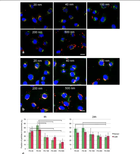
To find out if CPS at a single exposure of 25μg/ml ac-cumulate in lysosomes co-localization of fluorescent CPS (FluoSpheresW) and lysosomes identified by activity for cathepsin B and pH sensitive dye was performed. The majority of organelles stained with both LysoSensor and cathepsin B substrate but small organelles in the cellular periphery reacted only with LysoSensor (Add-itional file 3: Figure S3). Pictures taken immediately after removal of the particles after 4h of incubation and after 24h of post-incubation in medium showed that the 20 nm and 40 nm particles co-localized with lysosome mar-kers whereas many 200 nm and 500 nm FS were still located at the cell periphery (Figure 3a, co-localization with LysoSensor shown). After 24h particles of all sizes were seen inside the cells and only differences in the co-localization rates between the smallest particles and 500 nm particles were obvious (Figure 3b). Quantification of the co-localization on a pixel basis using Metamorph software (Figure 3c) also showed significant differences between the co-localization of small (<100 nm) and lar-ger particles after 4h, whereas after 24h only the co-localization rates between 20 nm and 40 nm versus 500 nm particles were found to be significantly different. There were no significant differences in the co-localization rate of the particles between the use of cathepsin B and of pH sensitive dye as lysosome marker.
Lysosome function & integrity
To assess lysosomal morphology and function, dye-release induced by loss of lysosome membrane integrity, measurements of pH by fluorescent dye, a screening
assay for lysosomal perturbation/dilation of the
endosomal-lysosomal system in drug development and enzymatic assays for lysosome activity, were used. Chloroquine, a drug known to increase intralysosomal pH and to inhibit the activities of arylsulfatase B and cathepsin B activity [38,39], was used as positive control. Table 2 Uptake rates of fluorescently labeled carboxyl
polystyrene particles (FS) by EAhy926 cells given as number of particles (N) and percentage of applied dose (%)
24h 48h 72h
Size (nm) N % N % N %
20 8.7*10^5 4.6 9.5*10^5 8.7 9.3*10^5 8.9
40 9.6*10^4 4.9 1.2*10^5 9.1 1.3*10^5 8.6
100 1.6*10^4 10.4 2.0*10^4 20.6 2.2*10^4 22.9
200 4.7*10^3 17.2 5.3*10^3 41.9 5.3*10^3 40.9
500 3.8*10^2 28.4 5.8*10^2 67.9 5.4*10^2 70
Figure 3Co-localization of fluorescently labeled CPS (FluoSpheres, 25μg/ml in DMEM + 2%FBS) and lysosomes identified by LysoSensor and cathepsin B activity by CV-(RR)2substrate in EAhy926 cells after 4h of exposure (a) and 24h (b) after particles were removed from the incubation medium.a: Co-labelling of FluoSpheres (FS red) with LysoSensor as marker for lysosomes (green). Scale bar: 10
μm. After 4h co-localization (yellow) is seen for 20 nm and 40 nm FS while 100 nm, 200 nm and 500 nm FS are seen predominantly at the cell periphery. b: After 24h more 100 nm-500 nm FS co-localize with lysosomes and only 500 nm FS were seen at the cell periphery. c: quantification of the co-localization by MetamorphWsoftware: the pH-dependent dye LysoSensor (Sensor, green) and enzymatic activity for the lysosomal enzyme cathepsin B (CatB, red) are used as markers for lysosomes. Co-localization rates of FS with LysoSensor were slightly higher than those with CatB substrate. After 4h significant differences in the co-localization rates were seen in the combinations: 20 nm FS versus 200 nm and 500 nm FS and 40 nm FS versus 100nm, 200 nm and 500 nm FS. After 24h only the differences between 20 nm and 40 nm FS on the one hand and 500 nm FS were significant. Particles with significant differences in the co-localization rates (p<0.05) are linked by brackets.
Fröhlichet al. Particle and Fibre Toxicology2012,9:26 Page 5 of 13
Changes in lysosomal membrane integrity, morphology and pH
Strong damage of lysosomal membranes leads to re-lease of the fluorescent dye Lucifer yellow, whereas minor damage can lead to changes in the lysosomal pH identified by decrease in the staining with the indi-cator dye LysoSensor. As it may be difficult to detect small increases in lysosomal pH based on fluorescence, the switch of the Acridine Orange fluorescence from red in healthy lysosomes to green in lysosomes with increased pH was used in addition. Finally, also an assay used in the screening for lysomotropic effects (lysosomal perturbation assay) was used. This assay is based on the increase in the green fluorescence of NBZ-PZ relative to the red fluorescence of propidium iodide as indication for dilatation of the endosomal-lysosomal system. The effect of 20μg/ml and 40 μg/ml CPS was investigated.
In the incubations with Lucifer Yellow the positive control (100μM chloroquine) after 24h led to leakage of the dye into the cytoplasm resulting in a diffuse cyto-plasmic staining instead of the punctate pattern in nor-mal cells (Additional file 3: Figure S1, 20μg/ml shown).
The staining of cells exposed to 20 nm and 200 nm CPS for 24h was not different from not exposed cells. After 72h the signal was too low for evaluation.
Also the staining with LysoSensor did not show differ-ences between 20 nm CPS, 200 nm CPS exposed cells (20 μg/ml and 40 μg/ml) and not exposed cells for 24h and 72h (data not shown).
Morphology of acridine orange stained cells exposed to 25 μM chloroquine showed enlarged lysosomes with marked increase in green fluorescence as indication for lysosomal swelling (Figure 4b). Cells exposed to 20 μg/ ml and 40μg/ml 20 nm and 200 nm CPS as well as not particle exposed cells showed occasionally lysosomes with increased green staining but no enlargement of the organelles (Figure 4a, c, d, 20μg/ml shown). To quantify these findings relative fluorescence in the red compared to the green channel was determined by fluorometry and a significant decrease in this ratio was seen for the positive control chloroquine (70.3 ± 2.2%). Neither in cells exposed to 20 μg/ml 20 nm CPS nor to those exposed to 20 μg/ml 200 nm CPS this ratio was signifi-cantly different from not exposed cells (91.37 ± 11.7% and 92.87 ± 13.2%, respectively).
In the lysosomal perturbation assay, a significant in-crease in the green fluorescence was only seen for the exposure to 20μg/ml and 40μg/ml 20 nm CPS for 24h (Figure 5a, 20 μg/ml shown) and the positive control (25μM chloroquine) at both time points. An increase in the amount of endosomal-lysosomal membranes was also seen when cells exposed for 24h to both concentra-tions of CPS were stained with anti-LAMP-1 antibody as marker for late endosomes and lysosomes. This increase to 135 ± 22% of the controls was not significant but was seen for all CPS-treated samples and at both concentra-tions of CPS (data not shown). After 72h of exposure to all particles, fluorescence in the green and red were simi-lar to those of the untreated controls in the lysosomal perturbation assay (Figure 5a). Similarly, also the increase in the anti-LAMP-1 staining of CPS exposed cells was no more seen (104 ± 6% of the controls).
Changes in enzyme activity
The assessment of cathepsin B activity in homogenates did not produce reliable data and the in-situ detection was used instead. The in-situ detection of cathepsin B and lysosomal sulfatase activity is based on the diffusion of substrates into the lysosomes, where they are metabo-lized and result in a fluorescent product. The fluores-cence was normalized to the intensity of the staining
with anti-LAMP-1 antibody, as marker for the
endosomal-lysosomal system since slight increases in LAMP-1 staining were seen upon exposure to CPS (see section lysosomal perturbation).
The decrease in lysosomal sulfatase activity was stron-gest after 24h of exposure to 20μg/ml CPS (Figure 5b). 20 nm, 200 nm and 500 nm CPS decreased lysosomal sulfatase activity significantly; the decrease was most pronounced for 20 nm CPS. After 72h of exposure still
Figure 5Enlargement of the endosomal-lysosomal system and changes in lysosomal enzyme activities in EAhy926 cells after 24h and 72h of exposure to 20μg/ml CPS of different sizes in DMEM + 2%FBS.25μM chloroquine was used as positive control (pos. co). a: Lysosomal/cytotoxicity assay, where an increase in the green signal is interpreted as perturbation in lysosome function. The signal is normalized to that of un-treated cells as 100%. Significant effects (asterisk) are seen only after incubation for 24h with 20 nm CPS. b: Changes in the reactivity with CV-(RR)2for cathepsin B (CatB) and with substrate for lysosomal sulfatases (Lyso). Arbitrary fluorescence data are normalized to the amount
of LAMP-1 staining and the signal of un-treated cells is set as 100%. In general, decreases in lysosomal activities were more pronounced after 24h of exposure than after 72h of exposure and the effect on lysosomal sulfatases was higher than that on cathepsin B. After 24h of exposure, significant decreases in the activity of lysosomal sulfatase were seen for all particles. After 72h, such significant decreases were only seen upon exposure to 20 nm CPS and 200 nm CPS. Significant changes are marked by asterisks.
Fröhlichet al. Particle and Fibre Toxicology2012,9:26 Page 7 of 13
significant decreases in lysosomal sulfatase activity were seen after exposure to 20 nm CPS and 200 nm CPS but not to 500 nm. Decrease of cathepsin B activity was not significant for these particles at both time points.
Discussion
In this study the intracellular distribution of CPS, as models for non- biodegradable NPs, and their effect on lysosomes of the endothelial cell line EAhy926 was stud-ied after 24h and 72h of exposure. These particles also served to identify assays, which could be used for the as-sessment of lysosomal effects of other NPs. 20μg/ml and 40 μg/ml reacted similarly. In addition to dilatation of the endosomal-lysosomal system (lysosome/cytotoxicity dual staining kit), decreases in the activities of lysosomal enzymes (cathepsin B, lysosomal sulfatase) were seen. Overall, the observed effects were small and more pro-nounced at 24h than at 72h of incubation suggesting that prolonged contact does not cause lysosomal damage.
In this study the intracellular localization and effects on lysosomes of CPS were studied at 50μg/ml as highest concentration in EAhy926 cells. These concentrations appear realistic for chronic effects upon repeated intra-venous exposure and EAhy926 cells as endothelial cell lines are relevant target cells. In the cytostatic Abraxa-neW the drug load accounts for 10% of the total nano-particle mass. Maximum serum concentrations for paclitaxel are 23μg/ml [40], corresponding to nanoparti-cle concentrations of 230 μg/ml. Imaging agents based on gadolinium chelates may reach blood concentrations of 1 mg/ml (http://www.berlex.com/html/products/pi/ Magnevist_PI.pdf ), but blood levels for iron oxide NPs in ResovistW range around 100 μg/ml [41]. The given concentrations are peak levels and not typical for chronic exposure. On the other hand, cellular accumula-tion has been shown in animal experiments [5-9,42] and may maintain intracellular levels of NPs at a lower level over a prolonged period of time.
For the studies on lysosomes, we used serum-reduced cells to slow down proliferation and, thereby, prevent di-lution of the intracellular nanoparticle concentration by cell division. Exposure to 0-1% FBS has been reported to decrease cell viability and induce apoptosis and necrosis in different cell lines [43-45]. In contrast to synthesis of RNA and DNA, protein synthesis was largely unaffected in L929 fibroblasts by serum deprivation but reduced in 3T3 fibroblasts [46,47]. To find out if serum deprivation displayed major effects on the EAhy926 cells used in this study, whole genome analysis was performed. As expected genes involved in pathways such as cell cycle, proliferation and apoptosis were changed but no effects on lysosomal genes were identified. The reduction of the
serum content from 10% to 2%, therefore, appears to be suitable for the study of lysosomes.
As prerequisite for lysosomal accumulation, intracellular localization of fluorescent CPS was studied. Already after 4h of exposure, 20 nm CPS showed a high rate of co-localization to endosomes and lysosomes. Consistent with studies by Lai et al. [48] the co-localization rate of 40 nm CPS was higher than that of 20 nm CPS. Co-localization of>40 nm CPS with lysosomes in our study increased with time and after 24h CPS also 500 nm CPS were loca-lized in lysosomes. Although different uptake mechanisms have been reported, most NPs, like for instance mesopor-ous silica particles, silicon dioxide particles, gold particles and quantum dots predominantly were localized in lyso-somes [49-52]. Size compared to surface charge appears to play only a minor role for lysosomal localization [53]. Nanoceria particles with positive and negative surface charge localized outside and inside lysosomes, respect-ively. Cytotoxicity data after 24h of exposure showed a much higher effect for lysosomal localization and the authors hypothesized that lysosomal localization is corre-lated to cytotoxicity on NPs in tumor cells. The authors do, however, not indicate if lysosomal changes are impli-cated in this tumor selective damage.
damage lysosomes. The size of quantum dots and fuller-enes is roughly in the same order of magnitude as that of the 20 nm CPS, but it is not likely that CPS act mainly by generation of oxygen species. In the concen-tration range, where lysosomal perturbation by 20 nm CPS was recorded in this paper (20μg/ml and 40μg/ml), no significant increase in intracellular oxygen radicals was recorded. The lack of generation of oxygen radicals by CPS is consistent with other data [37,57]. Although no dramatic damage of lysosomal morphology was detected, we identified changes in enzymatic activities, es-pecially of lysosomal sulfatase, upon exposure to CPS for 24h. These alterations were less pronounced at 72h of ex-posure. A more prominent decrease in enzyme activity at short incubation times than upon longer exposures is con-sistent with reported decreases in cathepsin B upon short exposure of macrophages to quartz D12 and to polystyr-ene microparticles [58,59]. Decreases in lysosomal sulfa-tase activities in this study were always greater than those in cathepsin B activity. This appears to be due to the spe-cificity of the substrate used for the assessment. The sub-strate SulfGreen is metabolized by all lysosomal sulfatases (MarkerGene LysoLife product information: http://www. markergene.com/product_sheets/pis1377.pdf ), whereas the specificity of the cathepsin B substrate for this prote-ase is higher. Provided that CPS acted on all lysosomal enzymes in a similar manner, changes in SulfGreen fluorescence are expected to be greater than those in CV-(RR)2(cathepsin B substrate) fluorescence.
In contrast to what was expected, damage by longer ex-posure to CPS was not higher than after shorter expos-ure. Polystyrene particles have a high potential for binding of proteins and form larger aggregates when in contact with protein-containing solutions [60,61]. It is, therefore, likely that the fraction of primary and poten-tially toxic particles rapidly declines inside the cell.
Conclusions
For the CPS investigated in this study only little interfer-ence with lysosomal function was seen. Changes were more prominent upon short than upon longer exposure. It may be suspected that by aggregation inside lysosomes the reactivity of CPS is decreased and toxicity reduced. The panel of lysosomal assays, which was validated for CPS in this study, can be used as a tool to identify the ac-tion of other types of NPs on lysosomes.
Methods
Particles
Carboxyl polystyrene (CPS) latex beads (20, 40, 100, 200, 500 nm), and the respective fluorescently labelled carboxyl polystyrene latex beads (FluoSpheresW) were obtained from Invitrogen. The fluorescently labelled particles contain dyes integrated in the core, which has
the advantage that fluorescent and non fluorescent particles have the same surface properties. CPS sus-pensions were put into an Elmasonic S40 water bath (ultrasonic frequency: 37 kHz, Elma, Singen) for 20 min prior to cell exposures and physicochemical characterization. To identify potential leakage of the dye from the FluoSpheresW after exposure for 24h-72h to cells at 37°C the supernatant of the cellular expo-sures was assessed for fluorescence. These measure-ments were performed only for the 500 nm spheres firstly because they contain the highest amount of fluorochrome and secondly because, according to the producer, it is not recommended to clean polystyrene spheres smaller than 300 nm by centrifugation. Super-natant containing the 500 nm FluoSpheresW were cen-trifuged at 20,000xg for 20 min in a Jouan 25 KRi centrifuge and fluorescence measured in a fluorescence plate reader (FLUOstar Optima, BMG Labortechnik) at 584 nm/612 nm. The fluorescence of the supernatant was 5 times higher than medium alone, this signal cor-responds to 0.05% of the fluorescence of the stem so-lution. As the fluorochromes in the particles are subjected to quenching, whereas the leached fluoro-chromes are not, the real amount of leached dye is expected to be much lower.
Physicochemical characterization of particles
Physicochemical characterization of the particles was performed by dynamic light scattering using a Malvern Zetasizer 3000 HS. Particles were diluted with respective medium to 200μg/ml and sonicated. After equilibration of the sample solution to 25°C, size and zeta potential were measured at 633 nm and a detection angle of 90°. NNLS software was used for sample analysis.
Cell culture
The human endothelial cell line EAhy926 (kind gift from Dr. C. J. Edgell) was cultured in DMEM, 10% fetal bo-vine serum (FBS), 2 mM L-glutamine and 1% penicillin/ streptomycin. Cell numbers were determined by cell counting and analyser system (CASYW TT, Innovatis). The cells were exposed to particles in DMEM + 2 mM L-glutamine for acute cytotoxicity studies and in DMEM, 2% fetal bovine serum (FBS), 2 mM L-glutamine for assessment of lysosome function. All cells were cultured at 37° C in a humid 95% air/5% CO2 at-mosphere. Cells were pre-cultured 24h in DMEM +10% FBS prior to the experiment. Subsequently, medium was removed and cells exposed to the particles suspended in DMEM + 2% FBS were cultured for up to 120h.
RNA isolation
Total RNA was isolated using RNeasy Mini kit (Qiagen, Stanford, CA, USA) according to the manufacturer’s
Fröhlichet al. Particle and Fibre Toxicology2012,9:26 Page 9 of 13
recommendations. The integrity of each RNA sample was evaluated using an Agilent 2100 Bioanalyzer (Agilent, Foster City, CA) and only RNAs with an RNA integrity number (RIN) above 9.5 were used for hybridizations.
Hybridization of microarrays
100 ng of total RNA for each sample were processed using the Affymetrix GeneChip Whole Transcript (WT) Sense Target Labeling Assay according to the manufac-turer’s instructions (Affymetrix). Double stranded cDNA was synthesized using a random hexamers tagged with a T7 promoter sequence. Using in vitro transcription, cRNA was generated from the double-stranded cDNA template using the Whole Transcript cDNA Synthesis and Amplification Kit (Affymetrix). cDNA was regener-ated using a reverse transcription reaction randomly primed with a mix containing dUTP. After hydrolysis of the cRNA with RNase H, the sense strand of cDNA was purified using the Affymetrix sample cleanup module, fragmented by incubation with UDG (uracil DNA glyco-sylase) and APE 1 (apurinic/apyrimidic endonuclease 1), and terminally biotin-labeled with terminal deoxynucleo-tidyl transferase using the WT Terminal Labeling Kit (Affymetrix), following the manufacturer’s instructions. Biotinylated sense strands were fragmented and hybri-dized to Affymetrix Human GeneChip 1.0 ST arrays (Affymetrix) using the Hybridization Control and Hybridization Wash and Stain kits (Affymetrix). The hybridization cocktail was incubated overnight at 45°C while rotating in a hybridization oven. After 16 h of hybridization, arrays were washed and stained in an Affymetrix GeneChip fluidics station 450, according to the Affymetrix-recommended protocol. Arrays were scanned on an Affymetrix GeneChip scanner.
Quantification of particle uptake
After 24h, 48h and 72h the incubation medium was removed, cells were washed three times with medium and removed from the plastic support by trypsin treatment (5 min with 0.25% trypsin/EDTA at 37°C). The action of trypsin was stopped with medium. One aliquot of the cell suspension was used for cell counting by CASY and an-other to measure fluorescence in a fluorescence plate reader (FLUOstar Optima, BMG Labortechnik) at 584 nm/612 nm.
To indicate particle uptake per cell the particle numbers in the stem solution used for the incubations were calculated according to the formula given by the producer (probes.invitrogen.com/media/pis/mp05000.pdf). The fluorescence signals of the stem solutions were mea-sured by serial dilution. Cell suspensions instead of medium alone were used for the dilution to account for autofluorescence or quenching effects caused by the cells.
Formazan bioreduction by MTS
CellTiter 96W AQueous Non-Radioactive Cell Prolifera-tion Assay (Promega) was used according to the manu-facturer’s instructions. In short, 20 μl of the combined MTS/PMS solution was added to 100 μl of each well. Plates were incubated for 2 hours at 37°C in the cell in-cubator. Absorbance was read at 490 nm on a plate reader (SPECTRA MAX plus 384, Molecular Devices).
Evaluation of oxidative stress by oxidation of dichlorodihydrofluorescein
Cells were grown for 24h in cell culture plates and loaded with 10μM 2,7-dichlorodihydrofluorescein diace-tate (Invitrogen) in medium for 30 min at 37°C. Subse-quently, cells were rinsed and cultured for 24h with 0-50 μg/ml CPS or 200μM H2O2as positive control. Fluores-cence was read with 485 nm excitation and 520 nm emisssion at a FLUOstar Optima (BMG Labortechnik).
Co-localization studies with lysosomes
channel the correlation of both signals was analysed. 50 cells were analysed for each particle.
Lysosomal membrane integrity
As positive control for lysosomal damage and interfer-ence with lysosome function chloroquine was used. After exposure for 24h to 20 nm and 200 nm CPS, to negative controls (medium) and to positive controls (25 μM chloroquine, Sigma-Aldrich) cells were washed and loaded with 1 μM Acridine Orange (Sigma-Aldrich) in PBS for 30 min at 37°C. After rinse in PBS cells fluores-cence was determined at a FLUOstar Optima (BMG Labortechnik) with 485 nm/520 nm for the green and 584 nm/612 nm for the red channel. The ratio of the signal in the red to the green channel was determined.
Alternatively, Lucifer yellow was used for assessment of lysosomal integrity. Cells were loaded with 0.1 mg/ml Lucifer yellow dilithium salt (Sigma) in culture medium at 37°C for 16h prior to the exposure to the particles. In-cubation with 100μM chloroquine was used as positive control. Images were taken with a LSM510 Meta con-focal laser scanning microscope (Zeiss) with ex 405 nm/ em LP 505 nm.
Assessment of lysosome function/pH
LysoSensor™ Green DND-189 (Invitrogen), an acidotro-pic probe, which accumulates in acidic compartments of cells as a result of protonization, was used at 1μM for 5 min at 37°C, according to the user manual. Accumula-tion in an acidic environment results in a pH-dependent increase in fluorescence. Staining performed after prein-cubation for 4h with CPS of different sizes was compared to cells preincubated in medium only. Quantification was performed at a FLUOstar Optima (BMG Labortechnik) with 485 nm/520 nm and pictures were taken with a LSM510 Meta confocal laser scanning microscope (Zeiss) with ex 488/em BP 505–550.
Lysosome/Cytotoxicity Dual staining Kit (Cayman), as indicator for perturbation of lysosome function, was used as indicated by the producer. The membrane permeable 4-nitro-7-(1-piperazinyl)-2,1,3-benzoxadiazole (NBZ-PZ) reacts with carboxylic acids in the lysosome and fluores-cence increases in an acid environment. Propidium iod-ide iod-identifies cells with loss of membrane integrity. Chloroquine was used at a concentration of 25 μM as positive control. Data were acquired at a FLUOstar Optima (BMG Labortechnik) with 485 nm/520 nm for the green and 584 nm/612 nm for the red channel.
Assessment of lysosome function/lysosomal enzymes
Cathepsin B activity
For detection in lysed cells the Cathepsin B Activity Assay Kit (PromoKine) with Ac-Arg-Arg labelled with amino-4-trifluoromethyl coumarine (Ac-RR-AFC) as substrate was
used according to the instructions given in the User Man-ual. Fluorescence was measured at a FLUOstar Optima (BMG Labortechnik) using a 400 nm/505 nm filter set.
For the detection of cathepsin B activity in-situ, cells were rinsed in PBS and exposed to the CV-(RR)2 sub-strate of the BIOMOL CV- Cathepsin B Detection Kit (Eubio). The stem solution in DMSO was diluted 1:5000 with medium and added for 20 min at 37°C. After this incubation, cells were rinsed in PBS and fluorescence quantified by fluorometric reading on a FLUOstar Op-tima (584 nm/612 nm). Thereafter, cells were fixed in 4% formalin and processed for co-labelling with LAMP-1 antibody. In several experiments, fluorescence data of unfixed and fixed samples were compared to exclude an influence of the fixation on the result 25 µM chloro-quine was used as positive control.
Lysosomal sulfatases
Activity in situ was assessed using the MarkerGene™ LysoLife™ Lysosomal Sulfatase Detection Kit (Eubio). After removal of the medium, the cells were washed once in PBS and incubated with the SulfGreen substrate (1.25 mM) in PBS for 4h at 37°C. After this incubation, cells were rinsed in PBS and fluorescence quantified by fluorometric reading on a FLUOstar Optima (485 nm/ 520 nm). Thereafter, cells were fixed in 4% formalin and processed for co-labelling with LAMP-1 antibody. In several experiments, fluorescence data of unfixed and fixed samples were compared to exclude an influence of the fixation on the result. 25 µM chloroquine was used as positive control.
Amount of lysosomes
For identification of changes in the
endosomal-lysosomal system immunoreactivity against the major lysosomal membrane glycoprotein LAMP-1, located in the membrane of late endosomes and lysosomes [62], was used. Cells were fixed in 4% formalin for 10 min at RT, washed three times in PBS, blocked for 30 min in 1% goat serum and incubated with the anti LAMP-1 antibody (1:1000, rabbit, Abcam) for 1h at RT. For visualization of the antibody binding, different secondary antibodies were used. Alexa 488-labelled secondary antibody (1:200, goat anti-rabbit IgG, Invitrogen) in co-staining experiments with cathepsin B activity and DyLight 594-labelled secondary antibody (1:200, goat anti-rabbit IgG, ThermoScientific) in combination with lysosomal sulfatase activity was added for 30 min at RT. For microscopical images cells were counterstained with 1 μg/ml Hoechst33342 for 15 min at RT. Data were acquired at a FLUOstar Optima (BMG Labortechnik). The following filter settings were used for the fluorometric evaluation: 485 nm/520 nm (SulfGreen substrate, LAMP-1 green detection), 584 nm/612 nm (cathepsin B substrate
Fröhlichet al. Particle and Fibre Toxicology2012,9:26 Page 11 of 13
CV-(RR)2, LAMP-1, red detection). For documentation of the staining, pictures were taken at a LSM510 Meta confocal laser scanning microscope (Zeiss) with 405/BP 420-480 for the blue channel, 488/BP 505–550 for the green channel (LysoSensor™, LAMP-1 detected with Alexa 488-labelled secondary antibody) and a 543/LP 560 for the red channel (CV-(RR)2, LAMP-1 detected with DyLight 594-labelled secondary antibody).
Statistics
For Data Analysis of microarrays CEL files were imported into Partek Genomic Suite6.4 software (Partek Inc) and robust multi-chip average (RMA) normalized (including background correction, quantilequintile normalization across all arrays, median polished summarization based on log transformed expression values). For detection of differ-entially expressed genes analysis of variance (ANOVA) was performed and genes with FDR5% and a fold change of at least 2 were considered to be significantly de-regulated.
All other data are represented as means ± S.D from three to six experiments. Data were analyzed with one-way analysis of variance (ANOVA) followed by Tukey-HSD post hoc test for multiple comparisons (SPSS 19 software). Differences between two samples were analyzed by independent t-test and Levine's Test for Equality of Variances. Results with p-values of less than 0.05 were considered to be statistically significant.
Additional files
Additional file 1: Table S1.Analysis of gene de-regulation in cells cultured in medium + 2% FBS compared to cells in medium + 10% FBS according to biological processes.A. Up-regulated genes in 2% FBS compared to 10% FBS. B. Down-regulated genes in 2% FBS compared to 10% FBS.
Additional file 2: Table S2.
Additional file 3: Figure S1.Staining of EAhy926 cells for lysosomal integrity using Lucifer yellow.Controls show a punctate staining, whereas cells treated with chloroquine show a diffuse cytoplasmic staining. The staining pattern of cells treated with 20μg/ml 20 nm and 200 nm carboxyl polystyrene particles (CPS) is similar to that of untreated controls. scale bar: 20μm. Figure 5s: Confocal image of LysoSensor (green) and cathepsin B substrate CV-(RR)2(red) double-stained EAhy926
cells. Co-localization of both staining is seen in yellow. In general more structures with acid content stained with LysoSensor than those with cathepsin B activity are seen. Arrows mark organelles with high cathepsin B activity and arrowheads indicate acidic structures with low cathepsin B activity. Scale bar: 10μm.
Competing interest
The authors disclose any financial competing interests.
Acknowledgements
We thank Diana Mujk for lysosomal assays, Hannes Seidl for microarray analysis and Sandra Blass for physico-chemical characterization of the particles. Editing of English language by Gabriella Salas is gratefully acknowledged. This work was supported by the FP6 European integrated project NanoBiopharmaceutics, the Research and Technology Development in Project Cluster NANO-HEALTH and the Austrian Research Science Grant P22576-B18.
Author details
1
Center for Medical Research, Medical University of Graz, Graz, Austria.
2Department of Internal Medicine, Division of Endocrinology and Nuclear
Medicine, Medical University of Graz, Graz, Austria.3Institute of Pharmaceutical Sciences, Department of Pharmaceutical Technology, Karl-Franzens-University of Graz, Graz, Austria.
Authors’contributions
EF: study design and writing of manuscript. CM, MA: experiments and data analysis. ER: particle characterization and draft of manuscript. EB: analysis of microarray data. TP: coordination and draft the manuscript. All authors read and approved the final manuscript.
Received: 20 February 2012 Accepted: 12 July 2012 Published: 12 July 2012
References
1. Nel A, Xia T, Madler L, Li N:Toxic potential of materials at the nanolevel.
Science2006,311:622–627.
2. Migliore L, Coppede F:Environmental-induced oxidative stress in neurodegenerative disorders and aging.Mutat Res2009,674:73–84. 3. Ranft U, Schikowski T, Sugiri D, Krutmann J, Kramer U:Long-term exposure
to traffic-related particulate matter impairs cognitive function in the elderly.Environ Res2009,109:1004–1011.
4. Stone V, Johnston H, Clift MJ:Air pollution, ultrafine and nanoparticle toxicology: cellular and molecular interactions.IEEE Trans Nanobiosci2007, 6:331–340.
5. Lin P, Chen JW, Chang LW, Wu JP, Redding L, Chang H, Yeh TK, Yang CS, Tsai MH, Wang HJ,et al:Computational and ultrastructural toxicology of a nanoparticle, Quantum Dot 705, in mice.Environ Sci Technol2008, 42:6264–6270.
6. Yang RS, Chang LW, Wu JP, Tsai MH, Wang HJ, Kuo YC, Yeh TK, Yang CS, Lin P:Persistent tissue kinetics and redistribution of nanoparticles, quantum dot 705, in mice: ICP-MS quantitative assessment.Environ Health Perspect
2007,115:1339–1343.
7. Chen J, Dong X, Zhao J, Tang G:In vivo acute toxicity of titanium dioxide nanoparticles to mice after intraperitioneal injection.J Appl Toxicol2009, 29:330–337.
8. Fabian E, Landsiedel R, Ma-Hock L, Wiench K, Wohlleben W, van Ravenzwaay B:Tissue distribution and toxicity of intravenously administered titanium dioxide nanoparticles in rats.Arch Toxicol2008, 82:151–157.
9. Lasagna-Reeves C, Gonzalez-Romero D, Barria MA, Olmedo I, Clos A, Sadagopa Ramanujam VM, Urayama A, Vergara L, Kogan MJ, Soto C: Bioaccumulation and toxicity of gold nanoparticles after repeated administration in mice.Biochem Biophys Res Commun2010,393:649–655. 10. Oberdorster G, Ferin J, Lehnert BE:Correlation between particle size,
in vivo particle persistence, and lung injury.Environ Health Perspect1994, 102(Suppl 5):173–179.
11. Mukae H, Vincent R, Quinlan K, English D, Hards J, Hogg JC, van Eeden SF: The effect of repeated exposure to particulate air pollution (PM10) on the bone marrow.Am J Respir Crit Care Med2001,163:201–209. 12. Ji JH, Jung JH, Kim SS, Yoon JU, Park JD, Choi BS, Chung YH, Kwon IH,
Jeong J, Han BS,et al:Twenty-eight-day inhalation toxicity study of silver nanoparticles in Sprague-Dawley rats.Inhal Toxicol2007,19:857–871. 13. Rossi EM, Pylkkanen L, Koivisto AJ, Vippola M, Jensen KA, Miettinen M, Sirola
K, Nykasenoja H, Karisola P, Stjernvall T,et al:Airway exposure to silica-coated TiO2 nanoparticles induces pulmonary neutrophilia in mice.
Toxicol Sci2010,113:422–433.
14. Yin XJ, Dong CC, Ma JY, Antonini JM, Roberts JR, Stanley CF, Schafer R, Ma JK:Suppression of cell-mediated immune responses to listeria infection by repeated exposure to diesel exhaust particles in brown Norway rats.
Toxicol Sci2004,77:263–271.
15. Geiser M, Rothen-Rutishauser B, Kapp N, Schurch S, Kreyling W, Schulz H, Semmler M, Im Hof V, Heyder J, Gehr P:Ultrafine particles cross cellular membranes by nonphagocytic mechanisms in lungs and in cultured cells.Environ Health Perspect2005,113:1555–1560.
17. Muhlfeld C, Gehr P, Rothen-Rutishauser B:Translocation and cellular entering mechanisms of nanoparticles in the respiratory tract.Swiss Med Wkly2008,138:387–391.
18. Sahay G, Alakhova DY, Kabanov AV:Endocytosis of nanomedicines.
J Control Release2010,145:182–195.
19. Doherty GJ, McMahon HT:Mechanisms of endocytosis.Ann Rev Biochem
2009,78:857–902.
20. Chithrani BD, Ghazani AA, Chan WC:Determining the size and shape dependence of gold nanoparticle uptake into mammalian cells.Nano Lett2006,6:662–668.
21. Faklaris O, Joshi V, Irinopoulou T, Tauc P, Sennour M, Girard H, Gesset C, Arnault JC, Thorel A, Boudou JP,et al:Photoluminescent diamond nanoparticles for cell labeling: study of the uptake mechanism in mammalian cells.ACS Nano2009,3:3955–3962.
22. Goya GF, Marcos-Campos I, Fernandez-Pacheco R, Saez B, Godino J, Asin L, Lambea J, Tabuenca P, Mayordomo JI, Larrad L,et al:Dendritic cell uptake of iron-based magnetic nanoparticles.Cell Biol Int2008,32:1001–1005. 23. Jaiswal JK, Mattoussi H, Mauro JM, Simon SM:Long-term multiple color imaging of live cells using quantum dot bioconjugates.Nat Biotechnol
2003,21:47–51.
24. Nativo P, Prior IA, Brust M:Uptake and intracellular fate of surface-modified gold nanoparticles.ACS Nano2008,2:1639–1644.
25. Rejman J, Oberle V, Zuhorn IS, Hoekstra D:Size-dependent internalization of particles via the pathways of clathrin- and caveolae-mediated endocytosis.Biochem J2004,377:159–169.
26. Stearns RC, Paulauskis JD, Godleski JJ:Endocytosis of ultrafine particles by A549 cells.Am J Respir Cell Mol Biol2001,24:108–115.
27. Olsson GM, Svensson I, Zdolsek JM, Brunk UT:Lysosomal enzyme leakage during the hypoxanthine/xanthine oxidase reaction.Virchows Archiv B
1989,56:385–391.
28. Maysinger D, Lovric J:Quantum dots and other fluorescent nanoparticles: quo vadis in the cell?Adv Exp Med Biol2007,620:156–167.
29. Moore MN, Readman JAJ, Readman JW, Lowe DM, Frickers PE, Beesley A: Lysosomal cytotoxicity of carbon nanoparticles in cells of the molluscan immune system: An in vitro study.Nanotoxicology2009,3:40–45. 30. Koehler A, Marx U, Broeg K, Bahns S, Bressling J:Effects of nanoparticles in
Mytilus edulis gills and hepatopancreas - a new threat to marine life?
Mar Environ Res2008,66:12–14.
31. Holz FG, Schutt F, Kopitz J, Eldred GE, Kruse FE, Volcker HE, Cantz M: Inhibition of lysosomal degradative functions in RPE cells by a retinoid component of lipofuscin.Invest Ophthalmol Vis Sci1999,40:737–743. 32. Garnett MC, Kallinteri P:Nanomedicines and nanotoxicology: some
physiological principles.Occup Med2006,56:307–311.
33. Frese MA, Schulz S, Dierks T:Arylsulfatase G, a novel lysosomal sulfatase.
J Biol Chem2008,283:11388–11395.
34. Greiner-Tollersrud O, Berg T:Lysosomal storage disorders. InLysosomes. Edited by Saftig P. New York: Springer Science, Landes Bioscience; 2005:60–73.
35. Anderson N, Borlak J:FEBS Lett.Drug-induced phospholipidosis2006, 580:5533–5540.
36. Fröhlich E, Meindl C, Roblegg E, Griesbacher A, Pieber TR:Cytotoxicity of nanoparticles is influenced by size, proliferation and embryonic origin of the cells used for testing.Nanotoxicology2012,6:424–439.
37. Fröhlich E, Samberger C, Kueznik T, Absenger M, Roblegg E, Zimmer A, Pieber TR:Cytotoxicity of nanoparticles independent from oxidative stress.J Toxicol Sci2009,34:363–375.
38. Bhattacharyya S, Solakyildirim K, Zhang Z, Linhardt RJ, Tobacman JK: Chloroquine reduces arylsulphatase B activity and increases chondroitin-4-sulphate: implications for mechanisms of action and resistance.Malar J
2009,8:303.
39. MacGregor RR, Hamilton JW, Kent GN, Shofstall RE, Cohn DV:The degradation of proparathormone and parathormone by parathyroid and liver cathepsin B.J Biol Chem1979,254:4428–4433.
40. http://www.fda.gov/ohrms/dockets/ac/06/briefing/2006-4235B2-, 01-01AbraxisBioscience-background.pdf.
41. http://www.pharmazie.com/graphic/A/42/1-24242.pdf.
42. Uchida M, Willits D, Muller K, Willis A, Jackiw L, Jutila M, Young M, Porter A, Douglas T, Uchida M, Willits D, Muller K, Willis A, Jackiw L, Jutila M, Young M, Porter A, Douglas T:Intra-cellular distribution of macrophage targeting ferritin iron oxide nano-composite.Adv Mater2009,21:458–462.
43. Hasan NM, Adams GE, Joiner MC:Effect of serum starvation on expression and phosphorylation of PKC-alpha and p53 in V79 cells: implications for cell death.Int J Cancer1999,80:400–405.
44. Mengual Gomez DL, Belaich MN, Rodriguez VA, Ghiringhelli PD:Effects of fetal bovine serum deprivation in cell cultures on the production of Anticarsia gemmatalis multinucleopolyhedrovirus.BMC Biotechnol2010, 10:68.
45. Oya N, Zolzer F, Werner F, Streffer C:Effects of serum starvation on radiosensitivity, proliferation and apoptosis in four human tumor cell lines with different p53 status.Strahlenther Onkol2003,179:99–106. 46. Zetterberg A, Larsson O:Kinetic analysis of regulatory events in G1
leading to proliferation or quiescence of Swiss 3T3 cells.Proc Natl Acad Sci USA1985,82:5365–5369.
47. Zetterberg A, Skold O:The effect of serum starvation on DNA, RNA and protein synthesis during interphase in L-cells.Exp Cell Res1969, 57:114–118.
48. Lai SK, Hida K, Man ST, Chen C, Machamer C, Schroer TA, Hanes J: Privileged delivery of polymer nanoparticles to the perinuclear region of live cells via a non-clathrin, non-degradative pathway.Biomaterials2007, 28:2876–2884.
49. Al-Rawi M, Diabate S, Weiss C:Uptake and intracellular localization of submicron and nano-sized SiO(2) particles in HeLa cells.Arch Toxicol
2011,85:813–826.
50. He Q, Zhang Z, Gao Y, Shi J, Li Y:Intracellular localization and cytotoxicity of spherical mesoporous silica nano- and microparticles.Small2009, 5:2722–2729.
51. Silver J, Ou W:Photoactivation of quantum dot fluorescence following endocytosis.Nano Lett2005,5:1445–1449.
52. Lévy R, Shaheen U, Cesbron Y, Sée V:Gold nanoparticles delivery in mammalian live cells: a critical review.Nano Rev2010, 1.
53. Asati A, Santra S, Kaittanis C, Perez JM:Surface-charge-dependent cell localization and cytotoxicity of cerium oxide nanoparticles.ACS Nano
2010,4:5321–5331.
54. van Dyke R:part III The lysosome in its cytoplasmic environment. Acidification of endosomes and lysosomes. Lysosomal membrane bodies. InSubcellular Biochemistry, Vol 27 Biology of the lysosome. 23rd edition. Edited by Lloyd J, Mason R. London, New York: Plenum Press; 1996. 55. Held P, Newick K, Shen D, Patton W:Automated Detection of
Drug-Induced Lysosomal Cytotoxicity - Automation of the Lyso-IDWRed Assay Using the EL406™Combination Washer Dispenser.Lab Manager Magazine
2010.
56. Ishiguro K, Ando T, Goto H:Novel application of 4-nitro-7-(1-piperazinyl)-2,1,3-benzoxadiazole to visualize lysosomes in live cells.Biotechniques
2008,45:467–468.
57. Swain WA, O'Byrne KJ, Faux SP:Activation of p38 MAP kinase by asbestos in rat mesothelial cells is mediated by oxidative stress.Am J Physiol Lung Cell Mol Physiol2004,286:L859–865.
58. Oh YK, Swanson JA:Different fates of phagocytosed particles after delivery into macrophage lysosomes.J Cell Biol1996,132:585–593. 59. Patzold S, Schmidt A, Seidel A:Loss of cathepsin B activity in alveolar
macrophages after in vitro quartz phagocytosis.J Toxicol Environ Health
1993,40:547–554.
60. Xia T, Kovochich M, Brant J, Hotze M, Sempf J, Oberley T, Sioutas C, Yeh JI, Wiesner MR, Nel AE:Comparison of the abilities of ambient and manufactured nanoparticles to induce cellular toxicity according to an oxidative stress paradigm.Nano Lett2006,6:1794–1807.
61. Mayer A, Vadon M, Rinner B, Novak A, Wintersteiger R, Fröhlich E:The role of nanoparticle size in hemocompatibility.Toxicology2009,258:139–147. 62. D'Souza MP, August JT:A kinetic analysis of biosynthesis and localization
of a lysosome-associated membrane glycoprotein.Arch Biochem Biophys
1986,249:522–532.
doi:10.1186/1743-8977-9-26
Cite this article as:Fröhlichet al.:Action of polystyrene nanoparticles of different sizes on lysosomal function and integrity.Particle and Fibre Toxicology20129:26.
Fröhlichet al. Particle and Fibre Toxicology2012,9:26 Page 13 of 13