INTRODUCTION
The lung is an important target organ for systemic inflammatory mediators re-leased after severe infection (1,2) and major trauma (3–5). Acute lung injury (ALI) and acute respiratory distress syn-drome (ARDS) have been the major causes of morbidity and mortality in in-tensive care units (6). During the past decade, despite improvements in sup-portive care, ALI/ARDS still carries high mortality rates between 26% and 35% (7). ALI/ARDS is characterized by noncar-diogenic pulmonary edema, and pul-monary and systemic inflammation with a spectrum of increasing severity of lung injury resulting in respiratory failure (8). ALI frequently has systemic components, of which several of the major triggering
conditions include sepsis, nonpulmonary trauma and shock. Conversely, diffuse injury and infection of the lung cause the systemic inflammatory response syn-drome (SIRS) and sepsis (9,10). There-fore, the interaction of pulmonary and systemic inflammation exaggerates the inflammatory process and the develop-ment of ALI.
Recent studies have demonstrated that the pattern-recognition receptors (PRRs) play an important role in the pathogene-sis of ALI/ARDS. PRRs are evolutionar-ily conserved receptors that sense not only pathogen-associated molecular pat-terns (PAMPs) that derived from invad-ing microbes, but also damage-associated molecular patterns (DAMPs) that are re-leased from dead cells, thereby
trigger-ing an inflammatory response to both in-fectious and noninin-fectious insults (11). Activation of PRRs results in initiation of several extracellular activating cascades, as well as various intracellular signaling pathways that cause inflammatory re-sponses. Figure 1 summarizes the role of PRRs in mediating infectious- and in-jury-induced ALI. This review will focus on recent advances in understanding of the role of PRRs in the mechanisms of ALI/ARDS.
BIOLOGY OF PRRS
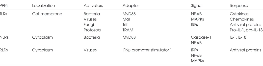
Three major subfamilies of PRRs have been reported: Toll-like receptors (TLRs), retinoic acid–inducible gene I (RIG-I)-like receptors (RLRs) and nucleotide-binging oligomerization domain (NOD)-like re-ceptors (NLRs) (12,13). A brief compari-son of the three subfamilies of PRRs is shown in Table 1.
Toll-Like Receptors
TLRs are the most extensively stud-ied family of PRRs. To date, 10 TLRs (TLRs 1–10) in humans and 12 TLRs
Acute Lung Injury
Meng Xiang and Jie Fan
Department of Surgery, School of Medicine, University of Pittsburgh and Surgical Research, VA Pittsburgh Healthcare System, Pittsburgh, Pennsylvania, United States of America
Acute lung injury (ALI) that clinically manifests as acute respiratory distress syndrome is caused by an uncontrolled systemic in-flammatory response resulting from clinical events including sepsis, major surgery and trauma. Innate immunity activation plays a central role in the development of ALI. Innate immunity is activated through families of related pattern recognition receptors (PRRs), which recognize conserved microbial motifs or pathogen-associated molecular patterns (PAMPs). Toll-like receptors were the first major family of PRRs discovered in mammals. Recently, NACHT–leucine-rich repeat (LRR) receptors and retinoic acid–inducible gene–like receptors have been added to the list. It is now understood that in addition to recognizing infectious stimuli, both Toll-like receptors and NACHT-LRR receptors can also respond to endogenous molecules released in response to stress, trauma and cell damage. These molecules have been termed damage-associated molecular patterns (DAMPs). It has been clinically observed for a long time that infectious and noninfectious insults initiate inflammation, so confirmation of overlap-ping receptor-signal pathways of activation between PAMPs and DAMPs is no surprise. This review provides an overview of the PRR-dependent mechanisms of ALI and clinical implication. Modification of PRR pathways is likely to be a logical therapeutic tar-get for ALI/acute respiratory distress syndrome.
© 2010 The Feinstein Institute for Medical Research, www.feinsteininstitute.org Online address: http://www.molmed.org
doi: 10.2119/molmed.2009.00097
Address correspondence and reprint requests to Jie Fan, Department of Surgery, School of Medicine, University of Pittsburgh, and Surgical Research, VA Pittsburgh Healthcare Sys-tem, University Drive, Pittsburgh, PA. 15240. Phone: 412-360-6204; Fax: 412-360-6389; E-mail: jif7@pitt.edu.
(TLRs 1–9 and TLRs 11–13) in mice have been defined (14). TLRs 3, 7, 8 and 9 are expressed intracellularly, whereas TLRs 1, 2, 4, 5, 6 and 10 are ex-pressed on the cell surface. TLRs are expressed on a range of immune cells including macrophages, dendritic cells, B cells and certain types of T cells, as well as on certain nonimmune cells, such as endothelial cells, smooth mus-cle cells and epithelial cells that lie at potential sites of entry, including the skin and the respiratory, intestinal and genitourinary tracts. The expression of TLRs is modulated by activation,
matu-ration or differentiation of the different cell types (15,16).
TLR proteins are a family of type I transmembrane receptors characterized by an NH2-terminal extracellular leucine-rich repeat (LRR) domain, which mediate the recognition of their respec-tive PAMPs, and a COOH-terminal intra-cellular tail containing a conserved re-gion called the Toll/interleukin 1 (IL-1) receptor (TIR) homology domain. The TIR domain is the defining motif of the TLR/IL-1 superfamily, and it is likely to be one of the earliest signaling domains to have evolved (17). TLRs can recognize
a diverse range of PAMPs, generate in-flammatory signals to coordinate innate immune responses and modulate adap-tive immune responses. The list of TLR ligands is growing. However, the ligand for TLR10 and mouse TLR8 remains un-known at present. Activation of TLRs initiates two major pathways: the MyD88-dependent pathway, which is used by all TLRs except TLR3, resulting in the activation of nuclear factor (NF)-κB and activator protein-1 (AP-1); and the TRIF- dependent pathway, which is initi-ated by TLR3 and TLR4, resulting in the activation of type I interferons (IFNs) (13,18,19). Expression of numerous proinflammatory cytokines, such as tumor necrosis factor (TNF)-α, IL-6, IL-12 and IFNs, is one of the major outcomes of the activation of the pathways (15). TLR signaling is summarized and shown in Figure 2.
RIG-Like Receptors
RLRs as DExD/H-containing RNA helicases are expressed in the cytoplasm in a variety of cells, including immune and nonimmune cells. Unlike membrane-bond TLR3, TLR7 and TLR9, which are localized on the endosome and recognize viral double stranded RNA, single-stranded RNA and DNA, respectively, RLRs are cytoplasmic proteins that recog-nize viral RNA produced as a conse-quence of viral replication (20,21). RLRs consist of three family members: RIG-I, melanoma differentiation-associated gene 5 (MDA5) and laboratory of genet-ics and physiology 2 (LGP2) (21,22). Figure 1.Role of PRRs in mediating inflammation and organ injury. Infection causes PAMP
release, but also causes tissue and cell damage and subsequent DAMP release. Similarly, injury caused by trauma or various other factors not only leads to DAMP release but also renders the patient more susceptible to infection and therefore PAMP release. In turn, the PAMPs and DAMPs act through PRRs, which include TLRs, NLRs and RLRs, to activate the in-nate immune system, yet they can also contribute to persistent and deleterious systemic inflammation and organ injury, including ALI.
Table 1.Comparison of the three subfamilies of PRRs.
PPRs Localization Activators Adaptor Signal Response
TLRs Cell membrane Bacteria MyD88 NF-κB Cytokines
Viruses Mal MAPKs Chemokines
Fungi Trif IRFs Antiviral proteins
Protozoa TRAM Pro–IL-1, pro–IL-18
NLRs Cytoplasm Bacteria MyD88 Caspase-1 IL-1, IL-18
NF-κB
RLRs Cytoplasm Viruses IFNβpromoter stimulator 1 IRFs Antiviral proteins
Structurally, RIG-I and MDA5 contain a DExD/H box RNA helicase domain and two caspase-recruiting domain (CARD)-like domains required for eliciting down-stream signaling pathways (23,24). The C-terminal region of RIG-I contains a re-pressor domain (RD), which inhibits downstream signaling. The MDA5 C-ter-minal region is similar to the RD of RIG-I; however, its function is not clear. LGP2 contains a DExD/H helicase domain and an RD, but lacks the CARD-like region.
LGP2 was suggested to play an inhibitory role in virus-induced response, because the LGP2 RD binds the RIG-I RD and suppresses signaling as a consequence of interfering with the self-association of RIG-I (20,25,26).
RIG-I is essential for the recognition of a series of RNA viruses, which include Sendai virus, Newcastle disease virus, in-fluenza virus, vesicular stomatitis virus and Japanese encephalitis virus (27). MDA5 is required for the recognition of
other RNA viruses, including picor-naviruses such as encephalomyocarditis virus, Mengo virus and Theiler virus (13,27). Thus, RIG-I and MDA5 have specificities in their detection of RNA viruses, presumably through recognition of distinct structures of viral RNA (24,28).
Recent studies revealed a pathway of RLR regulation of NF-κB. RIG-I/MDA5 CARD domains, through a CARD-con-taining adaptor protein, IFNβpromoter stimulator 1, also known as mitochondr-ial antiviral signaling protein, and CARD adaptor inducing IFNβ, ultimately acti-vate IRF3 and NF-κB (29,30). The role of RLRs in the mechanism of ALI has not been elucidated.
Nucleotide-Binding Oligomerization Domain–Like Receptors
The NLR family is a group of recently identified cytoplasmic PRRs that con-tain more than 23 members in humans (15). The major role of NLRs is to recog-nize cytoplasmic microbial PAMPs and/or endogenous danger signals and initiate immunological responses, al-though the physiological function of most NLRs is poorly understood at present (31). Members of the NLR fam-ily are categorized into at least five sub-families according to their N-terminal structure, including NODs (nucleotide-binding oligomerization domain-1), NALPs (NACHT-, LRR- and domain– containing proteins), IPAF (ICE-protease activating factor), NAIPs (neuronal apoptosis inhibitor factors) and class II transactivator (CIITA) (32).
The NLR family shares a domain organization consisting of a C-terminal LRR domain, a central nucleotide- binding NACHT domain, and an N-terminal protein–protein interaction domain com-posed of a CARD, pyrin domain (PYD) or baculovirus inhibitor of apoptosis re-peat (BIR) domain (33). NODs and IPAF contain CARD effector domains, whereas NALPs have PYD domains, and NAIPs possess BIR domains.
The functions of NOD1, NOD2, IPAF and NALP3 are more studied. NOD1 and NOD2 are the first NLRs that are reported Figure 2.TLR signaling (an example in macrophages and dendritic cells). TLR2 (TLR2 in
to have a direct function as PRRs in the recognition of peptidoglycan derived peptides. NOD1 senses
γ-D-glutamyl-meso-diaminopimelic acid (that is,-DAP) that is derived from Gram-negative bacteria (34), whereas NOD2 senses muramyl dipeptide, which is from both Gram-positive and Gram-negative bacteria (35,36). When NODs bind with PGN-derived peptides, they rapidly form oligomers, which lead to the recruitment of the receptor-interacting protein 2 (RIP2) kinase through CARD–CARD interactions (37). This complex of NOD1-RIP2 or NOD2-RIP2 then recruits the inhibitor of NK-κB kinase complex (IKK), leading to the activation of NF-κB. Activation NOD1 and NOD2 can also initiate an MAPK pathway that leads to the activation of p38 and ERK. In addition, NOD1 signal-ing can activate JNK as well (38).
NALP proteins are characterized by the presence of an N-terminal pyrin effector domain (39). Several NLRs, namely NALP1, NALP2 and NALP3, have an im-portant role in activation of proinflamma-tory caspases through formation of in-flammasome (37,40). The inin-flammasome is a multiprotein complex of more than 700 kDa that is responsible for the activa-tion of caspases 1 and 5, leading to the processing and secretion of the proin-flammatory cytokines IL-1βand IL-18 (41,42). Martinon et al. have presented different caspase activation platforms in which different components constitute the various inflammasomes (43). Two types of NALP inflammasome are better studied: the NALP1 inflammasome that is composed of NALP1, the adaptor pro-tein ASC, caspase-1 and caspase-5 (41), and the NALP2/NALP3 inflammasome that contains NALP2 or NALP3 CARDI-NAL, ASC and caspase-1 (44).
PRRS AND INFECTION-RELATED ALI
Role of TLRs in Activation of Cellular Inflammatory Responses
TLRs 1–10 are expressed in lung tissue (45), and individual TLRs are differen-tially regulated in specific lung cell pop-ulations in response to microbial
stimula-tion. TLR2, TLR4, TLR5 and TLR9 are the most likely to be involved in recognition of bacteria in the lungs (46–48). A study by Bernard et al. has shown that ALI/ ARDS induced by lipopolysaccharide (LPS) is a major cause of mortality among humans (49). The LPS membrane receptor complex is composed of several accessory molecules, which include phosphatidylinositol- anchored CD14, TLR4, MD2 and MD1 (50). The LPS-binding protein (LBP) enhances the bind-ing of LPS to its receptor (51). Absence of CD14, MD2 or LBP abrogates most LPS responses (51,52). Expression of func-tional TLR4 has been found in many cell types in the lung (45), and LPS-induced lethal shock and ALI have been shown to be TLR4 dependent (53–55). Thus, TLR4 plays a critical role in the mechanism of infection-related ALI.
A recent study has shown that respira-tory infections in the human lung initi-ated by TLR2 agonist lipoteichoic acid (LTA, a component of Gram-positive bac-teria) and TLR4 agonist LPS (a compo-nent of Gram-negative bacteria) exhibit different inflammatory responses (56). The study was performed on healthy subjects with LPS or LTA instillation into the contralateral lung. Alveolar macro -phages (AMφ) isolated from broncho -alveolar lavage fluid were analyzed by multiplex ligation-dependent probe amplification. The results show that whereas both LPS and LTA elicited neu-trophil recruitment, only LPS instillation was associated with activation of neu-trophils (PMN) (CD11b surface expres-sion and degranulation) and consistent rises of chemokine/cytokine levels. Moreover, LPS but not LTA activated AM, as reflected by enhanced expression of proinflammatory mediators and in-creased spontaneous cytokine release upon incubation ex vivo. Remarkably, only LTA induced C5a release. These data suggest that stimulation of TLR2 or TLR4 results in differential pulmonary inflammation, which may be of relevance for understanding the differences during Gram-positive and Gram-negative respi-ratory tract infection (56).
TLR and Endothelial Cell Activation
Pulmonary endothelium is a major component of the alveolar-capillary unit, and is susceptible to injury from noxious agents that are either inhaled or deliv-ered to the lung through the pulmonary circulation (57). In ALI, pulmonary en-dothelium plays a major role by (a) alter-ing metabolic activity to affect pul-monary and systemic homeostasis, (b) mediating polymorphonuclear PMN ad-hesion to promote PMN infiltration, (c) changing PMN barrier permeability to cause pulmonary edema and (d) secret-ing cytokines and chemokines to induce lung inflammation (58).
A recent study by Andonegui and col-leagues has shown that endothelial cells (ECs) are the key sentinel cells for de-tecting infection by Gram-negative bac-teria and recruiting PMN to peripheral tissues (53,59). Indeed, previous studies have shown that direct activation of cir-culating PMN with LPS is not sufficient to induce their sequestration within the lung (53). Interactions of PMN with ECs seems important for the process of PMN sequestration into the lung (53,60). LPS stimulates the CD14 and TLR4 complex, which in turn activates NF-κB (61) and increases the expression of adhesion molecule E-selection, intercellular adhe-sion molecule-1 (ICAM-1) and vascular cell adhesion molecule 1 (62,63). The TLR4 signaling also leads to production and release of various bioactive mole-cules, including IL-1β, IL-6, TNF-α, chemokines and nitric oxide (64), all of which are actively involved in the devel-opment of ALI.
upregulates TLR2 expression in ECs, and this process is enhanced by oxidant signal-ing generated by PMN NAD(P)H oxidase. The functional relevance of NAD(P)H oxi-dase in mediating TLR4- induced TLR2 ex-pression in ECs is evident by markedly el-evated and stable ICAM-1 expression as well as augmented PMN migration in re-sponse to sequential challenge with LPS and peptidoglycan (68). Thus, TLR2 acti-vation, signaled by TLR4 and as regulated by PMN NAD(P)H oxidase, is an impor-tant mechanism responsible for amplify-ing PMN transmigration to sites of infec-tion (Figure 3).
The TLR4-TLR2 interaction suggests a highly coordinated, oxidant-mediated up-regulation of TLR2 in response to LPS. When one considers the interactions of
the innate immune system as microbes are first encountered, the value of such temporal organization is significant. For example, Gram-negative bacteria persist in tissues and, if they are not immediately killed through the activation of PMN, complement and other antimicrobial fac-tors, they may spill out systemically and result in septic shock. Survival in the face of such infections depends on the innate immune system, which must be able to monitor and respond to pathogens over a prolonged period of time. Given the need for a prolonged response to bacterial in-fection, it has always seemed somewhat surprising that response to LPS is tempo-rally finite. This endotoxin tolerance means that within hours after exposure to LPS, innate immune cells are incapable of
responding again to a rechallenge (69). But it is now clear that as LPS sensitivity wanes, the immune system has at its dis-posal the capability of marshaling re-sponses via oxidative metabolites and their ability to upregulate other TLRs (70). The subsequent means of responding to bacteria depend on the ability of the in-nate immune system to destroy microbes and enhance the release of alternative im-mune stimuli. The TLRs that are utilized are the ones that bind the constituents of degrading bacteria, such as lipopeptides, PGN, heat shock proteins and CpG DNA. It seems plausible that activated PMN may even alter the phenomenon of LPS tolerance, at least in a localized context, by setting into action a positive feedback loop at sites to which PMN are chemoat-tracted (71). This would enhance inflam-matory responses locally and help fight infection. However, in a setting of post-trauma SIRS or ALI, the primed PMN ac-tivation serves as an amplifier to cause enhanced PMN infiltration and organ injury.
TLR and PMN Activation
Extensive PMN influx into the lungs is one of the characteristics of ALI. Stud-ies have shown that excessive induction of proinflammatory cytokines in PMN and delayed PMN apoptosis are associ-ated with higher mortality and more se-vere organ dysfunction in sepsis pa-tients (72,73). Human PMN express all TLR mRNA except TLR3. TLR2 are more abundant than TLR4 on PMN (74,75). PMN recruitment to the lung after LPS inhalation is primarily depen-dent on TLR4–NF-κB signaling (76,77). The PI3K/Akt pathway was also re-ported to be involved in TLR4-induced expression of IL-1β, TNF-αand chemo -kine macro phage inflammatory protein (MIP)-2 (78).
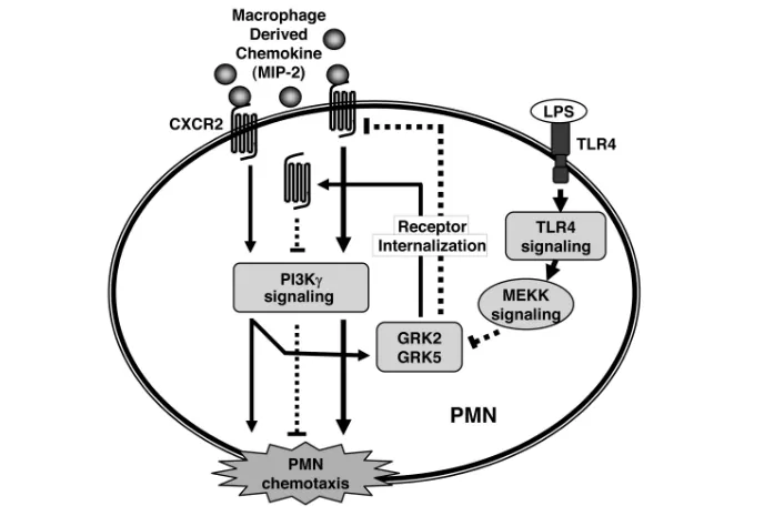
We have reported that TLR4, through regulating G-protein–coupled receptor kinases (GRKs), promotes PMN migra-tion. We demonstrated that MIP-2 induces GRK2 and GRK5 expression in PMNs through phosphoinositide-3-kinase (PI3K)-γsignaling, and LPS-activated Figure 3.Model of PMN NADPH oxidase-derived oxidant signaling in mediating the
signaling through the TLR4 pathway transcriptionally downregulates the ex-pression of GRK2 and GRK5 in response to MIP-2. The reduced expression of GRKs lowers chemokine receptor desen-sitization and markedly augments the PMN migratory response. These data in-dicate that TLR4 modulation of PMN surface chemokine receptor expression after the downregulation of GRK2 and GRK5 expression is a critical determinant of PMN migration (79) (Figure 4).
A recent study explored a novel role of mTOR complex 1 (mTORC1) in TLR2-and TLR4-induced PMN activation (80). Administration of rapamycin, an in-hibitor of mTORC1, decreased the sever-ity of lung injury after intratracheal LPS or PAM (a TLR2 ligand) administration, as determined by diminished neutrophil accumulation in the lungs, reduced inter-stitial pulmonary edema, and diminished
levels of TNF-αand IL-6 in bronchoalve-olar lavage fluid. These results indicate that mTORC1 activation is essential in TLR2- and TLR4-induced PMN activa-tion, as well as in the development and severity of ALI.
The E3 ubiquitin ligase Cblb has a cru-cial role in the prevention of chronic in-flammation and autoimmunity. How-ever, a recent study showed that Cblb also has an unexpected function in acute lung inflammation (81). Cblb attenuates the sequestration of PMN in the lungs after administration of LPS. In a model of polymicrobial sepsis in which acute lung inflammation depends on TLR4, the loss of Cblb expression accentuates acute lung inflammation and reduces survival. Cblb controls the association between TLR4 and the intracellular adaptor MyD88. Expression of WT Cblb, but not expression of a Cblb mutant that lacks E3
ubiquitin ligase function, prevents the activity of a reporter gene for NF-κB in monocytes that have been challenged with LPS. The downregulation of TLR4 expression on the cell surface of PMN is impaired in the absence of Cblb. These data reveal that Cblb regulates the TLR4-mediated acute inflammatory response that is induced by sepsis (81).
PMN apoptosis is a crucial limiting mechanism of inflammatory res-olution. Circulating PMN undergo con-stitutive apoptosis that results in the shutdown of secretary capacity and al-lows PMN recognition and removal by macrophages (82,83). Several inflamma-tory agents, such as LPS, TNF, IL-8, IL-6, IL-1 and granulocyte colony-stimulating factor (G-CSF), can delay apoptotic re-sponse, providing PMN with a longer life span, which in turn allows the PMN to accumulate at local tissue sites of in-flammation/infection (84,85). Protein 53 (p53) is a transcription factor that is im-portant in multicellular organisms, where it regulates the cell cycle and pro-motes apoptosis. Modulation of p53 by nutlin-3αdiminished the response of PMN and macrophages to stimulation through TLR2 or TLR4 as well as attenu-ated LPS-induced ALI. NF-κB has been reported as a modulator of apoptosis in inflammatory cells (86). p53 can nega-tively regulate NF-κB activity by decreas-ing binddecreas-ing of NF-κB to the promoters of genes for proinflammatory cytokines. In p53–/–mice, the inflammatory process and severity of ALI in response to LPS are enhanced (87).
TLR and AMφActivation
AMφaccount for approximately 95% of airspace leukocytes (88). Tissue dam-age induced by LPS is mediated mainly by inflammatory products released from AMφ(89,90), thus activated AMφplay a critical role in the development of ALI (91). LPS inhalation induces AMφto pro-duce and release inflammatory media-tors TNF-α, IL-1βand MIP-2 in a TLR4-dependent manner (92), which further result in the recruitment of PMN into the lower respiratory tract and activate other Figure 4.Model of TLR4 and chemokine receptor cross-talk. MIP-2 binding to CXCR2 induces
cell types, including epithelia and en-dothelia (93).
TLR4 is constitutively expressed in AMφ, and TLR2 can be induced in re-sponse to LPS or proinflammatory cy-tokines. The inducible TLR2 expression might be important in responding to other bacterial components from Gram-positive bacteria (48).
The role of CD44 in the regulation of LPS-TLR signaling in macrophages has recently been reported (94). CD44 is a transmembrane adhesion molecule and hemopoietic CD44 has an essential role in hyaluronan clearance and resolution of noninfectious lung injury. Following intratracheal LPS treatment, CD44–/– mice demonstrated an exaggerated in-flammatory response characterized by increased inflammatory cell recruitment, elevated chemokine expression in bron-choalveolar lavage fluid and a marked increase in NF-κB DNA-binding activity in lung tissue in vivoand in macrophages
in vitro. Furthermore, CD44–/–mice were more susceptible to LPS-induced shock. The study further found that the induc-tion of the negative regulators of TLR signaling IL-1R–associated kinase-M, Toll-interacting protein and A20 by intra-tracheal LPS in vivoand in macrophages
in vitrowas significantly reduced in CD44–/–mice. Collectively, these data suggest that CD44 plays a role in pventing exaggerated inflammatory re-sponses to LPS by promoting the expres-sion of negative regulators of TLR-4 signaling (94).
TLR and Epithelial Cell Activation
Impairment of the alveolar epithelial barrier is important in the development of ALI. Under physiologic conditions the epithelial barrier is less permeable than the endothelial barrier; thus, destruction of epithelial barrier integrity prompts a progressive influx of protein-rich fluid into the alveoli (95). On the other hand, the loss of epithelial integrity represents an impairment of physiologic transep-ithelial fluid transport and further in-hibits the reabsorption of alveolar edema (96). TLRs are critical for airway
epithe-lial cell recognition of inhaled pathogens and for innate immune signaling. In cul-tured human lung epithelial cells, mRNA of all TLRs has been detected (97,98). TLR2, TLR3, TLR5 and TLR6 have the highest expression, and the ligands for these TLRs increased IL-8 and vascular endothelial growth factor (VEGF) pro-duction in normal human bronchial ep-ithelial cells (99). TLR2 is a heterodimer with TLR1 and TLR6, and each of these is present on the airway epithelial sur-face. TLR3, which recognizes dsRNA or poly(I:C), is located in endosomes in un-stimulated human bronchial epithelial cells (100). TLR5 is also present on the airway epithelial surface where it can in-teract with epidermal growth factor re-ceptor (EGFR). Studies have shown that TLR ligands stimulated IL-8 and VEGF production via EGFR and the down-stream signaling that might include MAP kinases and NF-κB (101,102). Interest-ingly, heat shock proteins (Hsp), such as Hsp72 and Hsp90, appear to be inti-mately involved in the recognition of LPS. Extracellular Hsp72 released from virally infected airway epithelial cells in-duces IL-8 express in human bronchial epithelial cells, resulting in the recruit-ment and activation of PMN via TLR4 (103,104). Type II alveolar epithelial cells can also be activated by LPS mediated through TLR4 signaling and in turn pro-mote pulmonary inflammatory processes (105,106).
A study by Togbe et al. of whether
TLRgene dosage contributes to infec-tion has demonstrated that overexpres-sion of TLR4 augmented an LPS-induced bronchoconstrictive effect, as well as TNF-αand CXC chemokine ligand 1 (keratinocyte-derived chemokine) pro-duction (55). The study further showed that PMN recruitment, microvascular and alveolar epithelial injury with pro-tein leak in the airways, and damage of the lung microarchitecture were depen-dent on TLR4gene dose. Therefore, the TLR4 expression level determines the extent of acute pulmonary response to inhaled LPS, and TLR4 may thus be a valuable target for immunointervention
in acute lung inflammation as a result of infection.
NLRs and ALI
In addition to the TLRs, NLRs are critically involved in the sensing of bacterial pathogens. NOD1 senses diaminopimelic acid–containing pepti-doglycan present in Gram-negative bacteria, whereas NOD2 senses the muramyl dipeptide present in most or-ganisms. Because of the apparent lack of direct effects on cell signaling induced by activators of NLRs, it is suggested that their role in pathogen sensing is one of cooperation with the TLRs (107). However, studies suggested that the ac-tions of NOD1 vary between cell types and, unlike those seen with LPS, the in vivoeffects may be independent of leukocyte activation. For instance, Cartwright and colleagues have shown that although selective activation of NOD1 in macrophages has no apparent effect, in vascular cells NOD1 activation results in the profound induction of NOSII and shock in vivo(108).
NOD2 is thought to be important in the maintenance of a healthy gut barrier because individuals who carry a defec-tive NOD2 have an increased risk of Crohn disease and other intestinal disor-ders (109).
PRRS AND NONINFECTIOUS ALI
Although the importance of TLR fam-ily in sensing pathogens is well recog-nized, it is also plausible that they may function in noninfectious diseases be-cause TLR expression is also regulated in conditions other than infection. Indeed, growing evidence has shown that PRRs play a central role in the mechanisms of noninfectious ALI.
leads to the modification of endogenous mediator that gives them the ability to activate TLRs (110,111). Because of the association of many endogenous ligands with tissue injury, the nomenclature of DAMPs has been suggested. The best characterized DAMPs include those products released from cells in response to stress or undergoing abnormal death, including Hsp60, Hsp70, the extra do-main A of fibronectin, oligosaccharides of hyaluronic acid and high-mobility group box 1 (HMGB1). Most of these lig-ands act as agonists of TLR2 or TLR4, or both receptors (112).
THE ROLE OF TLR IN HEMORRHAGIC SHOCK–PRIMED ALI
Resuscitated hemorrhagic shock (HS) often promotes the development of lung injury by priming the immune system for an exaggerated inflammatory re-sponse to a second, often trivial, stimu-lus, the so-called “two hit hypothesis” (113).
We used a simplified animal model of the two-hit paradigm to address the mechanisms of HS-primed PMN migra-tion and lung inflammamigra-tion (114). In this model, animals are subjected to a nonse-vere resuscitated HS (hypotension at 40 mmHg for 1 hour, followed by a small dose of intratracheal LPS. Although nei-ther shock nor LPS alone induces injury, the combination caused lung PMN accu-mulation and increased 125I-albumin transpulmonary flux. Findings from this model have suggested that the mecha-nisms underlying the priming of PMN and inflammation involve a complicated receptor cross-talk process and interac-tion between PMN and AMφ, which is described below.
HS-Activated PMN Mediate TLR4 Signaling Upregulation of TLR2 in AMφ
We demonstrated that LPS-TLR4 sig-naling upregulates TLR2 expression in AMφ, and HS-activated PMN play a crit-ical role in the mechanism of TLR2 up-regulation (115). This cross-talk between TLR4 and TLR2 in AMφresults in the amplification of expression of cytokines
and chemokines in response to the bacte-rial products LPS and PGN, and subse-quently leads to enhanced PMN seques-tration in the lung. These findings reveal a novel mechanism underlying HS-primed lung injury, namely that HS-acti-vated PMN that were initially se-questered into the alveoli can instruct AMφto upregulate TLR2, thereby sensi-tizing AMφto TLR2 ligands and promot-ing enhanced lung inflammation.
How does HS-activated PMN enhance TLR4 upregulation of TLR2 in AMφ? We found that reactive oxygen species (ROS) derived from PMN NAD(P)H oxi-dase play an important role in amplify-ing the TLR2 upregulation (116). Studies have also shown that lack of endoge-nous NAD(P)H oxidase in the AMφ caused a decrease in TLR2 expression in response to LPS stimulation; however, the decrease was restored when the AMφwas coincubated with PMN iso-lated from wild-type mice subjected to HS. These results indicate that although the endogenous NAD(P)H oxidase in AMφis also involved in the signaling, the exogenous oxidants from PMN NAD(P)H oxidase are essential for in-ducing amplified TLR2 expression in AMφin response to LPS.
The TLR2 gene promoter contains multiple binding sites for transcrip-tional factors, which include NF-κB, CCAAT/enhancer binding protein, cAMP response element–binding pro-tein and STAT (signal transducer and activator of transcription) (117). Of these, NF-κB has been reported to regu-late TLR2 expression in response to cy-tokines and mycobacterial infection (117,118). It has been demonstrated that LPS-TLR4–induced TLR2 upregulation in AMφis largely mediated through the NF-κB signaling pathway, because the NF-κB inhibitor IKK-NBD significantly decreased LPS-induced TLR2 expression in AMφ(115). Although oxidants are in-volved in the NF-κB signal transduction pathway (119–121), their molecular tar-gets have not yet been defined. The con-tribution of redox regulation and loca-tion of potential redox-sensitive sites
within the NF-κB activation pathway are the subjects of controversy (119).
HS Augments Lung EC Activation: Role of Temporal Alterations of TLR4 and TLR2
Recently we reported that
HMGB1/TLR4 signaling mediates the HS-induced increase in TLR2 surface ex-pression and decrease in TLR4 surface expression in the lung as well as in mouse lung vascular ECs (MLVEC) (122). These alterations in TLR4 and TLR2 ex-pression result in HMGB1-mediated acti-vation of NAD(P)H oxidase and expres-sion of ICAM-1 in MLVEC that is TLR4 dependent in the early phase and switches to being TLR2 dependent in the late phase following HS. More impor-tantly, the HS-induced surface expression of TLR2 contributes to an enhanced acti-vation of MLVEC and augmented pul-monary PMN infiltration in response to the TLR2 agonist PGN. Thus, the study demonstrates a novel mechanism under-lying HS-augmented lung inflammation, namely that induction of increased TLR2 surface expression in lung endothelial cells, which is induced by HS/R and me-diated by HMGB1 activation of TLR4 signaling, is an important mechanism re-sponsible for EC-mediated inflammation and organ injury following HS (122).
HMGB1-TLR4 Signaling Mediates HS-Induced NAD(P)H Oxidase Activation in PMN
lethality in sepsis (123,124). However, re-cent studies suggest that HMGB1 acts as an early mediator of inflammation, con-tributing to the development of ALI after hemorrhage (128), and hepatic injury after liver ischemia-reperfusion (129).
We found in our study that HS/R acti-vates the TLR4-MyD88-IRAK4 signaling pathway through HMGB1, and further activates p38 MAPK and Akt pathways to initiate PMN NAD(P)H oxidase acti-vation. PMN NAD(P)H oxidase–derived oxidants, in turn, mediate TLR4-TLR2 cross-talk in AMφand sensitize AMφ re-sponse to TLR2 ligands, which act in a positive feedback manner to amplify pul-monary PMN infiltration and inflamma-tion (130).
Multisystem Interaction Mediates HS-Induced Lung Inflammation
We have also addressed a fundamental question regarding how HS globally reg-ulates PMN infiltration in the lungs. We have shown that HS, through alarmin HMGB1, induced IL-23 secretion from macrophages in an autocrine and TLR4 signaling–dependent manner. In turn, IL-23, through an IL-17–G-CSF–mediated mechanism, induced PMN egress from bone marrow. Therefore a sustained and HS-primed migration of PMN was maintained. We have also shown that
β-adrenergic–receptor activation by cate-cholamine of macrophages mediated the HS-induced release of HMGB1. These data indicate that HS, a global ischemia/ reperfusion stimulus, regulates PMN mobilization through a series of interact-ing pathways that include neuroendocrine and both innate and acquired immune systems (131).
Hyaluronan-TLR Signaling–Induced ALI
Hyaluronan (HA) is a massive sugar polymer in the extracellular matrix. Under physiologic conditions, HA exists as a high–molecular-weight polymer (>106 D) and undergoes dynamic regula-tion resulting in accumularegula-tion of lower molecular weight species (10–500 kD) after tissue injury. HA fragments can trigger innate immune responses in a
manner that overlaps with both Gram-positive and Gram-negative organism recognition pathways (132). It has been demonstrated that fragmented HA accu-mulates during tissue injury (133–135). CD44 is required to clear HA during tis-sue injury, and impaired clearance of HA results in unremitting inflammation. Ad-ditionally, fragmented HA stimulates the expression of inflammatory genes by in-flammatory cells at the injury site (136). Recently, Jiang et al. demonstrated that HA fragments require both TLR2 and TLR4 to stimulate mouse macrophages to produce inflammatory chemokines and cytokines. In a noninfectious lung injury model, mice deficient in both TLR2 and TLR4 showed an impaired transepithelial migration of inflamma-tory cells, increased tissue injury, ele-vated lung epithelial cell apoptosis and decreased survival (132). Lung epithelial cell overexpression of high molecular mass HA protected mice against ALI and apoptosis, in part through TLR-depen-dent basal activation of NF-κB. The exag-gerated injury in TLR2- and TLR4-defi-cient mice appears to be due to impaired HA-TLR interactions on epithelial cells. These studies demonstrate that
host–matrix component HA and TLR teractions provide signals that initiate in-flammatory responses, maintain epithe-lial cell integrity and promote recovery from ALI (136).
Role of TLR in ALI Induced by MechanicalVentilation
Mechanical ventilation (MV) provides life-saving support for many patients with respiratory failure (137). However, mechanical stresses produced by MV can induce lung injury, termed ventilator-in-duced lung injury (138). Evidence from animal experimental studies has demon-strated that MV per secan induce inflam-matory responses (139–141). A recent re-port has demonstrated that TLR4, but not TLR2, played a role in development of the inflammatory response after short-time MV (142). MV not only causes ven-tilator-induced lung injury in healthy an-imals, but also exacerbates damage in the
injured lung (143). TLR4 blockade re-duces pulmonary inflammation caused by the combination of LPS and MV (144). In the mechanism of hyperoxia-induced ALI, TLR3 expression and activation seems impotent. Exposure of human ep-ithelial cells to hyperoxia in the absence of an exogenous viral pathogen signifi-cantly increased TLR3 expression. In vivo
studies showed that both the absence of TLR3 via gene deletion in mice and the presence of an anti-TLR3 antibody in wild-type mice conferred significant pro-tection in a hyperoxia-mediated lung in-jury model (145).
Role of NLR in Danger Signal–Induced ALI
The inflammasome is a multiprotein complex that mediates the activation of caspase-1, which promotes secretion of the proinflammatory cytokines IL-1βand IL-18, as well as pyroptosis, a form of cell death induced by bacterial pathogens. Members of the NLR family, including NLRP1, NLRP3 and NLRC4, and the adaptor ASC are critical components of the inflammasome that link microbial and endogenous danger signals to caspase-1 activation.
with TLR2 and TLR4 on macrophages (147). The study showed that in macro -phages soluble biglycan induces the NLRP3/ASC inflammasome and subse-quent activation of caspase-1 and release of mature IL-1βwithout need for addi-tional costimulatory factors. This is caused by the interaction of biglycan with TLR2/4 and purinergic P2 ×4/P2 ×7 re-ceptors, which induces receptor coopera-tivity. Furthermore, ROS formation is in-volved in biglycan-mediated activation of the inflammasome. By signaling through TLR2/4 biglycan stimulates the expression of NLRP3 and pro–IL-1β mRNA. These results provide evidence for direct activation of the NLRP3 in-flammasome by biglycan and suggest a fundamental paradigm of how tissue stress and injury are monitored by innate immune receptors detecting the release of the extracellular matrix components and turning such a signal into a robust inflammatory response (147).
CLINICAL IMPLICATIONS
Identification of TLR Genetic Variations for Predicting Disease Susceptibility
Susceptibility and response to infec-tious disease is, in part, heritable. Po-tential associations between clinical outcome from sepsis and many inflam-matory cytokine gene polymorphisms, innate immunity pathway gene poly-morphisms and coagulation cascade polymorphisms have been observed. We may yet be able to tease out the complex influence of genetic variation on susceptibility and response to infec-tious disease (148).
In 2000, Arbour and colleagues re-ported that two polymorphisms of the
TLR4gene were present in a higher pro-portion of individuals who are hypore-sponsive to inhaled LPS (149). This find-ing led to a number of studies
investigating the potential impact of these TLR4 polymorphisms on the course of infectious diseases and the de-velopment of septic shock and TLR4 polymorphisms (150–152). These poly-morphisms do not seem to confer
sus-ceptibility to all Gram-negative infec-tions, because other groups (153,154) have shown no correlation in other infec-tious diseases such as meningococcal dis-ease, and again one should be mindful that very significant hypofunctioning TLR4 alleles are likely to have a very strong negative selection pressure across the generations (155). TLR-2 polymor-phisms have also been linked to suscepti-bility to staphylococcal infection (156) and lepromatous leprosy (157,158). Al-though some of these studies are still rel-atively small in scale, they reinforce the important role of TLRs in pathogen rec-ognition and immune response in hu-mans. CD14 polymorphisms, key acces-sory molecules for TLR signaling, have been associated with increased preva-lence of positive bacterial culture find-ings and sepsis attributed to negative infections in a critically ill population (159), as well as the suscepti-bility to chronic Chlamydia pneumoniae in-fection in patients with coronary artery disease (160).
Targeting PRRs for ALI Therapies
The discovery of the importance of PRRs in the pathogenesis of SIRS and organ injury, including ALI, has led to a therapeutic strategy targeting PRRs. TLRs are the most extensively studied family of PRRs, and thus recently developed new drugs mainly target TLRs and are either agonists of TLRs to enhance immune re-sponses against infectious agents or an-tagonists designed to reduce inflamma-tion due to infecinflamma-tion or autoimmune responses (161). The approaches to modu-lating TLR activity have focused on the following aspects: (a) ligands or ana-logues such as Eritoran (E5564) from Eisai (Woodcliff Lake, NJ, USA), a synthetic analogue of bacterial lipid A that inhibits LPS from activating cells through the TLR4/CD14/MD2 complex (162–164); (b) monoclonal antibodies, soluble receptors and other accessory proteins, such as a natural soluble form of TLR2, found in mouse plasma and breast milk, which acts to block TLR2 ligand stimulation (165), and a member of the TLR/IL-1 receptor
family (TIR8 or SIGIRR) that inhibits NF-κB signaling and may be an endoge-nous inhibitor of the TLR system (166); (c) signal transduction blockers; many of the key molecules in the signaling path-ways for each TLR have been identified and are considered potential drug targets (167–169). The structural bases of TIR do-main interactions between TLRs and adapters such as MyD88, Mal, TRAM and TRIF have been modeled, and small pep-tidic sequences based on the TIR domain BB loop or peptidomimetics of this region have been made that can block the inter-actions (110,169,170). (d) siRNA and anti-sense; studies with knockout mice suggest that deficiency in individual TLRs has limited consequences for animals under normal conditions, but exhibits impact under conditions of specific infectious challenge (110,171,172). However, siRNA sequences themselves may be ligands for intracellular TLRs (173); thus attention to the design of appropriate sequences is necessary.
Drugs targeting TLRs have not yet been clinically applied to the treatment of ALI, but because of the critical role of PRRs in the development of ALI, target-ing of PRRs has opened up a productive area for the therapy of ALI.
CONCLUSION
Current pharmacotherapy has not been highly successful in increasing pa-tient survival in cases of ALI/ARDS. Since PRRs were recognized, their signif-icance in the mechanisms of ALI has been quickly identified. The combined activation of these different receptors may result in complementary, synergistic or antagonistic effects that modulate the process of ALI. Therefore, modification of PRR pathways is likely to be a logical therapeutic target for ALI/ARDS. How-ever, a complete understanding of the role of PRRs in the mechanism of ALI re-quires further “decoding” of these multi-ple receptor interactions.
ACKNOWLEDGMENTS
R01-HL-079669, National Institutes of Health Center Grant P50-GM-53789, and a VA Merit Award.
DISCLOSURE
The authors declare that they have no competing interests as defined by Molec-ular Medicine, or other interests that might be perceived to influence the re-sults and discussion reported in this paper.
REFERENCES
1. Repine JE, Beehler CJ. (1991) Neutrophils and adult respiratory distress syndrome: two inter-locking perspectives in 1991. Am. Rev. Respir. Dis. 144:251–2.
2. Sauaia A, et al. (1994) Early predictors of postin-jury multiple organ failure. Arch. Surg. 129:39–45. 3. Ashbaugh DG, Bigelow DB, Petty TL, Levine BE. (1967) Acute respiratory distress in adults. Lancet. 2:319–23.
4. Faist E, Baue AE, Dittmer H, Heberer G. (1983) Multiple organ failure in polytrauma patients.
J. Trauma. 23:775–87.
5. Fowler AA, et al. (1983) Adult respiratory distress syndrome: risk with common predispositions.
Ann. Intern. Med. 98:593–597.
6. Edwards JC, Sedgwick AD, Willoughby DA. (1981) The formation of a structure with the fea-tures of synovial lining by subcutaneous injection of air: an in vivo tissue culture system. J. Pathol. 134:147–56.
7. Erickson SE, Martin GS, Davis JL, Matthay MA, Eisner MD. (2009) Recent trends in acute lung in-jury mortality: 1996–2005. Crit. Care Med. 37:1574–9.
8. Rubenfeld GD, et al. (2005) Incidence and out-comes of acute lung injury. N. Engl. J. Med. 353:1685–93.
9. Brun-Buisson C, et al. (1995) Incidence, risk fac-tors, and outcome of severe sepsis and septic shock in adults: a multicenter prospective study in intensive care units. French ICU Group for Se-vere Sepsis. JAMA. 274:968–74.
10. Coopersmith CM, et al. (2002) Inhibition of intes-tinal epithelial apoptosis and survival in a murine model of pneumonia-induced sepsis.
JAMA287:1716–21.
11. Kawai T, Akira S. (2007) Antiviral signaling through pattern recognition receptors. J. Biochem. 141:137–45.
12. Ohto U, Fukase K, Miyake K, Satow Y. (2007) Crystal structures of human MD-2 and its com-plex with antiendotoxic lipid IVa. Science. 316:1632–4.
13. Kumar H, Kawai T, Akira S. (2009) Toll-like re-ceptors and innate immunity. Biochem. Biophys.
Res. Commun. 388:621–5.
14. Trinchieri G, Sher A. (2007) Cooperation of
Toll-like receptor signals in innate immune defence.
Nat. Rev. Immunol. 7:179–90.
15. Becker CE, O’Neill LA. (2007) Inflammasomes in inflammatory disorders: the role of TLRs and their interactions with NLRs. Semin. Immunopathol. 29:239–48.
16. Basu S, Fenton MJ. (2004). Toll-like receptors: function and roles in lung disease. Am. J. Physiol.
Lung Cell. Mol. Physiol. 286:L887–92.
17. Kimbrell DA, Beutler B. (2001) The evolution and genetics of innate immunity. Nat. Rev. Genet. 2:256–67.
18. Creagh EM, O’Neill LA. (2006) TLRs, NLRs and RLRs: a trinity of pathogen sensors that co-operate in innate immunity. Trends Immunol. 27:352–7. 19. Yamamoto M, et al. (2003) Role of adaptor TRIF
in the MyD88-independent toll-like receptor sig-naling pathway. Science. 301:640–3.
20. Kawai T, Akira S. (2008) Toll-like receptor and RIG-I-like receptor signaling. Ann. N. Y. Acad.
Sci. 1143:1–20.
21. Kawai T, Akira S. (2006) TLR signaling. Cell Death Differ. 13:816–25.
22. Yoneyama M, Fujita T. (2008) Structural mecha-nism of RNA recognition by the RIG-I-like recep-tors. Immunity. 29:178–81.
23. Yoneyama M, et al. (2004) The RNA helicase RIG-I has an essential function in double-stranded RNA-induced innate antiviral responses. Nat. Im-munol. 5:730–7.
24. Kato H, et al. (2006) Differential roles of MDA5 and RIG-I helicases in the recognition of RNA viruses. Nature. 441:101–5.
25. Yoneyama M, et al. (2005) Shared and unique functions of the DExD/H-box helicases RIG-I, MDA5, and LGP2 in antiviral innate immunity.
J. Immunol. 175:2851–8.
26. Rothenfusser S, et al. (2005) The RNA helicase Lgp2 inhibits TLR-independent sensing of viral replication by retinoic acid-inducible gene-I.
J. Immunol. 175:5260–8.
27. Yoneyama M, Fujita T. (2007) RIG-I family RNA helicases: cytoplasmic sensor for antiviral innate immunity. Cytokine Growth Factor Rev. 18:545–51. 28. Gitlin L, et al. (2006) Essential role of mda-5 in type I
IFN responses to polyriboinosinic:polyribocytidylic acid and encephalomyocarditis picornavirus. Proc.
Natl. Acad. Sci. U. S. A. 103:8459–64.
29. Kawai T, et al. (2005) IPS-1, an adaptor triggering RIG-I- and Mda5-mediated type I interferon in-duction. Nat. Immunol. 6:981–8.
30. Michallet MC, et al. (2008) TRADD protein is an essential component of the RIG-like helicase an-tiviral pathway. Immunity. 28:651–61.
31. Ting JP, et al. (2008) The NLR gene family: a stan-dard nomenclature. Immunity. 28:285–7. 32. Ting JP, Kastner DL, Hoffman HM. (2006)
CATERPILLERs, pyrin and hereditary immuno-logical disorders. Nat. Rev. Immunol. 6:183–95. 33. Werts C, Girardin SE, Philpott DJ. (2006) TIR,
CARD and PYRIN: three domains for an antimi-crobial triad. Cell Death Differ. 13:798–815.
34. Girardin SE, et al. (2003) Nod1 detects a unique muropeptide from gram-negative bacterial pepti-doglycan. Science. 300:1584–7.
35. Inohara N, et al. (2003) Host recognition of bacte-rial muramyl dipeptide mediated through NOD2. Implications for Crohn′s disease. J. Biol.
Chem. 278:5509–12.
36. Girardin SE, et al. (2003) Nod2 is a general sensor of peptidoglycan through muramyl dipeptide (MDP) detection. J. Biol. Chem. 278:8869–72. 37. Martinon F, Agostini L, Meylan E, Tschopp J.
(2004) Identification of bacterial muramyl dipep-tide as activator of the NALP3/cryopyrin inflam-masome. Curr. Biol. 14:1929–34.
38. Yamamoto M, et al. (2002) Essential role for TIRAP in activation of the signalling cascade shared by TLR2 and TLR4. Nature. 420:324–9. 39. Groemping Y, Rittinger K. (2005) Activation and
assembly of the NADPH oxidase: a structural perspective. Biochem. J. 386:401–16.
40. Fritz JH, Ferrero RL, Philpott DJ, Girardin SE. (2006) Nod-like proteins in immunity, inflamma-tion and disease. Nat. Immunol. 7:1250–7. 41. Martinon F, Burns K, Tschopp J. (2002) The
in-flammasome: a molecular platform triggering ac-tivation of inflammatory caspases and processing of proIL-beta. Mol. Cell. 10:417–26.
42. Petrilli V, Papin S, Tschopp J. (2005) The inflam-masome. Curr. Biol. 15:R581.
43. Martinon F, Tschopp J. (2004) Inflammatory cas-pases: linking an intracellular innate immune system to autoinflammatory diseases. Cell. 117:561–74.
44. Agostini L, et al. (2004) NALP3 forms an IL-1beta-processing inflammasome with increased activity in Muckle-Wells autoinflammatory disor-der. Immunity. 20:319–25.
45. Zarember KA, Godowski PJ. (2002) Tissue ex-pression of human Toll-like receptors and differ-ential regulation of Toll-like receptor mRNAs in leukocytes in response to microbes, their prod-ucts, and cytokines. J. Immunol. 168:554–61. 46. Adamo R, Sokol S, Soong G, Gomez MI, Prince
A. (2004) Pseudomonas aeruginosa flagella acti-vate airway epithelial cells through asialoGM1 and toll-like receptor 2 as well as toll-like recep-tor 5. Am. J. Respir. Cell Mol. Biol. 30:627–34. 47. Branger J, et al. (2004) Role of Toll-like receptor 4
in gram-positive and gram-negative pneumonia in mice. Infect. Immun. 72:788–94.
48. Oshikawa K, Sugiyama Y. (2003) Gene expression of Toll-like receptors and associated molecules induced by inflammatory stimuli in the primary alveolar macrophage. Biochem. Biophys. Res. Com-mun. 305:649–55.
49. Bernard GR, et al. (1994) The American-European Consensus Conference on ARDS. Definitions, mechanisms, relevant outcomes, and clinical trial coordination. Am. J. Respir. Crit. Care Med. 149:818–24.
51. Jack RS, et al. (1997) Lipopolysaccharide-binding protein is required to combat a murine gram-negative bacterial infection. Nature. 389:742–5. 52. Jeyaseelan S, Chu HW, Young SK, Freeman MW,
Worthen GS. (2005) Distinct roles of pattern rec-ognition receptors CD14 and Toll-like receptor 4 in acute lung injury. Infect. Immun. 73:1754–63. 53. Andonegui G, et al. (2003) Endothelium-derived
Toll-like receptor-4 is the key molecule in LPS-induced neutrophil sequestration into lungs.
J. Clin. Invest. 111:1011–20.
54. Assier E, et al. (2004) NK cells and polymorphonu-clear neutrophils are both critical for IL-2-induced pulmonary vascular leak syndrome. J. Immunol. 172:7661–8.
55. Togbe D, et al. (2006) TLR4 gene dosage con-tributes to endotoxin-induced acute respiratory inflammation. J. Leukoc. Biol. 80:451–7. 56. Hoogerwerf JJ, et al. (2008) Lung inflammation
induced by lipoteichoic acid or lipopolysaccha-ride in humans. Am. J. Respir. Crit. Care Med. 178:34–41.
57. Orfanos SE, Mavrommati I, Korovesi I, Roussos C. (2004) Pulmonary endothelium in acute lung injury: from basic science to the critically ill. In-tensive Care Med. 30:1702–14.
58. Pittet JF, Mackersie RC, Martin TR, Matthay MA. (1997) Biological markers of acute lung injury: prognostic and pathogenetic significance. Am. J.
Respir. Crit. Care Med. 155:1187–205.
59. Tseng H, et al. (2006) Lipopolysaccharide-stimulated responses in rat aortic endothelial cells by a systems biology approach. Proteomics. 6:5915–28.
60. Andonegui G, Goyert SM, Kubes P. (2002) Lipopolysaccharide-induced leukocyte-endothelial cell interactions: a role for CD14 versus toll-like re-ceptor 4 within microvessels. J. Immunol. 169:2111–9. 61. Dauphinee SM, Karsan A. (2006)
Lipopolysac-charide signaling in endothelial cells. Lab. Invest. 86:9–22.
62. Tasaki O, et al. (1999) Selectin blockade worsened lipopolysaccharide-induced lung injury in a swine model. J. Trauma. 46:1089–95. 63. Burns JA, Issekutz TB, Yagita H, Issekutz AC.
(2001) The 41 (very late antigen (VLA)-4, CD49d/ CD29) and 51 (VLA-5, CD49e/CD29) integrins mediate 2 (CD11/CD18) integrin-independent neutrophil recruitment to endotoxin-induced lung inflammation. J. Immunol. 166:4644. 64. Huang H, Rose JL, Hoyt DG. (2004) p38
Mito-gen-activated protein kinase mediates synergistic induction of inducible nitric-oxide synthase by lipopolysaccharide and interferon-gamma through signal transducer and activator of tran-scription 1 Ser727 phosphorylation in murine aortic endothelial cells. Mol. Pharmacol. 66:302–11. 65. Flo TH, et al. (2001) Differential expression of
Toll-like receptor 2 in human cells. J. Leukoc. Biol. 69:474–81.
66. Visintin A, et al. (2001) Regulation of Toll-like re-ceptors in human monocytes and dendritic cells.
J. Immunol. 166:249–55.
67. Imler JL, Hoffmann JA. (2001) Toll receptors in innate immunity. Trends Cell Biol. 11:304–11. 68. Fan J, Frey RS, Malik AB. (2003) TLR4 signaling
in-duces TLR2 expression in endothelial cells via neu-trophil NADPH oxidase. J. Clin. Invest. 112:1234–43. 69. Dobrovolskaia MA, Vogel SN. (2002) Toll
recep-tors, CD14, and macrophage activation and deac-tivation by LPS. Microbes Infect. 4:903–14. 70. Latz ED, Golenbock T. (2003) Receptor “cross talk”
in innate immunity. J. Clin. Invest. 112:1136–7. 71. Lotze MT, Tracey KJ. (2005) High-mobility group
box 1 protein (HMGB1): nuclear weapon in the immune arsenal. Nat. Rev. Immunol. 5:331–42. 72. Everhart MB, et al. (2005) Intratracheal
adminis-tration of liposomal clodronate accelerates alveo-lar macrophage reconstitution following fetal liver transplantation. J. Leukoc. Biol. 77:173–80. 73. Sadikot RT, et al. (2003) Selective I kappa B ki-nase expression in airway epithelium generates neutrophilic lung inflammation. J. Immunol. 170:1091–8.
74. Hayashi F, Means TK, Luster AD. (2003) Toll-like receptors stimulate human neutrophil function.
Blood. 102:2660–9.
75. Kurt-Jones EA, et al. (2002) Role of toll-like recep-tor 2 (TLR2) in neutrophil activation: GM-CSF enhances TLR2 expression and TLR2-mediated interleukin 8 responses in neutrophils. Blood. 100:1860–8.
76. Schwartz MD, et al. (1996) Nuclear factor-kappa B is activated in alveolar macrophages from pa-tients with acute respiratory distress syndrome.
Crit. Care Med. 24:1285–92.
77. Moine P, et al. (2000) NF-kappaB regulatory mechanisms in alveolar macrophages from pa-tients with acute respiratory distress syndrome.
Shock. 13:85–91.
78. Abraham E. (2003) Neutrophils and acute lung injury. Crit. Care Med. 31:S195–9.
79. Fan J, Malik AB. (2003) Toll-like receptor-4 (TLR4) signaling augments chemokine-induced neutrophil migration by modulating cell surface expression of chemokine receptors. Nat. Med. 9:315–21. 80. Lorne E, et al. (2009) Participation of mammalian
target of rapamycin complex 1 in toll-like receptor 2- and 4-induced neutrophil activation and acute lung injury. Am. J. Respir. Cell Mol. Biol. 41:237–45. 81. Bachmaier K, et al. (2007) E3 ubiquitin ligase
Cblb regulates the acute inflammatory response underlying lung injury. Nat. Med. 13:920–6. 82. Savill J, Dransfield I, Gregory C, Haslett C. (2002)
A blast from the past: clearance of apoptotic cells regulates immune responses. Nat. Rev. Immunol. 2:965–75.
83. El Kebir D, Jozsef L, Filep JG. (2008) Neutrophil recognition of bacterial DNA and Toll-like recep-tor 9-dependent and -independent regulation of neutrophil function. Arch. Immunol. Ther. Exp.
(Warsz). 56:41–53.
84. Dunican AL, Leuenroth SJ, Grutkoski P, Ayala A, Simms HH. (2000) TNFalpha-induced suppres-sion of PMN apoptosis is mediated through in-terleukin-8 production. Shock. 14:284–8; discus-sion 288–9.
85. Dunican AL, Leuenroth SJ, Ayala A, Simms HH. (2000) CXC chemokine suppression of polymor-phonuclear leukocytes apoptosis and preserva-tion of funcpreserva-tion is oxidative stress independent.
Shock. 13:244–50.
86. Nakanishi C, Toi M. (2005) Nuclear factor-kappaB inhibitors as sensitizers to anticancer drugs. Nat.
Rev. Cancer5:297–309.
87. Liu G, Park YJ, Tsuruta Y, Lorne E, Abraham E. (2009) p53 Attenuates lipopolysaccharide-duced NF-kappaB activation and acute lung in-jury. J. Immunol. 182:5063–71.
88. Martin TR, Frevert CW. (2005) Innate immunity in the lungs. Proc. Am. Thorac. Soc. 2:403–11. 89. Saluk-Juszczak J, Wachowicz B. (2005) The
proinflammatory activity of lipopolysaccharide [in Polish]. Postepy. Biochem. 51:280–7. 90. Van Amersfoort ES, Van Berkel TJ, Kuiper J.
(2003) Receptors, mediators, and mechanisms involved in bacterial sepsis and septic shock.
Clin. Microbiol. Rev. 16:379–414.
91. Laskin DL, Pendino KJ. (1995) Macrophages and inflammatory mediators in tissue injury.
Annu. Rev. Pharmacol. Toxicol. 35:655–77. 92. Lohmann-Matthes ML, Steinmuller C,
Franke-Ullmann G. (1994) Pulmonary macrophages.
Eur. Respir. J. 7:1678–89.
93. Hollingsworth JW, et al. (2005) The critical role of hematopoietic cells in induced airway inflammation. Am. J. Respir.
Crit. Care Med. 171:806–13.
94. Liang J, et al. (2007) CD44 is a negative regula-tor of acute pulmonary inflammation and lipopolysaccharide-TLR signaling in mouse macrophages. J. Immunol. 178:2469–75. 95. Ware LB, Matthay MA. (2000) The acute
respi-ratory distress syndrome. N. Engl. J. Med. 342:1334–49.
96. Perl M, Lomas-Neira J, Chung CS, Ayala A. (2008) Epithelial cell apoptosis and neutrophil recruitment in acute lung injury-a unifying hy-pothesis? What we have learned from small in-terfering RNAs. Mol. Med. 14:465–75. 97. O’Neill LA, Greene C. (1998) Signal
transduc-tion pathways activated by the IL-1 receptor family: ancient signaling machinery in mam-mals, insects, and plants. J. Leukoc. Biol. 63:650–657.
98. Muir A, et al. (2004) Toll-like receptors in nor-mal and cystic fibrosis airway epithelial cells.
Am. J. Respir. Cell Mol. Biol. 30:777–83. 99. Li JM, Shah AM. (2004) Endothelial cell
super-oxide generation: regulation and relevance for cardiovascular pathophysiology. Am. J. Physiol.
Regul. Integr. Comp. Physiol. 287:R1014–30. 100. Guillot L, et al. (2005) Involvement of toll-like
receptor 3 in the immune response of lung ep-ithelial cells to double-stranded RNA and in-fluenza A virus. J. Biol. Chem. 280:5571–80. 101. Roberts PJ, Der CJ. (2007) Targeting the
102. Koff JL, Shao MX, Ueki IF, Nadel JA. (2008) Multiple TLRs activate EGFR via a signaling cascade to produce innate immune responses in airway epithelium. Am. J. Physiol. Lung Cell.
Mol. Physiol. 294:L1068–75.
103. Chase MA, et al. (2007) Hsp72 induces inflam-mation and regulates cytokine production in airway epithelium through a TLR4- and NF-kappaB-dependent mechanism. J. Immunol. 179:6318–24.
104. Wheeler DS, et al. (2009) Extracellular Hsp72, an endogenous DAMP, is released by virally infected airway epithelial cells and activates neutrophils via Toll-like receptor (TLR)-4. Respir. Res. 10:31. 105. dos Santos CC, et al. (2004) DNA microarray
analysis of gene expression in alveolar epithe-lial cells in response to TNFalpha, LPS, and cyclic stretch. Physiol. Genomics. 19:331–42. 106. Farberman MM, Hoffmann JW, Ryerse JS,
Demello DE. (2004) Diffusible signal to murine alveolar macrophages from lipopolysaccharide-and Escherichia coli-stimulated lung Type II ep-ithelial cells. Inflamm. Res. 53:475–83.
107. Takada H, Uehara A. (2006) Enhancement of TLR-mediated innate immune responses by peptidoglycans through NOD signaling. Curr.
Pharm. Des. 12:4163–72.
108. Cartwright N, et al. (2007) Selective NOD1 ago-nists cause shock and organ injury/dysfunction in vivo. Am. J. Respir. Crit. Care Med. 175:595–603. 109. Abreu MT, Fukata M, Arditi M. (2005) TLR
sig-naling in the gut in health and disease. J. Im-munol. 174:4453–60.
110. Jiang Z, et al. (2006) Details of Toll-like recep-tor:adapter interaction revealed by germ-line mutagenesis. Proc. Natl. Acad. Sci. U. S. A. 103:10961–6.
111. Levy RM, et al. (2007) Systemic inflammation and remote organ injury following trauma re-quire HMGB1. Am. J. Physiol. Regul. Integr.
Comp. Physiol. 293:R1538–44.
112. Mencin A, Kluwe J, Schwabe RF. (2009) Toll-like receptors as targets in chronic liver diseases.
Gut. 58:704–20.
113. Moore FA, Moore EE. (1995) Evolving concepts in the pathogenesis of postinjury multiple organ failure. Surg. Clin. North Am. 75:257–77. 114. Fan J, et al. (1998) Hemorrhagic shock primes
for increased expression of cytokine-induced neutrophil chemoattractant in the lung: role in pulmonary inflammation following lipopolysaccharide. J. Immunol. 161:440–7. 115. Fan J, Li Y, Vodovotz Y, Billiar TR, Wilson MA.
(2006) Hemorrhagic shock-activated neutrophils augment TLR4 signaling-induced TLR2 upregu-lation in alveolar macrophages: role in hemor-rhage-primed lung inflammation. Am. J. Physiol.
Lung Cell. Mol. Physiol. 290:L738-46.
116. Fan J, Li Y, Vodovotz Y, Billiar TR, Wilson MA. (2007) Neutrophil NAD(P)H oxidase is required for hemorrhagic shock-enhanced TLR2 up-regu-lation in alveolar macrophages in response to LPS. Shock. 28:213–8.
117. Musikacharoen T, Matsuguchi T, Kikuchi T, Yoshikai Y. (2001) NF-kappa B and STAT5 play important roles in the regulation of mouse Toll-like receptor 2 gene expression. J. Immunol. 166:4516–24.
118. Wang H, Yang H, Czura CJ, Sama AE, Tracey KJ. (2001) HMGB1 as a late mediator of lethal systemic inflammation. Am. J. Respir. Crit. Care Med. 164:1768–73.
119. Bowie A, O’Neill LA. (2000) Oxidative stress and nuclear factor-kappaB activation: a re-assessment of the evidence in the light of recent discoveries. Biochem. Pharmacol. 59:13–23. 120. Hayashi T, Ueno Y, Okamoto T. (1993)
Oxidore-ductive regulation of nuclear factor kappa B. In-volvement of a cellular reducing catalyst thiore-doxin. J. Biol. Chem. 268:11380–8.
121. Matthews JR, Wakasugi N, Virelizier JL, Yodoi J, Hay RT. (1992) Thioredoxin regulates the DNA binding activity of NF-kappa B by reduction of a disulphide bond involving cysteine 62. Nucleic Acids Res. 20:3821–30.
122. Li Y, et al. (2009) Hemorrhagic shock augments lung endothelial cell activation: role of temporal alterations of TLR4 and TLR2. Am. J. Physiol.
Regul. Integr. Comp. Physiol. 2009, Oct 14 [Epub ahead of print].
123. Wang HO, et al. (1999) HMGB-1 as a late media-tor of endotoxin lethality in mice. Science. 285:248–51.
124. Yang HM, et al. (2004) Reversing established sepsis with antagonists of endogenous high-mobility group box 1. Proc. Natl. Acad. Sci.
U. S. A. 101:296–301.
125. Matzinger P. (2002) The danger model: a re-newed sense of self. Science. 296:301–5. 126. Seong SY, Matzinger P. (2004) Hydrophobicity:
an ancient damage-associated molecular pattern that initiates innate immune responses. Nat.
Rev. Immunol. 4:469–78.
127. Zeh HJ 3rd, Lotze MT. (2005) Addicted to death: invasive cancer and the immune response to unscheduled cell death. J. Immunother. 28:1–9. 128. Kim JY, et al. (2005) HMGB1 contributes to the development of acute lung injury after hemor-rhage. Am. J. Physiol. Lung Cell. Mol. Physiol. 288:L958–65.
129. Tsung A, et al. (2005) Hepatic ischemia/ reperfusion injury involves functional TLR4 signaling in nonparenchymal cells. J. Immunol. 175:7661–8.
130. Fan J, et al. (2007) Hemorrhagic shock induces NAD(P)H oxidase activation in neutrophils: role of HMGB1-TLR4 signaling. J. Immunol. 178:6573–80.
131. Liu Y, et al. (2009) Interacting neuroendocrine and innate and acquired immune pathways regulate neutrophil mobilization from bone marrow following hemorrhagic shock. J. Im-munol. 182:572–80.
132. Jiang D, et al. (2005) Regulation of lung injury and repair by Toll-like receptors and hyaluro-nan. Nat. Med. 11:1173–9.
133. Termeer C, et al. (2002) Oligosaccharides of Hyaluronan activate dendritic cells via toll-like receptor 4. J. Exp. Med. 195:99–111.
134. McKee CM, et al. (1996) Hyaluronan (HA) frag-ments induce chemokine gene expression in alveolar macrophages. The role of HA size and CD44. J. Clin. Invest. 98:2403–13.
135. Noble PW, McKee CM, Cowman M, Shin HS. (1996) Hyaluronan fragments activate an NF-kappa B/I-NF-kappa B alpha autoregulatory loop in murine macrophages. J. Exp. Med. 183:2373–8. 136. Jiang D, Liang J. Li Y, Noble PW. (2006) The role of Toll-like receptors in non-infectious lung in-jury. Cell Res. 16:693–701.
137. Tobin MJ. (2001) Advances in mechanical venti-lation. N. Engl. J. Med. 344:1986–96.
138. The Acute Respiratory Distress Syndrome Network. (2000) Ventilation with lower tidal volumes as compared with traditional tidal vol-umes for acute lung injury and the acute respi-ratory distress syndrome. N. Engl. J. Med. 342:1301–8.
139. Zhang J, et al. (2003) Prolonged gene expression in mouse lung endothelial cells following trans-fection with Epstein-Barr virus-based episomal plasmid. Gene Ther. 10:822–6.
140. Tremblay LN, Miatto D, Hamid Q, Govindara-jan A, Slutsky AS. (2002) Injurious ventilation induces widespread pulmonary epithelial ex-pression of tumor necrosis factor-alpha and in-terleukin-6 messenger RNA. Crit. Care Med. 30:1693–700.
141. Haitsma JJ, et al. (2002) Exogenous surfactant reduces ventilator-induced decompartmental-ization of tumor necrosis factor alpha in ab-sence of positive end-expiratory pressure. Inten-sive Care Med. 28:1131–7.
142. Vaneker M, et al. (2008) Low-tidal-volume mechanical ventilation induces a toll-like receptor 4-dependent inflammatory response in healthy mice. Anesthesiology. 109:465–72. 143. Gajic O, et al. (2004) Ventilator-associated lung
injury in patients without acute lung injury at the onset of mechanical ventilation. Crit. Care Med. 32:1817–24.
144. Smith LS, et al. (2008) Effect of Toll-like receptor 4 blockade on pulmonary inflammation caused by mechanical ventilation and bacterial endo-toxin. Exp. Lung Res. 34:225–43.
145. Murray LA, et al. (2008) Deleterious role of TLR3 during hyperoxia-induced acute lung in-jury. Am. J. Respir. Crit. Care Med. 178:1227–37. 146. Gasse P, et al. (2009) Uric acid is a danger signal
activating NALP3 inflammasome in lung injury inflammation and fibrosis. Am. J. Respir. Crit.
Care Med. 179:903–13.
147. Babelova AK, et al. (2009) Biglycan: a danger signal that activates the NLRP3 inflammasome via toll-like and P2X receptors. J. Biol. Chem. 284:24035–48.