Open Access
Research
Comparative morphology of the axial complex and
interdependence of internal organ systems in sea urchins
(Echinodermata: Echinoidea)
Alexander Ziegler*
1, Cornelius Faber
2and Thomas Bartolomaeus
3Address: 1Institut für Immungenetik, Charité-Universitätsmedizin Berlin, Freie Universität Berlin, Thielallee 73, 14195 Berlin, Germany, 2Institut für Klinische Radiologie, Universitätsklinikum Münster, Westfälische Wilhelms-Universität Münster, Waldeyerstraße 1, 48149 Münster, Germany and 3Institut für Evolutionsbiologie und Zooökologie, Rheinische Friedrich-Wilhelms-Universität Bonn, An der Immenburg 1, 53121 Bonn, Germany
Email: Alexander Ziegler* - [email protected]; Cornelius Faber - [email protected]; Thomas Bartolomaeus - [email protected]
* Corresponding author
Abstract
Background: The axial complex of echinoderms (Echinodermata) is composed of various primary and secondary body cavities that interact with each other. In sea urchins (Echinoidea), structural differences of the axial complex in "regular" and irregular species have been observed, but the reasons underlying these differences are not fully understood. In addition, a better knowledge of axial complex diversity could not only be useful for phylogenetic inferences, but improve also an understanding of the function of this enigmatic structure.
Results: We therefore analyzed numerous species of almost all sea urchin orders by magnetic resonance imaging, dissection, histology, and transmission electron microscopy and compared the results with findings from published studies spanning almost two centuries. These combined analyses demonstrate that the axial complex is present in all sea urchin orders and has remained structurally conserved for a long time, at least in the "regular" species. Within the Irregularia, a considerable morphological variation of the axial complex can be observed with gradual changes in topography, size, and internal architecture. These modifications are related to the growing size of the gastric caecum as well as to the rearrangement of the morphology of the digestive tract as a whole.
Conclusion: The structurally most divergent axial complex can be observed in the highly derived Atelostomata in which the reorganization of the digestive tract is most pronounced. Our findings demonstrate a structural interdependence of various internal organs, including digestive tract, mesenteries, and the axial complex.
Background
"Das Dorsalorgan ist von jeher das Schmerzenskind der Anatomen gewesen." (Johannes Wagner, 1903)
All echinoderms (Echinodermata) possess an axial com-plex as part of their coelomic and haemal system. This organ complex is characterized by a structural, functional Published: 9 June 2009
Frontiers in Zoology 2009, 6:10 doi:10.1186/1742-9994-6-10
Received: 4 December 2008 Accepted: 9 June 2009
This article is available from: http://www.frontiersinzoology.com/content/6/1/10
© 2009 Ziegler et al; licensee BioMed Central Ltd.
and topographic interaction between various primary and secondary body cavities and is composed of derivatives of the three paired larval coeloms, i.e. the protocoel and the mesocoel, both surrounded by the lining epithelia of the metacoel. The different coelothelia rest on the connective tissue matrix that is crossed by numerous haemal spaces and lacunae. Primary and secondary body cavities can be distinguished at the ultrastructural level by their lining [1-3]: a primary body cavity is lined by extracellular matrix (ECM), whereas a secondary body cavity is lined by an epithelium consisting of basal lamina and epithelial cells. In echinoderms, the primary body cavities form the hae-mal system, whereas the secondary body cavities develop to constitute the coelom and are thus also termed coe-lomic cavities. Historically, the secondary body cavities found in echinoderms have been termed axocoel (proto-coel), hydrocoel (meso(proto-coel), and somatocoel (metacoel) [4].
The axial complex of sea urchins (Echinoidea) is part of this tripartite coelomic system. In its most basic form, the axial complex lies vertically within the oral-aboral axis – hence the term "axial complex" – of interradius CD (inter-ambulacrum 2) and is surrounded by the oral and aboral somatocoel. It consists of derivatives of axocoel and hydrocoel. During ontogenesis, these two cavities have different fates: while the right hydrocoel degenerates, the left hydrocoel gains connection to the left axocoel via the stone canal. During further ontogenesis, the left hydrocoel becomes the ring canal that gives rise to the radial canals of the ambulacral (or water vascular) system, and the stone canal connects the ring canal with the madreporic ampulla. The latter is connected to the exterior by a number of small ductules, the madreporic pore canals. These canals penetrate the madreporic plate, their distal regions being lined by an ectodermally derived epithe-lium, the epidermis. As in sea stars (Asteroidea), the lining of the proximal madreporic pore canal sections is of mes-odermal origin [5], since madreporic ampulla and axial coelom ontogenetically originate from the left larval axo-coel.
The axial coelom is an orally oriented part of the axocoel that partly enwraps the stone canal as well as the axial organ (see Fig. 1 for a representative example). The axial organ is a large space within the connective tissue matrix, lined by epithelial cells of the coelomic cavities that sur-round it. It constitutes in fact a hypertrophy of the mesenteries that attach part of the digestive tract to the cal-cite endoskeleton. Since the haemal structures of the axial complex are mainly located within the dorso-ventral mesentery, bounded by the lining of the somatocoel, the axial organ is surrounded by the somatocoel on one side and by the axocoel on the other. The axial organ is an inte-gral component of the echinoid haemal system [6]. An aboral extension of the axial organ, however, is
sur-rounded by the so-called dorsal sac, a derivative of the right larval axocoel. This aboral extension is termed the head process and consists of a large compartment within the connective tissue matrix between somatocoel and dor-sal sac. Axial organ and head process are crossed by numerous anastomosing haemal lacunae that aborally join with the anal haemal ring and the genital lacunae. In addition, the axial organ is crossed by numerous canal-iculi that constitute invaginations of the axial coelom and somatocoelomic epithelia. Adorally, the axial organ does not end entirely blindly, but extends into the peri-oesophageal haemal ring either directly or through con-necting haemal lacunae.
The various sub-structures forming the axial complex have been successfully homologized in all echinoderm taxa [7-13]. In sea urchins – see Fig. 2 for the present view on sea urchin phylogeny based largely on hard-part morphology and molecular data – the morphological data obtained for this structure are based to a large extent on findings in the more easily accessible "regular" sea urchins such as Arba-cia punctulata, Psammechinus miliaris, Sphaerechinus granu-laris, and Strongylocentrotus purpuratus. However, historical [14-18], as well as more recent [19,20] studies have revealed that the axial complex found in irregular sea urchins differs in its gross morphology and histology from that found in "regular" taxa. This is exemplified by the work of Kaburek and Hilgers [20], who reported that Schizaster canaliferus possesses a specialized axial complex exhibiting pronounced structural changes including loss of sub-structures. However, some descriptions of sea urchin soft tissues [21-24] indicate that there is good
rea-son to believe that the axial complex found in Schizaster
canaliferus might not be typical for all Irregularia. In the more primitive irregular species Echinoneus cyclostomus, for example, the gross morphology of the axial complex is more similar to the "regular" type. A number of questions therefore arise from previous studies: (i) What are the major changes affecting the architecture of the axial com-plex within the Echinoidea? (ii) Can significant differ-ences in its structure be observed also among the "regular" species? (iii) What could have caused the drastic changes found in some taxa? (iv) Does a better understanding of axial complex morphology reveal information about its function? And finally, (v) can characters be deduced from comparative observations of the axial complex that might be useful for phylogenetic inferences?
interdepend-ence of soft tissue organ systems in sea urchins that we believe to be ultimately responsible for the structural changes of the axial complex observed in irregular taxa.
Since part of the confusion regarding axial complex mor-phology can be attributed to the bewildering terminology applied by different authors and in different languages to the same anatomical entities, we also suggest a list of def-initions and provide a multilingual compilation for echi-noid axial complex components to facilitate further comparative studies.
Materials and methods
The specimens referred to in this study are listed in Tables 1 &2 together with information on the current systematic
classification of each species, the source of the data used in the study, specimen ID where applicable, and literature references [26-67].
Magnetic resonance imaging
Magnetic resonance imaging (MRI) was performed using the methods described by [24]. Imaging was carried out in Berlin, Germany and Würzburg, Germany using high-field small animal MRI scanners equipped with 7 T and 17.6 T super-conducting electromagnets. The resolution
of the datasets varied between 20 × 18 × 18 μm3 and (86
μm)3. Tables 1 &2 list the resolutions achieved for every
species analyzed by MRI. Image processing was carried out using ImageJ 1.38w and its Volume Viewer plugin. Semi-schematic representation of the echinoid axial complex
Figure 1
Semi-schematic representation of the echinoid axial complex. Semi-schematic representation of the apical region in interradius CD (interambulacrum 2) of Sphaerechinus granularis (Echinoidea: Echinoida) showing madreporic plate, ring canal, axial complex, and rectum [after Leipoldt [26] and Strenger [61], modified]. Aboral haemal ring, Aristotle's lantern, gonads, gonoducts, and spongy (or Tiedemann's) bodies not shown. Not to scale.
Anus
Rectum
Dorso-ventral
mesentery
Free
mesentery
Madreporic
plate
Madreporic
pore canal
Madreporic ampulla
Axial
coelom
Stone
canal
Dorsal sac
Head
process
Axial
organ
Somatocoel
Endoskeleton
Epidermis
Oesophagus
Outer marginal
haemal duct
Inner marginal
haemal duct
Ring canal
Radial canal
Axial
complex
Dissection
Dissection was performed on freshly fixed as well as museum specimens under direct observation through a stereo-microscope equipped with a digital camera for doc-umentation.
Histology
Histological analyses were performed on four echinoid species: Eucidaris tribuloides, Diadema setosum, Psammechi-nus miliaris, and Echinocyamus pusillus. Eucidaris tribuloides
was collected at Twin Cayes, Belize in June 2002.
Psam-mechinus miliaris and Echinocyamus pusillus were dredged from depths of 10–50 m in the North Sea off Helgoland,
Germany in April 2005. Diadema setosum was purchased at
a tropical fish store in Berlin, Germany in May 2005. One
juvenile of Eucidaris tribuloides, one juvenile and three
adults of Diadema setosum, five adults of Psammechinus
miliaris, and three adults of Echinocyamus pusillus were used in this study. For light microscopy, the specimens
were fixed in Bouin's fluid for 24 h, decalcified in 2% nitric acid, dehydrated in ethanol series, methylbenzoate and butanol, and embedded in paraplast (Kendall).
Com-plete series of 8 μm thick sections were prepared using a
microtome (Reichert-Jung 2050 Supercut) with steel blades (ThermoShandon Coated High Profile Disposable Blades) and later stained using an Azan staining technique [68]. Series of sections were digitally recorded with an Olympus BX 2 microscope equipped with a Color View II camera (Soft Imaging Systems).
Electron microscopy
For transmission electron microscopy (TEM), several
specimens of Psammechinus miliaris and Echinocardium
cor-datum were dredged from depths of 10–50 m in the North
Sea off Helgoland, Germany in April 2005. The specimens were dissected and their axial complexes were fixed at 4°C in 2.5% glutaraldehyde in 0.1 M sodium cacodylate buffer at pH 7.4 for 2 h. The tissue samples were washed thrice Current understanding of sea urchin phylogeny
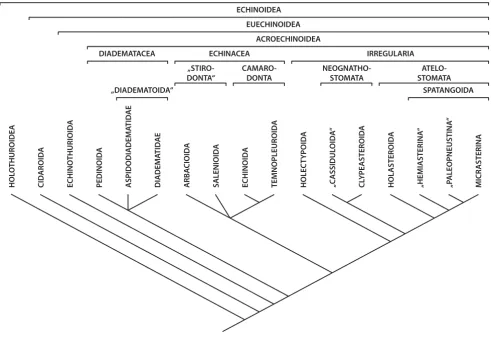
Figure 2
Current understanding of sea urchin phylogeny. The hypotheses of echinozoan and echinoid relationships are based on multiple sources of morphological and molecular datasets (for further references see the Materials and methods section). This tree has not been generated using a consensus or numerical technique and reflects the views and biases of the authors.
CID
AROID
A
ECHINO
THURIOID
A
PEDINOID
A
ASPIDODIADEMA
TID
AE
ARBA
CIOID
A
ECHINOID
A
TEMNOPLEUROID
A
HOLEC
T
Y
POID
A
HOLASTEROID
A
„HEMIASTERINA
“
DIADEM
A
TID
AE
MICR
ASTERINA
„P
ALEOPNEUSTINA
“
ECHINOIDEA
EUECHINOIDEA
ACROECHINOIDEA
„DIADEMATOIDA“
ECHINACEA
„STIRO-DONTA“
CAMARO-DONTA
IRREGULARIA
NEOGNATHO-STOMATA
ATELO-STOMATA
SPATANGOIDA
SALENIOID
A
HOL
O
THUROIDEA
ECHINOZOA
DIADEMATACEA
CL
Y
PEASTEROID
A
„C
ASSIDUL
OID
A
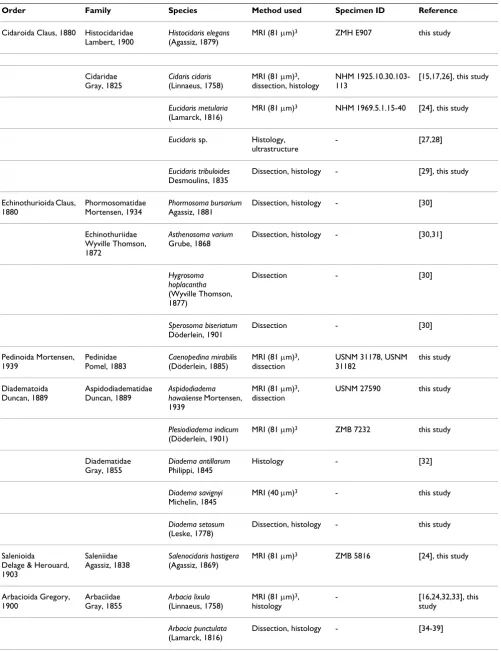
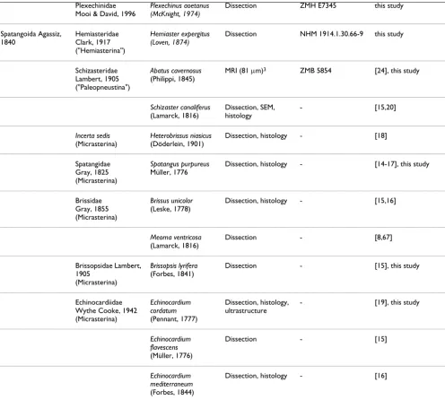
Table 1: List of "regular" sea urchin species included in this study.
Order Family Species Method used Specimen ID Reference
Cidaroida Claus, 1880 Histocidaridae Lambert, 1900
Histocidaris elegans
(Agassiz, 1879)
MRI (81 μm)3 ZMH E907 this study
Cidaridae Gray, 1825
Cidaris cidaris
(Linnaeus, 1758)
MRI (81 μm)3,
dissection, histology
NHM 1925.10.30.103-113
[15,17,26], this study
Eucidaris metularia
(Lamarck, 1816)
MRI (81 μm)3 NHM 1969.5.1.15-40 [24], this study
Eucidaris sp. Histology, ultrastructure
- [27,28]
Eucidaris tribuloides
Desmoulins, 1835
Dissection, histology - [29], this study
Echinothurioida Claus, 1880
Phormosomatidae Mortensen, 1934
Phormosoma bursarium
Agassiz, 1881
Dissection, histology - [30]
Echinothuriidae Wyville Thomson, 1872
Asthenosoma varium
Grube, 1868
Dissection, histology - [30,31]
Hygrosoma hoplacantha
(Wyville Thomson, 1877)
Dissection - [30]
Sperosoma biseriatum
Döderlein, 1901
Dissection - [30]
Pedinoida Mortensen, 1939
Pedinidae Pomel, 1883
Caenopedina mirabilis
(Döderlein, 1885)
MRI (81 μm)3,
dissection
USNM 31178, USNM 31182
this study
Diadematoida Duncan, 1889
Aspidodiadematidae Duncan, 1889
Aspidodiadema hawaiiense Mortensen, 1939
MRI (81 μm)3,
dissection
USNM 27590 this study
Plesiodiadema indicum
(Döderlein, 1901)
MRI (81 μm)3 ZMB 7232 this study
Diadematidae Gray, 1855
Diadema antillarum
Philippi, 1845
Histology - [32]
Diadema savignyi
Michelin, 1845
MRI (40 μm)3 - this study
Diadema setosum
(Leske, 1778)
Dissection, histology - this study
Salenioida
Delage & Herouard, 1903
Saleniidae Agassiz, 1838
Salenocidaris hastigera
(Agassiz, 1869)
MRI (81 μm)3 ZMB 5816 [24], this study
Arbacioida Gregory, 1900
Arbaciidae Gray, 1855
Arbacia lixula
(Linnaeus, 1758)
MRI (81 μm)3,
histology
- [16,24,32,33], this
study
Arbacia punctulata
(Lamarck, 1816)
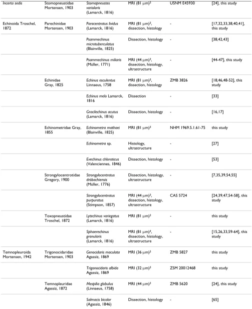
Incerta sedis Stomopneustidae Mortensen, 1903
Stomopneustes variolaris
(Lamarck, 1816)
MRI (81 μm)3 USNM E45930 [24], this study
Echinoida Troschel, 1872
Parechinidae Mortensen, 1903
Paracentrotus lividus
(Lamarck, 1816)
MRI (81 μm)3,
dissection, histology
- [17,32,33,38,40,41],
this study
Psammechinus microtuberculatus
(Blainville, 1825)
Dissection, histology - [38,42,43]
Psammechinus miliaris
(Müller, 1771)
MRI (44 μm)3,
dissection, histology, ultrastructure
- [44-47], this study
Echinidae Gray, 1825
Echinus esculentus
Linnaeus, 1758
MRI (81 μm)3,
dissection, histology
ZMB 3826 [18,46,48-52], this
study
Echinus melo Lamarck, 1816
Dissection - [33]
Gracilechinus acutus
(Lamarck, 1816)
Dissection, histology - [16,17]
Echinometridae Gray, 1855
Echinometra mathaei
(Blainville, 1825)
MRI (81 μm)3 NHM 1969.5.1.61-75 this study
Echinometra sp. Histology, ultrastructure
- [27]
Evechinus chloroticus
(Valenciennes, 1846)
Dissection, histology - [53]
Strongylocentrotidae Gregory, 1900
Strongylocentrotus dröbachiensis
(Müller, 1776)
Dissection, histology, ultrastructure
- [7,35,39,54,55]
Strongylocentrotus purpuratus
(Stimpson, 1857)
MRI (44 μm)3,
dissection, histology, ultrastructure
CAS 5724 [24,39,47,54-58], this
study
Toxopneustidae Troschel, 1872
Lytechinus variegatus
(Lamarck, 1816)
MRI (81 μm)3 - this study
Sphaerechinus granularis
(Lamarck, 1816)
MRI (81 μm)3,
dissection, histology, ultrastructure
- [15,26,33,59-64], this
study
Temnopleuroida Mortensen, 1942
Trigonocidaridae Mortensen, 1903
Genocidaris maculata
Agassiz, 1869
MRI (36 μm)3 ZMB 5827 this study
Trigonocidaris albida
Agassiz, 1869
MRI (32 μm)3 ZSM 20012468 this study
Temnopleuridae Agassiz, 1872
Mespilia globulus
(Linnaeus, 1758)
MRI (44 μm)3 ZMB 5620 [24], this study
Salmacis bicolor
(Agassiz, 1846)
Dissection, histology - [65]
The table provides information on every species studied so far with regard to the axial complex, the method(s) used to infer axial complex anatomy, the specimen ID of museum specimens where applicable, and the respective references. Numbers in brackets behind "MRI" represent the resolution of the dataset. An overview of scanning parameters is provided by [24].
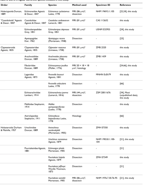
Table 2: List of irregular sea urchin species included in this study.
Order Family Species Method used Specimen ID Reference
Holectypoida Duncan, 1889
Echinoneidae Agassiz & Desor, 1847
Echinoneus cyclostomus
Leske, 1778
MRI (86 μm)3,
dissection
NHM 1969.5.1.105 [22,24], this study
"Cassiduloida" Agassiz & Desor, 1847
Cassidulidae
Agassiz & Desor, 1847
Cassidulus caribaearum
Lamarck, 1801
MRI (81 μm)3 CAS 112632 this study
Echinolampadidae Gray, 1851
Echinolampas depressa
Gray, 1851
MRI (81 μm)3 USNM E32955 [24], this study
Apatopygidae Kier, 1962
Apatopygus recens
(Mortensen, 1948)
Dissection - [23]
Clypeasteroida Agassiz, 1835
Clypeasteridae Agassiz, 1835
Clypeaster rosaceus
(Linnaeus, 1758)
MRI (81 μm)3 ZMB 2520 this study
Arachnoididae Duncan, 1889
Arachnoides placenta
(Linnaeus, 1758)
MRI (81 μm)3 ZMB 1439 this study
Fibulariidae Duncan, 1889
Echinocyamus pusillus
(Müller, 1776)
MRI 20 × 18 × 18
μm3, histology
- [24,66], this study
Laganidae Agassiz, 1873
Peronella lesueuri
Agassiz, 1841
Dissection MNHN EcEh79 this study
Peronella orbicularis
Leske, 1778
Dissection - [66]
Echinarachniidae Lambert, 1914
Echinarachnius parma
(Lamarck, 1816)
MRI (44 μm)3,
dissection
ZSM 20011676 [34], Mooi
(unpublished data), this study
Mellitidae Stephanini, 1914
Mellita
quinquesperforata
(Leske, 1778)
Dissection - this study
Astriclypeidae Stephanini, 1911
Echinodiscus bisperforatus Leske, 1778
Histology - [66]
Holasteroida Durham & Melville, 1957
Urechinidae Duncan, 1889
Antrechinus nordenskjöldi
(Mortensen, 1905)
Dissection ZMH E7350 this study
Urechinus naresianus
Agassiz, 1879
Dissection NHM
1903.8.1.100-104
[21], this study
Pourtalesiidae Agassiz, 1881
Echinosigra phiale
Mortensen, 1905
Dissection - [21]
Pourtalesia hispida
Agassiz, 1879
Dissection ZMH E7349 this study
Pourtalesia jeffreysi
Wyville Thomson, 1873
Dissection - [21]
Pourtalesia wandeli
Mortensen, 1905
MRI (86 μm)3,
dissection
in the same buffer, subsequently post-fixed for 1 h in 1%
OsO4 buffered in 0.1 M sodium cacodylate, dehydrated in
an acetone series and embedded in Araldite. Silver inter-ference coloured sections (75 nm) were made with a dia-mond knife using an ultramicrotome (LEICA UC6), automatically stained with 2% uranyl acetate and 2% lead citrate using a semi-automatic ultrastaining machine (Phoenix), and observed with a transmission electron microscope (Philips CM 120 BioTWIN). Micrographs were made on digital imaging plates (Ditabis) and elec-tronically processed with the software Adobe Photoshop CS3.
Systematic classification
The systematic classification used throughout this study is based upon results obtained by [23,69-75]. Sea cucum-bers (Holothuroidea) constitute the sister taxon to sea urchins, while Cidaroida are the most primitive taxon within the Echinoidea and sister taxon to Euechinoidea. The "Regularia" are a paraphyletic clade, while the Echina-cea and the Irregularia each form a monophyletic taxon (Fig. 2). "Hemiasterina" and "Paleopneustina" presuma-bly are paraphyletic clades. Resolution at the base of the Euechinoidea as well as for the Echinacea is still consid-ered relatively poor [75].
Plexechinidae Mooi & David, 1996
Plexechinus aoetanus (McKnight, 1974)
Dissection ZMH E7345 this study
Spatangoida Agassiz, 1840
Hemiasteridae Clark, 1917 ("Hemiasterina")
Hemiaster expergitus (Loven, 1874)
Dissection NHM 1914.1.30.66-9 this study
Schizasteridae Lambert, 1905 ("Paleopneustina")
Abatus cavernosus
(Philippi, 1845)
MRI (81 μm)3 ZMB 5854 [24], this study
Schizaster canaliferus
(Lamarck, 1816)
Dissection, SEM, histology
- [15,20]
Incerta sedis
(Micrasterina)
Heterobrissus niasicus
(Döderlein, 1901)
Dissection, histology - [18]
Spatangidae Gray, 1825 (Micrasterina)
Spatangus purpureus
Müller, 1776
Dissection, histology - [14-17], this study
Brissidae Gray, 1855 (Micrasterina)
Brissus unicolor
(Leske, 1778)
Dissection, histology - [15,16]
Meoma ventricosa
(Lamarck, 1816)
Dissection - [8,67]
Brissopsidae Lambert, 1905
(Micrasterina)
Brissopsis lyrifera
(Forbes, 1841)
Dissection - [15], this study
Echinocardiidae Wythe Cooke, 1942 (Micrasterina)
Echinocardium cordatum
(Pennant, 1777)
Dissection, histology, ultrastructure
- [19], this study
Echinocardium flavescens
(Müller, 1776)
Dissection - [15]
Echinocardium mediterraneum
(Forbes, 1844)
Dissection, histology - [16]
The table provides information on every species studied so far with regard to the axial complex, the method(s) used to infer axial complex anatomy, the specimen ID of museum specimens where applicable, and the respective references. Numbers in brackets behind "MRI" represent the resolution of the dataset. An overview of scanning parameters is provided by [24].
Results
Definition of components of the echinoid axial complex In its primary structure, the echinoid axial complex con-sists of the following components: madreporic ampulla, dorsal sac, head process, pulsating vessel, axial coelom, axial organ, canaliculi, haemal lacunae, and stone canal (Fig. 1). The designations and definitions referring to components of the echinoid axial complex as well as the
developmental origin of each structure are provided in Table 3. A trilingual compilation of synonymous terms to facilitate comparative studies is provided in Table 4 with references [76-85].
Morphological findings
In this section, the results derived from our own analyses are combined with results derived from the literature
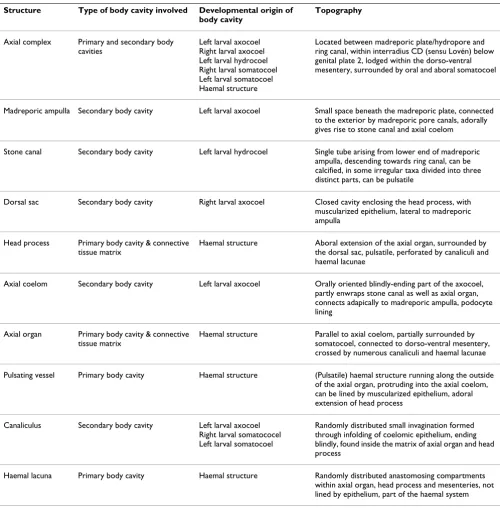
Table 3: Definition of technical terms assigned to the axial complex of echinoids used in this manuscript.
Structure Type of body cavity involved Developmental origin of body cavity
Topography
Axial complex Primary and secondary body cavities
Left larval axocoel Right larval axocoel Left larval hydrocoel Right larval somatocoel Left larval somatocoel Haemal structure
Located between madreporic plate/hydropore and ring canal, within interradius CD (sensu Lovén) below genital plate 2, lodged within the dorso-ventral mesentery, surrounded by oral and aboral somatocoel
Madreporic ampulla Secondary body cavity Left larval axocoel Small space beneath the madreporic plate, connected to the exterior by madreporic pore canals, adorally gives rise to stone canal and axial coelom
Stone canal Secondary body cavity Left larval hydrocoel Single tube arising from lower end of madreporic ampulla, descending towards ring canal, can be calcified, in some irregular taxa divided into three distinct parts, can be pulsatile
Dorsal sac Secondary body cavity Right larval axocoel Closed cavity enclosing the head process, with
muscularized epithelium, lateral to madreporic ampulla
Head process Primary body cavity & connective tissue matrix
Haemal structure Aboral extension of the axial organ, surrounded by the dorsal sac, pulsatile, perforated by canaliculi and haemal lacunae
Axial coelom Secondary body cavity Left larval axocoel Orally oriented blindly-ending part of the axocoel, partly enwraps stone canal as well as axial organ, connects adapically to madreporic ampulla, podocyte lining
Axial organ Primary body cavity & connective tissue matrix
Haemal structure Parallel to axial coelom, partially surrounded by somatocoel, connected to dorso-ventral mesentery, crossed by numerous canaliculi and haemal lacunae
Pulsating vessel Primary body cavity Haemal structure (Pulsatile) haemal structure running along the outside of the axial organ, protruding into the axial coelom, can be lined by muscularized epithelium, adoral extension of head process
Canaliculus Secondary body cavity Left larval axocoel
Right larval somatococel Left larval somatocoel
Randomly distributed small invagination formed through infolding of coelomic epithelium, ending blindly, found inside the matrix of axial organ and head process
Haemal lacuna Primary body cavity Haemal structure Randomly distributed anastomosing compartments
within axial organ, head process and mesenteries, not lined by epithelium, part of the haemal system
available on the axial complex as well as its mesenterial suspension. For a given species, these different contribu-tions may vary considerably, and Tables 1 &2 provide a compilation.
Cidaroida
In lateral view, the axial complex found in Cidaris cidaris
and Eucidaris metularia is almost straight and it lies directly underneath the madreporic plate (Figs. 3A, 4). Through-out its entire course it maintains more or less the same width, only slightly bulging in the middle. It is suspended by the dorso-ventral and the free mesentery (Fig. 5). The dorso-ventral mesentery is strongly developed and con-nects axial complex, oesophagus, and the peripharyngeal (or lantern) coelom with the endoskeleton. The free mesentery connects the peripharyngeal coelom with axial complex and rectum. The axial complex is thus attached to two mesenteries over its entire length in Cidaris cidaris and Eucidaris metularia (Fig. 6A, B). In the juvenile specimen of Eucidaris tribuloides, however, the free mesentery is lacking at the level of the axial organ (Fig. 7B).
A madreporic plate is present in cidaroid species and numerous madreporic pore canals connect the exterior with the madreporic ampulla underneath the madreporic plate. Below the madreporic ampulla lies the roundish-elongated dorsal sac (Fig. 7A). Its epithelium is only slightly muscularized and mainly glandular according to [17] and [26]. However, the presence of glands at this location seems unlikely due to the enclosed nature of the dorsal sac.
The head process, located within the dorsal sac (Fig. 8), is lined by myoepithelial cells. It extends adorally into the pulsating vessel of the axial organ. The madreporic ampulla opens adorally into the axial coelom and the stone canal. The axial coelom is a blindly-ending cavity that is located between stone canal and axial organ (Fig. 9) and that extends towards Aristotle's lantern. The width of the axial coelom does not vary much, remaining large
towards the adoral end in Cidaris cidaris [17,26]. In
Eucidaris tribuloides, the axial coelom in general is reduced to a thin cavity (Fig. 7B). It is lined by an epithelium with numerous podocytes. These podocytes are found to be restricted to that side of the axial coelom in Eucidaris sp. that borders the axial organ [27].
In all cidaroid species analyzed to date, the axial organ is a haemal structure composed of a connective tissue matrix filled with coelomocytes and anastomosing haemal lacu-nae. Canaliculi extend into its interior from the lining somatocoelomic and axocoelomic epithelia. The axial organ sends out a well-developed haemal lacuna towards the perioesophageal haemal ring that is in close contact with the ring canal. The stone canal is a tubular structure
connecting the madreporic ampulla with the ring canal. At its adoral end, the stone canal forms several dilatations that converge with the ring canal. Ultrastructural
investi-gations of the stone canal of Eucidaris sp. reveal a
pris-matic epithelium with ciliated, myoepithelial, and granulated cells that probably represent secretory neurons [28].
Echinothurioida
The axial complex of echinothurioids is straight in lateral view, located directly underneath the madreporic plate. Schurig [30] and Sarasin & Sarasin [31] report it to be
spi-rally winding in Phormosoma bursarium, Asthenosoma
var-ium, Hygrosoma hoplacantha, and Sperosoma biseriatum (Fig. 4). It is suspended by two mesenteries (Fig. 5), of which the dorso-ventral mesentery is strongly developed, con-necting the axial complex, the oesophagus, and the peripharyngeal coelom with the endoskeleton. The free mesentery connects the axial complex with the rectum
and terminates halfway down the axial complex in
Phor-mosoma bursarium and Asthenosoma varium [30,31] (Fig.
5).
A madreporic ampulla lies underneath the madreporic plate and opens into the axial coelom and the stone canal. Sarasin & Sarasin [31] report the madreporic ampulla of Asthenosoma varium to be connected by a canal to another small cavity lying underneath the madreporic plate.
How-ever, these structures were not reported for Phormosoma
bursarium [30], and we believe this isolated observation to be an artefact that will not be further considered in this analysis. The dorsal sac (Fig. 8) lies underneath the madreporic plate and is lined by a slightly muscularized epithelium that is mainly glandular [30,31]. However, in analogy to cidaroids, the presence of glands at this loca-tion seems unlikely.
The head process is lined by a muscularized epithelium as well and continues adorally in the form of the also mus-cularized pulsating vessel [30]. The axial coelom parallels stone canal and axial organ (Fig. 9) and extends from the madreporic ampulla towards the ring canal where it ends blindly. Its width at the base is comparable to its middle
part, at least in Asthenosoma varium and Phormosoma
bursa-rium.
The axial organ is a structure composed of a haemal tissue mesh crossed by anastomosing haemal lacunae and canal-iculi. As in Cidaroida, the stone canal forms several dilata-tions at its junction with the ring canal.
Pedinoida
In lateral view, the axial complex found in Caenopedina
Table 4: Trilingual list of technical terms used in publications dealing with the axial complex.
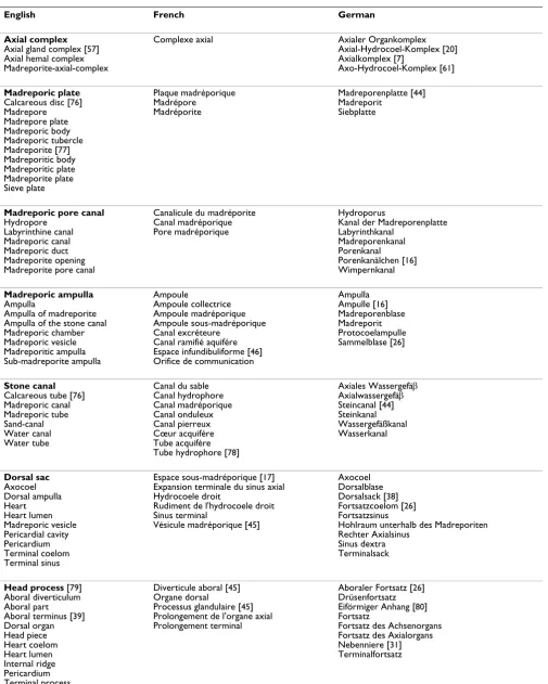
English French German
Axial complex Axial gland complex [57] Axial hemal complex Madreporite-axial-complex
Complexe axial Axialer Organkomplex
Axial-Hydrocoel-Komplex [20] Axialkomplex [7]
Axo-Hydrocoel-Komplex [61]
Madreporic plate Calcareous disc [76] Madrepore Madrepore plate Madreporic body Madreporic tubercle Madreporite [77] Madreporitic body Madreporitic plate Madreporite plate Sieve plate Plaque madréporique Madrépore Madréporite
Madreporenplatte [44] Madreporit
Siebplatte
Madreporic pore canal Hydropore
Labyrinthine canal Madreporic canal Madreporic duct Madreporite opening Madreporite pore canal
Canalicule du madréporite Canal madréporique Pore madréporique
Hydroporus
Kanal der Madreporenplatte Labyrinthkanal
Madreporenkanal Porenkanal Porenkanälchen [16] Wimpernkanal
Madreporic ampulla Ampulla
Ampulla of madreporite Ampulla of the stone canal Madreporic chamber Madreporic vesicle Madreporitic ampulla Sub-madreporite ampulla Ampoule Ampoule collectrice Ampoule madréporique Ampoule sous-madréporique Canal excréteure
Canal ramifié aquifére Espace infundibuliforme [46] Orifice de communication
Ampulla Ampulle [16] Madreporenblase Madreporit Protocoelampulle Sammelblase [26]
Stone canal Calcareous tube [76] Madreporic canal Madreporic tube Sand-canal Water canal Water tube
Canal du sable Canal hydrophore Canal madréporique Canal onduleux Canal pierreux Cœur acquifére Tube acquifére Tube hydrophore [78]
Axiales Wassergefäβ Axialwassergefäβ Steincanal [44] Steinkanal Wassergefäßkanal Wasserkanal Dorsal sac Axocoel Dorsal ampulla Heart Heart lumen Madreporic vesicle Pericardial cavity Pericardium Terminal coelom Terminal sinus
Espace sous-madréporique [17] Expansion terminale du sinus axial Hydrocoele droit
Rudiment de l'hydrocoele droit Sinus terminal
Vésicule madréporique [45]
Axocoel Dorsalblase Dorsalsack [38] Fortsatzcoelom [26] Fortsatzsinus
Hohlraum unterhalb des Madreporiten Rechter Axialsinus
Sinus dextra Terminalsack
Head process [79] Aboral diverticulum Aboral part Aboral terminus [39] Dorsal organ Head piece Heart coelom Heart lumen Internal ridge Pericardium Terminal process
Diverticule aboral [45] Organe dorsal
Processus glandulaire [45] Prolongement de l'organe axial Prolongement terminal
Aboraler Fortsatz [26] Drüsenfortsatz Eiförmiger Anhang [80] Fortsatz
Axial coelom Axial canal Axial gland lumen Axial sinus Axocoel Central cavity Central lumen Lumen of axial gland
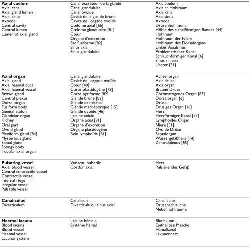
Canal excréteur de la glande Canal glandulaire
Canal ovoïde
Cavité de la glande brune Cavité de l'organe ovoïde Cœlome axial [66] Cœlome glandulaire [81] Cœur
Organe d'excrétion Sac fusiforme [82] Sinus axial Sinus glandulaire
Axialcoelom Axialer Hohlraum Axialkanal Axialsinus Axocoel Drüsenhohlraum
Höhle des sichelförmigen Bandes [44] Hohlraum
Hohlraum der Niere Hohlraum des Dorsalorgans Linker Axialsinus
Problematischer Kanal Schlauchförmiger Kanal [6] Sinus sinistra
Ureter [31]
Axial organ Axial gland Axial haemal duct Axial haemal vessel Brown gland Central plexus Dorsal organ Fusiform body Genital stolon Glandular organ Kidney Oral part Ovoid gland Plexiform gland [84] Mysterious gland Septal gland Spongy body Tubular axial organ
Canal glandulaire Cavité de l'organe ovoïde Cœur [40]
Corps plastidogène [78] Corps pyriforme [82] Glande brune [82] Glande excrétrice Glande madréporique [15] Glande ovoïde [46] Lacune axiale Organe axial [81] Organe d'excrétion Organe plastidogène Rein lymphoïde [81]
Achsenorgan Axialdrüse Axialorgan Braune Drüse
Chromatogenes Organ [83] Dorsalorgan [6]
Drüse
Drüsiges Organ [16] Herz
Herzförmiger Kanal [44] Lymphoides Organ Niere [31] Ovoide Drüse Septalorgan
Wassergefäßherz [14] Zentralplexus [85]
Pulsating vessel Axial blood vessel Central contractile vessel Contractile vessel Internal ridge Irregular vessel Pulsatile vessel
Vaisseau pulsatile Cordon axial
Herz
Pulsierendes Gefäβ
Canaliculus Diverticulum
Canalicule
Diverticule du sinus axial
Canaliculus Drüsenschläuche Nebenhohlräume
Haemal lacuna Blood lacuna Blood vessel Haemal vessel Lacunar system
Lacune hémale Système hémal
Blutlakune Epithellose Masche Hämalkanal Lakunennetz
List of terms that have been used so far in publications dealing with the echinoid (as well as echinoderm) axial complex including madreporic plate and madreporic pore canals. The terms used in this study are printed in bold. The author(s) that first established a certain term appear after it in brackets. Where no reference is provided, the creator (author) of the respective term could not be identified with certainty.
the upper part broadens, the lower part is of the same size as the middle part of the axial complex. It is suspended by two mesenteries (Fig. 6C), the dorso-ventral and the free mesentery. The former is strongly developed and connects axial complex, oesophagus, and the peripharyngeal coe-lom with the endoskeleton, while the free mesentery con-nects the axial complex with the rectum and terminates halfway down the axial complex, thus resembling the sit-uation found in Echinothurioida (Fig. 5). A madreporic
plate is present and numerous madreporic pore canals connect the exterior with the madreporic ampulla under-neath the madreporic plate.
Aspidodiadematidae
The axial complex of Aspidodiadema hawaiiense and
Plesio-diadema indicum is mostly straight when viewed laterally and only slightly oblique in its upper half (Figs. 3C, 4). On the level of the first curvature of the oesophagus, the Vertical magnetic resonance imaging (MRI) sections of various sea urchin specimens
Figure 3
Vertical magnetic resonance imaging (MRI) sections of various sea urchin specimens. The virtual sections depict the axial complex (arrow). A Cidaris cidaris (Cidaroida). B Caenopedina mirabilis (Pedinoida). C Plesiodiadema indicum (Aspidodia-dematidae). D Salenocidaris hastigera (Salenioida). E Strongylocentrotus purpuratus (Echinoida). F Lytechinus variegatus (Echinoida). G Genocidaris maculata (Temnopleuroida). H Echinoneus cyclostomus (Holectypoida). I Cassidulus caribearum ("Cassiduloida"). J Echinolampas depressa ("Cassiduloida"). K Echinarachnius parma (Clypeasteroida). Tables 1 & 2 list resolutions for each MRI dataset.
A
B
C
D
0.5 cm
E
F
G
H
I
J
K
0.5 cm
0.5 cm
0.5 cm
0.5 cm
0.5 cm
0.5 cm
0.5 cm
0.5 cm
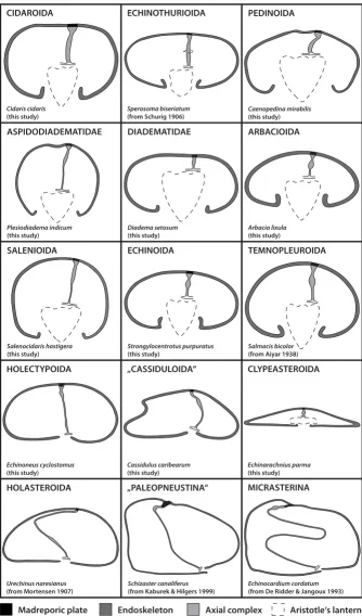
Schematic representation of the axial complex in selected echinoid taxa Figure 4
Schematic representation of the axial complex in selected echinoid taxa. The drawings concentrate on the gross morphology of the axial complex. All other internal organs are omitted, the ring canal is depicted in part only, and the anus is not shown. The legend indicates every structure shown.
Cidaris cidaris
(this study)
CIDAROIDA ECHINOTHURIOIDA
Sperosoma biseriatum
(from Schurig 1906)
DIADEMATIDAE ARBACIOIDA
ECHINOIDA TEMNOPLEUROIDA
HOLECTYPOIDA „CASSIDULOIDA“ CLYPEASTEROIDA
HOLASTEROIDA „PALEOPNEUSTINA“ MICRASTERINA
Diadema setosum
(this study)
Arbacia lixula
(this study)
Strongylocentrotus purpuratus
(this study)
Salmacis bicolor
(from Aiyar 1938)
Echinoneus cyclostomus
(this study)
Cassidulus caribearum
(this study)
Echinarachnius parma
(this study)
Urechinus naresianus
(from Mortensen 1907)
Echinocardium cordatum
(from De Ridder & Jangoux 1993)
Schizaster canaliferus
(from Kaburek & Hilgers 1999)
Madreporic plate Endoskeleton Axial complex Aristotle‘s lantern
PEDINOIDA
ASPIDODIADEMATIDAE
Caenopedina mirabilis
(this study)
Plesiodiadema indicum
(this study)
Salenocidaris hastigera
Schematic representation of the mesenterial suspension of the axial complex in various higher sea urchin taxa Figure 5
Schematic representation of the mesenterial suspension of the axial complex in various higher sea urchin taxa. The drawings demonstrate the impact of the gastric caecum on the architecture of the axial complex. Upper three lines: dorso-ventral mesentery always depicted on the right-hand side. Dashed lines indicate course of the oesophagus or the axial complex (Micrasterina). Lower line: Ventral view of the digestive tract and the axial complex in the vicinity of the oesophagus of highly derived irregular sea urchin taxa (Atelostomata) – note the extension of the mesentery. The grey-scale legend denominates every structure shown. dt = digestive tract, gc = gastric caecum, oe = oesophagus, re = rectum.
CIDAROIDA ECHINOTHURIOIDA & PEDINOIDA ASPIDODIADEMATIDAE
DIADEMATIDAE & ECHINACEA „CASSIDULOIDA“
CLYPEASTEROIDA
HOLASTEROIDA „PALEOPNEUSTINA“ MICRASTERINA
Madreporic plate Endoskeleton Axial complex Urechinus
naresianus (from Mortensen 1907)
Schizaster
canaliferus (from Koehler 1883)
Echinocardium
cordatum (from De Ridder & Jangoux 1993)
Mesenteries HOLECTYPOIDA
HOLASTEROIDA MICRASTERINA
gc
gc
gc gc gc
oe oe
oe
oe oe oe
oe
oe
oe
re re re
re
dt
dt
Horizontal magnetic resonance imaging (MRI) sections of various sea urchin specimens Figure 6
Horizontal magnetic resonance imaging (MRI) sections of various sea urchin specimens. The virtual sections were made at the level of gonads, upper digestive tract, and axial complex (arrow). Note the differing mesenterial suspensions of the axial complex. A Cidaris cidaris (Cidaroida). B Eucidaris metularia (Cidaroida). C Caenopedina mirabilis (Pedinoida). D Aspidodia-dema hawaiiense (AspidodiaAspidodia-dematidae). E Strongylocentrotus purpuratus (Echinoida). F Lytechinus variegatus (Echinoida). G Mespilia globulus (Temnopleuroida). H Echinoneus cyclostomus (Holectypoida). Tables 1 & 2 list resolutions for each MRI dataset.
A
B
C
D
E
F
G
H
0.5 cm
0.5 cm
0.5 cm
0.5 cm
0.5 cm
0.5 cm
0.5 cm
Horizontal light-microscopic sections through the echinoid axial complex Figure 7
axial complex tapers out adorally towards the ring canal. The axial complex is attached to the dorso-ventral and the free mesentery (Fig. 6D). The dorso-ventral mesentery is strongly developed and connects axial complex, oesopha-gus, and the peripharyngeal coelom with the endoskele-ton. The free mesentery connects the axial complex with the rectum and terminates halfway down the axial com-plex (Fig. 5). In its lower half, it appears that the axial complex consists only of the stone canal and the haemal lacuna(e) that connect(s) the axial organ and the peri-oesophageal haemal ring. The axial organ itself seems to be restricted to the upper half of the axial complex. It is spindle-shaped and bulges in its middle part (Figs. 3C, 4).
Diadematidae
In lateral view, the axial complex found in Diadema
seto-sum is extending straight down from the madreporic plate
to the ring canal. The relatively large Aristotle's lantern found in this species (as well as in most other diadema-tids) squeezes the axial complex between lantern and the apical endoskeleton (Fig. 4). The axial complex is sus-pended by the dorso-ventral and the free mesentery. The former is strongly developed and connects axial complex, oesophagus, and the peripharyngeal coelom with the
endoskeleton, while the free mesentery connects the rec-tum with the axial complex in its uppermost part close to dorsal sac and head process. It is not present in the middle and lower parts of the axial complex (Fig. 5). In the
juve-nile specimen of Diadema setosum, the dorso-ventral
mesentery interconnects peripharyngeal coelom, oesophagus, axial complex as well as the rectum (Fig. 7C, D).
The madreporic plate is perforated by numerous madreporic pore canals that connect the exterior with the madreporic ampulla. Right underneath the madreporic ampulla lies the dorsal sac that encloses the compact and roundish-elongated head process. The dorsal sac extends further adorally, paralleling axial coelom and stone canal in its lower part (Figs. 7C, 8). The head process is more prominent in its adapical part and is lined by a strong myoepithelium. It sends out the pulsating vessel into the
lumen of the axial coelom in Diadema setosum and
Dia-dema antillarum. The axial coelom is a large cavity located between axial organ and stone canal (Fig. 8). It ends blindly towards the adoral end of the axial complex.
The axial organ bulges in the middle part of the axial com-plex and shows considerable infolding into the axial coe-lom (Fig. 7D). The stone canal is a tubular structure that constantly decreases in diameter on its way down towards the ring canal. It joins the ring canal trough a number of small canals.
Arbacioida
In lateral view, the axial complex is straight, extending down from the madreporic plate to the ring canal [33,34] (Fig. 4). In the vicinity of the oesophagus, the adoral part of the axial complex tapers out and reaches the surface of Aristotle's lantern. Its mesenterial suspension resembles the diadematid/echinacean type (Fig. 5). The madreporic plate is perforated by numerous madreporic pore canals. A pulsating vessel constitutes the adoral extension of the head process. Schematic representations of horizontal sec-tions of the axial organ, axial coelom as well as stone canal of Arbacia lixula and Arbacia punctulata illustrate the inter-nal morphology of the arbacioid axial complex (Fig. 9).
The axial organ of Arbacia lixula is reported to be
sur-rounded by a large number of haemal lacunae [35]. The
axial complex found in Stomopneustes variolaris (incerta
sedis, presumably sister taxon to Arbacioida) resembles
the arbacioid/echinoid gross morphology.
Salenioida
The axial complex found in Salenocidaris hastigera (Figs.
3D, 4) is comparable in its lateral aspect to the axial com-plex found in aspidodiadematids. The mesenterial sus-pension resembles the diadematid/echinacean type (Fig. 5).
Comparative morphology of the echinoid axial complex at the level of head process and dorsal sac
Figure 8
Comparative morphology of the echinoid axial com-plex at the level of head process and dorsal sac. Sche-matic representation of the axial complex at the level of head process and dorsal sac based on light-microscopic sections. Note that although changes in shape do occur, the internal composition remains largely the same. For better compari-son, the stone canal is shown towards the top of each image. The legend indicates every structure shown.
Cidaris cidaris (from Prouho 1887)
Eucidaris tribuloides (this study)
Phormosoma bursarium (from Schurig 1906)
Diadema setosum (this study)
Psammechinus miliaris (this study)
Echinocyamus pusillus (this study)
Spatangus purpureus (from Hamann 1887) Schizaster canaliferus
(from Kaburek & Hilgers 1999)
Heterobrissus niasicus (from Wagner 1903)
CIDAROIDA CIDAROIDA ECHINOTHURIOIDA
DIADEMATIDAE ECHINOIDA CLYPEASTEROIDA
MICRASTERINA MICRASTERINA
„PALEOPNEUSTINA“
Comparative morphology of the echinoid axial complex at the level of axial organ and axial coelom Figure 9
Comparative morphology of the echinoid axial complex at the level of axial organ and axial coelom. Schematic representation of the axial complex at the level of axial organ and axial coelom based on light-microscopic sections. Note that although changes in shape do occur, the internal composition remains largely the same. For better comparison, the stone canal is depicted towards the top of each image. The legend indicates every structure shown.
Cidaris cidaris
(from Prouho 1887)
Eucidaris tribuloides
(this study)
Eucidaris sp.
(from Welsch & Rehkämper 1987)
Phormosoma bursarium
(from Schurig 1906)
Asthenosoma varium
(from Sarasin & Sarasin 1887)
Diadema antillarum
(from Millott & Vevers 1967)
Diadema setosum
(this study)
Arbacia lixula
(from Hamann 1887)
Arbacia punctulata
(from Millott 1966)
Psammechinus miliaris
(this study)
Echinus esculentus
(from Perrier 1875)
Sphaerechinus granularis
(from Leipoldt 1893)
Salmacis bicolor
(from Aiyar 1938)
Echinocyamus pusillus
(this study)
Brissus unicolor
(from Hamann 1887)
Echinocardium cordatum
(this study)
Spatangus purpureus
(from Hamann 1887)
Heterobrissus niasicus
(from Wagner 1903)
Schizaster canaliferus
(from Kaburek & Hilgers 1999)
CIDAROIDA CIDAROIDA CIDAROIDA ECHINOTHURIOIDA
ECHINOTHURIOIDA DIADEMATIDAE DIADEMATIDAE ARBACIOIDA
ARBACIOIDA ECHINOIDA ECHINOIDA ECHINOIDA
ECHINOIDA TEMNOPLEUROIDA CLYPEASTEROIDA
MICRASTERINA MICRASTERINA MICRASTERINA MICRASTERINA
„PALEOPNEUSTINA“
Strongylocentrotus purpuratus
(from Boolootian & Campbell 1964)
Echinoida
The axial complex found in the Echinoida is straight in lat-eral view, situated slightly shifted from the oral-aboral axis underneath the madreporic plate (Figs. 3; 4; 6E, F). At the junction with the oesophagus, the axial complex grad-ually tapers out towards the ring canal. Above this junc-tion, a conspicuous swelling is visible that is widest in its middle part, giving the axial complex a spindle-shaped appearance. The mesenterial suspension resembles the diadematid/echinacean type (Figs. 5; 6E, F).
The madreporic plate is perforated by numerous madreporic pore canals that are lined by a ciliated epithe-lium and that merge adorally to form the madreporic ampulla (Fig. 8). The latter is lined by a ciliated epithe-lium as well. The diameter of the madreporic ampulla becomes gradually smaller towards its adoral end and it basally diverges into stone canal and axial coelom. The dorsal sac lies laterally underneath the madreporic ampulla and surrounds the globular head process (Fig. 8). The dorsal sac is a closed cavity and is not connected to either madreporic ampulla, stone canal or the axial coe-lom. Boolootian & Campbell [56], however, report a
divergent morphology of the dorsal sac in
Strongylocentro-tus purpuraStrongylocentro-tus in which the dorsal sac communicates with the somatocoel via a small slit and is divided into two contractile chambers. We believe this isolated finding to be an artefact and will not consider it any further in our analysis.
A strong pulsating vessel is present in all echinoid species studied so far, its ultrastructure resembling that of the head process. The axial coelom is located in-between stone canal and axial organ (Fig. 9), extending adorally before ending blindly above the ring canal. The axial organ of Echinoida is spindle-shaped in vertical section and shows characteristic infolding, with an enlarged sur-face towards the axial coelom with numerous digitations that protrude into the lumen of the latter.
The stone canal is located at the lateral edge of the axial complex (Fig. 9). After its gradual opening at the lower end of the madreporic ampulla, it runs down towards the ring canal, while exhibiting a diameter that varies only slightly and a shape that is almost circular in cross-section. At the lower end of the axial complex, the haemal lacuna(e) of the axial organ adorally merge(s) with the perioesophageal haemal ring while the stone canal directly opens into the ring canal. In numerous echinoid species of Echinoida the stone canal was found to pulsate.
Echinoida: ultrastructural findings
In Psammechinus miliaris, a ciliated, pseudostratified mon-olayer lines the dorsal sac (Fig. 10A). While only some of the peripheral lining cells are epithelio-muscle cells, those
resting on the matrix of the head process form a strong myoepithelium (Fig. 11E). Here, the epithelio-muscle cells contain strong, basally located bundles of myofila-ments that form a regular network of rectangularly arranged outer circular and inner longitudinal bundles that are embedded in the voluminous matrix of the head process. The perikarya deeply extend into the dorsal sac lumen, which indicates that the organ was apparently fixed during contraction. The state of contraction can be estimated from the position of the apical adhaerens junc-tions between adjacent cells. Intraepithelial nerve fibre processes were found interspersed among the lining cells, but podocytes were never found.
The head process contains primary body cavities lined by the matrix of the head process. Numerous cells with homogenous cytoplasma and relatively big nuclei float inside this cavity, and some haematocytes could be recog-nized by their large number of lysosomes and residual bodies.
The axial organ begins at the level of the first curvature of the oesophagus and is surrounded by numerous haemal lacunae (Fig. 11B). That part of the axial organ which bulges into the axial coelom contains numerous deep
crypts and invaginations in Psammechinus miliaris, so that
the axocoelomic surface of the axial organ is tremen-dously enlarged. Inside the canaliculi, the entire lumen seems to be occupied by microvilli (Fig. 10B). Only a few cells are found inside the lumen, most of them coelomo-cytes. Haemotocytes at different stages of differentiation are present within the matrix. They contain residual bod-ies and lysosomes of different rank.
Numerous podocytes rest on the axial coelomic side of the matrix (Figs. 10C, 11F). Their pedicels are bridged by elec-tron-dense diaphragmata. Each podocyte has a single cil-ium with a (9 × 2)+2 axoneme that adheres to the cell body by a basal body, short rootlets and an accessory cen-triole perpendicular to the basal body. A circle of 9–12 strong microvilli surrounds each cilium (Fig. 10D). Addi-tional microvilli emanate from the surface of the
peri-karyon and only seldom from the pedicels. In Echinometra
sp., the podocytes within the axial coelom lining are irreg-ularly distributed and not restricted to the side covering the axial organ [27].
The stone canal lining in Psammechinus miliaris is a
epithelium: some cells of the stone canal lining contain large, electron-densely stained vesicles. These possible secretory granules lie near the apex while other cellular components are located more basally. However, degrad-ing cells can rarely be found among the basal portions of the lining cells, and epithelio-muscle cells, muscle cells or podocytes were never seen to be part of the stone canal epithelium. The somatocoel is lined by a flat epithelium which is only slightly muscularized (Figs. 10D, 11D). The cilia of the somatocoel epithelium show the characteristic composition with a dense microvilli fringe surrounding the axoneme. The epithelial cells are connected to each other via adhaerens junctions and do not show the spe-cializations of the dorsal sac and axial coelom lining.
Temnopleuroida
In lateral view, the axial complex found in Mespilia
globu-lus and Salmacis bicolor is straight, extending down from the madreporic plate to the ring canal (Fig. 4) [65]. In the vicinity of the oesophagus, the adoral part of the axial complex tapers out and reaches the apical surface of Aris-totle's lantern. The mesenterial suspension of the axial complex resembles the diadematid/echinacean type (Figs. 5, 6G). A schematic representation of a horizontal section through the axial complex of Salmacis bicolor at the level of the axial organ is shown in Fig. 8. In Genocidaris maculata, the course of the axial complex is slightly oblique (Fig. 3G).
Irregularia
Most obviously, irregular sea urchin species are distin-guished from animals belonging to "regular" orders by their secondarily developed bilateral symmetry. However, their analysis demonstrates that the shape of the axial complex is primarily not dependent on features of the endoskeleton, but is influenced by the architecture of sev-eral internal organs as detailed below.
Holectypoida
The axial complex found in Echinoneus cyclostomus extends
from the madreporic plate down to the ring canal in an almost straight line (Figs. 3H, 4). The madreporic plate can be seen bulging slightly into the interior of the ani-mal. In the upper fourth of the axial complex, a conspicu-ous swelling can be seen, which is interpreted as the axial organ [22]. Close to the ring canal, the lower fourth of the axial complex shows a swollen region which has been seen with MRI but has also been described by [22]. Unfor-tunately, histological data are currently not available for this taxon.
The mesenterial suspension of the axial complex resem-bles the aspidodiadematid type (Fig. 5). The dorso-ventral mesentery extends between oesophagus, axial complex, gastric caecum, pentagonal apical membrane (or aboral
sinus), and the endoskeleton (Figs. 5, 6H). At the apical pole this mesentery joins the pentagonal apical mem-brane which forms the connection between the gonoducts and the axial complex. This membrane appears to be present in all irregular sea urchin taxa [8]. A second mesentery suspends the axial complex towards the rectum and also forms part of the pentagonal apical membrane. The gastric caecum is not in close contact with the axial complex (Fig. 5).
"Cassiduloida"
The axial complex found in Cassidulus caribearum and
Echinolampas depressa extends from the madreporic plate
down to the ring canal. It is bent anteriorly towards ambu-lacrum III (Figs. 3I, J; 4). Jensen [23] has reported similar
findings for Apatopygus recens. The madreporic plate can
be seen bulging only slightly into the interior of the ani-mal. In the upper third, a conspicuous swelling of the axial complex can be seen which is interpreted as the axial organ. During its entire path, the axial complex is attached to the dorso-ventral mesentery. This structure extends between oesophagus, axial complex, gastric caecum, pen-tagonal apical membrane and the endoskeleton (Fig. 5). A second, smaller mesentery suspends the axial complex towards rectum and aboral endoskeleton as well as the
pentagonal apical membrane. The gastric caecum of
Cas-sidulus caribearum and Echinolampas depressa is reduced to numerous smaller sacs that are located further anteriorly [24] and that are therefore not in close contact with the
axial complex. In Cassidulus caribearum, a slightly swollen
region can be seen in the adoral part of the axial complex (Fig. 3I). Unfortunately, histological data are currently not available for this taxon.
Clypeasteroida
Due to the flattened aspect of clypeasteroids (sea biscuits and sand dollars), the axial complex is considerably min-iaturized. It is located underneath the madreporic plate and has a straight (Fig. 4) to bean-shaped form in lateral view. The madreporic plate is perforated by numerous madreporic pore canals that connect the madreporic
ampulla to the exterior. In Echinocyamus pusillus (and all
other taxa of the Fibulariidae), the madreporic ampulla is in contact with the exterior via a single canal, the
hydro-pore (sensu Mortensen [86]). Except for a small lateral
Horizontal electron-microscopic sections of the echinoid axial complex Figure 10
Horizontal electron-microscopic sections of the echinoid axial complex. A-D Psammechinus miliaris (Echinoida) and E, F Echinocardium cordatum (Micrasterina). A Dorsal sac epithelium with myoepithelial cells. B Heavily ciliated canaliculus inside the axial organ. C Axial coelom epithelium with podocyte and haemal lacunae. D Somatocoelomic epithelium. E Axial coelom epithelium with podocyte and haemal lacunae. F Heavily ciliated canaliculus inside the axial organ. ac = axial coelom, aj = adhae-rens junctions, bl = basal lamina, cf = collagenous fibers, ci = cilium, cm = circumciliary microvillum, ds = dorsal sac, ec = epi-thelial cell, hl = haemal lacuna, mf = myofibrils, mv = microvillum, nu = nucleus, pe = pedicel, po = podocyte, so = somatocoel.
1 μm
1 μm
2.5 μm
C
D
ac
pe
po
hl bl
po ec
ac
hl
mv ci
cf
bl
aj
ec
cm
ci
so nu
aj
1 μm
2 μm
F
ec
cf ec nu
ci
mv
ac bl
pe
hl ec
cm ci
ac
bl
5 μm
E
mf
bl ds
mf
ec nu
A
B
developed dorsal sac that surrounds the muscularized head process (Figs. 7G, 8). Furthermore, Cuénot [48]
reports that the dorsal sac of Echinodiscus bisperforatus
communicates with the somatocoel via a small slit. Such
a connection could not be found in Echinocyamus pusillus.
At least in the latter species, a pulsating vessel could not be detected (Figs. 7H, 9).
The axial coelom originates from the madreporic ampulla directly underneath the madreporic plate. The axial organ
can be found to be straight (Echinocyamus pusillus) or
bean-shaped (Echinarachnius parma, Mellita
quinquesperfo-rata) in lateral view (Fig. 3K). The stone canal's diameter
continuously decreases before passing into the ring canal in Echinocyamus pusillus. In the few clypeasteroid taxa that Schematic representation of sections through the axial complex of Sphaerechinus granularis (Echinoida)
Figure 11
Schematic representation of sections through the axial complex of Sphaerechinus granularis (Echinoida). A Hor-izontal section through the echinoid axial complex at the level of head process and dorsal sac. B HorHor-izontal section through the echinoid axial complex at the level of the axial organ. The insert between A and B (compare Fig. 1) roughly outlines where the virtual sections were made. C Schematic representation of the columnar epithelium of the stone canal. D Schematic repre-sentation of the flat epithelium of the somatocoel. E Schematic reprerepre-sentation of the myoepithelium that lines head process and pulsating vessel. F Schematic representation of the specialized epithelium with podocytes found in the axial coelom. ac = axial coelom, ao = axial organ, bl = basal lamina, ca = canaliculus, ci = cilium, ct = connective tissue, dm = dorso-ventral mesen-tery, ds = dorsal sac, ec = epithelial cell, fm = free mesenmesen-tery, hl = haemal lacuna, hp = head process, pv = pulsating vessel, sc = stone canal, so = somatocoel.
A
B
C
D
E
F
C
C
F
F
E
D
D
D
A
B
ds
hp
sc
fm
dm
hl
so
so
ac
sc
dm
ca
ao
so
so
pv
hl
bl
ct
ci
ec
possess a gastric caecum, this highly reduced structure is not in close contact with the axial complex.
Holasteroida
In lateral view, the axial complex found in Holasteroida is C-shaped and extends between madreporic plate and ring canal (Fig. 4). In its upper half, the axial complex runs par-allel to the aboral endoskeleton towards interambu-lacrum 5. The madreporic plate is slightly infolded. The conspicuous swelling right underneath the madreporic plate, interpreted as the axial organ, is in an horizontal position. In its middle section, the previously horizontally oriented axial complex bends towards the mouth, comes into close contact with the oesophagus adorally, and leads in a straight line down towards the ring canal. Another swelling can be observed in its lower third, at least in Ure-chinus naresianus (Fig. 4). Unfortunately, no histological data are currently available for this taxon.
The axial complex is held in place by the dorso-ventral mesentery that attaches digestive tract, axial complex, gas-tric caecum, and pentagonal membrane to the calcite endoskeleton. Between gastric caecum and axial complex, this mesentery is very thin, so that both structures are in close contact with each other in the aboral part of the ani-mals. A second, smaller mesentery connects the axial com-plex with rectum, pentagonal apical membrane and endoskeleton. The mesenterial strand that interconnects the first loop of the digestive tract is extending half-ways along the gut loop (Fig. 5). Mortensen [21] describes the gross morphology of the axial complex in four holasteroid species: Echinosigra paradoxa, Pourtalesia jeffreysi, Pour-talesia wandeli, and Urechinus naresianus. The axial
com-plexes found in Antrechinus nordenskjoldi and Pourtalesia
hispida closely resemble this description. The large gastric caecum is in close contact with the upper part of the axial complex (Fig. 5).
"Hemiasterina"
The axial complex found in Hemiaster expergitus closely
resembles the holasteroid and paleopneustine axial com-plex in its gross morphology.
"Paleopneustina"
In lateral view, the axial complex found in Abatus
caverno-sus and Schizaster canaliferus is C- to L-shaped and extends down from the madreporic plate to the ring canal close to the mouth (Fig. 4). The following description is based on Schizaster canaliferus [20]. The madreporic plate is charac-terized by two calcareous ridges protruding into the inte-rior of the endoskeleton. The axial complex is attached to the right ridge from where it is extending towards interam-bulacrum 5. It then bends down towards the mouth and extends along the gastric caecum and later the oesophagus before finally reaching the ring canal. It is primarily
sus-pended by the dorso-ventral mesentery that attaches digestive tract, axial complex, gastric caecum, and pentag-onal apical membrane to the calcite endoskeleton. Between gastric caecum and axial complex this mesentery is very thin, so that both structures are in close contact in the aboral part of the specimen (Fig. 5). A second, smaller mesentery attaches the axial complex to rectum, pentago-nal apical membrane and endoskeleton.
The madreporic ampulla is reduced to several broadened madreporic pore canals inside the right calcareous ridge, and according to Kaburek & Hilgers [20], the dorsal sac as
well as a pulsating vessel are not present in Schizaster
canaliferus. However, the structure of the head process
resembles that in the "regular" species (Fig. 8). The axial organ is located directly behind the right calcareous ridge and is oriented horizontally, parallel to the endoskeleton. A large uniform axial coelom is not present. The stone canal is tri-partite: its upper part bears a tall-prismatic epi-thelium, its middle region is muscularized and filled with coelomocytes, while its lower part close to the ring canal is lined by a flat epithelium and is only slightly filled with
coelomocytes. The stone canal of Schizaster canaliferus is
not dilated at its junction with the ring canal and at least its muscularized middle part can pulsate. The mesenterial strand that interconnects the first loops of the digestive tract is extending half-ways along the first gut loop (Fig. 5, bottom line). The large gastric caecum is in close contact with the upper part of the axial complex (Fig. 5).
Micrasterina
the digestive tract is extending far towards ambulacrum III, "dragging" the axial complex along (Fig. 5).
The madreporic plate is perforated by a small number of horizontal and vertical madreporic pore canals [16,18]. Underneath and within the right calcareous ridge, a small madreporic ampulla can be seen. Parallel to this structure,
a dorsal sac can be found at least in Echinocardium
mediter-raneum, Heterobrissus niasicus, and Spatangus purpureus [16-18]. It contains the head process (Fig. 8), which is not
pul-satile in H