BioMed Central
Cell Division
Open Access
Review
Eg5 steps it up!
Megan T Valentine
1, Polly M Fordyce
2and Steven M Block*
1,3Address: 1Department of Biological Sciences, Stanford University, Stanford CA 94305, USA, 2Department of Physics, Stanford University, Stanford
CA 94305, USA and 3Department of Applied Physics, Stanford University, Stanford CA 94305, USA
Email: Megan T Valentine - [email protected]; Polly M Fordyce - [email protected]; Steven M Block* - [email protected] * Corresponding author
Abstract
Understanding how molecular motors generate force and move microtubules in mitosis is essential to understanding the physical mechanism of cell division. Recent measurements have shown that one mitotic kinesin superfamily member, Eg5, is mechanically processive and capable of crosslinking and sliding microtubules in vitro. In this review, we highlight recent work that explores how Eg5 functions under load, with an emphasis on the nanomechanical properties of single enzymes.
Review
Eg5 motors slide microtubules during cell division
Eg5, a member of the Kinesin-5 subclass of kinesins, is a plus-end-directed tetrameric kinesin-family protein that influences the assembly and organization of the mitotic spindle, a self-assembled and dynamic microtubule-based structure that orchestrates chromosome segregation in dividing cells (Figure 1) [1-3]. Eg5 action is essential: when it is depleted from the cytoplasm of meiotically-mature Xenopus laevis eggs, abnormal monopolar spindles form, preventing successful division. Homologous pro-teins (referred to generically as 'Eg5' herein) with similar loss-of-function phenotypes have been identified across organisms [4-7].
During metaphase, the mitotic spindle maintains con-stant size and shape despite poleward movement of microtubules that is coupled to minus-end disassembly at the spindle pole, a process known as "poleward flux" [8,9]. The mechanism driving poleward translocation remains controversial, but likely involves both microtu-bule polymerization in the mid-zone as well as motor-mediated microtubule sliding [10-12].
The tetrameric structure of Eg5 makes it a particularly attractive candidate for binding antiparallel microtubules and sliding them apart [13,14]. However, dissecting the role of Eg5 in poleward flux is challenging, since its selec-tive removal or inhibition often leads to serious mitotic defects. In Xenopus egg extracts, experiments are tractable as it is possible to stabilize bipolar spindles while moni-toring the movement of microtubules using fluorescence speckle microscopy [11,15]. Such experiments have indi-cated that Eg5 is required for poleward translocation of spindle microtubules [16]. Moreover, biochemical deple-tion of Eg5 significantly decreases flux rate, and pharma-cological inhibition of Eg5 produces a dose-dependent slowing [11]. Flux persists in spindles in which microtu-bule depolymerization has been blocked through chemi-cal treatment by hexylene glycol or the addition of pole-disrupting reagents, providing further evidence that Eg5-mediated sliding, and not depolymerization, dominates flux generation in egg extracts [16,17].
While Eg5 may be essential for generating flux in Xenopu s-derived spindles, where flux is fast relative to chromo-some movement, its role in higher eukaryotes, where flux is relatively slow, is less clear [18,19]. Inhibition of Eg5 in
Published: 15 December 2006
Cell Division 2006, 1:31 doi:10.1186/1747-1028-1-31
Received: 06 December 2006 Accepted: 15 December 2006
This article is available from: http://www.celldiv.com/content/1/1/31 © 2006 Valentine et al; licensee BioMed Central Ltd.
mammalian PtK1 cells results in only a minor reduction in the flux rate, suggesting that depolymerization may be more important in this system [20]. Consistent with this, depletion of the Kinesin-13 subfamily depolymerizing proteins KIF2A and MCAK in human U2OS cells elimi-nated poleward flux and reduced poleward chromosome
velocity at anaphase, albeit with no deleterious effect on overall mitotic progression [19]. Flux rates vary signifi-cantly among different cell types and between mitotic and meiotic systems, suggesting there may not be a single dominant mechanism or function for poleward flux in all cells [8,18,21].
Schematic depicting Eg5 activity in the mitotic spindle
Figure 1
Cell Division 2006, 1:31 http://www.celldiv.com/content/1/1/31
Establishing Eg5 processivity is critical to understanding its function
Intriguingly, the distribution of Eg5 in Xenopus egg extracts is static with respect to poleward-fluxing microtu-bules. Two competing models have been proposed to explain this effect. In the first, ensembles of Eg5 motors transiently bind to a microtubule, stroke, and detach, thereby pushing microtubules poleward without main-taining prolonged contact with the tubulin substrate. To prevent diffusion away from the spindle, Eg5 motors are proposed to interact with a non-microtubule-based matrix in the spindle [22-24]. Although the molecular identity of the proposed "spindle matrix" is unknown, several candidate filaments are required for spindle func-tion, including the nuclear/mitotic apparatus protein (NuMA) [25], lamin-B [26], and the branched polyelec-trolyte, poly(ADP-ribose) [27]. Alternatively, if Eg5 tetramers are mechanically processive, taking multiple steps along the microtubule before detaching, they could simultaneously move towards the plus ends of the two antiparallel microtubules they crosslink [22]. This would slide both microtubules toward opposing poles while the Eg5 motors remained stationary, as if walking on juxta-posed treadmills.
The key distinction between these models is the amount of time Eg5 motors remain bound to the microtubule dur-ing the kinetic cycle. Early solution biochemistry experi-ments sought to resolve this controversy by measuring the "chemical processivity", or the number of ATP molecules consumed per diffusional encounter with the microtu-bule, which scales with the ratio of the rate of catalysis,
kcat, to the equilibrium binding constant, K50%MTs [28]. Dimeric Xenopus-derived truncation mutants were found to be less chemically processive than either kinesin mon-omers or ncd dimers, both of which are known to be mechanically nonprocessive. From these data, it was con-cluded that Eg5 is "slightly if at all processive." The dose-responsive hyperbolic slowing of flux in response to phar-macological inhibition of Eg5 [11] – resembling the slow-ing of glidslow-ing actin filaments when the number of drivslow-ing nonprocessive myosin motors is reduced- was taken as further evidence of nonprocessivity [29]. Although these data have been widely interpreted as evidence of non-processivity, a lack of mechanical data precluded a defini-tive determination.
A clever in vitro fluorescence assay demonstrated that full length Eg5 tetramers, in the absence of secondary matrix proteins, were capable of simultaneously binding two microtubules and moving toward the plus-ends of both, once again raising the possibility of mechanical processiv-ity and reviving the debate (Figure 2A) [13]. This study provided the first direct evidence that purified Eg5 motors were capable of providing structural integrity and motive
force to microtubules. For efficient sliding, it seemed pos-sible that Eg5 motors remained microtubule-bound for sustained periods; however, these experiments were per-formed under multiple motor conditions, so the mechan-ical processivity of single motors remained unresolved.
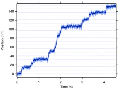
Optical trapping measurements allow the direct observa-tion of individual motors as they move along a microtu-bule and provide a definitive measurement of mechanical processivity. Although extensively used to characterize the biophysical properties of conventional kinesin, optical trapping assays have not been applied widely to other kinesin-related proteins, largely because traditional assays rely on especially fortuitous surface interactions specific to conventional kinesin. Measurements of mitotic kinesins require the development of new assays using polymer-coated surfaces and stereospecific attachment schemes to create robust, functionalized and protein-resistant sur-faces [30-32]. Using one such in vitro assay (Figure 2B), it was shown that individual dimeric human Eg5 proteins walk processively, taking 8 steps on average before disso-ciation [30]. Frequent runs of multiple steps were observed with clear transitions between each step (Figure 3), and statistical tests verified that single motors were suf-ficient to power movement. As expected for a processive enzyme, the step size is 8.1 nm, identical to that of con-ventional kinesin and the spacing between tubulin het-erodimers in the microtubule lattice. A subsequent study using single-molecule fluorescence confirmed that full-length GFP-tagged Eg5 tetramers move processively on microtubules as well [33].
In light of these single-molecule experiments, previous conclusions of Eg5 nonprocessivity must be reconsidered. Chemical processivity measurements should never be mistaken as evidence of true mechanically-processive movement [34], and may be particularly poor estimators for motors with small run lengths. The hyperbolic slowing of Eg5-driven flux as a function of inhibitor concentration that was initially interpreted as evidence of nonprocessiv-ity may instead indicate that inhibited motors remain weakly associated with microtubules, acting as a brake [33,35,36]. Further experiments investigating the mechanical basis of inhibition will be required to fully understand this result. While Eg5's mechanical processiv-ity and abilprocessiv-ity to crosslink and slide microtubules in vitro
certainly does not rule out the presence of a static spindle matrix, the immobilization of Eg5 within the spindle can no longer be used as evidence supporting its existence.
Eg5's load-dependent mechanochemistry shows key distinctions as compared to conventional kinesin
informative motor properties: the force-velocity relation-ship and stall force. Taken together, the mechanical prop-erties of Eg5 show several important distinctions from those of conventional kinesin, a cargo transporter. Through a comparative analysis we may begin to gain insight into how physiology influences motor function, and how small changes in protein structure or organiza-tion give rise to the distinct mechanical properties of all kinesin family members.
Both Eg5 and kinesin velocities display a Michaelis-Menten dependence on ATP concentration, but the over-all velocity of Eg5 is significantly slower, with a maximal stepping rate of 100 nm/s, compared to approximately
650 nm/s for conventional kinesin [30,37]. Eg5 is also much less processive, taking ~8 steps at a time at saturat-ing ATP and zero load, while kinesin takes 50 steps or more under similar conditions [30,38].
The most significant difference between the two motors lies in their response to applied force. At fixed ATP condi-tions, both kinesin and Eg5 velocities remain roughly constant for assisting loads and slow monotonically for hindering loads; however, while kinesin slows by a factor of ~8 from its maximal value at -5 pN, Eg5 is significantly less sensitive to force, slowing by only a factor of three (Figure 4) [30,37]. While both kinesin and Eg5 dimers can sustain hindering loads as high as -7 pN, kinesin motors
Schematic showing in vitro assay designs for Eg5 motor studies
Figure 2
Cell Division 2006, 1:31 http://www.celldiv.com/content/1/1/31
tend to stall and step backwards, maintaining their grip, whereas Eg5 dimers dissociate, making collection of data above -5 pN difficult [30,37,39]. Eg5 motors may eventu-ally stall at extremely high loads: a linear extrapolation of the force-velocity curve to zero velocity suggests the stall force would be approximately -9 pN, near the theoretical maximum allowed for work produced by the hydrolysis of ATP [40]. Future studies at higher forces, inspired by recent work that probed the effect of the sudden applica-tion of large superstall loads to convenapplica-tional kinesin [39], may be required to probe this regime and may reveal addi-tional information about Eg5 mechanochemistry.
Structural differences may be responsible for unusual force response
Several recent studies have revealed structural properties that could contribute to Eg5's unique mechanochemical characteristics. The crystal structure of the motor domain for a human-derived Eg5 monomer bound to ADP showed a novel, ordered neck linker configuration that is docked perpendicular to the long edge of the protein via a series of hydrogen bonds [41]. In all previous kinesin fam-ily member structures, the neck linker was either disor-dered or docked parallel to the long edge of the protein, and in the ADP-bound state, the neck linker is typically floppy [42-45]. The residues involved in Eg5's novel dock-ing are highly conserved among Kinesin-5 family mem-bers, suggesting that this conformation may be specific to this subclass of motors [41].
Although a rigid neck linker would impart molecular stiff-ness, perhaps allowing controlled microtubule sliding under constant tension, it could also hinder each motor head's diffusional search for the next tubulin binding site. A unique mode of motility might provide a much-needed mechanical compromise, allowing processive motion under significant load even with an inflexible neck linker. Based on a series of ensemble FRET measurements, Rosen-feld, et al. proposed three different rigid neck linker con-formations for the ATP-, ADP- and no nucleotide-bound states, and further showed that Eg5 likely moves in two sequential steps [46]. First, ATP binding docks the neck linker parallel to the motor domain, then, upon hydroly-sis, the entire motor domain rolls forward along the microtubule. This two-step mode of motility could be crit-ical for Eg5 function and may influence its relative insen-sitivity to applied force.
Interestingly, a recent report indicates that full-length GFP-tagged Eg5 motors display an unusual mode of motility: processive directional movement interrupted by periods of diffusion as tetramers move along a single cov-erslip-bound microtubule [33]. One-dimensional diffu-sion along the tubulin lattice has been reported for other kinesin-related proteins such as monomeric KIF1A [47,48] and MCAK, a centromere-associated depolymer-izer [49]; localization arises from the electrostatic attrac-tion of the motor to the highly negatively-charged microtubule. Further experiments will be required to determine the role of electrostatic interactions in Eg5, to demonstrate whether the diffusive state is specific to
tetra-Comparison of the force-dependence of the velocities of Eg5 and conventional kinesin
Figure 4
Comparison of the force-dependence of the veloci-ties of Eg5 and conventional kinesin. Eg5 (red, left axis) [30] and conventional kinesin (blue, right axis) [37] velocity as a function of force, as measured with a force-clamped optical trap. Positive forces indicate that load was applied toward the plus-end of the microtubule, assisting motion; negative forces hinder translocation. Conventional kinesin slows much more dramatically than Eg5 does under hindering load.
120
100
80
60
40
20
0
Eg5 Velocity (nm/s)
-4 -2 0 2 4
Force (pN)
600
400
200
0
Kinesin Velocity (nm/s)
Representative trace of position of single Eg5 dimer moving in vitro
Figure 3
Representative trace of position of single Eg5 dimer moving in vitro. Record shows motion of a bead-attached Eg5 dimer held in an optical trap, and walking along microtu-bules in 8.1-nm steps. Position (light blue) and smoothed position (dark blue) are plotted as a function of time; dotted lines are placed every 8.1 nm to guide the eye. Experimental conditions: 2 mM ATP, 4 pN load applied toward the micro-tubule plus-end (assisting motion).
140
120
100
80
60
40
20
0
Position (nm)
4 3
2 1
0
meric motors moving along an isolated microtubule or exists for dimers or tetramers bridging two microtubules, and to determine how much force motors undergoing dif-fusion can withstand before they detach. This diffusive mode could be important to Eg5's function by increasing the total time each motor remains localized to the spindle and therefore the likelihood that tetramers will form crosslinks in cells.
Coordination within and among Eg5 motors
Some of the biggest questions about Eg5 motility sur-round coordination: Do individual Eg5 dimers move by a hand-over-hand mechanism, like conventional kinesin does [50-52]? Are the four motor heads of single Eg5 tetramers coordinated? Finally, do ensembles of Eg5 motors work together to generate force in cell division, and how is the force generated by Eg5 motors balanced against forces generated by other spindle motors (such as ncd, dynein, and the chromokinesins) [53-55]?
Conventional kinesin likely works in isolation to tote cargo long distances in cells. Such highly processive move-ment requires that the catalytic cycles of the two motor heads be tightly coordinated to prevent simultaneous dis-sociation. It is proposed that intramolecular strain trans-mitted by the neck linker regulates the biochemical state of each motor head, keeping the two heads biochemically out-of-phase and allowing one head to maintain a tight grip on the microtubule at all times, even under consider-able load [56-58].
By contrast, Eg5 probably works in small ensembles. The limited processivity of Eg5 may arise because ensembles of motors must work together within the spindle. By tak-ing multiple steps, stak-ingle motors maintain sustained con-tact with their microtubule substrates and aid in de novo
spindle assembly. By dissociating quickly, however, motors detach from the microtubule before stalling and slowing other motors in the ensemble, promoting effi-cient sliding once spindles are formed. These short run lengths may prove to be a consequence of reduced head-head coordination resulting from the rigid nature of the neck linker. Additional experiments will be required to unravel the extent of catalytic coordination, and deter-mine if Eg5 dimers walk hand-over-hand.
In native tetramers, another level of coordination is possi-ble: the opposing pairs of motors heads located at either end of the coiled-coil stalk could cooperate to enhance processivity. Based on the average run length for the dimer, and assuming each pair of dimers in the homote-tramer moves independently, the tehomote-tramer should remain attached to the spindle for ~64 steps, on average [30]. This simple model would predict a run length of ~520 nm, sur-prisingly similar to the ~580-nm average run length of
GFP-Eg5 tetramers moving along a single microtubule in vitro [33]. In principle, either linear or torsional strain within the extended stalk domain could allow the pairs of dimers to communicate, thereby modulating tetrameric run lengths. Direct mechanical measurements of single full-length Eg5 tetramers moving on two microtubules will be necessary to probe this possibility. Finally, new in
vitro assays capable of measuring the forces exerted by
ensembles of motors are required to fully understand how mixed populations of motors work together to organize and move microtubules in cells.
Outlook
Single-molecule measurements of the motor proteins that generate force during mitosis are indispensable for eluci-dating the physical basis of cell division. Establishing whether or not these motors are processive and how they respond to force is critical to developing predictive com-putational models and to understanding how ensembles of motors cooperate to balance forces during each stage of division. Although many mitotic motor proteins have been identified, little nanomechanical characterization has been performed and many important questions remain. The new in vitro assays [13,30] reviewed here should allow complete characterization of Kinesin-5 sub-class members, as well as rapid expansion into new sub-classes of motor proteins. These new data will permit unprece-dented comparative studies of diverse kinesin family members and shed new light on both how protein struc-ture influences motor function and how biochemical energy is harnessed into productive work by all mecha-noenzymes.
Competing interests
The author(s) declare that they have no competing inter-ests.
Acknowledgements
This work was supported by grants to S.M.B. from the National Institutes of Health (NIH). M.T.V. was supported by a Career Award at the Scientific Interface from the Burroughs Wellcome Fund. P.M.F. was supported by a predoctoral fellowship from the National Science Foundation (NSF) and a Lieberman fellowship. We thank Susan Gilbert, Steve Rosenfeld, Duane Compton and members of the Block lab for helpful discussions, and in par-ticular, Jennifer Shanoski and Nick Guydosh for careful reading of the man-uscript.
References
1. Cole DG, Saxton WM, Sheehan KB, Scholey JM: A "slow" homote-trameric kinesin-related motor protein purified from
Dro-sophila embryos. J Biol Chem 1994, 269(37):22913-22916.
2. Sawin KE, LeGuellec K, Philippe M, Mitchison TJ: Mitotic spindle organization by a plus-end-directed microtubule motor. Nature 1992, 359(6395):540-543.
3. Wittmann T, Hyman A, Desai A: The spindle: a dynamic
assem-bly of microtubules and motors. Nat Cell Biol 2001, 3:E28-E34.
4. Hagan I, Yanagida M: Kinesin-related cut 7 protein associates
with mitotic and meiotic spindles in fission yeast. Nature 1992,
Cell Division 2006, 1:31 http://www.celldiv.com/content/1/1/31
5. Le Guellec R, Paris J, Couturier A, Roghi C, Philippe M: Cloning by differential screening of a Xenopus cDNA that encodes a
kinesin-related protein. Mol Cell Biol 1991, 11(6):3395-3398.
6. Heck MM, Pereira A, Pesavento P, Yannoni Y, Spradling AC, Gold-stein LS: The kinesin-like protein KLP61F is essential for
mito-sis in Drosophila. J Cell Biol 1993, 123(3):665-679.
7. Blangy A, Lane HA, d'Herin P, Harper M, Kress M, Nigg EA: Phos-phorylation by p34cdc2 regulates spindle association of human Eg5, a kinesin-related motor essential for bipolar
spindle formation in vivo. Cell 1995, 83(7):1159-1169.
8. Rogers GC, Rogers SL, Sharp DJ: Spindle microtubules in flux. J Cell Sci 2005, 118(6):1105-1116.
9. Maddox P, Straight A, Coughlin P, Mitchison TJ, Salmon ED: Direct observation of microtubule dynamics at kinetochores in Xenopus extract spindles: implications for spindle mechan-ics. J Cell Biol 2003, 162(3):377-382.
10. Sawin KE, Mitchison TJ: Poleward microtubule flux mitotic
spin-dles assembled in vitro. J Cell Biol 1991, 112(5):941-954.
11. Miyamoto DT, Perlman ZE, Burbank KS, Groen AC, Mitchison TJ:
The kinesin Eg5 drives poleward microtubule flux in
Xeno-pus laevis egg extract spindles. J Cell Biol 2004, 167(5):813-818.
12. Rogers GC, Rogers SL, Schwimmer TA, Ems-McClung SC, Walczak CE, Vale RD, Scholey JM, Sharp DJ: Two mitotic kinesins cooper-ate to drive sister chromatid separation during anaphase. Nature 2004, 427(6972):364-370.
13. Kapitein LC, Peterman EJG, Kwok BH, Kim JH, Kapoor TM, Schmidt
CF: The bipolar mitotic kinesin Eg5 moves on both
microtu-bules that it crosslinks. Nature 2005, 435(7038):114-118.
14. Sharp DJ, McDonald KL, Brown HM, Matthies HJ, Walczak C, Vale RD, Mitchison TJ, Scholey JM: The bipolar kinesin, KLP61F, cross-links microtubules within interpolar microtubule
bun-dles of Drosophila embryonic mitotic spinbun-dles. J Cell Biol 1999,
144(1):125-138.
15. Waterman-Storer CM, Desai A, Bulinski JC, Salmon ED: Fluores-cent speckle microscopy, a method to visualize the dynamics
of protein assemblies in living cells. Curr Biol 1998,
8(22):1227-1230.
16. Shirasu-Hiza M, Perlman ZE, Wittmann T, Karsenti E, Mitchison TJ:
Eg5 causes elongation of meiotic spindles when
flux-associ-ated microtubule depolymerization is blocked. Curr Biol 2004,
14(21):1941-1945.
17. Mitchison TJ: Mechanism and function of poleward flux in
Xenopus extract meiotic spindles. Philos Trans R Soc Lond B Biol
Sci 2005, 360(1455):623-629.
18. Ganem NJ, Compton DA: Functional roles of poleward
micro-tubule flux during mitosis. Cell Cycle 2006, 5(5):481-485.
19. Ganem NJ, Upton K, Compton DA: Efficient mitosis in human
cells lacking poleward microtubule flux. Curr Biol 2005,
15:1827-1832.
20. Cameron LA, Yang G, Cimini D, Canman JC, Kisurina-Evgenieva O, Khodjakov A, Danuser G, Salmon ED: Kinesin 5-independent
poleward flux of kinetochore microtubules in PtK1 cells. J
Cell Biol 2006, 173(2):173-179.
21. Maddox P, Desai A, Oegema K, Mitchison TJ, Salmon ED: Poleward microtubule flux is a major component of spindle dynamics
and anaphase a in mitotic Drosophila embryos. Curr Biol 2002,
12(19):1670-1674.
22. Kapoor TM, Mitchison TJ: Eg5 is static in bipolar spindles
rela-tive to tubulin: evidence for a static spindle matrix. J Cell Biol
2001, 154(6):1125-1134.
23. Scholey JM, Rogers GC, Sharp DJ: Mitosis, microtubules, and the
matrix. J Cell Biol 2001, 154(2):261-266.
24. Wells WA: Searching for a spindle matrix. J Cell Biol 2001,
154(6):1102-1104.
25. Dionne MA, Howard L, Compton DA: NuMA is a component of
an insoluble matrix at mitotic spindle poles. Cell Motil
Cytoskel-eton 1999, 42(3):189-203.
26. Tsai MY, Wang S, Heidinger JM, Shumaker DK, Adam SA, Goldman RD, Zheng Y: A mitotic lamin B matrix Induced by RanGTP
required for spindle assembly. Science 2006,
311(5769):1887-1893.
27. Chang P, Jacobson MK, Mitchison TJ: Poly(ADP-ribose) is
required for spindle assembly and structure. Nature 2004,
432(7017):645-649.
28. Crevel IMTC, Lockhart A, Cross RA: Kinetic evidence for low
chemical processivity in ncd and Eg5. J Mol Biol 1997,
273(1):160-170.
29. Uyeda TQP, Kron SJ, Spudich JA: Myosin step size : Estimation from slow sliding movement of actin over low densities of
heavy meromyosin. J Mol Biol 1990, 214(3):699-710.
30. Valentine MT, Fordyce PM, Krzysiak TC, Gilbert SP, Block SM: Indi-vidual dimers of the mitotic kinesin motor Eg5 step
proces-sively and support substantial loads in vitro. Nat Cell Biol 2006,
8(5):470-476.
31. deCastro MJ, Fondecave RM, Clarke LA, Schmidt CF, Stewart RJ:
Working strokes by single molecules of the kinesin-related
microtubule motor ncd. Nat Cell Biol 2000, 2(10):724-729.
32. deCastro MJ, Ho CH, Stewart RJ: Motility of dimeric ncd on a metal-chelating surfactant: Evidence that ncd Is not
proces-sive. Biochemistry 1999, 38(16):5076-5081.
33. Kwok BH, Kapitein LC, Kim JH, Peterman EJG, Schmidt CF, Kapoor
TM: Allosteric inhibition of kinesin-5 modulates its
proces-sive directional motility. Nat Chem Biol 2006, 2(9):480-5.
34. Jiang W, Hackney DD: Monomeric kinesin head domains hydro-lyze multiple ATP molecules before release from a
microtu-bule. J Biol Chem 1997, 272(9):5616-5621.
35. Crevel IMTC, Alonso MC, Cross RA: Monastrol stabilises an
attached low-friction mode of Eg5. Curr Biol 2004,
14(11):R411-412.
36. Krzysiak TC, Wendt T, Sproul LR, Tittmann P, Gross H, Gilbert SP, Hoenger A: A structural model for monastrol inhibition of
dimeric kinesin Eg5. EMBO J 2006, 25:2263-2273.
37. Block SM, Asbury CL, Shaevitz JW, Lang MJ: Probing the kinesin
reaction cycle with a 2D optical force clamp. Proc Natl Acad Sci
U S A 2003, 100(5):2351-2356.
38. Block SM, Goldstein LS, Schnapp BJ: Bead movement by single
kinesin molecules studied with optical tweezers. Nature 1990,
348(6299):348-352.
39. Carter NJ, Cross RA: Mechanics of the kinesin step. Nature 2005,
435(7040):308-312.
40. Block SM: Nanometres and piconewtons: the
macromolecu-lar mechanics of kinesin. Trends Cell Biol 1995, 5(4):169-175.
41. Turner J, Anderson R, Guo J, Beraud C, Fletterick R, Sakowicz R:
Crystal structure of the mitotic spindle kinesin Eg5 reveals a
novel conformation of the neck-linker. J Biol Chem 2001,
276(27):25496-25502.
42. Nitta R, Kikkawa M, Okada Y, Hirokawa N: KIF1A alternately uses
two loops to bind microtubules. Science 2004,
305(5684):678-683.
43. Kikkawa M, Sablin EP, Okada Y, Yajima H, Fletterick RJ, Hirokawa N:
Switch-based mechanism of kinesin motors. Nature 2001,
411(6836):439-445.
44. Kull FJ, Sablin EP, Lau R, Fletterick RJ, Vale RD: Crystal structure of the kinesin motor domain reveals a structural similarity to
myosin. Nature 1996, 380(6574):550-555.
45. Sablin EP, Kull FJ, Cooke R, Vale RD, Fletterick RJ: Crystal struc-ture of the motor domain of the kinesin-related motor ncd. Nature 1996, 380(6574):555-559.
46. Rosenfeld SS, Xing J, Jefferson GM, King PH: Docking and
rolling--a model of how the mitotic motor Eg5 works. J Biol Chem 2005,
280(42):35684-35695.
47. Okada Y, Hirokawa N: A processive single-headed motor:
Kinesin superfamily protein KIF1A. Science 1999,
283(5405):1152-1157.
48. Okada Y, Hirokawa N: Mechanism of the single-headed proces-sivity: Diffusional anchoring between the K-loop of kinesin
and the C terminus of tubulin. Proc Natl Acad Sci U S A 2000,
97(2):640-645.
49. Helenius J, Brouhard G, Kalaidzidis Y, Diez S, Howard J: The depo-lymerizing kinesin MCAK uses lattice diffusion to rapidly
tar-get microtubule ends. Nature 2006, 441(7089):115-119.
50. Asbury CL, Fehr AN, Block SM: Kinesin moves by an asymmetric
hand-over-hand mechanism. Science 2003,
302(5653):2130-2134.
51. Kaseda K, Higuchi H, Hirose K: Alternate fast and slow stepping
of a heterodimeric kinesin molecule. Nat Cell Biol 2003,
5(12):1079-1082.
52. Yildiz A, Tomishige M, Vale RD, Selvin PR: Kinesin walks
Publish with BioMed Central and every scientist can read your work free of charge "BioMed Central will be the most significant development for disseminating the results of biomedical researc h in our lifetime."
Sir Paul Nurse, Cancer Research UK
Your research papers will be:
available free of charge to the entire biomedical community
peer reviewed and published immediately upon acceptance
cited in PubMed and archived on PubMed Central
yours — you keep the copyright
Submit your manuscript here:
http://www.biomedcentral.com/info/publishing_adv.asp
BioMedcentral
53. Chakravarty A, Howard L, Compton DA: A Mechanistic Model for the Organization of Microtubule Asters by Motor and
Non-Motor Proteins in a Mammalian Mitotic Extract. Mol Biol Cell
2004, 15(5):2116-2132.
54. Cytrynbaum EN, Scholey JM, Mogilner A: A force balance model
of early spindle pole separation in Drosophila embryos.
Bio-phys J 2003, 84(2):757-769.
55. Gaglio T, Saredi A, Bingham JB, Hasbani MJ, Gill SR, Schroer TA, Compton DA: Opposing motor activities are required for the
organization of the mammalian mitotic spindle pole. J Cell Biol
1996, 135(2):399-414.
56. Rosenfeld SS, Fordyce PM, Jefferson GM, King PH, Block SM: Step-ping and stretching - How kinesin uses internal strain to walk
processively. J Biol Chem 2003, 278(20):18550-18556.
57. Guydosh NR, Block SM: Backsteps induced by nucleotide
ana-logs suggest the front head of kinesin is gated by strain. Proc
Natl Acad Sci U S A 2006, 103(21):8054-8059.
58. Hancock WO, Howard J: Kinesin's processivity results from mechanical and chemical coordination between the ATP
hydrolysis cycles of the two motor domains. Proc Natl Acad Sci
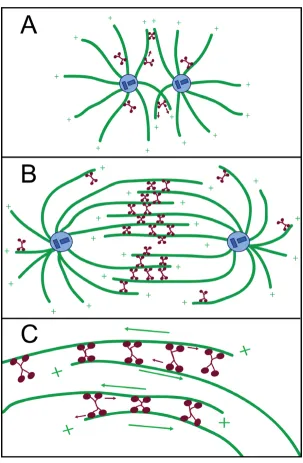
![Figure 2in vitro coated beads through a biotinlyated PentaHis antibody. Coverslip surfaces are precoated with poly-L-lysine-cal trapping assay used to observe processive movement of Eg5 dimers [30]](https://thumb-us.123doks.com/thumbv2/123dok_us/381330.1530813/4.612.144.467.88.455/biotinlyated-pentahis-coverslip-surfaces-precoated-trapping-processive-movement.webp)