Open Access
Research article
Effects of aging on vibration detection thresholds at various body
regions
Meg Stuart*, A Bulent Turman, Jacqueline Shaw, Natalie Walsh and
Vincent Nguyen
Address: School of Biomedical Sciences, University of Sydney, Lidcombe, 2141, NSW, Australia
Email: Meg Stuart* - [email protected]; A Bulent Turman - [email protected]; Jacqueline Shaw - [email protected]; Natalie Walsh - [email protected]; Vincent Nguyen - [email protected]
* Corresponding author †Equal contributors
Abstract
Background: The ability to detect sinusoidal vibrations on the skin surface is dependent on the activation of two classes of receptors. The density of such receptors varies across the skin surface and is a factor in determining the sensory acuity of each skin area. However, the acuity of many sensory systems is known to deteriorate with advancing age. The aim of this study was to determine if vibrotactile sensibility of several skin surfaces deteriorated equally with advancing age.
Methods: Vibration detection thresholds for two frequencies of vibration (30 Hz and 200 Hz) were determined using a method of limits protocol, in two groups of healthy adults, one group aged 17 to 27 years and the other aged 55 to 90 years. Sinusoidal vibrations were generated by a computer and delivered to the skin surface via the probe (diameter = 2 mm) of a mechanical vibrator. Four skin sites (palmar surface of the tip of the middle finger, volar surface of the forearm, lateral aspect of the shoulder, cheek just caudal to the zygoma) were tested.
Results: The fingertip was the most sensitive site for vibrotactile detection at both frequencies in a substantial majority of subjects. The older group of subjects showed significantly higher detection thresholds for both frequencies at all sites, except the fingertip, when compared to young subjects.
Conclusion: The study confirms the deterioration of vibrotactile acuity at several skin sites previously reported in the literature. However, there appears to be no significant reduction in vibrotactile detection at the fingertips in older subjects. This may reflect the high receptor density of this area, or the functional importance of vibrotactile sensibility of the fingertips or some combination of both of these factors.
Background
Inputs arising from various groups of peripheral receptors play a role in vibrotactile sensibility. Four separate chan-nels have been proposed to mediate the sense of touch from the glabrous skin of humans [1]. The ability to detect high frequency (60–1000 Hz) vibration principally de-pends on the P channel, associated with Pacinian
corpus-cles (PC). However, by manipulating stimulus parameters a second channel the NPI channel, may also be responsive to vibrotactile stimuli in this range. The NPII channel, as-sociated with Meissner's corpuscles in glabrous skin and hair follicle receptors in the hairy skin, and their rapidly adapting (RA) afferents fibres, appear to best respond to low frequency (2–100 Hz) vibration. The fourth channel, Published: 25 February 2003
BMC Geriatrics 2003, 3:1
Received: 12 August 2002 Accepted: 25 February 2003
This article is available from: http://www.biomedcentral.com/1471-2318/3/1
the NPIII channel responds best to frequencies in the range of 0.4–100 Hz [1]. There are considerable differenc-es in the density of receptors and receptive field sizdifferenc-es among the glabrous skin locations, the hands and the lips being most densely populated [2,3] and having small re-ceptive fields [3,4]. Even within the hand there is a signif-icant increase in the density of receptors when moving from the proximal part of the finger to the fingertip [2,5], which accounts for the outstanding sensitivity of this re-gion. The receptor density is also associated with cortical magnification, i.e. densely populated body regions having larger cortical representation [6].
The hairy skin shows location specific variations in recep-tor density and field size, presumably reflecting the func-tional roles played by the afferent input from these skin surfaces. The hairy skin of the forearm has a predomi-nance of slowly adapting afferent units, with rapidly adapting units being only sparsely distributed [7]. The skin of the face also has a predominance of slowly adapt-ing mechanoreceptive units, some fast adaptadapt-ing type I or RA units with receptive field sizes similar to those seen in the hand, and an absence of fast adapting type II or PC units [3]. The hairy skin surfaces examined in this study were chosen for their proximity on the somatosensory homunculus to the area for the glabrous hand [8]. Altera-tions to the somatosensory input to one area on the homunculus are known to affect the sensitivity of sur-rounding areas [9].
Deteriorating effects of advancing age have been demon-strated for most sensory modalities [10,11]. Vibrotactile sensibility has also been reported to show age related changes [12–20]. Although initial studies (for review see [14]) were implemented using rather crude stimulating devices, some of the more recent methodologies allowed quantitative evaluation of this sensory modality [14,15,20,21]. However, most of these quantitative stud-ies also had limitations. They either tested a single loca-tion or skin type [14–17] and/or used a single vibraloca-tion test frequency [16,17,19,20], which did not allow the as-sessment of separate afferent channels involved in vibro-tactile signalling. In this study our aim was to investigate the effects of aging on vibrotactile sensibility in various body regions and, by using different frequencies, deter-mine the changes in vibration information processing sig-nalled through separate channels of afferent units.
Methods
SubjectsTesting was performed on 22 young adults (11 females and 11 males; 17–27 years old, µ ± δ, 20.2 years ± 2.2) and 22 elderly adults (11 females and 11 males; 55–90 years old, µ ± δ, 68.6 years ± 10.6). All subjects were in good general health, without any history of upper limb nerve
le-sions, peripheral vascular disease or diabetes mellitus. Testing was carried out in a temperature-controlled room, no measures were made of skin temperature. The study was approved by the Human Ethics Committee of the University of Sydney and conforms to the guidelines for human experimentation of the National Health and Med-ical Research Council of Australia.
Apparatus
Sinusoidal vibration was delivered perpendicular to the skin surface at each test site via a 2 mm diameter perspex©
probe attached to the shaft of a mechanical vibrator (GWV4). The vibrator was mounted on an isolated rigid trunnion (Gearing and Watson, T4), in turn attached to a series of adjustable levers allowing the vibrator to be posi-tioned at a range of heights and orientations. Sinusoidal waveforms were generated by a computer equipped with the Labview® application, passed to a linear power
ampli-fier (Gearing and Watson, PA30) and then delivered to the mechanical vibrator. The use of the Labview® application
allowed instantaneous alterations in the frequency and amplitude of the sinusoids. Although online measure-ments of the dampening effect of applied loads on dis-placement of the vibrator probe were not made, offline measurements using a linear displacement transducer (OD4, Schlumberger Industries) indicated that there was no reduction in the peak-to-peak amplitude of the sinu-soid under loads equivalent to those experienced during data collection. As the mechanical vibrator did not emit any audible sound at the frequencies and amplitudes used in this series of experiments auditory masking was not required.
Experimental procedure
Subjects were tested at four sites bilaterally:
i) palmar surface of the distal one third of the distal pad of the middle finger,
ii) anterior surface of the forearm, in the midcubital line, at the junction of the proximal and middle one third of the forearm,
iii) lateral aspect of the shoulder at the junction of the proximal and distal halves of the deltoid muscle, and
iv) immediately inferior to the zygomatic bone on the face.
plate (300 mm2) suspended from the rigid trunnion. The
plate was positioned parallel to the skin surface and acted as a guard to maintain a constant indentation of the probe into the skin of the testing site and to limit the spread of surface waves across the skin. The probe and the rigid sur-round were separated by a gap of 2 mm on each side.
Two different frequencies of vibrotactile stimulation were selected for preferential activation of separate afferent classes, 30 Hz to preferentially activate rapidly adapting (RA) units and 200 Hz to activate the Pacinian (PC) units.
At each site and at each frequency subjects were initially allowed to experience an amplitude of vibration above the threshold for detection. The amplitude of the test fre-quency was then gradually decreased (descending mode) and subjects were asked to indicate the point at which the vibration could no longer be detected. The amplitude of the sinusoidal waveform was then gradually increased from a subthreshold level (ascending mode) and subjects were asked to indicate the point at which vibration could again be detected. Sinusoidal vibration amplitude in both the ascending and descending modes was incremented and decremented respectively, in steps of 0.17 µm for 200 Hz vibration and 1.05 µm for 30 Hz vibration. On aver-age, three step changes in amplitude were made during each second. Threshold was determined by averaging the results of six trials (three descending and three ascending). This method of limits procedure has been shown to be as re-liable as, and more time efficient than the two-alternative forced-choice procedure in a group of adult subjects aged 22 to 56 years [22]. Furthermore, in four elderly subjects we studied the vibration detection thresholds for 30 Hz and 200 Hz vibration on the tip of the index finger and on the thenar eminence using both methods. A t-test compar-ison revealed no significant differences in the detection thresholds obtained using the method of limits and the two-alternative forced-choice procedure (fingertip 30 Hz p = 0.48, fingertip 200 Hz p = 0.70, thenar eminence 30 Hz p = 0.32, thenar eminence 200 Hz p = 0.28). As the
time course of the testing procedure was much shorter with the method of limits, taking into account the number of sites tested and the age of elderly subjects, this method was considered to be more suitable for the study.
Statistical Analysis
For each group of subjects, group means and standard er-rors were calculated for the detection threshold of each test frequency at each site. To analyse the effects of aging on vibrotactile sensibility, a three way ANOVA with re-peated measures on two variables, formatted as A × B × C with repeated measures on variables B and C, was used to analyse all the data. Variable A was defined as age, firstly young vs. elderly, and then as old (55–64 years, n = 9) vs.
older (65–74 years, n = 6) vs. oldest (over 75 years, n = 7),
variable B was defined as frequency (30 Hz vs. 200 Hz) and variable C was defined as test site.
Results
In the substantial majority of subjects tested, the detection thresholds for both low and high frequency vibration were lowest in the glabrous skin of the fingertip, an obser-vation that reflects the high innerobser-vation density at this site [2]. At 30 Hz vibration, the fingertip was the most sensi-tive site for detection in forty-one subjects, while in the re-maining three subjects (all within the young group) the cheek was the most sensitive site. At 200 Hz vibration the fingertip was the most sensitive site for detection in forty subjects. In the remaining subjects, the cheek was the most sensitive site in three (2 young, 1 elderly) and the shoulder in one young subject. The least sensitive site for vibration detection was the shoulder in twenty subjects, the forearm in seventeen subjects, and the cheek in seven subjects. At the group level, the threshold of vibration de-tection at the fingertip at both 30 and 200 Hz was signifi-cantly (p < 0.001) lower than detection threshold at each of the other sites. Pooled results for all subjects at each site are shown in Table 1.
Table 1: Vibration detection thresholds (dB) form the four sites tested in all young and elderly subjects Stimulus and
age group
Group size Fingertip Forearm Shoulder Cheek
n µ SE µ SE µ SE µ SE
30 Hz
young 22 22.58 0.88 32.09 0.92 32.35 0.90 29.10 0.94
elderly 22 25.51 1.34 40.59 1.32 43.46 1.43 40.65 1.79
200 Hz
young 22 13.80 1.02 23.20 0.89 23.12 0.76 21.78 0.86
In the glabrous skin, i.e. fingertips, the detection thresh-olds for high frequency vibration were lower than those for low frequency vibration in all subjects. These results conform with previous observations on frequency tuning curves obtained from both psychophysical human and electrophysiological animal studies (for review see [23]). The hairy skin locations also showed similar frequency sensitivities, i.e. a greater sensitivity to high frequency vi-bration, however as expected the detection thresholds were significantly higher than the glabrous skin. Figure 1 shows the results obtained from an individual subject at both frequencies for all sites tested. The threshold levels shown at various sites are representative of the results ob-tained from the majority of subjects.
Although in general, the vibration detection thresholds at the fingertip and at the forearm were lower in the descend-ing mode compared to the ascenddescend-ing mode, these differ-ences were not statistically significant. At the shoulder and at the cheek, the vibration detection thresholds were low-er in the ascending mode compared to the descending mode, but again these differences were not statistically sig-nificant. Accordingly, data obtained from the ascending and the descending modes were combined for further analysis.
Effect of age on vibration detection thresholds
ANOVA revealed statistically significant main effects for age (F 1,42 = 15.57, p < 0.001), for site (F 1,42 = 31.33, p < 0.001) and for frequency (F 1,42 = 39.04, p < 0.001). Fol-low up t-tests within the elderly group revealed
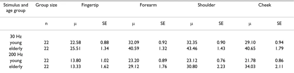
Figure 1
Detection thresholds at several body sites for two frequencies of vibration (30 and 200 Hz) in one elderly sub-ject (male, aged 79 years). Detection thresholds for two frequencies of vibration (30 and 200 Hz). The y-axis represents the peak to peak amplitude (microns) of vibration. The results for this single subject are typical for results from all subjects in the older group.
0
20
40
60
80
100
120
140
30Hz
200Hz
Vibration frequency (Hz)
Ampl
it
ude (mi
crons)
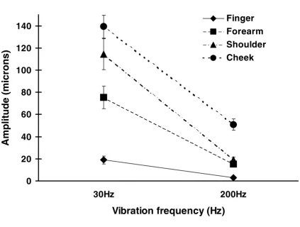
Figure 2
Comparison of vibration detection thresholds in young and elderly subjects, for two frequencies of vibration at several body sites. The histograms show the mean and standard error of vibration detection thresholds of all young (open columns) and all elderly subjects (filled columns) at each site tested, for two frequencies of vibration (30 Hz – upper panel, 200 Hz – lower panel). The y-axis represents the peak to peak amplitude of vibration detection thresholds (dB re: 1 µm peak-to-peak). Statistically significant comparisons are indicated by an asterisk (*).
30 Hz
0
10
20
30
40
50
Tip Forearm
Shoulder
Cheek
Young
Elderly
*
*
*
200 Hz
0
10
20
30
40
50
Tip Forearm
Shoulder
Cheek
*
*
*
D
e
te
c
ti
o
n thre
s
h
o
ld
(d
B
re
:
1
µ
m
pe
a
k
-t
o-p
ea
k
significantly elevated thresholds for the detection of vibra-tion at both frequencies and at all sites, except the finger-tip (p < 0.01). When data obtained from young and elderly subjects were further analysed, those differences that were significant appeared to be the result of deterio-ration in detection thresholds at all sites, except the finger-tip, with aging. The differences in sensitivity between elderly fingertips and other sites were significantly greater, (F 1,42 = 15.65, p < 0.001), than the differences between young fingertips and other sites. Furthermore, it appeared that this discrepancy was further affected by frequency (F
1,42 = 12.47, p < 0.01), with greater deterioration of
thresholds for 30 Hz vibration at all sites, except the cheek. Figure 2 shows the comparison of vibration detec-tion thresholds between young and elderly subjects at each site. For all sites, apart from the fingertips, the elderly group showed higher detection thresholds at both fre-quencies. It should be noted that at 30 Hz vibration there was considerable variability within the elderly group. For this frequency at the fingertip, although the elderly group had higher detection threshold values overall, the statisti-cal analysis did not reveal any significant differences be-tween the two groups.
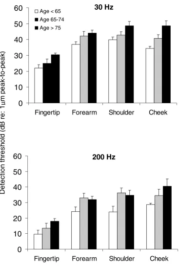
Analysis of data from the elderly group
Figure 3 shows the group means and standard errors for vibration detection thresholds at each site for the three subgroups of elderly subjects. Analysis of these data re-vealed the same main effects for frequency (F1,19 = 38.30, p < 0.001) and for site (F1,19 = 34.92, p < 0.001) within the elderly group, as that revealed by the analysis of data from young versus elderly groups. A main effect for age (F
1,19 = 7.53, p < 0.02) was seen, with thresholds for
vibra-tion detecvibra-tion in two of the elderly groups (older, aged 65– 74 and oldest, aged 75–90) being significantly (p < 0.04) elevated when compared to the thresholds in the remain-ing group (old, aged under 65), for both frequencies and at all sites, except the fingertip. At the fingertip, although overall there was an increase in the detection threshold values with increasing age, there were no statistically sig-nificant differences between those aged under 65 and those in the older (aged 65–74) and oldest (aged 75–90) groups (p = 0.06 - 0.24).
When the fingertips of the older (aged 65–74) group were compared to those of the oldest (aged 75–90) group, there were no significant differences for the 200 Hz vibration detection threshold (p = 0.32), but at 30 Hz the fingertip detection thresholds of the oldest group were significantly (p = 0.02) higher than those of the older group. At the re-maining sites (forearm, shoulder and cheek) there was greater deterioration for the detection of 30 Hz vibration compared to detection of 200 Hz with progressive aging. This effect was more pronounced at the shoulder and at the cheek, than at the forearm.
Discussion
Comparison to previous work
The overall results of this study are in agreement with pre-vious work, the hairy skin locations demonstrate higher detection thresholds for both 30 Hz and 200 Hz vibration [24], the hairy skin of the cheek showing almost none of the U-function described by Verrillo and further demon-strated by Barlow [25].
Direct comparisons of absolute thresholds reported here and those reported by others are difficult for two reasons. Firstly, the contactor size (0.04 mm2) used in the current
study is smaller than that reported by most other groups. The detection threshold for high frequency vibrations is known to be affected by contactor size. Spatial summa-tion in the Pacinian system in the presence of a large con-tactor leads to lowered detection thresholds [21]. However, detection thresholds for low frequency vibra-tions appear to be somewhat independent of contactor size, even for very large contactors [14,21,26]. Secondly, the gap between the contactor and the rigid surround used here was larger than most other studies (2 mm vs 1 mm). A larger gap has been shown to elevate detection threshold for low frequency vibrations due the enhanced sensitivity of this class of receptors to the presence of an edge in their receptive field [27]. The relatively small con-tactor and larger concon-tactor-rigid surround gap used here may account for the generally higher detection thresholds seen in this study in comparison to others.
For the glabrous skin of the fingertip of young subjects, detection thresholds for 30 Hz vibration reported here are comparable to those reported by Mountcastle et al, 1972 [28], Ferrington et al, 1977 [29], Barlow, 1987 [25] and Horch et al, 1992 [18]. While each of these groups used different contactor sizes to the present study, the critical similarity appears to be the size of the contactor-surround gap. Both the current work and the Ferrington group used relatively large gaps, while the Mountcastle and Horch groups used no surround. The thresholds reported by oth-er groups [26,21,30,31] using similar or largoth-er contactor sizes and a smaller contactor-rigid surround gap are con-siderable lower. Detection thresholds for 200 Hz vibra-tion at the fingertip are comparable to the work of Gescheider et al, 1994 [26] using a smaller contactor and a similar gap. However, most other studies [21,24,28,30,31] used larger contactor sizes, known to significantly improve detection for high frequency vibra-tion [21].
Figure 3
Comparison of vibration detection thresholds in all elderly subjects, for two frequencies of vibration at several body sites. The histograms show the mean and standard error of vibration detection thresholds of all elderly subjects – sub-jects aged under 65 (open columns), subsub-jects aged between 65 and 74 (hatched columns) and subsub-jects aged over 75 (filled col-umns) at each site tested, for two frequencies of vibration (30 Hz – upper panel, 200 Hz – lower panel). The y-axis represents the peak to peak amplitude of vibration detection thresholds (dB re: 1 µm peak-to-peak).
30 Hz
0
10
20
30
40
50
60
Fingertip
Forearm
Shoulder
Cheek
Age < 65
Age 65-74
Age > 75
200 Hz
0
10
20
30
40
50
60
Fingertip
Forearm
Shoulder
Cheek
D
e
te
ct
io
n
t
h
re
sh
o
ld
(
d
B
r
e
:
1
µ
m
p
eak
-t
o-p
eak
than the present study. For 200 Hz vibration the thresh-olds reported here are comparable to previous reports for the forearm [24,32] and considerably lower than those for the cheek [25]. There are no previous reports of vibrotac-tile detection thresholds for the hairy skin of the shoulder region.
Comparison of detection thresholds between young and elderly subjects
The overall results of this study are in agreement with pre-vious studies [13–21], that aging has a significant deteriorating effect on vibration sensibility, and that great-er detgreat-erioration is obsgreat-erved on the proximal sites tested. However, a number of previous studies reported that only a small percentage (5%–24%) of subjects showed deteri-oration in vibration sensibility with advancing age [33– 35]. Even though these impairments were particularly prominent beyond the eighth decade of life, the vibrotac-tile sensibility appeared to be normal on the upper ex-tremities compared to the lower exex-tremities [35]. Our results also appear to be in agreement with these studies, as we were unable to demonstrate a deterioration of vibro-tactile detection at the fingertips in elderly subjects. The reasons for the differential effect of aging on detection threshold at various body regions are not clear. Several hy-potheses have been put forward to account for the mech-anism of aging related deterioration in mechanoreception. These include, changes in the me-chanical properties of the skin, decrease in density and al-terations in the morphology of mechanoreceptors, decline in the number of spinal root afferent fibres, chang-es and irregularitichang-es in internodal distancchang-es as a rchang-esult of demyelination and remyelination that may influence the input transmission synchrony, and diminished circula-tion and ischaemia at the peripheral and spinal cord levels (for review see [15]). Some or all of these factors may be responsible for the age related changes observed in vibro-tactile sensibility at various body regions.
Similar methodology to that described here has been used by other groups to measure the detection thresholds of the glabrous skin of young and older subjects. Both Goble [21] and Gescheider's [26] groups found a deterioration of detection thresholds at the fingertip for both high and low frequency vibrations in older subjects, with greater deterioration of thresholds for high frequency vibrations. Increasing the size of the contactor caused small improve-ments in the ability to detect low frequency vibrations, but made an insignificant difference to the thresholds for high frequency vibrations. Verrillo [14] found a deteriora-tion of detecdeteriora-tion thresholds at the thenar eminence for high frequency vibration, but no change in low frequency detection thresholds, with aging. Similarly, Gescheider et al, 1994 [26] demonstrated age related changes in detec-tion thresholds for high frequency vibradetec-tions at the thenar
eminence (low frequency thresholds were not reported in this paper). Each of these latter studies used a very large contactor. However, in the present study we were unable to demonstrate a deterioration in detection thresholds for high and low frequency vibrations at the fingertip. There is a 3–6 fold increase in the RA and PC type mechanore-ceptive unit density between the palm of the hand and the fingertip [2]. Furthermore, a recent study on the distribu-tion of Pacinian corpuscles in the hand of human cadav-ers reported that while 44–60% of corpuscles of the hand are found in the fingers, only 8–18% are located in the th-enar and hypothth-enar regions [5]. Therefore, the different results reported for vibrotactile sensibility in the upper ex-tremities may be related to the variability in receptor den-sity within this body part. If aging affects a proportion of afferent units and their associated receptors in a given ar-ea, it is likely that the densely populated regions, such as the fingertips, may retain their sensitivity compared to less densely populated areas, such as the thenar eminence.
Vibration detection threshold differences amongst elderly subjects
Our analysis of data obtained from elderly subjects indi-cates a more significant deterioration of detection thresh-olds in the older (age 65–74) and oldest (age >75) groups compared to the old (age <65) group at all sites tested, ex-cept the fingertip. Therefore, it appears that on proximal body sites there is a progressive decline in the sensitivity to vibration with increasing age. Such progressive changes with aging have previously been reported [14]. Verrillo [14] described a more prominent decrease in sensitivity to high frequency vibration but not to low frequencies, measured on the thenar eminence, in a group with a mean age of 65 years. Possible mechanisms for this deteriora-tion include reducdeteriora-tion in the number of Pacinian corpuscles, and more favourably, structural changes in the corpuscle morphology [14]. Either of these hypotheses would be particularly relevant for body regions less dense-ly populated with receptors. In our results, measurement of both high and low frequency vibration detection thresholds on proximal body sites also indicated a pro-gressive deterioration with aging. However, at the finger-tip, particularly at 200 Hz, no significant difference could be established between values obtained from the three elderly groups. This conflicting outcome is likely to be due to the different hand regions tested in the present and in Verrillo's studies [14]. It is most likely that gradual changes in receptor density and/or structure throughout aging would result in progressive deterioration in vibro-tactile sensibility, more profoundly so in proximal regions where receptor densities are lower.
re-tained at the fingertips. There are an increasing number of studies indicating that the adult central nervous system is capable of undergoing plastic changes (for review see [36]). Alteration of inputs to the central nervous system may occur not only as a result of peripheral or central le-sions, but also following increased stimulation of a partic-ular body region. Evidence also appears to suggest that active forms of peripheral stimulation involving attention are most effective in inducing changes in the central rep-resentation. It has been shown that when monkeys per-form a vibration frequency discrimination task with a particular finger, the cortical representation for the trained finger increases compared with control fingers [36,37]. Mapping of the cortical representation of the trained skin location revealed a significantly greater (1.5–3 times) area of representation compared to equivalent skin locations on control fingers of the same or opposite hemisphere, and on fingers of passively stimulated control monkeys [37]. Evidence from human studies also suggests similar consequences. For example, results from a neurophysio-logical imaging study indicate that the sensorimotor cor-tical representation of the left hand of string players is greater than the right hand representation and the repre-sentations in non-violin playing control subjects [38]. In another study, it has been reported that the cortical representation of the fingertip in Braille readers is larger than the representation of the fingertip of sighted control subjects [39].
The functional implications of a larger cortical representa-tion can be assessed by means of psychophysical meth-ods. The preliminary results of a study from our laboratory suggest lower thresholds for vibration detec-tion in the string playing fingers of adult violinists. Previ-ously, Stevens, Foulke & Patterson [40] assessed the tactile acuity in blind and age-matched sighted subjects, and re-ported that blind subjects showed better performance at the fingertips than sighted subjects. Although this study further suggested that the acuity at the fingertip declined in both blind and sighted subjects as a function of age, it should be noted that the tests applied in this study were based on discriminatory aspects of tactile sensation rather than vibrotactile detection measurements. It may well be the case that the afferent channels conveying different as-pects of mechanoreception are differentially affected dur-ing agdur-ing. It is therefore possible that, as the most frequently and attentively used body part, especially for texture discrimination, grasp and slip detection, the corti-cal representations of fingertips may be functionally re-tained throughout life. Such central changes may occur through various mechanisms, such as expansion of repre-sentations or synaptic strengthening of neural connections.
Conclusions
In summary, the results of this study confirm the changes in vibration detection sensibility throughout aging. How-ever, there appears to be no significant deterioration of this function at the fingertips. The preservation of detec-tion thresholds at the fingertips reflects the funcdetec-tional im-portance of the skin of the fingertip throughout life in exploration and manipulation of the environment, and is consistent with central mechanisms of plasticity likely to result from such use. The exact mechanisms of this process are not clear at present, but may be established in the near future with more advanced techniques, such as magnetic resonance imaging, that can detect central representation-al changes.
Competing interests
None declared.Authors' contributions
MS participated in data collection, analysis and prepara-tion of the manuscript. BT conceived of the study and par-ticipated in its design and preparation of the manuscript, JS participated in data collection and analysis, NW partic-ipated in data collection and subject recruitment, VN par-ticipated in data collection and subject recruitment. All authors have read and approved the final manuscript.
Acknowledgements
This research was supported by Sydney University Internal Mechanism B Grant 32 304 024 and Sydney University Research Grant S000 4645.
References
1. Bolanowski SJ Jr, Gescheider GA, Verrillo RT and Checkosky CM
Four channels mediate the mechanical aspects of touch. Jour-nal of the Acoustical Society of America 1988, 84:1680-1694
2. Johansson RS and Vallbo AB Tactile sensibility in the human hand: relative and absolute densities of four types of mechanoreceptive units in glabrous skin.Journal of Physiology
1979, 286:283-300
3. Johansson RS, Trulsson M, Olsson KA and Westberg K-G Mech-anoreceptor activity from the human face and oral mucosa. Exp Brain Res 1988, 72:204-208
4. Johansson RS and Vallbo AB Spatial properties of the population of mechanoreceptive units in the glabrous skin of the human hand.Brain Research 1980, 184:353-366
5. Stark B, Carlstedt T, Hallin RG and Risling M Distribution of hu-man Pacinian corpuscles in the hand. A cadaver study.J Hand Surg [Br] 1998, 23:370-372
6. Mountcastle VB Central nervous mechanisms in mechanore-ceptive sensibility.In: Handbook of Physiology: The Nervous System Sensory Processes, part 2.(Edited by: Brookhart JM, Mountcastle VB) Mar-yland, USA: American Physiological Society 1984, III:789-878
7. Vallbo AB, Olausson H, Wessberg J and Kakuda N Receptive field characteristics of tactile units with myelinated afferents in hairy skin of human subjects.Journal of Physiology 1995, 483: 783-795
8. Penfield W and Rasmussen T The cerebral cortex in man: a clin-ical study of localisation of function New York.Macmillan 1950, 9. Recanzone GH, Jenkins WM, Hradek GT and Merzenich MM Pro-gressive improvement in discriminative abilities in adult owl monkeys performing a tactile frequency discrimination task. Journal of Neurophysiology 1992, 67:1015-1030
Publish with BioMed Central and every scientist can read your work free of charge
"BioMed Central will be the most significant development for disseminating the results of biomedical researc h in our lifetime."
Sir Paul Nurse, Cancer Research UK
Your research papers will be:
available free of charge to the entire biomedical community
peer reviewed and published immediately upon acceptance
cited in PubMed and archived on PubMed Central
yours — you keep the copyright
Submit your manuscript here:
http://www.biomedcentral.com/info/publishing_adv.asp
BioMedcentral 11. Verrillo RT and Verrillo V Sensory and perceptual performance.
In: Aging and Human Performance(Edited by: Charness N) Chichester: John Wiley and Sons 1985, 1-33
12. Perret E and Regli F Age and the perceptual threshold for vibra-tory stimuli.Europ Neurol 1970, 4:65-76
13. Potvin AR, Syndulko K, Tourtellotte WW, Lemmon JA and Potvin JH
Human neurologic function and the aging process.Journal of the American Geriatrics Society 1980, 28:1-9
14. Verrillo RT Age related changes in the sensitivity to vibration. Journal of Gerontology 1980, 35:185-193
15. Kenshalo DR Somesthetic sensitivity in young and elderly humans.Journal of Gerontology 1986, 41:732-742
16. Merchut MP and Toleikis SC Aging and quantitative sensory thresholds. Electromyography and Clinical Neurophysiology 1990,
30:293-297
17. Wiles PG, Pearce SM, Rice PJS and Mitchell JMO Vibration percep-tion threshold: influence of age, height, sex, and smoking, and calculation of accurate centile values.Diabetic medicine
1990, 8:157-161
18. Horch K, Hardy M, Jimenez S and Jabaley M An automated tactile tester for evaluation of cutaneous sensibility.J of Hand Surg [Am] 1992, 17:829-837
19. de Neeling JN, Beks PJ, Bertelsmann FW, Heine RJ and Bouter LM
Sensory thresholds in older adults: reproducibility and refer-ence values.Muscle & Nerve 1994, 17:454-461
20. Williams G, Gill JS, Aber V and Mather HM Variability in vibration perception threshold among sites: a potential source of er-ror in biothesiometry.British Medical Journal 1988, 296:233-235 21. Goble AK, Collins AA and Cholewiak RW Vibrotactile threshold
in young and old observers – the effects of spatial summation and the presence of a rigid surround.Journal of the Acoustical So-ciety of America 1996, 99:2256-2269
22. Gerr FE and Letz R Reliability of a widely used test of peripher-al cutaneous vibration sensitivity and a comparison of two testing protocols.Br J Ind Med 1988, 45:635-639
23. Darian-Smith I The sense of touch: performance and peripher-al neurperipher-al processes.In: Handbook of Physiology Section I: The Nervous System Sensory Processes, part 2.(Edited by: Brookhart JM, Mountcastle VB) Maryland, USA: American Physiological Society 1984, III:739-788 24. Verrillo RT and Gescheider GA Perception via the sense of
touch.In: Tactile aids for the hearing impaired(Edited by: Summers IR) London: Whurr Publishers 1992,
25. Barlow SM Mechanical frequency detection thresholds in the human face.Experimental Neurology 1987, 96:253-261
26. Gescheider GA, Bolanowski SJ, Hall KL, Hoffman KE and Verrillo RT
The effects of aging on information-processing channels in the sense of touch: 1. Absolute sensitivity.Somatosensory & Mo-tor Research 1994, 11:345-357
27. Verrillo RT The effect of surface gradients on vibrotactile thresholds.Sensory Processes 1979, 3:27-36
28. Mountcastle VB, Lamotte RH and Carli G Detection thresholds for stimuli in humans and monkeys: comparison with thresh-old events in mechanoreceptive afferent nerve fibres inner-vating the monkey hand.Journal of Neurophysiology 1972, 35: 122-136
29. Ferrington DG, Nail BS and Rowe M Human tactile detection threshold: modification by inputs from specific tactile recep-tor classes.J Physiol 1977, 272:415-433
30. Bernstein LE, Schechter MB and Goldstein MH Child and adult vi-brotactile thresholds for sinusoidal and pulsatile stimuli.J Acoust Soc Ame 1986, 80:118-124
31. Donahue AM and Letowski T Vibrotactile performance by nor-mal and hearing-impaired subjects using two commercially available vibrators.Audiology 1985, 24:362-373
32. Verrillo RT Vibrotactile thresholds for hairy skin.Journal of Ex-perimental Psychology 1966, 72:47-50
33. Plumb CS and Meigs JW Human vibration perception. Part I, Vi-bration perception at different ages (normal values).Arch Gen Psychiat 1961, 4:611-614
34. Steinberg FU and Graber AL The effect of age and peripheral cir-culation on the perception of vibration.Archives of Physical Med-icine and Rehabilitation 1963, 44:645-650
35. Cosh JA Studies on the nature of vibration sense.Clin Sci 1953,
12:131-151
36. Recanzone GH, Merzenich MM, Jenkins WJ, Grajski KA and Dinse HR
Topographic reorganisation of the hand representation in
cortical area 3b of owl monkeys trained in a frequency-dis-crimination task.Journal of Neurophysiology 1992, 67:1031-1056 37. Recanzone GH, Merzenich MM and Jenkins WM Frequency
dis-crimination training engaging a restricted skin surface re-sults in an emergence of a cutaneous response zone in cortical area 3a.Journal of Neurophysiology 1992, 67:1057-1070 38. Elbert T, Pantev C, Weinbruch C, Rockstroh B and Taub E
In-creased cortical representation of the fingers in the left hand in string players.Science 1995, 270:305-307
39. Pascual-Leone A and Torres F Plasticity of the sensorimotor cor-tex representation of the reading finger in Braille readers. Brain 1993, 116:39-52
40. Stevens JC, Foulke E and Patterson MQ Tactile acuity, aging and Braille reading in long-term blindness.Journal of experimental psychology: applied 1996, 2:91-106
Pre-publication history
The pre-publication history for this paper can be accessed here: