Place Field Plasticity and Directionality in a Spatial Memory Task
Patrick D. Martin
A thesis submitted for the degree of Doctor of Philosophy.
ProQuest Number: 10016770
All rights reserved
INFORMATION TO ALL USERS
The quality of this reproduction is dependent upon the quality of the copy submitted. In the unlikely event that the author did not send a complete manuscript and there are missing pages, these will be noted. Also, if material had to be removed,
a note will indicate the deletion.
uest.
ProQuest 10016770
Published by ProQuest LLC(2016). Copyright of the Dissertation is held by the Author. All rights reserved.
This work is protected against unauthorized copying under Title 17, United States Code. Microform Edition © ProQuest LLC.
ProQuest LLC
789 East Eisenhower Parkway P.O. Box 1346
C o n te n ts ... 2
Acknowledgements... 6
Abstract ... 7
Abbreviations ... 9
Chapter 1: Background... 10
1.1 Why the Hippocampus? ... 11
1.2 Outline of the T h e s is ... 12
Chapter 2: Anatomy ... 14
2.1 Gross Anatomy of the Hippocampal Formation ... 15
2.2 Layers of the Hippocampal Form ation... 16
2.3 Cells of the Hippocampal F orm ation... 17
2.4 Connections Within the Hippocampal Formation ... 19
Trisynaptic Pathway First Synapse of the Trisynaptic Pathway Second Synapse of the Trisynaptic Pathway Third Synapse of the Trisynaptic Pathway Extensions to the Trisynaptic Pathway 2.5 Afferents to the Hippocampal F o rm a tion... 23
Septal Formation Afferents Other Subcortical Afferents Cortical Afferents 2.6 Efferents from the Hippocampal Formation ... 25
Subicular Formation Efferents Entorhinal Cortex Efferents Other Efferents 2.7 Anatomical Models of Hippocampal Function ... 27
Chapter 3: Physiology... 29
3.1 Theta R h yth m ... 30
3.2 Large Irregular A ctivity ... 31
3.3 Hippocampal Ripple ... 31
3.4 Long Term P otentiation... 32
3.5 Ammon’s Horn Complex Spike Units ... 33 Observation of Place Fields
CA1 and CA3 Place Fields Dorsal and Ventral Place Fields
Place Fields are Controlled by Multiple Distal Cues Place Fields Appear Insensitive to Cue Removal Place Fields Differ in Different Environments
Place Fields Depend on Both Visual and Vestibular Inputs Place Fields and Environment Scaling
Multiple Place Fields
Quantitative Properties of Place Fields
Place Fields Are Abolished by Restricting Movement Complex Spike Firing is Correlated with Motor Behaviors Complex Spike Firing May be Correlated with Sensory Stimuli
3.6 Ammon's Horn Theta Units ... 45
Theta Unit Firing is Strongly Correlated with Motor Behaviors Theta Unit Firing is Weakly Correlated with Spatial Location 3.7 Subicular Formation Units ... 46
Postsubicular Unit Firing is Strongly Correlated with Head Direction Head Direction Firing Can be Entrained by a Single Cue Head Direction Firing is Controlled by External and Internal Cues Subicular Unit Firing is Weakly Correlated with Spatial Location 3.8 Dentate Gyrus Granule U n its ... 50
Granule Unit Firing is Weakly Correlated with Spatial Location 3.9 Entorhinal Cortex Units ... 52
Entorhinal Unit Firing is Weakly Correlated with Spatial Location Chapter 4: Function ... 54
4.1 Spatial Map H ypothesis... ... ... 55
4.2 Working Memory Hypothesis ... 59
4.3 Relational Memory H ypothesis... 61
4.4 Comparison of the Hypotheses ... 63
Chapter 5: General M e th o d s ... 67
5.1 Introduction... 68
5.2 S u b je c ts ... ... 68
5.3 E nvironm ent... 69
Structure of the Environment Cues in the Environment 5.4 Training ... 71
Familiarization Static Goal Shifting Goal Movable Center Delay of Reward Delay of Choice Segregation Cylinder Cue Removal Perceptual-Perceptual and Perceptual-Memory Tasks Fixed Platform Covers Coverage Paradigm Priming Trial 5.5 Implantation ... 78
Electrodes and Microdrives Surgery 5.6 R ecording... 80
Amplification and Digitization Unit Screening
Trial Running Trial Verification
5.7 Initial A n a lysis... 83
Correcting for Spatial Distortion Clustering Waveforms using Envelopes Verifying the Waveform Envelope Complex Spike Units and Theta Units Creation of Event Lists 5.8 Histology ... 87
5.9 Introduction to R e su lts... 88
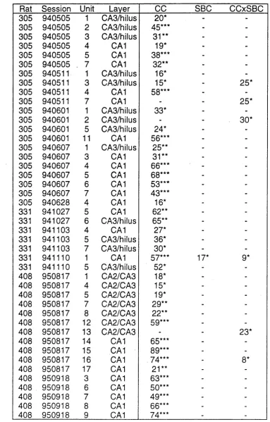
Chapter 6: Effects of GO’S and SBC’s on Hippocampal U n it s ... 89
6.1 Rationale ... 90
6.2 Analysis ... . 90
6.3 R e su lts... 92
6.4 Discussion ... 94
Complex Spike Unit Firing is Governed by the CC’s Complex Spike Unit Firing is Not Governed by the SBC’s Some Complex Spike Unit Firing is Governed by the CCxSBC Interaction Some Complex Spike Units Fail to Display Spatial Firing Relative Spatial Specificity of CA1 and CA2/CA3/Hilar Units Chapter 7: Effects of Cue Removal on Hippocampal U n it s ... 98
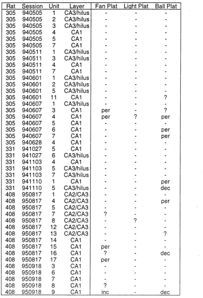
7.1 Rationale ... 99
7.2 Analysis ... 100
7.3 R e su lts... 105
7.4 Discussion ... 107
Place Fields Display Persistence of Firing After Cue Removal Place Fields Display Decreases in Firing After Cue Removal Place Fields Display Increases in Firing After Cue Removal Quantitative Analysis of Place Field Responses to Cue Removal Effects of Cue Removal are Reversed by Cue Replacement Dynamics of Two Lobed Place Fields Postulated Mechanisms of Place Field Dynamics Place Fields in CA1 and CA2/CA3/Hilus Chapter 8: Effects of the Direction of Travel on Hippocampal Units . 113 8.1 Rationale ... 114
8.2 A nalysis... 115
8.3 R e su lts... 119
8.4 Discussion ... 121 Place Fields Display Directionality in the CCE Spatial Memory Task Directionality Persists after Cue Removal
Peak Directional Firing Occurs in Multiple Adjacent Directions Peak Direction of Unit Firing is not Uniformly Distributed Unit Directionality May be Correlated Across Start Platforms Directionality is not Due to Geometric Constraints
Relative Directionality of CA1 and CA2/CA3/Hilar Units
Chapter 9: Examination of Spatial Firing Seen During Error Trials . . 125
9.1 Rationale ... : : ... 126
9.2 Analysis ... 127
9.3 R e su lts... 129
9.4 Discussion ... 130
Hippocampal Error Trial Firing Can Predict the Rat’s Choice Hippocampal Error Trial Firing Can Reflect the CC’s Hippocampal Error Trial Firing May Reflect a Separate Environment Correlation Between Trial Type and Error Trial Firing Chapter 10: Conclusions... 133
10.1 Summary of Findings... 134
Place Field Dynamics in a Spatial Memory Task Place Field Directionality in a Spatial Memory Task Place Fields and Error Trials Differences in Spatial Firing Between CA1 and CA2/CA3/Hilus 10.2 Implications for the Spatial Map Hypothesis ... 139
10.3 Implications for the Working Memory H ypothesis... 143
10.4 Implications for the Relational Memory Hypothesis... 146
10.5 Future D irections... 147
References ... 149
Acknowledgements
I would like to thank Alan Ainsworth and Dulcie Conway for constructing the cue controlled environment, Alan Ainsworth for making the microdrive
assemblies, Michael Recce and Andrew Speakman for writing data collection and data analysis programs used in this study, Andrew Speakman and Jim Dennett for frequently assisting me with the use of the computer network, Clive Parker for constructing and maintaining the headstage and second stage amplifiers, and Penny Ainsworth and Steve Burton for assistance with
histology. I would also like to thank Neil Burgess for helpful suggestion concerning data analysis. I am extremely grateful to Kate Jeffery for reading the draft manuscript and suggesting numerous improvements. Any errors herein are entirely my responsibility. Most of all I would like to thank John O’Keefe for allowing me to spend vast amounts of time, money, and lab resources in pursuing numerous detours and side alleys, many completely unrelated to neuroscience, during my PhD.
Abstract
In order to determine whether hippocampal units display firing which is modulated by the demands of a spatial memory task rats were trained in an enclosed "cue controlled environment" (CCE) consisting of four platforms and six spatial cues that identified one of the four platforms as the goal. Rats learned to select the goal platform at the end of the trial even when the cues defined the goal platform were removed mid-way through the trial. .
In a previous study (O’Keefe and Speakman, 1987a) place fields were shown to be controlled by the spatial cues such that rotation of the cues caused concomitant rotation of the fields. They also found that the place fields continued to fire after removal of the cues. Thus although the place fields were controlled by the six spatial cues they were not dependent on them.
In the present study we confirm the above findings and report two new responses. Some hippocampal place units were observed to increase or initiate place specific firing after cue removal. Others decreased or ceased place specific firing after cue removal. Thus place field location and place field intensity appears to be governed by the presence or absence of the six spatial cues.
Previous work has shown that place fields show directionality when a rat traverses an open field in a restricted and stereotypical manner (McNaughton et al, 1983a; Marcus et al, 1995). Rats trained on the CCE spatial memory task were shaped to run in a raster pattern to insure uniform platform
coverage. The place fields recorded in this situation were found to display directionality oriented with respect to the six spatial cues. Moreover the
directionality of place fields was found to persist after the six spatial cues were removed. Thus place field directionality appears to be initially configured by the six spatial cues but is subsequently independent of them.
Abbreviations
CCE cue controlled environment CC controlled cue
SBC static background cue PP perceptual-perceptual trial
PP1 first two minute control phase of the PP trial, cues are present PP2 second two minute test phase of the PP trial, cues are present PM perceptual-memory or cue removal trial
1.1 Why the Hippocampus?
In 1953 the patient HM underwent bilateral removal of mesial temporal lobe structures in an effort to treat epilepsy (Corkin, 1984). He subsequently displayed a profound anterograde amnesia. This finding led to a surge of interest in the mesial temporal lobe structures. The most prominent of these structures is the hippocampus. The conclusion initially drawn was that the locus of the mnemonic deficit was the hippocampus. However Mishkin (1978) found that both the amygdala and hippocampus had to be removed in order to produce a memory deficit in monkeys. Furthermore later MRI studies have shown that HM sustained extensive damage to mesial temporal lobe
structures other than the hippocampus and retains five eighths of his
hippocampus (O’Keefe, personal communication). This has cast some doubt as to the identity of the structures participating in the anterograde amnesia of HM.
With the spotlight now on the mesial temporal lobe structures more patients were identified with damage to these structures (Squire and Zola-Morgan,
1988; Squire, 1992; Bechara et al, 1995; Kartsounis et al, 1995). They were found to display various memory deficits. McLardy (1970) reviewed the
patient pool available at the time and concluded that they invariably sustained damage to structures adjacent to the hippocampus as a result of their diverse diseases. He concluded the mnemonic functions often ascribed to the
hippocampus proper were located in structures adjacent to the hippocampus.
Despite the uncertainty regarding the locus of the mnemonic deficit numerous neuropsychologists were inspired to perform a large number of studies in which the hippocampi of standard laboratory animals were lesioned, stimulated, recorded from, and otherwise manipulated while the animal performed a variety of mnemonic tasks. Numerous hypotheses of
During this period in which research into the possible mnemonic role of the hippocampus was at a peak O’Keefe and Dostrovsky published a paper in which they observed a correlation between the rat’s location and single unit hippocampal discharge (O’Keefe and Dostrovsky, 1971). They found that units in the CA1 layer of Ammon’s Horn fired when the rat was in specific location on a box and failed to fire in all other locations. They termed the location in which the unit fired a "place field" and the unit itself was called a "place unit". Numerous other workers soon observed place units and began exploring their properties. This growing body of data prompted O’Keefe and Nadel (1978) to propose that the hippocampus was not involved in generalized mnemonic functions but rather was exclusively concerned with spatial
processing.
1.2 Outline of the Thesis
The aim of this thesis is to examine whether hippocampal units display firing which is modulated by the demands of a spatial memory task.
This thesis is organized into four main parts. The first part consists of Chapters 2, 3, and 4. Together these form an overview of the anatomy, physiology, and hypothesized functions of the hippocampus respectively, the background to the research described herein.
The second part consists of Chapter 5, in which the methods used to collect the data in this thesis are detailed. Only one experimental paradigm was employed to gather this data, namely recording from rat hippocampal place cells while animal performed a spatial memory task.
The third part consists of Chapters 6, 7, 8, and 9, in which the data is
analyzed in four different ways. In each of these four chapters the rational for the analysis procedure is introduced, followed by a description of the analysis paradigm, the results of the analysis, and a discussion of these results.
conclusions as to the function of the hippocampus, drawing on three currently influential hypotheses of hippocampal function. Lastly possible fruitful
2.1 Gross Anatomy of the Hippocampal Formation
Ramon y Cajal (1911) and Lorente de No (1933, 1934) first provided detailed descriptions of the hippocampal formation, including the hippocampus proper and the surrounding structures. In rodents the hippocampal formation lies just beneath the cortex, occupying a large fraction of the total brain area. In
primates the hippocampal formation is located in the medial temporal cortex, occupying a considerably smaller fraction of the total brain area. (Figure 1.)
This thesis will be predominantly concerned with the rat unless stated otherwise. Opinions vary as to which structures constitute the hippocampal formation. In this thesis the convention of Amaral and Witter (1989) will be followed. Thus the structures that will be considered part of the hippocampal formation are Ammon’s Horn, dentate gyrus, subiculum, and entorhinal cortex.
In the rat the two interlocking C shapes of Ammon’s Horn and dentate gyrus start at the midline of the hippocampus, just posterior to the septum. They curve both ventrally and posteriorly around the dorsal surface of the thalamus to their deepest and most lateral extent adjacent to the amygdala. This
complex three dimensional morphology has lead to the adoption of the septal- temporal axis nomenclature. This axis follows the curves of the hippocampus. The septal pole or direction is located at the midline or septum. The temporal pole or direction is located laterally and posteriorly, just below the medial temporal lobe, and adjacent to the amygdala. The subiculum lies posterior and medial to the mass formed by the interlocking Ammon’s Horn and dentate gyrus. The entorhinal cortex lies posterior and lateral to the mass formed by the interlocking Ammon’s Horn and dentate gyrus. (Figure 2.)
Ammon’s Horn is a C shaped layer of cells that is subdivided into subfields, known as CA1, CA2, and CA3 where CA is an abbreviation for Cornu
Ammonis. The CA2 layer is physically small and functionally obscure.
being more closely associated with the hilus of the dentate gyrus than
Ammon’s Horn (Blackstad, 1956). The latter classification will be followed in this thesis. (Figure 3.)
The dentate gyrus is composed of a C shaped layer of cells that curves around the lower blade of Ammon’s Horn such that the two C shapes
interlock. The dentate gyrus is subdivided into the upper or buried blade and the lower or exposed blade. Enclosed by the C shaped layer of dentate gyrus cells is the dentate hilus. (Figure 3.)
The subicular complex lies beside the CA1 layer. The subiculum is divided into either four or five subdivisions by different authors (Swanson, 1979; Amaral et al, 1991). The prosubiculum is the area between the end of the CA1 layer and the subiculum. However some authors consider the
prosubiculum to be the area in which the CA1 and subiculum overlap and consequently do not recognize the prosubiculum as a separate area (Witter and Groenewegen, 1990). Posterior to the prosubiculum is the subiculum proper, the presubiculum, and the parasubiculum. The postsubiculum is an invagination of the subiculum present only at the septal end. (Figure 3.)
The entorhinal cortex lies beside the parasubiculum. Based on cell
morphology the entorhinal cortex can be divided into lateral and medial halves (Blackstad 1956). The medial entorhinal cortex lies between the subiculum and the lateral entorhinal cortex. The lateral entorhinal cortex is lies between the medial entorhinal cortex and the perirhinal fissure. (Figure 3.)
2.2 Layers of the Hippocampal Formation
Ammon’s Horn is divided into five layers. Descending from the ventricular surface these are the alveus, striatum oriens, striatum pyramidale, striatum radiatum, and striatum lacunosum-moleculare. In the CA3 cell layer there is one further layer, the striatum lucidum, located just below the striatum
The dentate gyrus is subdivided into three layers, the molecular layer, granule layer, and polymorph layer or hilus. The hilus or polymorph layer is the
innermost zone, curving around with the curve of the dentate and enclosed by the granule layer.
There is little detailed histological information on the various components of the subiculum. However most authors agree the prosubiculum, subiculum, and the septally located postsubiculum consist of a single diffuse layer of loosely packed cells (Blackstad, 1956; Swanson, 1979; Amaral et al, 1991). Blackstad (1956) states the presubiculum and parasubiculum consist of six layers. However this is not readily apparent in photomicrographs. (Figure 3.)
The medial and lateral entorhinal cortex consists of six cell layers (Blackstad, 1956; Stewart, 1976; Alonso and Garcia-Austt, 1987; Amaral et al, 1987). Layers I, II, and III are termed superficial and layers IV, V, and VI are termed deep. Between the superficial and deep layers is a cell free zone called the lamina dissecans.
2.3 Cells of the Hippocampal Formation
Ammon’s Horn contains two main classes of cells, pyramidal neurons and non-pyramidal cells. In Ammon’s Horn the soma of pyramidal neurons are organized into the C shaped striatum pyramidale. Ammon’s Horn pyramidal neurons have both basilar and apical dendrites and an axon emerging from the soma base. (Figure 5.)
In the CA3 layer the pyramidal cell soma is relatively larger than the CA1 pyramidal cell soma. CA3 pyramidal neurons also have large spines close to the soma. These spines are found in the striatum lucidum, the layer peculiar to CA3.
axons ramify widely and contact approximately 1500 pyramidal neurons as well as approximately 60 other interneurons.
The dentate gyrus consists predominantly of granule cells. Granule cells are physically smaller than pyramidal cells and have only apical dendrites
extending up into the molecular layer and an axon extending downwards into the polymorph layer or hilus. The axons of the dentate granule cells are known as mossy fibers due to the numerous varicosities along their length giving them a fuzzy mossy appearance.
There are basket cells in the dentate gyrus with soma located on the border between the granule and polymorph layers (Amaral, 1978). In this case the term "basket" is a misnomer as the axons do not form basket like plexuses that envelope the soma of the granule cells but rather display beaded axons that are interspersed with the granule cells.
In the hilus of the dentate there are numerous cell types including pyramidal, mossy, pyramidal basket, pyramidal-like stellate, giant aspiny stellate, large spiny stellate, wavy multipolar, and peanut cells among others (Amaral, 1978; Ribak et al, 1985). However the two main cell types are pyramidal cells and the mossy cells. Both apical and basal dendrites of the hilar pyramidal neurons display thorny excrescences at their proximal end. The hilar mossy cells also have numerous thorny excrescences on their soma and dendrites that give them the appearance of being covered by moss.
The prosubiculum, subiculum, postsubiculum, presubiculum, and
parasubiculum are chiefly composed of pyramidal neurons (Blackstad, 1956; Amaral et al, 1991).
The medial and lateral entorhinal cortexes do not differ greatly in terms of their cell composition (Blackstad, 1956; Germroth et al, 1989; Schwerdtfeger et al,
pyramidal neurons, spinous multipolar neurons, spinous bipolar cells, and spinous tripolar cells.
2.4 Connections Within the Hippocampal Formation
Trisynaptic Pathway
Within the hippocampus there exists a series of three synaptic connections. These run from entorhinal cortex to dentate gyrus, from dentate gyrus to CA3, and then from CA3 to CA1. This trisynaptic pathway was first proposed by Andersen et al (1971b). It has since been shown to be an oyersimplification.
Howeyer it is still useful as a means of characterizing the major excitatory pathways within the hippocampal formation as well as organizing the exceptions and extensions to the basic trisynaptic idea.
First Synapse of the Trisynaptic Pathway
The first synapse of the trisynaptic pathway is that between entorhinal cortex and dentate gyrus. The entorhinal cortex giyes rise to the perforant path, so named because it perforates the hippocampal fissure. When the entorhinal cortex is lesioned extensiye degeneration of terminals is seen in the dentate gyrus and regio inferior of Ammon’s Horn (Blackstad, 1958). Andersen et al (1971b) found that the perforant pathway forms predominantly excitatory synapses on the dendrites of the dentate granule cells, Amaral and Witter (1989) showed the entorhinal cortex projects to a large septo temporal extent of the dentate gyrus.
The perforant path has since been shown to excite Ammon’s Horn pyramidals directly (Steward, 1976; Yeckel and Berger, 1990). Yeckel and Berger claim the entorhinal to Ammon’s Horn projection is at least as strong as the
The perforant path also synapses on inhibitory basket cells. The entorhinal projection to the basket cells of Ammon’s Horn was demonstrated
anatomically by Seress and Ribak (1985) and electrophysiologically by
Schwartzkroin and Mathers (1978). By stimulating the perforant path Mizumori et al (1989a) and Jung and McNaughton (1993) exploited the lower threshold of basket cell discharge to distinguish basket cells from granule cells during acute single unit recording.
Stewart (1976) has also shown a projection from the entorhinal cortex to the molecular layer of the subiculum.
Second Synapse of the Trisynaptic Pathway
The second synapse of the trisynaptic pathway is that between dentate gyrus and CA3. Dentate gyrus granule cells give rise to axons know as mossy fibers due to the large number of varicosities along their length that give them a mossy appearance under the microscope (Andersen et al, 1966). The mossy fibers synapse on the proximal apical dendrites of CA3 pyramidals. These synapses have been shown to be excitatory. The mossy fiber system is topographically organized in a lamellar fashion in which a given septo temporal extent of dentate gyrus will synapse with the corresponding point in Ammon’s Horn, with the exception of the septal hippocampus. Near the septum the mossy fibers from the dentate turn abruptly upon reaching the border between CA3 and CA1 where upon they course in a caudal direction for several millimeters before proceeding into Ammon’s Horn and forming synapses (Gaarskjaer, 1978; Gaarskjaer, 1986; Amaral and Witter, 1989).
et al (1985) claimed that cells unambiguously identified as mossy cells project to the contralateral hilus.
Third Synapse of the Trisynaptic Pathway
The third synapse of the trisynaptic pathway is that between the CA3 and CA1 layers. In Ammon’s Horn the CA3 layer projects to the ipsilateral CA1 cell layer via Schaffer collateral associational fibers. Andersen et al (1971b) stimulated Schaffer collateral associational fibers and recorded population spikes along the septo-temporal extent of the ipsilateral CA1. They found the population response was maximal at the same septo temporal beam as the stimulation site and dropped dramatically as the recording electrode was moved in either the septo or temporal direction. They concluded that the connection was divided into lamella organized perpendicular to the septo- temporal axis.
This conclusion was contested by Amaral and Witter (1989) who used neuroanatomical tracers to show that the CA3 to CA1 Schaffer collateral associational fibers reached across the postulated lamina of Andersen et al. According to Amaral and Witter the ipsilateral projection from CA3 to CA1 changes in a regular fashion as a function of the particular transverse level observed. Moving septally the CA3 associational fibers terminate
preferentially in the CA1 areas closest to the CA1/CA3 border, and deeply in the striatum radiatum and encompassing the striatum oriens. Moving
temporally the CA3 associational fibers terminate preferentially in the CA1 areas closest to the CAI/subiculum border, and in the superficial levels of the striatum radiatum. The septal temporal extent of the CA3/CA1 associational system is approximately 90% of the long axis of the hippocampus.
Buzsâki and Eidelberg (1982) stimulated the contralateral commissural and ipsilateral associational pathways arising in the CA3 layer and recorded individual units in the CA1 layer. They found both pathways effectively produced negative field potentials at one location in CA1. Moreover both pathways could be tetanized by high frequency stimulation. They concluded that both the ipsilateral associational and contralateral commissural projections converged onto individual CA1 neurons and formed excitatory synapses.
Andersen et al (1963) described a synaptic loop between pyramidal cells and basket cells in Ammon’s Horn. (Figure 6.) Pyramidal axon collaterals make excitatory synapses on basket cells. Andersen et al (1963) determined that the majority of basket cells gave rise to inhibitory afferents that synapse on the soma of pyramidal cells. Thus pyramidal cell firing excites basket cells that then recurrently inhibit the originally active pyramidal neurons. Using
intracellular labelling Sik et al (1995) found the lateral spread of the basket cell axon was comparatively large, extending to approximately 1500 pyramidal neurons in the local area. Thus one basket cell could modulate the firing of numerous pyramidal units.
Extensions to the Trisvnaptic Pathway
The trisynaptic pathway as first postulated by Andersen et al (1971b) does not include three of the major projection pathways in the hippocampal formation, namely the projection from Ammon’s Horn to the subicular formation, the projection from Ammon’s Horn to the entorhinal cortex, and the projection from the subicular formation to the entorhinal cortex.
The second pathway in the hippocampal formation not included in the trisynaptic pathway is the projection from Ammon’s Horn to the entorhinal cortex (Swanson and Cowan, 1977; Beckstead, 1978; Tamamaki and Nojyo, 1995). As the entorhinal cortex is the first structure in the trisynaptic pathway it can be seen that the connectivity loops around onto itself. The functional implications of this looping are discussed below.
The last major pathway in the hippocampal formation not included in the trisynaptic pathway is the projection from the subicular formation to the entorhinal cortex (Swanson and Cowan, 1977; Sorensen and Shipley, 1979;Van Groen and Wyss, 1990). This projection is weaker than the projection between the CA1 layer and the entorhinal cortex (Tamamaki and Nojyo, 1995).
While the trisynaptic pathway captures the essence of the feedforward hippocampal formation connectivity, there exists evidence for feedback connections as well. Scharfman (1994) found CA3 pyramidal neurons innervated hilar mossy cells in the slice preparation. Sik et al (1994) found inhibitory feedback projections from CA1 to CA3 and hilus. They speculate that this pathway allows for the synchronization of activity in Ammon’s Horn.
2.5 Afferents to the Hippocampal Formation
Septal Formation Afferents
to the hippocampal formation are postulated to provide a pacemaker input to the hippocampal neurons. (See below.)
Other Subcortical Afferents
Room and Groenewegen (1986b) examined subcortical afferents to the entorhinal cortex. They found the magnocellular basal forebrain, basolateral amygdaloid complex, claustrum, nucleus reuniens of the thalamus, supra- mammillary region of the hypothalamus, ventral tegmental area of the mesencephalon, dorsal raphe nucleus, nucleus centralis superior, locus
coeruleus, and medial septum gave rise to projections to the entorhinal cortex in the cat.
Magloczky et al (1994) found the supra-mammillary bodies of the
hypothalamus project to CA3 pyramidals and dentate granule cells in a widely diffuse fashion. Moore and Halaris (1975) demonstrate a projection from the raphe nuclei to Ammon’s Horn and the dentate gyrus. Van Groen and Wyss (1990) found various thalamic nuclei project to the presubiculum,
parasubiculum, and postsubiculum.
Thus it appears that while a minority of subcortical structures synapse on various components of the hippocampal formation the majority of the subcortical inputs to the hippocampal formation are channelled through the entorhinal cortex.
Cortical Afferents
The entorhinal cortex receives projections from the anterior piriform cortex, diagonal band nucleus of Broca, retrosplenial cortex, infralimbic cortex,
prepiriform cortex, olfactory bulb, olfactory cortex, prelimbic cortex, infralimbic cortex, cingulate cortex, and insular cortex (Beckstead, 1978; Sorensen,
1985; Room et al, 1985; Room and Groenewegen, 1986a).
et al (1985) found the infralimbic cortex projects to the subiculum in the cat. Witter and Groenewegen (1984a) demonstrated a weak projection from the perirhinal cortex to Ammon’s Horn and the dentate gyrus.
Thus in common with subcortical inputs it appears that the majority of the cortical inputs to the hippocampal formation are channelled through the entorhinal cortex. A minority pass through the subicular complex or travel directly to the hippocampal formation.
2.6 Efferents from the Hippocampal Formation
Subicular Formation Efferents
The most intriguing efferent pathway originating in the subicular formation (subiculum and prosubiculum) is the one to the nucleus accumbens (Kelley and Domesick, 1982) since this pathway has been implicated in the control of locomotion (Mogenson and Nielsen, 1984; Annett et al, 1989; Schacter et al, 1989). Moreover the nucleus accumbens has been shown to display
increases in activity following feeding and drinking (Mark et al, 1992).
Swanson and Cowan (1977) found the subiculum projected to the mammillary bodies, perirhinal cortex, ventral hypothalamus, and ipsilateral lateral septal complex. Finch et al (1984a) found the subiculum projected to the posterior cingulate cortex. Sorensen (1980) and Vogt and Miller (1983) found a connection from the subiculum to the retrosplenial cortex. Ganteras and Swanson (1992) found the subiculum also projected to the amygdala, bed nuclei of the stria terminalis, and the rostral hypothalamus.
(1990) found a further projection from the presubiculum to the retrosplenial cortex.
Swanson and Cowan (1977) found the postsubiculum has a bilateral projection to both the cingulate and retrosplenial cortices. Vogt and Miller (1983) found a connection from the postsubiculum to the retrosplenial cortex and visual cortex areas 17 and 18b. Van Groen and Wyss (1990) examined the postsubiculum further and found an additional projection to adjacent perirhinal cortex.
Maren and Fanselow (1995) implanted electrodes in the ventral angular bundle and recorded monosynaptic responses in the basolateral amygdala. They note that ventral angular bundle fibers arise from CA1, ventral
subiculum, and lateral entorhinal cortex. However they concluded that the majority of the hippocampal efferents to the basolateral amygdala originate in the subiculum.
Entorhinal Cortex Efferents
Kosel et al (1982) found projections from the entorhinal cortex to the inferior temporal cortex, perirhinal cortex, and auditory cortex. Sorensen (1985) found a projection from the entorhinal cortex to retrosplenial cortex, cingulate cortex, prelimbic cortex, infralimbic cortex, perirhinal cortex, p re piriform cortex, and insular cortex as well as the subcortical lateral thalamic nucleus, accumbens nucleus, and striatum.
Swanson and Kohler (1986) studied connections between entorhinal cortex and cortical and subcortical areas in the rat. They found the entorhinal cortex projects to the olfactory cortex, cingulate gyrus, medial prefrontal cortex, perirhinal cortex, and insular cortex. They also found connections between the entorhinal cortex and the caudoputamen, nucleus accumbens, and the basolateral complex of the amygdala.
unrestricted way station the entorhinal cortex is more specialized for efferents to other cortical areas.
Other Efferents
Raisman et al (1966) found Ammon’s Horn projects to the anterior thalamic nuclei, mammillary nucleus, medial and lateral septum, diagonal band nucleus, and the nucleus accumbens. Meibach and Siegel (1977) and Swanson and Cowan (1977) confirmed the projection from Ammon’s Horn to the septum. Swanson and Cowan (1977) also found a weak projection from the CA1 layer to the perirhinal cortex and another weak projection from the CA3 layer to the cingulate cortex. In general Ammon’s Horn efferents are weaker than
subicular formation and entorhinal cortex efferents.
The dentate gyrus is not thought to project outside the hippocampus (Raisman et al, 1966; Swanson and Cowan, 1977).
2.7 Anatomical Models of Hippocampal Function
A number of models of hippocampal function have been put forward based predominantly on the postulated function of its efferent and afferent structures. The first of these is the Papez circuit postulated to mediate emotion (Papez,
1937). The Papez circuit consists of the reciprocal connections between medial temporal structures and the hypothalamus. Papez considered the hippocampal formation and cingulate gyrus to be the necessary medial temporal structures and the mammillary bodies to be the requisite
Based on the topology of the connections between the hippocampal formation and cortical structures Teyler and DiScenna (1984, 1986) proposed that it serves to construct an index of neocortical areas active during a learning episode. Upon subsequent activation of a subset of these neocortical areas the index stored in the hippocampal formation is reactivated and leads to the retrieval of the entire neocortical representation.
The trisynaptic pathway loops back onto itself via a direct projection from CA1 to the entorhinal cortex. There is also an indirect loop from CA1 to the
entorhinal cortex via the subiculum. It has been proposed that this physical loop of connectivity is designed to facilitate the maintenance of electrical loops of activity in the hippocampus and related structures, thus creating a pattern completion or memory device (McNaughton and Nadel, 1990; Mizumori et al, 1989b; Buzsaki et al, 1990).
Risold and Swanson (1996) proposed that information processing in different regions of the hippocampus mediates different behaviors. This postulate was based on the observation that different hippocampal regions were connected via the lateral septal nucleus to hypothalamic systems which mediated
different behaviors. The CA3 region was found to project to the lateral supramammillary nucleus. The bulk of CA1 and subiculum was found to project to the medial zone nuclei of the hypothalamus thought to be involved in social behavior. The ventral tip of CA1 and subiculum was found to project to the medial preoptic nucleus and hypothalamic periventricular region.
3.1 Thêta Rhythm
When a rat runs, rears, or jumps a regular sinusoidal EEG wave termed "rhythmic slow activity" (RSA) or the "theta rhythm" can be recorded in
Ammon’s Horn and dentate gyrus (Vanderwolf, 1969). This rhythm consisted of a 6 to 12 Hz sinusoid with an amplitude of 300 pV or greater. Later work showed that there was two types of theta rhythm, atropine sensitive and atropine resistant (Vanderwolf, 1975). Atropine sensitive theta rhythm was found to occur during motionless awake alert behaviors and tonic REM sleep and could be abolished by the injection of atropine or other muscarinic
cholinergic antagonists. Atropine resistant theta rhythm was found to occur during voluntary movement and phasic REM sleep and was not affected by muscarinic cholinergic antagonists but could be abolished by the application of anesthetics (ether, urethane) and
morphine (Vanderwolf et al, 1978).
In the rat the hippocampal theta varies with depth (Winson, 1976). Starting at the striatum oriens it increases in amplitude until it reaches a local maximum at the level of the stratum pyramidale. As the recording electrode moves further the theta wave shifts phase and drops in amplitude until at the level of the hippocampal fissure it is greatly attenuated. It then picks up in amplitude until it peaks at the level of the granule cell layer. At this level the theta is substantially greater in amplitude than at the level of the stratum pyramidale and 180° phase reversed.
The hippocampal theta pacemaker is thought by some to be located in the medial septum (Stewart and Fox, 1990). Cholinergic and GABAergic fibers originate in the septum and project to the hippocampal pyramidals and
interneurons (Freund and Antal, 1988). It is this synchronized septally driven variation in the membrane potentials of numerous hippocampal cells that is thought to give rise to the extracellular voltage changes characteristic of theta.
cholinergic agonist carbachol. It was possible to record a rhythmic slow wave in the dentate gyrus of the slice with a frequency that was comparable to that of theta found in intact hippocampus, despite the absence of septal afferents.
3.2 Large Irregular Activity
When a rat is sleeping or resting quietly the hippocampus displays irregular bursts of cell firing that summate to form a large low frequency electrical events (Vanderwolf, 1969; O’Keefe and Nadel, 1978; Ylinen et al, 1995a). This EEG activity has been termed large-amplitude irregular activity (LIA) (Vanderwolf, 1969) or the sharp wave (Ylinen et al, 1995a). A sharp wave consists of a relatively high amplitude and long duration EEG wave.
Successive sharp waves occur at irregular intervals, they do not display rhythmicity.
Positive going monophasic sharp waves are observed in the stratum oriens above the pyramidal cell layer. The amplitude increases as the electrode approaches the cell layer. Sharp waves reverse polarity at the level of the stratum pyramidale, displaying biphasic waveforms in the pyramidal layer itself. They then became relatively smaller in amplitude and negative going in the stratum radiatum. Sharp waves are postulated to arise as a result of spontaneous and synchronous discharge of numerous pyramidal neurons. (O’Keefe and Nadel, 1978). Sharp waves form a useful marker for positioning electrodes in the CA1 cell layer as the sharp wave polarity is determined by the location of the recording electrode with respect to the stratum pyramidale.
3.3 Hippocampal Ripple
Found at the level of the CA1 cell layer is a characteristic EEG wave referred to as the "hippocampal ripple" (O’Keefe, 1976; O’Keefe and Nadel, 1978). Ripples are observed during LIA riding the sharp wave. They consist of a burst of 4 to 10 low amplitude waves of 4 to 8 ms duration.
(O’Keefe and Nadel, 1978). An alternate hypothesis is that the ripple arises as a result of a barrage of inhibitory inputs from basket cells resulting in a train of inhibitory post-synaptic potentials in the pyramidals (Buzsaki et al, 1992; Ylinen et al, 1995a).
The presence of the ripple on the sharp wave is another phenomenon that can be exploited to locate electrodes in the CA1 cell layer. When the amplitude of the ripple is at its maximum the recording electrode is located close to the stratum pyramidale.
3.4 Long Term Potentiation
Long term potentiation is the term given to the increase in the extracellular field potential following stimulation of afferent fibers either by a paired pulse or by a burst of pulses (Bliss and Collingridge, 1993). LTP was first described in the projection from the entorhinal cortex to the dentate gyrus (Bliss and Lomo,
1973) but has since been found in many limbic and neocortical pathways (Racine et al, 1983; Bear and Kirkwood, 1993; Fox and Daw, 1993). The stimulation parameters originally used to induce LTP were not very
physiologically plausible. However subsequent studies found that stimulation at the theta frequency (Larson et al, 1986; Greenstein et al, 1988) or artificial sharp waves (Buzsaki et al, 1987) produced LTP. Furthermore the ease with which LTP can be induced varies at different phases of the theta rhythm (Pavlides et al, 1988), providing further evidence for a physiological role. The phenomenon of LTP can last for several weeks in chronic recording setups (Bliss and Gardner-Medwin, 1973).
In order to induce LTP at a synapse the postsynaptic cell must be depolarized when the action potential reaches the presynaptic terminal and causes
necessary for the induction of LTP (Kelso et al, 1986) and is postulated to underlie some forms of learning and memory.
3.5 Ammon’s Horn Complex Spike Units
Ranck (1973) first described extracellular single unit recording in Ammon’s Horn of the hippocampus in the freely moving rat. Based on this study he divided the units of Ammon’s Horn into two broad groups, complex spike units and theta units.
Complex spike units would fire single spikes or multiple "complex spikes" of decreasing amplitude. (Figure 7.) The duration of their extracellularly
recorded waveform was greater than that of theta units and the magnitude of the first phase was greater than the second. Their firing frequency, counting a complex spike burst as one spike, was lower than theta units and they were never observed to fire continuously for seconds at a time. The longest burst observed was typically less than two seconds. Occasionally a complex spike unit would fail to fire for minutes at a time. Relative to theta units complex spike units displayed low frequency, irregular firing.
Complex spike units are found in association with other large amplitude spikes in the stratum pyramidale of Ammon’s Horn. Thus the complex spike unit identified electrophysiologically is postulated to be one and the same as the pyramidal cell identified histologically (Ranck, 1973; Fox and Ranck, 1975; Fox and Ranck, 1981). Complex spikes have been recorded in areas other than Ammon’s Horn such as the stratum granulosum of the dentate gyrus.
However in this thesis the term "complex spike" will be used to refer to units recorded in Ammon’s Horn unless stated otherwise.
Observation of Place Fields
the unit fired came to be known as a "place field" and the unit was referred to as a "place unit" (O’Keefe, 1976).
By taking into account the work by Ranck (1973) in classifying Ammon’s Horn cell types into pyramidal cells and basket cells O’Keefe (1979) determined that place fields were identified primarily with pyramidal cells. This conclusion was based on the observation that units displaying place fields also displayed spike bursts of decreasing amplitude and that the histological location of the units displaying place fields was the same location of the complex spike pyramidal cells of Ranck, namely the stratum pyramidale. Thus pyramidal cells display location specific firing.
CA1 and CA3 Place Fields
Initially due to methodological constraints units displaying place fields were only described in the CA1 field. Subsequent work revealed units displaying place fields in CA3 (Olton et al, 1978). According to Olton et al (1978) place fields in CA3 were essentially identical to those seen in CA1. However with the advent of more precise techniques to quantify the identity of individual units and the spatial location of the rat this view was modified. McNaughton et al (1983a) found that place units in CA3 were less spatially exact than those in CA1. They put forward the hypothesis that CA1 place unit responses were constructed from the intersection of a number of CA3 place units. This would account for their higher spatial selectivity. Muller et al (1987) stated that the spatial specificity of CA1 and CA3 place fields was about the same. Barnes et al (1990) claimed that CA3 place fields are more spatially specific relative to CA1 place fields. The resolution of this issue must await further experimentation.
Dorsal and Ventral Place Fields
their overall firing rate was lower. Jung et al (1994) also compared Ammon’s Horn dorsal and ventral place units. They concluded that a significantly smaller fraction of the ventral units displayed fields and the ventral units had fields that were significantly less spatially specific. Contradictions between these two studies must await further experimentation to be resolved.
Place Fields are Controlled bv Multiple Distal Cues
In the first description of place fields O’Keefe and Dostrovsky (1971) examined their dependence on environmental cues. The location of the place field was insensitive to the removal of single cues. However it was altered when large changes were made to the environment. O’Keefe and Conway (1978) also found that place field firing could be maintained by a subset of the
environmental cues. Thus it appeared the place specificity of place units is determined by a constellation of cues.
O’Keefe and Conway (1978) found that rotations of controlled cues within a circular curtained enclosure reliably rotated CA1 place fields. If a subset of the cues were removed before placing the rat in the environment the place fields were unaffected. However if all of the controlled cues were removed before placing the animal in the enclosure the place specific firing of the units was altered or abolished.
Muller and Kubie (1987) recorded place units in a standard cylindrical
apparatus with a single polarizing cue (a white card). When the polarizing cue was rotated the place fields would rotate in register. Breese et al (1989) also found that a polarizing cue card could rotate place fields.
Place Fields Appear Insensitive to Cue Removal
goal. However if the cues were not present from the start of the trial the rat would chose an arm at random, and performed at chance levels.
O’Keefe and Speakman (1987a) succeeded in recording units in rats while they performed the above task. They found that place field firing was set by the environment cues and rotated in synchrony when the environment cues were rotated. Moreover when the controlled cues were removed the place fields persisted. They concluded that Ammon’s Horn place specific firing was initiated but not maintained by the environmental cues.
Quirk et al (1990) recorded the response of Ammon’s Horn place units when rats were placed in an environment that was initially lit but then darkened half way through the recording session. For the majority of place units darkening the environment did not significantly affect the spatial specificity of their firing. However for a small minority of place units darkness abolished the spatial specificity of their firing. They contrasted these results with a second
recording paradigm in which the rat was placed in the same environment, but it was darkened throughout recording. They found about half of the units had vastly changed place fields in the darkened environment. They concluded that the spatial specificity of hippocampal units depended on recently viewed visual information but could be maintained in the absence of visual information by either non-visual information or by the rat constantly tracking the previous and current locations ("dead reckoning").
Markus et al (1994) recorded place units while the rat performed an eight arm radial maze task in an environment that was alternately lit and darkened between trials. They found a higher proportion of Ammon’s Horn complex spike units displayed place fields in the lit trials than in the darkened trials. Moreover the place units that were observed in darkness were less specific and reliable than their lit counterparts. However some place fields were found to appear only when the trials were run in darkness.
environment has been exited and a new one entered. The place units specific to the dark situation may be neurons coding for the new environment.
Alternately changes observed in place unit firing after cue removal may simply be due to the degradation of place unit firing due to the paucity, of visual information.
Place Fields Differ in Different Environments
O’Keefe and Conway (1978) recorded place fields in two environments. They found that single place units had place fields in either the first, the second, or both environments. For those units with fields in two environments there was no clear relationship between the shape or location of the two place fields. They concluded a single place unit can participate in the representation of different environments.
Kubie and Ranck (1983) recorded place cells in three different environments, an eight arm maze, a operant conditioning cage, and the rat’s home cage. All environments were centered in the same room and thus occupied the same location in real space. They found a given unit would have completely dissimilar place firing in the three environments. Moreover upon rotation of the eight arm maze about the center Kubie and Ranck found the fields failed to rotate whereas upon rotation of the operant chamber and the home cage about the center the fields also rotated. This differential response to the rotation of the environment clearly shows that the same place units were responding differently to different environments.
Muller and Kubie (1987) recorded place units in two environments, a circular enclosure and a square enclosure of comparable size and color. They also found that for a given unit the location and shape of its place fields in one environment did not resemble that seen in the other.
Place Fields Depend on Both Visual and Vestibular Inputs
apparatus in which the floor and walls could be rotated independently or in concordance. If the floor was rotated and the walls were not rotated 100% of Ammon’s Horn fields would rotate in register with the floor. Sharp et al claim this is due to the rat being able to view the walls going round while the floor rotated, therefore the vestibular and visual inputs are in agreement as to the magnitude and direction of the rotation. If the lights were turned off and the floor rotated only 40% of the fields would rotate in register with the floor. They maintain this is due to the rat not being able to view the walls going round while the floor rotated. Thus the vestibular input indicates rotation but the visual input was unavailable. If both the floor and walls were rotated less than 20% of the fields would rotate in register with the floor and walls. They assert this is due to the rat not being able to view the walls going round while the floor rotated, therefore the vestibular input indicates rotation but the visual input does not. Sharp et al concluded that rats use both vestibular and rotational information to set up place fields. If one form of information is unavailable the stability of place fields is moderately impaired. However if one form of information contradicts the other the stability of place fields is greatly impaired.
Knierim et al (1995) compared place fields recorded in a cylinder with a single polarizing cue. They pre-trained two groups of rats to forage for pellets in the cylinder. One group was disoriented before foraging and the second was not. They found the rats that were pre-trained with repeated disorientation had unstable place fields in the cylinder that did not reliably rotate when the polarizing cue was rotated. The rats in which pre-training did not involve repeated disorientation had stable place fields in the cylinder that reliably rotated when the polarizing cue was rotated. They concluded that by disorienting the rat before training it was prevented from learning that the polarizing cue is a stable directional reference. Consequently the effect it later had over place unit firing was minimal.
Place Fields and Environment Scaling
units (36%) were also scaled up in size while remaining in the same angular location. The place fields of other units (52%) were completely altered by the scaling of the cylinder. When barriers were placed within place fields they were found to attenuate or abolish place field firing. Breese et al (1989) also found that a barrier placed within a place field attenuated the firing of the corresponding place unit.
Wilson and McNaughton (1993) further explored the dynamics of place unit response upon environment scaling. They placed rats in an environment with a partition in the middle and recorded from place units. Half way through the recording session the partition was raised. They found that place units that fired in the original environment continued to do so in the new expanded environment. However they also found that units that did not display place fields in the original environment began to fire in the new half of the
environment. Initially the units fired in a manner that was not spatially robust but they soon settled down and began to display clear place fields. It appears as though when the environment was increased in size the hippocampal place units not already involved in the spatial representation were recruited in order to "fill in" the newly opened space.
O’Keefe and Burgess (1996) studied the dependence of place fields on the environment shape. They recorded place fields in four differently shaped boxes in the same room, namely a small square, horizontal rectangle, vertical rectangle, and large square. They found some place fields that remained in a constant location relative to two orthogonal walls, for example tucked up in a corner. Thus as the rat was moved from one box to another differently shaped box the field would remain in the same corner. However the location of other place fields was dependent on two opposite walls. If the two walls were pulled further apart the place field would be elongated and in some cases pulled apart into two separate components. The striking observation of the dependence of place fields on one or more of the edges of the
The environment scaling manipulation of Muller and Kubie (1987), the partition removal manipulation of Wilson and McNaughton (1993), and wall
manipulation of O’Keefe and Burgess (1996) are comparable in design. The experiments of Muller and Kubie (1987) can be characterized as ones in which all of the walls of the environment were moved simultaneously, the
experiments of Wilson and McNaughton (1993) can be characterized as ones in which only one wall of the environment was moved, whereas in the
experiments of O’Keefe and Burgess (1996) all of the environment walls were moved. Thus the scaling of place fields seen by Muller and Kubie (1987) and the elongation and separation of fields seen by O’Keefe and Burgess (1996) may have taken place via the same mechanism.
However in the partition removal experiment of Wilson and McNaughton (1993) the fields were found to be unchanged. This may have been due to how the experiments were run. In the studies of Muller and Kubie (1987) and O’Keefe and Burgess (1996) the rat was removed from one environment and placed in another, differently shaped environment. In the study of Wilson and McNaughton (1993) the rat was in the environment when the partition was removed. The fact that the rat was in place when the environment was manipulated may have caused it to view the new environment as essentially the same as the old environment. Thus the fields were relatively unchanged. In the studies of Muller and Kubie (1987) and O’Keefe and Burgess (1996) the rat was physically removed from the old environment. Thus the spatial map may have been partially or completely dissociated from the old environment such that when the rat was replaced in the new environment the hippocampal fields could display novel firing more readily.
Place Fields and Directionalitv
O’Keefe (1976) and McNaughton et al (1983a) observed that the firing rate of some place units was greater when the rat ran through the unit’s place field in one of two directions on a radial arm maze. However it is possible the
firing in rats run trained to run around randomly in a high walled cylindrical enclosure.
Muller et al (1994) performed a more extensive analysis to test the hypothesis that place unit directional specificity is due solely to geometric constraints of an environment. They assumed the firing rate of a place unit smoothly decreased from the maximum in the center and postulated that the illusion of direction specific firing arose as a result of the inhomogeneous distribution of rat traversals within this region. They found this assumption was valid when place unit recording was performed in a high walled cylinder but invalid when recording was performed on an eight arm radial maze. They concluded that there was an intrinsic directionally component to place unit firing for fields recorded on environments consisting of narrow arms.
The directional specificity of place field firing was further examined by Markus et al (1995). They also found place fields displayed more directional
selectivity on radial arm mazes than on an open circular platform. However they also observed that shaping a rat to traverse a circular open field between reward sites in a stereotypical and invariant manner caused place field to show directionality. They conclude it is the rat’s behavior in an environment rather than the geometry of the environment that determines whether place fields recorded in the environment display directionality.
Topoqraphv of Place Fields
Other areas of the brain such as visual and somatosensory cortex (Kandel et al, 1991) and auditory cortex (Takahashi, 1989) demonstrate clear topographic correspondence between the external world and the neural representation. Researchers have examined hippocampal place fields in an effort to see whether there is a similar topographic organization. Using single unit
(1987a) observed that the fields produced by physically close units recorded on one electrode are spread throughout the environment. There was no relationship between the locations of place fields in the environment and the physical locations of their corresponding place units in the hippocampus. This finding was disputed by Eichenbaum et al (1989). They found a slight bias towards the grouping of place fields recorded simultaneously from one electrode.
Goal Location and Place Fields
It has been claimed that the locations of place fields are independent of the goal location (O’Keefe and Speakman, 1987b; Speakman and O’Keefe, 1990). Rats were trained on a spatial memory task with a given goal and the place fields were recorded. The rats were then trained to select a new goal. The locations of the place fields that were previously recorded were unchanged.
However Eichenbaum et al (1987), Deadwyler et al (1989), Breese et al
(1989), and Wiener et al (1989) found the strength of hippocampal firing could be modulated by goal location. Eichenbaum et al (1987) and Wiener et al (1989) described hippocampal "goal-approach" units which fired preferentially when the rat approached a reward cup. However it is possible that the increased hippocampal firing near the reward cup may simply be due to their being a place field in that location. Breese et al (1989) and Deadwyler et al (1989) described place fields which shifted such that they were superimposed onto water ports which had recently delivered water.
Multiple Place Fields
O’Keefe and Speakman (1987a) showed numerous units with double and triple fields. The potentials were recorded on two closely spaced wires
spikes were recorded on four closely spaced wires ("tetrode") and interrupt driven analogue to digital recording was used to capture the entire spike waveform (O’Keefe and Recce, 1993). Recce (1994) demonstrated that the use of tetrodes made it theoretically possible to separate spikes
unambiguously. He concluded that using stereotrodes to record place fields leads to the illusion of multiple place fields due to erroneously classifying two or more place units as one. Since each unit gives rise to its own place field the result is a pseudo double field. Nevertheless studies using tetrodes still uncovered what appear to be single units that give rise to double or multiple fields. Muller et al (1987) found a double place field that remained in a
constant location even as the waveform of the unit was altered by moving the electrode. Recording in a symmetrical environment with two cards Sharp et al (1990) found some units would express two fields. In some instances one field would become attenuated after repeated recording sessions. Jung and McNaughton (1993) found that in rare instances CA3 units would display two fields.
Quantitative Properties of Place Fields
O’Keefe (1976) observed that place units had tonic firing rates of 1 Hz or less outside their place fields. Within the fields the frequency of place unit firing could rise to as high as 30 Hz. Muller et al (1987) found Ammon’s Horn place fields occupied an average of 22% of the total environment area. The CA1 place units had a maximal firing rate of 15 Hz whereas the CA3 place units had a maximal firing rate of 21 Hz. The field centers were uniformly
Place Fields Are Abolished bv Restricting Movement
Foster et al (1989) found that when a rat was restrained within a place field the within field firing was abolished. They hypothesized that place unit firing represents the association between movement and spatial translation.
Complex Spike Firing is Correlated with Motor Behaviors
Ranck (1973) found complex spike units would fire when the rat groomed, ate, moved its body, and drank. The strongest motor correlated firing occurred during sniffing behavior when there was no object present. This work was done prior to the discovery of place fields. Thus it is possible that what were thought to be complex spike motor responses may actually have been place specific firing. However there is evidence that the unit firing within a place field can be modulated by various motor behaviors such as grooming, eating, and drinking (O’Keefe, personal communication). Furthermore Wiener et al (1989) examined hippocampal neuronal firing and found correlations between hippocampal neuronal firing and the speed, direction, and turning angle of the rat within place fields.
McNaughton et al (1983a) and Recce (1994) found the firing rate of complex spike units increased in linear proportion to running speed when the rat traversed the unit’s place field.
Complex Spike Firing Mav be Correlated with Sensorv Stimuli