HYPOHYDROTIC ECTODERMAL DYSPLASIA (Christ-Siemens-Touraine syndrome)
.B.Prabhavathi*1, Lakshmi Sowjanya . 1, J.Mastanaiah 2, B.Samyuktha Rani 1
1
Dept. of pharmacology, st.ann’s college of pharmacy, ayunipalli, chirala.
2
Dept. of pharmacology, Sana College of pharmacy, kodad,
algonda dist.
Correspondence author:
N.B.Prabhavathi Asst.professor
St.Ann’s college of pharmacy Nayunipalli, Chirala.
Summary
The present review mainly discusses the detailed disease information. Hypohydrotic ectodermal Dysplasia (Christ - Siemens Touraine syndrome) is a rare genetic disorder that affects several ectodermal structures. The condition is usually inherited as
X – Linked recessive trait, in which gene is carried by females and manifested in males. The manifestations may vary in individuals and usually involves skin, hair, nail, sweat and sebaceous glands. The incidence in male is estimated at 1 in 100,000 births, the carriers-incidence is probably around 17.3 in 100,000 women most patients with EDA have a normal life expectancy and normal intelligence. However, the lack of sweat glands may lead to hyperthermia, followed by brain damage or death in early infancy, if unrecognized. Thus an early diagnosis is important. Families with EDA should therefore be offered genetic counseling. Currently the genes and gene products are defined, but the function of the proteins is not fully known. Once the exact function is known, we can understand the embryogenesis and morphogenesis of the ectodermal structures. It is quite possible that these kinds of observations of gene function and interaction may form the basis of new therapeutic methods in the future.
Introduction
Ectodermal dysplasia (ED) is not a single disorder, it is a large and complex group of disorders defined by the abnormal development of two or more structures derived from the embryonic ectoderm layer [1,2]. These are congenital, diffuse and non progressive disorders. More than 192 distinct disorders have been described till date. Most common of them are X-linked recessive anhidrotic (Christ-Siemens-Touraine syndrome) and hidrotic ectodermal dysplasias (Clouston syndrome) [3]. It is also rare and non progressive and presents a triad of partial or total absence of sweat glands, hypotrichosis, and hypodontia [4]. In addition, there are other signs and symptoms that can be found depending on the involvement of the ectodermal tissue [5]. The ectoderm, one of three germ layers present in the developing embryo, gives rise to the central nervous system, peripheral nervous system, sweat glands, hair, nails, and tooth enamel [6]. As a result, patients of ED exhibit the following clinical sign: hypotrichosis, hypohidrosis, and cranial abnormalities. The patients often exhibit a smaller than normal face because of frontal bossing, a depressed nasal bridge, the absence of sweat glands results in very smooth, dry skin and/or hyperkeratosis of hands and feet. Oral traits may express themselves as anodontia, hypodontia, and conical teeth. Anodontia also manifests itself by a lack of alveolar ridge development [7, 8]. The earliest recorded cases of ED were described in 1792. Since then, nearly 200 different pathologic clinical conditions have been recognized and defined as ED. These disorders are considered relatively rare, 1 in 10,000 to 1 in 100,000 births [9, 10].
Classification
abnormalities or onychodysplasias, and (4) eccrine gland dysfunction or dyshidrosis.
Overall, the ectodermal dysplasias were classified into either group A disorders, which were manifested by defects in at least 2 of the 4 classic ectodermal structures as defined above, with or without other defects, and group B disorders, which were manifested by a defect in one classic ectodermal structure (1-4 from above) in combination with (5) a defect in one other ectodermal structure (ie, ears, lips, dermatoglyphics). Eleven group A subgroups were defined, each with a distinct combination of 2 or more ectodermal defects (eg, 2-4, 1-2-3, 1-2-3-4 from above). The group B disorders were indicated as 1-5, 2-5, 3-5, or 4-5 (from above).
Lamartine reclassified the ectodermal dysplasias into the following 4 functional groups based on the underlying pathophysiologic defect: those are
(1) cell-to-cell communication and signaling, (2) Adhesion,
(3) Development, and (4) Other [14].
Priolo and Laganà reclassified the ectodermal dysplasias into 2 main functional groups:
(1) defects in developmental regulation/epithelial-mesenchymal interaction and
(2) defects in cytoskeleton maintenance and cell stability[15].
Other classification systems categorize the ectodermal dysplasias based on defects in cell-cell communication and signaling, adhesion, transcription regulation, or development [16].
Etiopathology
Molecular genetic pathogenesis:
The molecular pathogenesis of hypohidrotic ectodermal dysplasia (HED) is poorly understood. The gene responsible for X-linked HED, EDA, produces ectodysplasin-A, a protein that is important for normal development of ectodermal appendages including hair, teeth, and sweat glands. Evidence is accumulating that ectodysplasin-A is important in several pathways that involve ectodermal-mesodermal interactions during embryogenesis. Defects in the molecular structure of ectodysplasin-A may inhibit the action of enzymes necessary for normal development of the ectoderm and/or its interaction with the underlying mesoderm and that leads to HED.
EDA
ormal allelic variants. EDA comprises 12 exons, eight of which encode the transmembrane protein ectodysplasin-A [17]
ormal gene product. Ectodysplasin-A has 391 amino acid residues with a short collagenous domain (Gly-X-Y) that is homologous to the protein in the tabby mouse [18],demonstrated that ectodysplasin-A is a trimeric type II protein that colocalizes with cytoskeletal structures at the lateral and apical surfaces of cells, suggesting that it is a novel member of the tumor necrosis factor (TNF)-related ligand family that plays a role in early epithelial-mesenchyme interactions. Several isoforms of ectodysplasin are expressed in keratinocytes, hair follicles, and sweat glands.
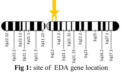
Pathologic allelic variants. More than 60 mutations have been identified in EDA, including nucleotide substitutions (missense, nonsense, and splicing), small deletions and insertions, and gross deletions[19].
Fig 1: site of EDA gene location
EDAR
ormal allelic variants. The human EDAR gene as 12 exons. EDAR
is homologous to the mouse downless gene.
ormal gene product. EDAR encodes a 448-amino acid protein that contains a single transmembrane domain with type 1 membrane topology. The protein probably functions as a multimeric receptor and is related to the TNFR family. It forms a ligand-receptor pair with ectodysplasin.
Pathologic allelic variants. Several mutations have been identified in the EDAR gene, including deletions and transitions [21-24]. Those responsible for autosomal recessive HED exhibit loss of function, while those responsible for autosomal dominant HED exhibit a dominant negative effect [25]. At least two of the dominant negative mutations are not associated with the HED phenotype.
Abnormal gene product. The defective proteins encoded by mutations in EDAR are unable to bind with ectodysplasin.
Fig :2 site of EDAR gene location
EDARADD
Toll/interleukin receptor signaling [Headon et al 2001]. It also contains a Traf-binding consensus sequence. It is coexpressed with tumor necrosis factor receptor superfamily member EDAR in epithelial cells during the formation of hair follicles and teeth. It interacts with the death domain of EDAR and links the receptor to signaling pathways downstream.
Pathologic allelic variants. A transition at nucleotide 424 of the
EDARADD gene, leading to a glutamine-to-lysine (p.Glu142Lys) amino acid substitution in the encoded protein, has been identified in an inbred family with autosomal recessive HED [26]. Another family with autosomal dominant HED has been found to have a heterozygous c.335T>G mutation in the EDARADD gene, indicating that both recessive and dominant forms of HED can be caused by
EDARADD mutations [27].
Abnormal gene product. The EDARADD mutation alters the charge of an amino acid in the resultant gene, rendering it incapable of performing its function.
Fig 3: site of location of EDARADD gene
Table 1: Hypohidrotic Ectodermal Dysplasia: Genes and Databases
Gene Symbol Chromosomal Locus Protein Name
EDA Xq12-q13.1 Ectodysplasin-A
EDAR 2q11-q13 Tumor necrosis factor receptor superfamily member EDAR
EDARADD 1q42.2-q43 Ectodysplasin-A receptor-associated adapter protein
Pathological changes:
Ectodermal dysplasia results from the abnormal morphogenesis of cutaneous or oral embryonal ectoderm (ie, hair, nails, teeth, eccrine glands). Note the following:
• Hair defects: A reduction in the number of hair follicles in conjunction with structural hair shaft abnormalities may be seen. Structural hair shaft abnormalities may result from aberrations in hair bulb formation and include longitudinal grooving, hair shaft torsion, and cuticle ruffling. Hair bulbs may be distorted, bifid, or small.
• Eccrine defects: Eccrine sweat glands may be absent or sparse and rudimentary, particularly in patients with hypohidrotic ectodermal dysplasia [28,29].
• Other secretory gland defects: Hypoplasia of the salivary and lacrimal glands may occur. In some patients, mucous glands may be absent in the upper respiratory tract and in the bronchi, esophagus, and duodenum.
• Dental defects: Abnormal morphogenesis or absence of teeth may occur [30].
• Nail dystrophy: Abnormal nail plate formation may result in brittle, thin, ridged, or grossly deformed nails.
Clinical features
EDA is characterized by the triad of signs comprising sparse hair (atrichosis or hypotrichosis), abnormal or missing teeth (anodontia or hypodontia) and inability to sweat due to lack of sweat glands (anhidrosis or hypohidrosis). The lack of teeth and the special appearance were reported to be major concerns. Most patients with EDA have a normal life expectancy and normal intelligence. However, the lack of sweat glands may lead to hyperthermia, followed by brain damage or death in early infancy, if unrecognized. Thus an early diagnosis is important. Families with EDA should therefore be offered genetic counseling.
Craniofacial structures
cranial bones of all affected patients is such that the resemblance among the patients is bigger than when compared with their own unaffected sib [31].
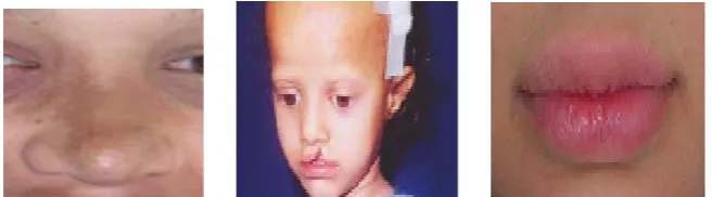
Clinically, the forehead appears square,(fig 2) with frontal bossing, and there is a prominent supra-orbital ridge. The nose has a depressed nasal bridge and is called a saddle nose.(fig 1) The midface is depressed and hypoplastic, giving it a “dished-in” appearance. The cheekbones are high and broad, although they appear flat and depressed as well. The chin may be pointed and the lips everted and protuberant [32]. (fig 3)
In non-treated patients with EDA, craniofacial deviations from the norm increased with advancing age [33] with a tendency toward a Class III pattern with anterior growth rotation [34]. Cephalometric analysis and anthropometry studies have been performed. The quantitative findings show reduced facial dimensions, decreased lower facial height, variable pattern in facial widths, the maxilla has been relatively more retruded than the mandible, the nasal alar width and mouth width were significantly smaller [35].
This remarkable variability in facial dimensions and harmony found in patients with ED probably corresponds to the different kinds of dysplasia, with different expression of the interested genes [36].
Fig 3:abnormality in nose Fig 4:abnormality in forehead Fig 5:abnormality in lips
Oral structures
to exclude EDA. The screening limit for the first tooth to erupt is 15 months. Besides the delay in teething, the teeth appear radiagraphically abnormal in shape and structure.
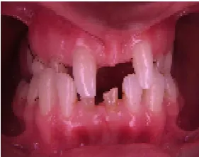
The enamel layer is thin and the cervical area of the tooth is constricted. Enamel is rarely hypoplastic. If at that stage aplasia of several teeth is seen, the patient should be referred to a geneticist in a pediatric unit with a suspescion of EDA diagnosis. Tooth crowns are small and abnormal in shape. Upper incisors and cuspids are always conical or pointed.
Taurodontism, frequently on the second deciduous molars, is a common feature .Not only the shape is abnormal, but also the number. A severe hypodontia is a universal feature amongst affected individuals. All lacked some deciduous teeth and permanent teeth. The number varies from four to twenty. A few patients have congenital anodontia. There are generally more teeth in the maxilla than in the mandible, although both jaws can be toothless [38]. Most often the lower incisors and premolars are missing, followed by the upper premolars and incisors. The upper cuspids and first upper and lower molars are formed.
The edentulous EDA patients do not have any alveolar processes either [39]. In those patients with some natural teeth, there is a striking difference in the intra-oral height and breadth of the bone.
In areas where no teeth have developed, the alveolar bone is missing and the bone ridge is very thin in contrast to the normal alveolus surrounding an occasional tooth.
Fig 6: abnormality in teeth Fig 7: abnormality in teeth
Hair, nails, skin and skintags
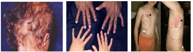
Abnormalities of hair are present in all affected individuals. Most individuals have sparse, fine, slowly growing scalp hair (fig 6). Some individuals are completely bald by their middle teens, whereas other individuals have normal amounts of scalp hair, though it may exhibit an abnormal texture. Sparse eyebrows and eyelashes were always found. Most individuals show decreased body hair, pubic hair, and/or axillary hair, but these features are more variable. However, beard and moustache hair are normal. Electron microscopy of hairs from affected and unaffected individuals showed no abnormalitites.
About half of the affected individuals exhibit mild fingernail abnormalities and nail dystrophy. Slow nail growth and split nails are most often reported. A few individuals had a longitudinal ridging, thinning and superficial peeling. Nail problems occur more frequently in older individuals. This suggests that the nail beds are more susceptible to progressive injury with age. Toenails were generally normal (fig 7).
Most individuals report dry skin. Affected individuals have a smooth, almost velvety skin texture. Theskin of patients also seems to be “thinner” than expected for age. Some infants may have a premature look because of the thin skin. Scaling in the neonate may form a clue to diagnosis(fig 8).
areas on their body. Common sites of sweating include palms, soles and axillae. Because of the reduced number of sweat glands, there is a danger of hyperthermia. In this way EDA has been associated with sudden infant death. The hyperthermia may also lead to brain damage, and is probably the cause of the rare cases of EDA reported with mental retardation. Subcutaneous fat is often diminished and over one third of the boys have abnormalities of the breast, including absent or accessory nipples [40]. Episodes of hyperpyrexia and severe respiratory infections are life-threatening components in EDA. The delay in teething often leads to the diagnosis in childhood. After the first critical years of life the patients experience a general improvement in health. The lack of teeth is the most important factor in determining the quality of life in these patients, particularly in later life. They all suffer greatly from their abnormal facial and dental appearance.
Fig 8: abnormality in hair Fig 9: abnormality in nails Fig 10: abnormality in skin
Clinical diagnosis
Hypohidrotic ectodermal dysplasia (HED) can be diagnosed after infancy in most affected individuals by the presence of three cardinal features:
• Hypotrichosis (sparseness of scalp and body hair). In addition, the scalp hair has thin shafts and is lightly pigmented. Note: Although hair shafts can be brittle and twisted (pili torti) or have other anomalies on microscopic analysis, these findings are not sufficiently sensitive to be of diagnostic benefit [41]. Secondary sexual hair (beard and pubic hair) is normal.
• The function of sweat glands may be assessed by bringing the skin into contact with an iodine solution and raising ambient temperatures to induce sweating. The iodine solution turns color when exposed to sweat and can be used to determine the amount and location of sweating.
• The number and distribution of sweat pores can be determined by coating parts of the body (usually the hypothenar eminences of the palms) with impression materials commonly used by dentists.
• While skin biopsies have been used to determine the distribution and morphology of sweat glands, noninvasive techniques are equally effective.
• Hypodontia (congenital absence of teeth):
• An average of nine permanent teeth develop, typically the canines and first molars [42].
• Teeth are smaller than average and often have conical crowns.
• Dental radiographs are essential to determine the extent of hypodontia and are useful in the diagnosis of mildly affected individuals.
Note: Anthropometric variations (measurements of facial form and tooth size) in HED are subtle and have not proven clinically useful.
Carrier detection for X-linked HED
• Because carriers for X-linked HED show mosaic patterns of sweat pore function and distribution, use of an iodine solution to assess sweat gland function or impression materials to assess number and distribution of sweat pores is particularly useful.
• Carriers frequently will also display some degree of hypodontia [43].
Molecular Genetic Testing Genes
• EDA is the only gene known to be associated with X-linked HED. Ninety-five percent of individuals with HED have the X-linked form.
Clinical testing: • Sequence analysis
• EDA. In males with X-linked HED, direct sequencing of the eight exons with flanking intron sequences of EDA identifies approximately 95% of mutations, including missense and nonsense mutations and smaller deletions.
• EDAR. Sequence analysis of the EDAR coding region is available on a clinical basis.
• EDARADD. Sequence analysis of the EDARADD coding and flanking intronic regions is available on a clinical basis.
• Duplication/deletion testing. Sequence analysis of EDA cannot detect exonic, multiexonic or whole gene deletions in females, and additional testing using methods that detect deletions is required.
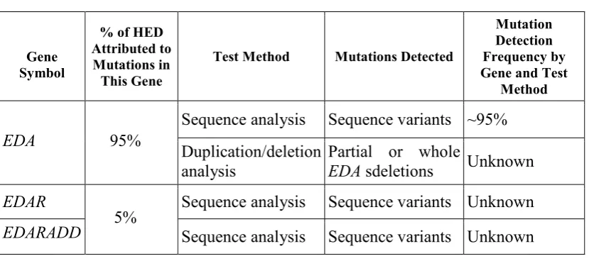
Table 1. Summary of Molecular Genetic Testing Used in Hypohidrotic Ectodermal Dysplasia
Gene Symbol
% of HED Attributed to Mutations in This Gene
Test Method Mutations Detected
Mutation Detection Frequency by Gene and Test
Method
Sequence analysis Sequence variants ~95%
EDA 95%
Duplication/deletion analysis
Partial or whole
EDA sdeletions Unknown
EDAR Sequence analysis Sequence variants Unknown
EDARADD 5% Sequence analysis Sequence variants Unknown
1. For males affected with X-linked HED, mutations detected include intragenic deletions, since lack of amplification by PCR prior to sequence analysis can suggest a putative exonic or whole gene deletion on the X chromosome in affected males. Confirmation requires deletion analysis.
3. Deletion analysis is used to detect exonic deletions in females and to confirm exonic deletions in affected males
4. Testing that detects deletions/duplications not readily detectable by sequence analysis of genomic DNA; a variety of methods including quantitative PCR, real-time PCR, multiplex ligation-dependent probe amplification (MLPA), or array GH may be used.
Testing Strategy:
Confirming the diagnosis in a proband:
• If the proband's findings are classic and are consistent with X-linked inheritance (i.e., males generally more severely affected than females, no male-to-male transmission), initial testing should be for EDA mutations:
o If the affected individual is male, sequence analysis is
sufficient as it detects both sequence variants and deletions.
o If the affected individual is female, sequence analysis
should be performed first; if no mutation is identified, deletion testing should be performed next.
• If the proband's findings are classic and consistent with autosomal recessive inheritance, or mild and consistent with autosomal dominant inheritance, testing should be done for EDAR
and EDARADD mutations.
Carrier testing for relatives at risk for X-linked HED or autosomal recessive HED requires prior identification of the disease-causing mutation(s) in the family.
Prenatal Diagnosis
The use of linked markers on DNA from chorionic villi has greatly improved the safety of prenatal diagnosis of X-linked EDA. This new method of prenatal diagnosis has major advantages as well as disadvantages. It permits the diagnosis to be made in the first trimester of pregnancy prior to the development of the affected structures, thereby allowing an early determination of an affected pregnancy. It is technically simpler and may present a lower risk to the pregnancy than the fetoscopy and multiple skin biopsies. Disadvantages to a linkage based indirect analysis include the need for the sampling of previously affected individuals. The counseling of families is more complex since one is dealing with the probabilities of an affected fetus, rather than a more definitive diagnosis based on direct observation. However, these statistical concepts are difficult for many families to comprehend fully.
The identification of mutations in the family will further improve the accuracy of prenatal diagnosis [44]. However, EDA is a disorder which in most cases is associated with a normal life expectancy and a normal intelligence. Prenatal diagnosis will therefore probably not be an option in most families with EDA.
The diagnostic procedure in ectodermal dysplasia patient is explained by showing a case report.
Case report
A male newborn with 10 days of life from the city of Caxias do Sul, state of Rio Grande do Sul, southern Brazil, was admitted to the Neonatal ICU of the Teaching Hospital at the Universidade de Caxias do Sul (UCS).
On August 20th, 1998. Patient presented with history of episodes of persistent hyperthermia since the first days of life. At physical examination, ES presented with dry mucous membranes, dry and desquamative skin, hyperthermia (39ºC), and umbilical granuloma.
omphalitis, which also presented negative. We started empiric therapy with oxacillin and gentamicin despite patient presenting normal for the examinations carried out.
On August 25th, 1998 patient peak temperatures persisted and the above examinations were carried out for a second time; once more, results were negative. We replaced oxacillin and gentamicin with vancomicin, amikacin, and cefotaxime.
On the 14th day of hospital stay the patient still did not present any improvement of clinical status. Consequently, we carried out exams to rule out HIV and congenital neonatal infection (STORCH). Again, patient presented negative for both exams. He also presented normal for ultra sonography examination of the abdomen.
Only on September 9th of that same year we suspected that the patient had anhidrotic ectodermal dysplasia. Biopsy of a specimen taken from the dorsum of the patient and histopathological report confirmed the diagnosis. Specimen submitted to anatomopathological exam indicated absence of eccrine and sebaceous gland structures and hypoplasia of follicular structures.
Patient was discharged from the hospital on September 12th, 1998. The mother was instructed regarding procedures for control of temperature and use of emollient for dry skin.
One year later, the clinical signs of anhidrotic ectodermal dysplasia were more evident. At physical examination, patient presented typical facies with frontal boss, small nose, lip protrusion, erythematous malar region rash, and rhinorrhea. Patient also presented hypotrichosis; thin and sparse hair; depigmentation of the hair; and sparse eyebrow hair. The skin was dry, pale, thin, and with protruding vessels.
relation to the mandible, we only found tooth germs of the first molar teeth (partial anodontia). Currently, the patient is being followed-up at the outpatient Dermatology and Pediatric clinic of the Teaching Hospital at UCS [45].
Treatment
There is no specific treatment for this disorder.
Medical Care
The care of affected patients depends on which ectodermal structures are involved. Note the following:
• For patients with anhidrosis/hypohidrosis, advise air conditioning for home, school, and work. Encourage frequent consumption of cool liquids to maintain adequate hydration and thermoregulation. Finally, advise patients to wear cool clothing. • For patients with dental defects, advise early dental
evaluation and intervention and encourage routine dental hygiene. Dentures may be indicated as early as age 2 years. Multiple replacements may be needed as the child grows, and dental implants may eventually be required [46,47,48,49,50]. Advise orthodontic treatment for cosmetic reasons and to ensure adequate nutritional intake.
• Patients with xerosis or eczematous dermatitis may benefit from the use of topical emollients.
• Patients with severe alopecia can wear wigs to improve their appearance.
• Patients with scalp erosions should be treated with topical and systemic antibiotics as needed. General scalp care may involve the use of weekly dilute bleach baths or acetic acid soaks to minimize bacterial colonization of the scalp. Application of special scalp dressings may be helpful.
• Use artificial tears to prevent damage to the cornea in patients with reduced lacrimation.
• Protect nasal mucosa with saline sprays followed by the application of petrolatum.
• Allogeneic stem cell transplantation has been performed in a small number of patients with autosomal dominant ectodermal dysplasia with immunodeficiency (EDA-ID); poor engraftment and post-transplant complications were common [51, 52].
Surgical Care
Early repair of cleft lip or palate may lessen facial deformities and improve speech. Other mid facial defects or hand/foot deformities may be surgically corrected in order to improve function and reduce physical disfigurement.
Conclusion
Ectodermal dysplasia is a genetic disorder. There is no pharmacological treatment for ectodermal dysplasia it is necessary to develop the researches for the development of drug therapy for ectodermal dysplasia. This review represents that it is mandatory to develop the awareness to the research scientists for developing the drug for ED.
References
Study of 15 Cases,” Archives of Medical Research, ; 37, 403-409.
2. Yavuz, I., S. Kıralp ve Z. Başkan, “Hypohyrotic Ectodermal Dysplasia: A Case Report,” Qinttessence International, 2008; 39, 81-86.
3. Shah KN, Mckinster CD. Ectodermal Dysplasia. eMedicine
Article last updated: Nov 8, 2006.
4. Rook A, Wilkinson DS, Ebling FJG. Textbook of dermatology. In: Harper JI, ed. 6th ed. Great Britain: By Champion; 1998.p. 391-5.
5. Kirtikant CS, Dipak DU. Unusual cutaneous manifestations of anhidrotic ectodermal dysplasia. J Dermat 1990; 17: 380-4. 6. Lamartine J. Towards a new classification of ectodermal
dysplasia. Clinical & Experimental Dermatology 2003; 28: 351-354.
7. Priolo M, Silengo M, Lerone M, Ravazzolo R. Ectodermal dysplasias: not only ‘skin’ deep. Clin Genet 2000; 58: 415-430. 8. Geza T, William S, Samir A. Ectodermal dysplasia. Qintessence
Int 2003; 34: 482-483.
9. Buyse M. Birth Defects Encyclopedia, Blackwell Scientific Publications: Inc, USA. 1990; Volume I.
10.Priolo M., Lagana C. Ectodermal dysplasias: a new clinical-genetic classification. J.Med. Genet. 2001; 38: 579-585.
11.Pinheiro M, Freire-Maia N. The ectodermal dysplasias. Arch Dermatol. Apr 1982; 118(4):215-6.
12.Pinheiro M, Freire-Maia N. Ectodermal dysplasias: a clinical classification and a causal review. Am J Med Genet. Nov 1 1994;53(2):153-62.
13.Freire-Maia N, Lisboa-Costa T, Pagnan NA. Ectodermal dysplasias:how many? Am J Med Genet. Nov 15 2001; 104(1):84.
14.Lamartine J. Towards a new classification of ectodermal dysplasias. Clin Exp Dermatol. Jul 2003; 28(4):351-5.
15.Priolo M, Lagana C. Ectodermal dysplasias: a new clinical-genetic classification. J Med Genet. Sep 2001; 38(9):579-85. 16.Itin PH, Fistarol SK. Ectodermal dysplasias. Am J Med Genet C
Semin Med Genet. Nov 15 2004; 131C(1):45-51.
deletion mutations in collagenous repeats. Hum Mol Genet. 1998;7: 1661–9.
18.Ezer S, Bayes M, Elomaa O, Schlessinger D, Kere J. Ectodysplasin is a collagenous trimeric type II membrane protein with a tumor necrosis factor-like domain and co-localizes with cytoskeletal structures at lateral and apical surfaces of cells. Hum Mol Genet. 1999; 8:2079–86.
19.Visinoni AF, de Souza RL, Freire-Maia N, Gollop TR, Chautard-Freire-Maia EA. X-linked hypohidrotic ectodermal dysplasia mutations in Brazilian families. Am J Med Genet A. 2003; 122A:51–5.
20.Chen Y, Molloy SS, Thomas L, Gambee J, Bachinger HP, Ferguson B, Zonana J, Thomas G, Morris NP. Mutations within a furin consensus sequence block proteolytic release of ectodysplasin-A and cause X-linked hypohidrotic ectodermal dysplasia. Proc atl Acad Sci U S A. 2001; 98:7218–23.
21.Shimomura Y, Sato N, Miyashita A, Hashimoto T, Ito M, Kuwano R. A rare case of hypohidrotic ectodermal dysplasia caused by compound heterozygous mutations in the EDAR gene.
J Invest Dermatol. 2004; 123:649–55.
22.Chassaing N, Bourthoumieu S, Cossee M, Calvas P, Vincent MC. Mutations in EDAR account for one-quarter of non-ED1-related hypohidrotic ectodermal dysplasia. Hum Mutat. 2006; 27:255–9.
23.Mégarbané H, Cluzeau C, Bodemer C, Fraïtag S, Chababi-Atallah M, Mégarbané A, Smahi A. Unusual presentation of a severe autosomal recessive anhydrotic ectodermal dysplasia with a novel mutation in the EDAR gene. Am J Med Genet A. 2008; 146A:2657–62.
24.Van der Hout AH, Oudesluijs GG, Venema A, Verheij JB, Mol BG, Rump P, Brunner HG, Vos YJ, van Essen AJ. Mutation screening of the Ectodysplasin-A receptor gene EDAR in hypohidrotic ectodermal dysplasia. Eur J Hum Genet. 2008; 16:673–9.
25.Valcuende-Cavero F, Martinez F, Pérez-Pastor G, Oltra S, Ferrer I, Tomás-Cabedo G, Moreno-Presmanes M. Autosomal-dominant hypohidrotic ectodermal dysplasia caused by a novel mutation. J Eur Acad Dermatol Venereol. 2008; 22:1508–10. 26.Headon DJ, Emmal SA, Ferguson BM, Tucker AS, Justice MJ,
dysplasia implicates a death domain adapter in development.
ature. 2001; 414:913–6.
27.Bal E, Baala L, Cluzeau C, El Kerch F, Ouldim K, Hadj-Rabia S, Bodemer C, Munnich A, Courtois G, Sefiani A, Smahi A. Autosomal dominant anhidrotic ectodermal dysplasias at the EDARADD locus. Hum Mutat. 2007; 28:703–9.
28.Rouse C, Siegfried E, Breer W, Nahass G. Hair and sweat glands in families with hypohidrotic ectodermal dysplasia: further characterization. Arch Dermatol. Jul 2004;140(7):850-5.
29.Berg D, Weingold DH, Abson KG, Olsen EA. Sweating in
ectodermal dysplasia syndromes. A review. Arch
Dermatol. Aug 1990; 126(8):1075-9.
30. Clauss F, Maniere MC, Obry F, et al. Dento-craniofacial phenotypes and underlying molecular mechanisms in hypohidrotic ectodermal dysplasia (HED): a review. J Dent Res. Dec 2008; 87(12):1089-99.
31.Bergendal B., Koch G., Kurol J., Wänndahl G.: Consensus conference on ectodermal dysplasia with special reference to dental treatment. The Institute for Postgraduate Dental Education, Jönköping, Sweden (1998).
32. Johnson E.R., Roberts M.W., Guckes A.D., Bailey L.J., Phillips C.L., Wright J.T.: Analysis of craniofacial development in children with hypohidrotic ectodermal dysplasia. Am. J. Med. Genet. (2002) 112: 327-34.
33. Clarke A., Burn J.: Sweat testing to identify female carriers of X-linked hypohidrotic ectodermal dysplasia. J. Med. Gen. (1991) 28: 330-333.
34. Bondarets N., Jones R.M., McDonald F.: Analysis of facial growth in subjects with syndromic ectodermal dysplasia: a longitudinal analysis. Orthod. Craniofac. Res. (2002) 5: 71-84. 35.Sforza C., Dellavia C., Vizzotto L., Ferrario V.F.: Variations in
facial soft tissues of Italian individuals with ectodermal dysplasia. Cleft Palate Craniofac. J. (2003) 41(3): 62-67.
36. Ruhin B., Martinot V., Lafforgue P., Catteau B., Manouvrier-Hanu S., Ferri J.: Pure ectodermal dysplasia: retrospective study of 16 cases and literature review. Cleft Palate Craniofac. J. (2001) 38: 504-18.
38. Aswegan A.L., Josephson K.D., Mowbray R., Pauli R.M., Spritz R.A., Williams M.S.: Autosomal dominant hypohydrotic ectodermal dysplasia in a large family. Am. J. Med. Genet. (1997) 72: 462-467.
39.Soderholm A.L., Kaitila I.: Expression of X-linked hypohidrotic ectodermal dysplasia in six males and in their mothers. Clin. Gen. (1985) 28: 136-144.
40. Goodship J., Malcolm S., Clarke A., Pembrey M.E.: Possible genetic heterogeneity in X-linked hypohidrotic ectodermal dysplasia. J. Med. Genet. (1990) 27: 422-425.
41.Rouse C, Siegfried E, Breer W, Nahass G. Hair and sweat glands in families with hypohidrotic ectodermal dysplasia: further characterization. Arch Dermatol. 2004; 140:850–5.
42.Lexner MO, Bardow A, Hertz JM, Nielsen LA, Kreiborg S. Anomalies of tooth formation in hypohidrotic ectodermal dysplasia. Int J Paediatr Dent. 2007; 17:10–8.
43.Cambiaghi S, Restano L, Paakkonen K, Caputo R, Kere J. Clinical findings in mosaic carriers of hypohidrotic ectodermal dysplasia. Arch Dermatol. 2000; 136:217–24.
44. Kere J., Srivastava A.K., Montonen O., et al.: XLinked anhidrotic (hypohidrotic) ectodermal dysplasia is caused by mutation in a novel transmembrane protein. Nature Genetics (1996) 13: 409-416.
45.Breno F. de Araújo, Adelar B. Nora, et al., Anhidrotic ectodermal dysplasia syndrome in the neonatal period - case report, J Pediatr (Rio J) 2001; 77(1): 55-58.
46.Hickey AJ, Vergo TJ. Prosthetic treatments for patients with ectodermal dysplasia. J Prosthet Dent. Oct 2001;86(4):364-8. 47.Imirzalioglu P, Uckan S, Haydar SG. Surgical and prosthodontic
treatment alternatives for children and adolescents with ectodermal dysplasia: a clinical report. J Prosthet Dent. Dec 2002; 88(6):569-72.
48.Dhanrajani PJ, Jiffry AO. Management of ectodermal dysplasia: a literature review. Dent Update. Mar 1998;25(2):73-5.
50.Tarjan I, Gabris K, Rozsa N. Early prosthetic treatment of patients with ectodermal dysplasia: a clinical report. J Prosthet Dent. May 2005; 93(5):419-24.
51.Dupuis-Girod S, Cancrini C, Le Deist F, et al. Successful allogeneic hemopoietic stem cell transplantation in a child who had anhidrotic ectodermal dysplasia with immunodeficiency.
Pediatrics. Jul 2006; 118(1):e205-11.