ATI-IFLAMMATORY AD HEPATOPROTECTIVE ACTIVITIES OF
ETHAOLIC EXTRACT OF
EUPHORBIA THYMIFOLIA
LI.
S. K. Singh*, T. Prabha, B. Kavitha, H. S. Chouhan and S. K. Bharti Pharmaceutical Chemistry Research Lab., Department of Pharmaceutics, Institute of Technology, Banaras Hindu University, Varanasi-221005, India.
Summary
Euphorbia thymifolia Linn. (Euphorbiaceae) is a small prostate, hispidly pubescent, annual weed, which is commonly found in India and tropical countries.The plant is reported to be used in traditional medicine for the treatment of diarrhea, jaundice, pain etc. The aim of the present study was to evaluate the anti-inflammatory and hepatoprotective activities of ethanolic extract of whole plant. The anti-inflammatory effect was evaluated by carrageenan and cotton pellet induced rat models and hepatoprotective activity was accessed on CCl4-induced liver damage
in rats. A dose dependent anti-edematogenic activity of ethanolic extract of E. thymifolia (dose of 200 mg/kg, p.o.) was observed and was comparable to Ibuprofen (dose of 50 mg/kg, p.o.; p < 0.01) used as standard. The extract of the plant, screened for its hepatoprotective activity in CCl4 (0.5 ml/kg, p.o.) induced
liver damage in rats at a dose of 100 mg/kg, p.o. The extract significantly (P < 0.01) normalized the serum enzymes alanine amino transferase (ALT), asparate amino transferase (AST), and lipid peroxidation (LPO) elevated by CCl4-toxicity, which
was also supported by the histological examination of liver tissues. The hepatoprotective effect of the extract was comparable to that of silymarin (50 mg/kg, p.o.) used as reference standard. The preliminary phytochemical screening of the ethanolic extract showed the presence of phenolics, terpenes and flavonoids. Thus, the results validate the use of E. thymifolia in traditional medicines for the treatment of jaundice and inflammation related disorders.
Keywords: Anti-inflammatory, Euphorbia thymifolia, Euphorbiaceae, Hepatoprotective
*
Corresponding Author:
Dr. Sushil K. Singh, Professor,
Department of Pharmaceutics, Institute of Technology, Banaras Hindu University, Varanasi-221005.
Tel.: +915426702736 Fax: +91542368428.
Introduction
Euphorbia thymifolia Linn. (Euphorbiaceae), also known as Chamaesyce thymifolia L., is a small prostate, hispidly pubescent, annual weed with horizontally spreading branches occurring in red and green forms. It is widely distributed in India and throughout tropics. This plant is reported to have antibacterial [1-2], anti-fungal [3-4], antioxidant [5-6], antiviral [5,7-8]. It is also used for astringent actions, as stimulant, in worm infection [9], pain and jaundice [10-11]. The presence of polyphenolics [12], flavonoids [7] and alkaloids [1] has been reported from Euphorbia thymifolia Linn. The plants of genus Euphorbia contain diversified classes of compounds in both free and conjugated form viz. tannins, phenolics [13-14], flavonoid glycosides [15], terpenes [16-19] and steroids [20]. Plants of genus Euphorbia are reported to be used as purgative, anthelmintic, antianaphylactic [21] anti-arthritic [22], antidiarrhoeal [23], anti-inflammatory [24-25], in warts treatment, antiasthmatic [26], astringent, narcotic, diuretic [27], and immunosuppressive properties [28]. The genus Euphorbia is also reported for cytotoxic activity [16, 18, 29], antinociceptive activity [30], antidepressant [17] and antioxidant activities [5-6, 31]. Oxidation processes are important for living organisms, the reduced oxygen species mediate important physiological processes and may induce cellular damage including hepatotoxicity. A great deal of interest is being generated towards the bioactive polyphenolics from natural source to be used as dietary source of anti-oxidants. In the present study, the phenolics rich ethanolic extract of E. thymifolia was used to evaluate its anti-inflammatory and hepatoprotective activities, which was not reported earlier.
Materials and methods
Plant material, extraction and isolation
The whole plants of Euphorbia thymifolia were collected from Tamil Nadu and were authenticated by Prof. K.N. Dubey (Department of Botany, Banaras Hindu University, Varanasi, India). A voucher specimen (specimen No. PCRL 36) has been deposited in the Pharmacutical Chemistry Research Laboratory, Department of Pharmaceutics, Institute of Technology, Banaras Hindu University, Varanasi, India, for future reference. The shade-dried powder of whole plant was passed through sieve No. 40 & 600 g was extracted (soxhlation) with petroleum ether (60-80ºC) for 24 h and subsequently with ethanol (30 h). The extractives were concentrated in vacuo (total yield 11 % w/w). Qualitative determination of ethanolic extract was done for the presence of tannins, flavonoids, sterols, terpenes and alkaloids using standard methods [32].
Animals
Male albino rats (Charles foster strain) weighing 100-140 g were obtained from M/S Asian Fauna Store, Varanasi. Animals were randomly housed in groups of five in polypropylene cages at an ambient temperature with a 12 h light: 12 h dark cycle. The animals were allowed free access to laboratory diet (M/s Hindustan Lever Ltd., Mumbai, India) and water ad libitum. The animals were fasted overnight before the experiment. Experiments were performed in accordance with the current guidelines for the care of laboratory animals and the ethical guidelines for the investigation of experimental pain in conscious animals [33].
Reagents and Chemicals
Lambda-carrageenan (Sigma, Type IV, Steinheim, Germany) and carbon tetrachloride (CCl4) were
purchased from Merck India Ltd, Mumbai. Silymarin was obtained from Ranbaxy Laboratories Limited, India. Span diagnostic kits were used for the spectrometric enzyme assays. All other chemicals used were of analytical grade.
Acute toxicity studies
were observed for 24 h for any signs of acute toxicity such as increased/decreased motor activity, tremors, convulsion, sedation, lacrimation etc. No mortality of the animals was observed even after 24 h. Hence, the LD50 cut off value of the test compounds was fixed as 2000 mg/kg and
1/10th of cut off value (i.e. 200 mg/kg) was taken as maximum screening dose for the evaluation of anti-inflammatory and hepatoprotective activity.
Anti-inflammatory activity
Carageenan- induced hind paw edema in rats
The inhibitory activity of the ethanolic extract of E. thymifolia on carrageenan-induced rat paw oedema was determined according to the method of Winter et al.[34]. The experimental animals were divided into five groups, each containing five animals. Group-I was marked as control (received an equal volume of distilled water), group-II as standard (received Ibuprofen, 50 mg/kg, p.o.) and groups III-V were given ethanolic extract of 50, 100 & 200 mg/kg p.o. respectively. After 30 min., all the groups were injected with carrageenan (0.1 ml, 1% w/v in normal saline) subcutaneously in the subplanter region of the left hind paw. The paw volume was measured by the difference of volume with mercury plethysmometer before and 1, 2 and 3 h after carrageenin injection. Percentage of inhibition of edema was determined for each animal by comparison with control and calculated by the following formula [35]:
% Inhibition = 1- dt dc
× 100
where dt is the difference in paw volume in the drug-treated group and dc the difference in paw volume in the control group.
Cotton pellet induced Granuloma
The method of Mossa et al. [36] was used for this study. This involves surgical insertion of sterilized cotton pellet (30 mg in weight) subcutaneously into the groin of rats using ether as an anaesthetic agent. Five groups of five rats in each group were included in the study. After shaving off fur, the animals were anaesthetized and administered the same doses of extracts, vehicle or Ibuprofen as in the carrageenan-induced rat paw oedema test.
After the surgical insertion, sterilized cotton pellet (30 mg) was implanted in the groin region of each rat. The extracts (50, 100 or 200 mg/kg), vehicle and Ibuprofen (50 mg/kg) were administered to respective groups of the animals for seven consecutive days. All the animals were sacrificed on the eighth day with an over dose of ether. The pellet and the surrounding granuloma were dissected out carefully, made free from extraneous tissues and dried overnight in an oven at 60ºC to a constant weight. The weight of the granuloma tissue was obtained
by determining the difference between the initial (30 mg) and the final weight of the cotton pellet with its attached granulomatous tissue. The mean weight of the granuloma tissue formed in each group and the percentage inhibition were determined.
Carbon tetrachloride-induced liver damage
The rats were randomly divided into four groups of five animals each. Group I was marked as control and given daily single dose of 1% of polyethyleneglycol (PEG) at a dose of 2 mL/kg, p.o. Group II -intoxicated with a single dose of CCl4 (CCl4: PEG in 1:1 ratio; 0.5 mL/kg, p.o.). Group
III & IV received a daily single dose of silymarin (50 mg/kg in 1% of PEG, p.o.) and ethanolic extract in 1% PEG (100 mg/kg, p.o.) for 7 consecutive days respectively. Group III served as standard. All animals except control received CCl4 on fifth day. Rats were sacrificed after 24 h of
CCl4 administration and blood was collected by cardiac puncture and was centrifuged at 2500 rpm
Histopathology
Histopathological analysis was carried out by fixing liver tissue in 10% buffered formalin saline, processed, embedded in paraffin wax to cut into 3 µm using microtome, and then stained with both haematoxylin and eosin. Observations were made under 400x magnification in light field microscope.
Statistical analysis
The mean ± standard error of the mean (S.E.M.) was determined for each parameter. The data was subjected to one-way analysis of variance (ANOVA) followed by Dunnett’s t-test. The results were considered significant if p < 0.05.
Results
Preliminary phytochemical analysis showed the presence of tannins, flavonoids, and terpenes in the ethanolic extract of E. thymifolia.
Anti-inflammatory activity:
Carrageenan induced inflammation
In carrageenan induced animal models, the ethanolic extract of E. thymifolia at the doses of 50, 100, and 200 mg/kg (p.o.), inhibited the edema formation in first hour by 29.14% (p < 0.05), 29.43%, and 45.71% (p < 0.01) respectively in a dose dependent manner. The effect also extended and significantly increased in second and third hour (p < 0.05). The reference drug (Ibuprofen) group significantly inhibited the edema formation by 59.71%, 67.57% and 68.92% at first, second and third hour respectively (p < 0.05). The inhibition of paw edema produced by ethanolic extract (200 mg/kg, p.o.) in third hour was found to be comparable to that of Ibuprofen (50 mg/kg, p.o.; p < 0.01). Results are shown in Table 1.
Table1. Effects of ethanolic extract of E. thymifolia on carrageenan- induced edema in rats.
Mean increase in paw volume (mL) at time T (h) Groups Dose (mg/kg,
p.o.) T(1) T (2) T (3)
Control - 0.700 + 0.026 (-) 0.740 + 0.023(-) 0.740 + 0.023(-)
Ibuprofen 50 0.282 ± 0.127***
(59.71)
0.240 ± 0.094** (67.57)
0.230 + 0.027** (68.92)
50 0.496 ±
0.065*(29.14)
0.490 ± 0.098*(33.78)
0.460 + 0.083* (37.84) 100 0.494 ± 0.063**
(29.43)
0.462 ± 0.061** (37.57)
0.390 + 0.062** (47.30) Ethanolic
extract
200 0.380 ± 0.163** (45.71)
0.340 ± 0.126** (54.05)
0.310 + 0.049** (58.11) Values were expressed as mean ± S.E.M. (n= 5). *p < 0.05, **p < 0.01 are compared with the control group (ANOVA followed Dunnett’s t-test.). Values given in parentheses represent Percentage of Inhibition.
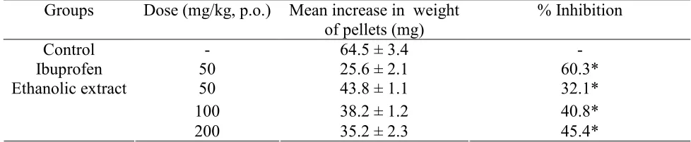
Cotton pellet induced granuloma
Table 2. Effects of ethanolic extract of E. thymifolia on cotton pellet induced granuloma in rats
Values were expressed as mean ± S.E.M. (n= 5).*p < 0. 05 is compared with the control group (ANOVA followed Dunnett’s t-test).
Hepatoprotective Activity:
The results of hepatoprotective activity of ethanolic extract (100 mg/kg, p.o.) in the CCl4-induced
liver damage are shown in Table 3. The CCl4-intoxication significantly elevated the ALT, AST
and LPO levels in the serum against the control group. The ethanolic extract & silamyrin (standard) treated groups showed significant fall in the elevated ALT, AST and LPO levels.
Table 3. Effect of ethanolic extract of E. thymifolia on the biochemical parameters of CCl4 -intoxicated rats (n = 5, mean ± S.E.M.)
Groups Dose ALT (U/L) AST (U/L) LPO (% Inhibition)
Control 2.0 mL/kg, p.o 31.69 ± 1.13 a, b 23.09 ± 1.01 a, b 18.8 ± 1.96 a, b CCl4 0.5 mL/kg, p.o 160.04 ± 2.0 b, c 129.60 ± 6.42 b, c 85.65 ± 1.64 b, c
Std. + CCl4 50 mg/kg, p.o 11.37 ± 1.92 a, c 75.35 ± 3.03 a, c 41.17 ± 4.86 a, c
Ethanolic extract + CCl4
100 mg/kg, p.o. 131.21 ± 2.14a,b,c 97.14 ± 1.80 a, b, c 31.31 ± 1.90 a, d Where a = p < 0.01 when compared with CCl4 group; b = p < 0.01 when compared with standard,
silymarin; c = p < 0.01 when compared with control group; d = p < 0.05 when compared with control group.
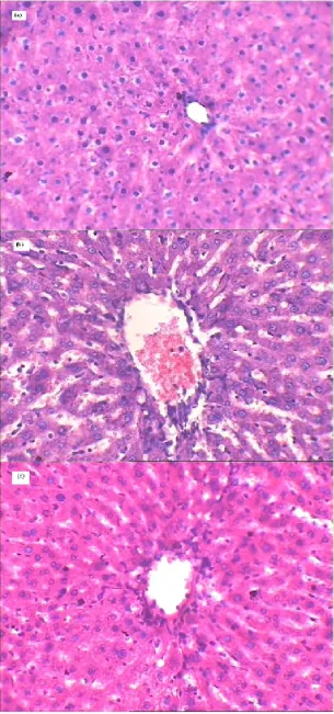
Histopathological examination:
Observations revealed that the CCl4-intoxication brought critical changes in the normal
architecture of rat liver as compared to liver of control group. A massive tissue necrosis, disruption of cytoplasm and cells boundary, and the difference in size of heapatocytes were observed in the CCl4 treated rats. The ethanolic extract treatment normalized these changes in CCl4-induced liver
damage and the effect was comparable to that of the silymarin treated group used as standard. Groups Dose (mg/kg, p.o.) Mean increase in weight
of pellets (mg)
% Inhibition
Control - 64.5 ± 3.4 -
Ibuprofen 50 25.6 ± 2.1 60.3*
50 43.8 ± 1.1 32.1*
100 38.2 ± 1.2 40.8*
Ethanolic extract
Figure 1. Histopathology of (a) normal rat liver (b) CCl4-intoxicated rat liver (c) ethanolic extract
Discussionand conclusion
The study provides a pharmacological basis for the remedial usage of E. thymifolia in inflammation related disorders. Carrageenan induced edema in the hind paw (acute inflammation) and cotton pellet granuloma (chronic inflammation) are widely employed in screening the new antiinflammatory compounds [37]. These studies investigated the antiinflammatory effect of ethanolic extracts of E. thymifolia on acute and chronic phases of inflammation. The carrageenan-induced inflammatory process in the rat involves three phases: an initial, second and third phases caused by the release of histamine and serotonin; bradykinin and prostaglandins respectively [38-39]. In the present study the anti-inflammatory activity of ethanolic extract of E. thymifolia took place at 1, 2, and 3 h after carrageenan injection, suggesting that its action mechanism may involve multiple anti-inflammatory factors and mediators [40]. This is in consistent with the generally believed thinking that herbal preparations usually have multitargets.
The cotton pellet granuloma method has been widely employed to assess the transudative, exudative and proliferative components of chronic inflammation [41]. The dryweight of the implanted cotton pellet correlates well with the amount of granulomatous tissue formed [42]. E. thymifolia reduced the dryweights of implanted cotton pellets, indicating that it may inhibit the proliferative phases of inflammation. In this study, the ethanolic extract of E. thymifolia inhibited the granuloma formation compared to control group. This may be due to the ability of E. thymifolia in reducing the number of fibroblasts and synthesis of collagen and mucopolysaccharide, which are natural proliferative agents of granulation tissue formation.
These studies suggest that the probable anti-inflammatory activity of E. thymifolia may be due to its flavonoids (qurecetin and its derivatives) and hydrolysable tannin constituents (viz. corilagin, 1,2,3,4,6-penta-O-galloyl-β-D-glucose, gerannin), and their effects on these mediators [43-47]. Further, ethanolic extract of E. thymifolia showed ameliorative effect on liver functions, as reflected by histopathological examination and the reduced level of serum enzymes viz. ALT, AST and LPO in CCl4 damaged liver. Carbon tetrachloride causes hepatic damage due to free
radical CCl3•, formed from CCl4 by the activation of the NADPH-Cyt. P450 system of liver
endoplasmic reticulam [48]. This leads to functional and morphological changes in the cell membrane and results in leakage of hepatic enzymes and thus increases blood serum transaminases and phosphatase activity [49-50]. This cellular necrosis is mediated by lipid peroxidation of membrane lipids due to CCl3• radicals and resulting in release of peroxides [51].
Therefore, the hepatoprotection may be due to marked antioxidant and inhibitory lipid peroxidation (LPO) activity of E. thymifolia [5-6] and also strong inhibitory effect of hydrolysable tannins on ADP plus NADPH-induced lipid peroxidation in microsome [52-53].
Acknowledgements
SKB, TP, B K and HSC are thankful to UGC, New Delhi, India, for the award of Research Fellowship.
References
1. Jabbar A, Khan GAMS. Antimicrobial alkaloids from Euphorbia thymifolia. Pak J Sci Ind Res 1965;
8:293.
2. Khan NH, Rahman M, Kamal MS. Antibacterial activity of Euphorbia thymifolia Linn. Indian J Med
Res 1988; 87:395-397.
3. Lal S, Gupta I. Control of sarcoptic mange with Chotidudhi (Euphorbia prostrato Ait. and Euphorbia
thymifolia Linn.) A preliminary report (abstract). Indian J Pharmacol 1970; 2:28.
4. Rao VR, Gupta I. In vitro studies on the antifungal activity of some indigenous drugs against
5. Lin CC, Cheng HY, Yang CM, Lin TC. Antioxidant and antiviral activities of Euphorbia thymifolia L. J Biomed Sci 2002; 9:656-664.
6. Singh SK, Prabha T. Antioxidant activity of ethanolic extract of Euphorbia thymifolia Linn. Indian J
Pharm Sci 2005; 67: 736-738.
7. Amaral ACF, Kuster RM, Goncalves JLS, Wigg MD. Antiviral investigation on the flavonoids of
Chamaesyce thymifolia. Fitoterapia 1999; 70: 293-295.
8. Yang CM, Lin TC, Chiang LC, Lin CC. Euphorbia thymifolia suppresses herpes simplex virus-2
infection by directly inactivating virus infectivity. Clin Exp Pharmacol Physiol 2007; 32: 346-349.
9. Kirtikar KR, Basu BD. Indian medicinal plants. New Delhi, India, Periodical expert Book Agency. pp.
1981; 2199-2210.
10. Khare CP. Indian herbal remedies: rational western therapy, ayurvedic, and other traditional usage,
botany. Springer; Berlin, 2004; 210.
11. Ragupathy S, Steven NG, Maruthakkutti M, Velusamy B, Ul-Huda MM. Consensus of the 'Malasars'
traditional aboriginal knowledge of medicinal plants in the Velliangiri holy hills, India, Journal of Ethnobiology and Ethnomedicine 2008; 4: 8.
12. Lee SH, Tanaka T, Nonaka G, Nishioka I. Hydrolysable tannins from Euphorbia thymifolia.
Phytochemistry 1990; 29: 3621-3625.
13. Yoshida T, Chen L, Shingu T, Okuda T. Tannins and related polyphenols of Euphorbiaceaous plants.
IV. Euphorbins A and B, novel dimeric dehydroellagitannins from Euphorbia hirta L. Chem Pharm
Bull 1988; 36: 2940-2949.
14. Yoshida T, Namba O, Chen L, Okuda T. Euphorbin C, a hydrolysable tannin dimmer of highly
oxidized structure from Euphorbia hirta. Chem Pharm Bull 1990; 38: 1113-1115.
15. Liu Y, Murakami N, Ji H, Abreu P, Zhang S. Antimalarial Flavonol Glycosides from Euphorbia hirta.
Pharmaceutical Biology 2007; 45: 278-281.
16. Fatope MO, Lu Zeng, Ohayaga JE, Guoen Shi, McLaughlin JL. Selectively cytotoxic diterpenes from
Euphorbia poisonii. J Med Chem 1996; 39:1005-1008.
17. Lanhers MC, Fleurentin J, Dorfman P. Neurophysiological effects of Euphorbia hirta L.
(Euphorbiaceae). Phytother Res 1996; 10: 670-676.
18. SmithKielland I, Dornish JM, Malterud KE, Hvistendahl G, Romming C. Cytotoxic triterpenoids from
the leaves of Euphorbia pulcherrima. Planta Med 1996; 62: 322-325.
19. Ahmad VU, Hussain H, Bukhari IA, Hussain J, Jassbi AR, Dar A. Antinociceptive diterpene from
Euphorbia decipiens. Fitoterapia 2005; 76:230-232.
20. Tanaka R, Kasubuchi K, Kita S, Matsunaga S. Obtusifoliol and related steroids from the whole herb of
Euphorbia chamaesyce. Phytochemistry 1999; 51:457-463.
21. Youssouf MS, Kaiser P, Tahir M, et al. Anti-anaphylactic effect of Euphorbia hirta.. Fitoterapia 2007;
78:535-539.
22. Bani S, Kaul A, Khan B, et al. Anti-arthritic activity of a biopolymeric fraction from Euphorbia
tirucall. J. Ethnopharmacol. 2007; 110:92-98.
23. Hore SK, Ahuja V, Mehta G, et al. Effect of aqueous Euphorbia hirta leaf extract on gastrointestinal
motility. Fitoterapia 2006; 77:35-38.
24. Singla AK, Pathak K. Topical anti-inflammatory effects of Euphorbia prostrata on
carrageenan-induced footpad oedema in mice. J. Ethnopharmacol.1990; 29:291-294.
25. Bani S, Kaul A, Jaggi BS, Suri KA, Suri OP, Sharma OP. Anti-inflammatory activity of the
hydrosoluble fraction of Euphorbia royleana latex. Fitoterapia 2000; 71:655-662.
26. Chabia M, Michel VF, Frossard N, et al. Anti-proliferative effect of Euphorbia stenoclada in human
airway smooth muscle cells in culture. J. Ethnopharmacol. 2007; 109:134-139.
27. Johnson PB, Abdurahman EM, Tiam EA, Abdu-Aguye I, Hussaini IM. Euphorbia hirta leaf extracts
increase urine output and electrolytes in rats. J. Ethnopharmacol. 1999; 65:63–69.
28. Bani S, Kaul A, Khan B, et al. Immunosuppressive properties of an ethyl acetate fraction from
Euphorbia royleana. J. Ethnopharmacol. 2005; 99:185-192.
29. Whelan LC, Ryan MF. Ethanolic extracts of Euphorbia and other ethnobotanical species as inhibitors
of human tumour cell growth. Phytomedicine 2003; 10:53-58.
30. Vamsidhar I, Mohammed AH, Nataraj B, Rao CM, Ramesh M. Antinociceptive activity of Euphorbia
31. Barla A, Ozturk M, Kultur S, Oksuz S. Screening of antioxidant activity of three Euphorbia species from Turkey. Fitoterapia 2007; 78: 423–425.
32. Trease GE, Evans WC. A Textbook of Pharmacognosy, 13th ed., Bailliere Tindall Ltd., London. 1989;
343-383.
33. Zimmerman M. Ethical guidelines for investigation of experimental pain in conscious animal. Pain
1983; 16:109-110.
34. Winter CA, Risley EA, Nuss CW. Carrageenan-induced edema in hind paw of the rat as an assay for
anti-inflammatory drugs. Proc. Soc. Exp. Biol. Med. 1962; 111:544-547.
35. Kouadio F, Kanko C, Juge M, et al. Analgesic and anti-inflammatory activities of an extract from
Parkia biglobosa used in traditional medicine in the Ivory Coast. Phytother Res 2000; 14:635–637.
36. Mossa JS, Rafatullah S, Galal AM, Al-Yahya MA. Pharmacological studies of Rhus retinorrharaI.
International Journal of Pharmacognosy 1995; 33: 242–246.
37. Billingham ME, Davis GE. Experimental models of arthritis in animals as screening tests for drugs to
treat arthritis in man. In: Vane JR, Ferriera SH, (Eds.), Antiinflammatory Drugs. Hand Book of Experimental Pharmacology, Springer- Verlag, New York, 1979; pp. 108–144.
38. Di Rosa M. Biological properties of carrageenan. Journal of Pharmacy and Pharmacology 1972; 24:
89–102.
39. Crunkhorn P, Meacock SCR. Mediators of the inflammation induced in the rat paw by carrageenin. Br.
J. Pharmacol. 1971; 42: 392-402.
40. Martelli AE. Inflammation and anti-inflammatories. Spectrum publications, New York, 1977; pp. 177.
41. Spector WG. The granulomatous inflammatory exudates. International Review of Experimental
Pathology 1969; 8: 1–55.
42. Swingle KF, Shideman FE. Phases of the inflammatory response to subcutaneous implantation of a
cotton pellet and their modification by certain antiinflammatory agents. Journal of Pharmacology and Experimental Therapeutics 1972; 183:226-234.
43. Kang DG, Moon MK, Choi DH, et al. Vasodilatory and anti-inflammatory effects of the
1,2,3,4,6-penta-O-galloyl-β-D-glucose (PGG) via a nitric oxide–cGMP pathway. Eur. J. Pharmacol. 2005;
524:111-119.
44. Lin SY, Wang CC, Lu YL, Wud WC, Hou WC. Antioxidant, antisemicarbazide- sensitive amine
oxidase, and anti-hypertensive activities of geraniin isolated from Phyllanthus urinaria. Food Chem. Toxicol., 2008; 46: 2485-2492.
45. Morikawa K, Nonaka M, Narahara M, et al. Inhibitory effect of quercetin on carrageenaninduced
inflammation in rats. Life Sci. 2003; 74: 709-721.
46. Singh A, Jain R, Singla AK. Use of a flavonoid-containing extract of the plant Euphorbia prostrata in
the manufacture of a medicament for the treatment of an anorectic or colonic disease. 1998; US patent no. 5858371.
47. Zhao L, Zhang SL, Tao JY, et al. Preliminary exploration on anti-inflammatory mechanism of corilagin
(beta-1-O-galloyl-3,6-(R)-hexahydroxydiphenoyl-D-glucose) in vitro. Int. Immunopharmacol. 2008; 8:
1059-1064.
48. Reckangale RO, Glende EA, Ugazio G, Koch RR, Srinivasan S. New data in support of lipid
peroxidation of carbon tetrachloride liver injury. Isr J Med Sci 1974; 10: 301-307.
49. Dwivedi Y, Rastogi R, Chander R, et al. Hepatoprotective activity of picroliv against carbon
tetrachloride induced liver damage in rats. Indian J. Med. Res. 1990; 92B: 195-200.
50. Tiegs G, Wendel A. Leukotriene-mediated liver injury. Biochem. Pharmacol. 1988; 37:2569-2573.
51. Reckangale RO, Ghosal, AK. Quantitative estimation of peroxidative degradation of rat liver
microsomal and mitochondrial lipids after carbon tetrachloride poisoning. Exp. Mol. Pathol. 1966; 5:413-426.
52. Kimura Y, Okuda H, Mori K, Okuda T, Arichi S. Studies on the activities of tannins and related
compounds from medicinal plants and drugs. IV. Effects of various extracts of Geranii herba and geraniin on liver injury and lipid metabolism in rats fed peroxidized oil. Chemical & Pharmaceutical Bulletin 1984; 32(5): 1866-1871.
53. Ohkawa H, Onishi N, Yagi K. Assay for lipid peroxides in animal tissue by thiobarbituric acid reaction.